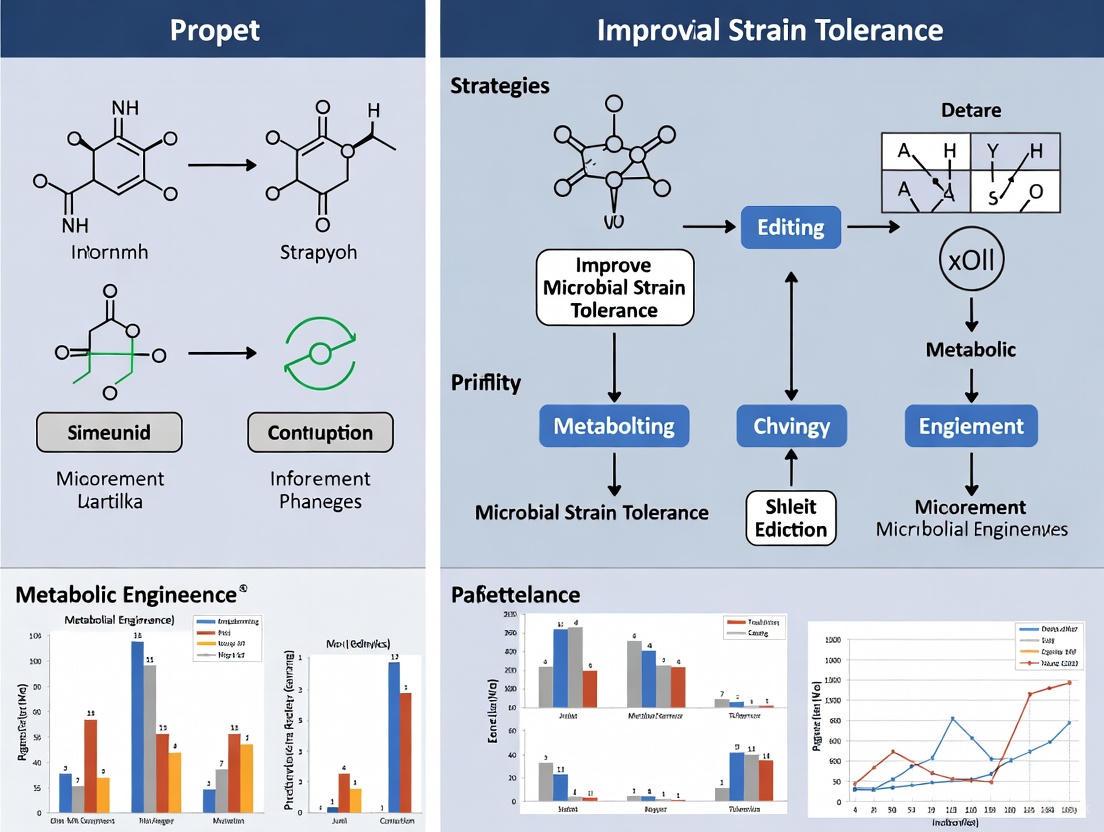
Advanced Strategies for Enhancing Microbial Strain Tolerance: From Cell Engineering to AI-Driven Discovery
This article provides a comprehensive overview of cutting-edge strategies to enhance microbial tolerance, a critical bottleneck in industrial biomanufacturing and antibiotic development.
Advanced Strategies for Enhancing Microbial Strain Tolerance: From Cell Engineering to AI-Driven Discovery
Abstract
This article provides a comprehensive overview of cutting-edge strategies to enhance microbial tolerance, a critical bottleneck in industrial biomanufacturing and antibiotic development. Tailored for researchers and drug development professionals, it systematically explores the foundational principles of microbial stress responses, details practical engineering methodologies at the cell envelope, intracellular, and extracellular levels, and discusses frameworks for troubleshooting and optimizing strain performance. Further, it examines advanced validation techniques and comparative analyses of different engineering approaches, integrating recent advances in synthetic biology, omics technologies, and generative AI to offer a holistic guide for developing robust microbial cell factories.
Understanding Microbial Tolerance: Core Concepts and Industrial Imperatives
Core Definitions and Key Distinctions
In the field of industrial microbiology, precisely defining microbial survival strategies is crucial for developing robust processes. Tolerance and resistance represent two fundamentally different mechanisms by which microbial populations survive antimicrobial agents or harsh production conditions.
Innate Tolerance is the inherent, non-specific ability of a bacterial strain to survive transient exposure to a lethal stressor without an increase in the Minimum Inhibitory Concentration (MIC). This phenotype is not acquired through genetic mutation or horizontal gene transfer in response to the stressor, but is a natural characteristic of the strain or species [1] [2]. It often involves physiological states that limit the lethal effect of the stressor, such as slow growth, dormancy, or a general stress response [1].
Acquired Resistance occurs when a previously susceptible microorganism gains the ability to grow in the presence of an antimicrobial agent, leading to an increase in the MIC. This is a genetically inherited trait that arises via de novo mutation or the acquisition of resistance genes through horizontal gene transfer (e.g., conjugation, transformation, or transduction) [3].
The following table summarizes the core distinctions:
| Feature | Innate Tolerance | Acquired Resistance |
|---|---|---|
| Genetic Basis | Innate, chromosomally encoded characteristics of a species or strain [3]. | Acquired via mutation or horizontal gene transfer of mobile genetic elements [3]. |
| Effect on MIC | No change in MIC [1]. | Increased MIC [1]. |
| Primary Mechanism | Stress response, slow growth, dormancy, efflux pumps, membrane impermeability [1] [4]. | Drug inactivation, target site modification, enhanced efflux [3]. |
| Population Effect | Can be homogeneous or heterogeneous (e.g., persister cells) [1]. | Typically homogeneous within the selected population. |
| Key Metric | Minimum Duration for Killing (MDK) [1]. | Minimum Inhibitory Concentration (MIC) [1]. |
Experimental Protocols for Quantification
Distinguishing between tolerance and resistance requires specific, quantitative laboratory methods.
Protocol 1: Quantifying Tolerance with the MDK Metric
The Minimum Duration for Killing (MDK) is a standardized metric for quantifying tolerance, measuring the time required to kill a certain percentage of the population at a lethal antibiotic concentration [1].
Methodology:
- Preparation: Prepare a 96-well plate with exponential dilutions of an antibiotic, ensuring concentrations reach at least 20x the known MIC. One column should remain antibiotic-free as a growth control [1].
- Inoculation: Dilute a stationary-phase bacterial culture to a precise density. For measuring MDK99, inoculate each well with approximately 100 CFU (Colony Forming Units) [1].
- Inoculation-Incubation Cycle: Inoculate plate rows at set time intervals and incubate the plate with shaking at 37°C. This creates a series of exposures from short to long durations [1].
- Antibiotic Neutralization: After the final incubation, thoroughly wash away the antibiotic via centrifugation or enzymatically inactivate it (e.g., using β-lactamase for ampicillin) to prevent carryover effects [1].
- Assessment of Regrowth: Incubate the washed plate under optimal growth conditions. The absence of regrowth in a well indicates that the exposure time was sufficient to kill the initial inoculum.
- Data Analysis: The MDK99 is determined as the shortest exposure time that prevents regrowth in at least 95% of the replicate wells at a concentration significantly above the MIC [1].
Protocol 2: Distinguishing Tolerance from Resistance
This workflow uses both MIC and MDK measurements to clearly differentiate the phenotypes.
Key Quantitative Metrics Table:
| Metric | What It Measures | Interpretation in Tolerance vs. Resistance |
|---|---|---|
| Minimum Inhibitory Concentration (MIC) | The lowest concentration of an antimicrobial that prevents visible growth [1]. | Resistance: Significantly increased MIC. Tolerance: Unchanged MIC [1]. |
| Minimum Duration for Killing (MDK) | The shortest duration of exposure to a lethal concentration of antimicrobial required to kill a given percentage (e.g., 99% for MDK99) of the population [1]. | Tolerance: Significantly increased MDK. This is the definitive metric for confirming a tolerance phenotype [1]. |
| Time-Kill Curve | A plot of viable cell count (CFU/mL) over time under a lethal antimicrobial concentration [1]. | Tolerance: A slower, often mono-phasic, killing curve. Persistence: A biphasic killing curve, indicating a small, tolerant subpopulation [1]. |
The Scientist's Toolkit: Essential Research Reagents
Successful experimentation requires carefully selected reagents and controls.
Research Reagent Solutions Table:
| Reagent / Material | Function in Experiment | Key Considerations |
|---|---|---|
| 96-Well Microtiter Plates | Platform for high-throughput MDK assays and MIC determinations [1]. | Use U-bottom or flat-bottom plates compatible with centrifugation washes [1]. |
| Lethal Concentration Antibiotics | Applied at high concentrations (e.g., 10-100x MIC) to stress the population for MDK assays [1]. | Concentration must be confirmed via prior MIC testing. Use a stock solution of known purity and concentration. |
| Specific Neutralizing Agents | Inactivates antibiotic residues after exposure to prevent carryover during regrowth assessment (e.g., β-lactamase for penicillins) [1] [5]. | Neutralization efficacy must be validated to ensure it does not affect bacterial viability [5]. |
| Culture Collection Strains | Reference microorganisms (e.g., E. coli ATCC, S. aureus ATCC) for controlled experiments and as quality controls [5]. | Use strains from approved collections. Maintain records of passage number to prevent phenotypic drift [5]. |
| Automated Robotic System | For precise, high-throughput pipetting and inoculation of time-course experiments [1]. | Enables accurate timing for MDK assays. Manual pipetting can be used but increases variability. |
| Phycocyanobilin | Phycocyanobilin, MF:C33H38N4O6, MW:586.7 g/mol | Chemical Reagent |
| SZM-1209 | SZM-1209, MF:C31H29F5N4O5S2, MW:696.7 g/mol | Chemical Reagent |
Troubleshooting Guides and FAQs
Frequently Asked Questions:
Q1: In an industrial fermentation context, why is it important to distinguish between a tolerant strain and a resistant one? The distinction has major implications for process design and contamination control. A resistant contaminant will grow continuously in the presence of a biocide or antibiotic, leading to persistent contamination. A tolerant contaminant may be killed eventually with prolonged exposure, indicating that the treatment is effective but the duration or concentration needs optimization. Furthermore, for a production strain, engineering for robustness (the ability to maintain stable production under stress) is a more holistic goal than just tolerance, as it encompasses both survival and functional performance [4].
Q2: Our time-kill curve data is noisy and not reproducible. What are the key factors to control? Noisy data often stems from inconsistent initial culture conditions. Key factors to standardize include [5]:
- Culture Age and Preparation: Always use cultures in the same growth phase (e.g., mid-exponential or early stationary). Do not use cultures more than five passages from the original seed stock.
- Inoculum Size: Precise standardization of the starting CFU/mL is critical for MDK calculations.
- Neutralization Validation: Ineffective antibiotic neutralization after exposure can prevent regrowth of survivors, leading to overestimation of killing. Always include controls to confirm neutralization efficacy [5].
Q3: Can the use of disinfectants like quaternary ammonium compounds (QACs) in our facility promote antibiotic tolerance or resistance? Laboratory studies indicate a concerning correlation. Bacteria can develop tolerance or resistance to QACs through mechanisms like efflux pumps and biofilm formation, and some of these mechanisms (e.g., multidrug efflux pumps) can also confer resistance to clinically important antibiotics [2]. While conclusive evidence from real-world settings is still being gathered, the potential risk necessitates the prudent and rotated use of disinfectants to minimize selective pressure [2].
Q4: What are some modern strategies to improve the tolerance and robustness of industrial microbial cell factories? Several engineering strategies are employed [6] [4]:
- Transcription Factor Engineering: Modifying global regulators (e.g., RpoD, CRP) to reprogram the cellular stress response on a genome-wide scale [4].
- Adaptive Laboratory Evolution (ALE): Passaging microbes over many generations under the target stress to naturally select for enhanced tolerance mutations [6].
- Membrane Engineering: Altering membrane lipid composition to improve integrity against solvents or other chemical stresses [6].
- Computational & Systems Biology: Using genome-scale models and machine learning to predict and design genetic modifications that enhance robustness [6] [4].
Core Concepts: Understanding Product Toxicity in Microbial Bioprocessing
What is "product toxicity" in the context of microbial bioproduction? Product toxicity refers to the phenomenon where the compounds being produced by a microbial cell factory—or the intermediates created during the synthesis pathway—inhibit the microorganism's own growth and metabolic activity. This occurs because these chemicals can disrupt essential cellular structures like the cell membrane, interfere with enzyme function, or alter internal pH. Ultimately, this self-toxicity places a fundamental limit on the final yield and productivity of the biomanufacturing process [7].
What are the primary mechanisms through which toxic products damage microbial cells? Toxic end-products and intermediates employ several mechanisms to impair cellular function. The cell envelope, comprising the membrane and cell wall, is the primary target. Hydrophobic compounds can integrate into and disorganize the lipid bilayer, compromising its role as a selective barrier. Furthermore, these chemicals can denature proteins, including critical enzymes, and disrupt energy metabolism by interfering with the proton motive force across the membrane [7].
Troubleshooting Guides: Identifying and Overcoming Toxicity Issues
Symptom: Unexpectedly Low Final Product Titer
Problem: Your bioproduction process is yielding a much lower final product concentration (titer) than anticipated based on model predictions or small-scale tests.
Investigation & Resolution:
| Step | Action | Rationale & Details |
|---|---|---|
| 1 | Repeat the Experiment | Rule out simple human error in protocol execution, such as incorrect reagent volumes or accidental omission of steps [8]. |
| 2 | Analyze Cell Viability & Physiology | Check for classic signs of toxicity: a steep drop in cell viability coinciding with product accumulation, or changes in cell morphology observed under a microscope [7] [9]. |
| 3 | Review Bioreactor Data | Compile and overlay online bioreactor data (e.g., dissolved oxygen, pH) with offline measurements (e.g., cell density, product titer). Look for correlations between the onset of product accumulation and physiological stress markers [9]. |
| 4 | Isolate the Variable | Systematically test which factor is causing the issue. A highly effective approach is to supplement the culture with a sub-lethal dose of the pure end-product and observe if it replicates the growth inhibition [7]. |
Symptom: Process Performance Deteriorates at Scale-Up
Problem: Your process works excellently in small-scale bioreactors but fails to maintain productivity and yield when moved to a larger production vessel.
Investigation & Resolution:
| Step | Action | Rationale & Details |
|---|---|---|
| 1 | Audit Scale-Up Parameters | Do not assume parameters like agitation and aeration scale linearly. Gradients in pH, nutrients, and dissolved oxygen are common in large tanks and can exacerbate product toxicity [10]. |
| 2 | Assess Raw Material Consistency | Variability in the quality of media components between small and large batches can lead to unexpected interactions with the toxic product or the organism's stress response [10]. |
| 3 | Mitigate Shear Stress | Increased agitation in large bioreactors can generate shear forces that damage cells already weakened by product toxicity, creating a compounded stress effect [10]. |
| 4 | Model Large-Scale Conditions | Use advanced sensor data and scale-down models to simulate the heterogeneous conditions of a large bioreactor at a small scale. This allows for efficient testing of strain robustness and process optimization [9]. |
Frequently Asked Questions (FAQs) for Researchers
Q1: What are the most promising synthetic biology strategies for enhancing microbial tolerance? Research focuses on two main areas: intracellular and extracellular engineering. Intracellular strategies include global transcription machinery engineering (gTME) to reprogram cellular stress responses, and engineering of membrane composition (e.g., altering lipid saturation) to fortify the cell against hydrophobic compounds. Extracellular strategies involve promoting biofilm formation to create a protective microenvironment and designing synthetic microbial consortia to distribute the metabolic burden of toxin production and tolerance [7].
Q2: How can I quickly determine if my observed low yield is due to product toxicity or another factor like media composition? The most direct diagnostic test is a sub-lethal spiking experiment. Add a known, non-lethal concentration of your pure target product or key intermediate to a growing culture during its early exponential phase. If this addition immediately replicates the observed growth inhibition or drop in productivity, product toxicity is a likely culprit. If not, the issue may lie with nutrient limitation, substrate inhibition, or other process parameters [7] [8].
Q3: Beyond genetic engineering, what process-level solutions can mitigate toxicity effects? Several bioprocess engineering strategies can help:
- In-situ Product Removal (ISPR): Continuously extracting the toxic product from the fermentation broth as it is produced relieves the stress on the cells.
- Two-Phase Systems: Using a water-immiscible organic solvent as a second phase can create a reservoir for the toxic product, keeping its aqueous concentration low.
- Controlled Feeding Strategies: Feeding substrates slowly can modulate the rate of toxic intermediate formation, preventing a sudden, lethal accumulation.
Q4: What biosafety considerations are critical when engineering more robust microbial strains? Enhanced tolerance must be balanced with environmental safety. A comprehensive biosafety assessment should evaluate the engineered strain's pathogenicity (e.g., cytotoxicity, hemolytic activity), immunogenicity (ability to trigger an immune response), and environmental persistence (survival in non-contained settings). Principal component analysis has shown that 84% of biosafety risk variation is strain-specific, not tied to taxonomy, meaning each novel strain requires its own risk assessment [11].
Experimental Protocols for Tolerance Engineering and Assessment
Protocol: Laboratory Evolution for Enhanced Tolerance
Objective: To generate a microbial population with improved tolerance to a toxic end-product through adaptive evolution.
Workflow:
Methodology:
- Inoculation: Start with a flask of standard growth medium inoculated with the wild-type strain.
- Initial Challenge: After one growth cycle, transfer a sample of this culture into fresh medium containing a sub-lethal concentration (e.g., IC~10~) of the toxic product.
- Growth Monitoring: Monitor the culture's growth (e.g., by optical density at 600 nm, OD600) until it reaches the late exponential phase.
- Serial Passaging: Use a sample of this grown culture to inoculate the next passage, gradually increasing the concentration of the toxic product in each subsequent batch.
- Isolation: Continue this serial passaging for dozens to hundreds of generations. Plate the evolved culture to isolate single colonies.
- Screening: Screen these isolated colonies for improved growth and production under high product concentrations compared to the original ancestor.
Protocol: Assessing Pathogenicity and Immunogenicity
Objective: To quantitatively evaluate the biosafety profile of an engineered, toxin-tolerant microbial strain.
Key Assays and Metrics:
Methodology: This protocol is based on a comprehensive, multi-parameter framework for biosafety assessment [11].
Pathogenicity Assessment:
- Hemolytic Activity: Culture the strain on blood agar plates. A clear zone around colonies indicates red blood cell lysis.
- Cytotoxicity: Co-culture the strain with human cell lines (e.g., HEp-2 epithelial cells) and measure cell viability using assays like MTT or lactate dehydrogenase (LDH) release.
- Enzyme Production: Test for the secretion of potentially damaging extracellular enzymes like proteases, lipases, and DNases using specific substrate plates or assays.
- Scoring: Each parameter is scored (e.g., 0-5), and a composite Pathogenicity Index (PI) is calculated.
Immunogenicity Assessment:
- In Vitro: Isolate Peripheral Blood Mononuclear Cells (PBMCs) from healthy donors. Co-culture these with heat-inactivated microbial cells and measure the production of pro-inflammatory cytokines (IL-1β, IL-6, TNF-α) via ELISA.
- In Vivo: Administer heat-inactivated cells to animal models (e.g., BALB/c mice) and measure serum cytokine levels at multiple time points post-administration.
- Scoring: Results are used to calculate an Immunostimulation Index (ISI).
Data Presentation: Quantitative Biosafety Profiles
Table 1: Pathogenicity Indicators Across Microbial Taxa [11]
| Taxonomic Group | # of Strains Tested | Growth at 37°C (Mean Score) | Hemolytic Activity (Mean Score) | Extracellular Enzymes (Mean Score) | Antibiotic Resistance (Mean Score) | Cell Adhesion (Mean Score) |
|---|---|---|---|---|---|---|
| Gram-Negative Bacteria | 12 | 4.1 | 3.5 | 3.8 | 3.2 | 2.2 |
| Fungi | 8 | 3.8 | 2.9 | 3.5 | 2.5 | 3.8 |
| Gram-Positive Bacteria | 12 | 3.2 | 2.5 | 1.8 | 2.8 | 1.0 |
| Actinomycetes | 8 | 2.5 | 1.2 | 1.5 | 1.8 | 0.2 |
Note: Scores are on a relative scale (e.g., 0-5), with higher values indicating greater potential for that pathogenicity trait.
Table 2: Key Reagents for Tolerance and Biosafety Research
| Research Reagent / Material | Function in Experiment |
|---|---|
| Blood Agar Plates | To assess hemolytic activity, a key indicator of potential pathogenicity [11]. |
| HEp-2 Cell Line | A human epithelial cell line used for cytotoxicity and cellular adhesion assays [11]. |
| PBMCs (Peripheral Blood Mononuclear Cells) | Primary human immune cells used for in vitro immunogenicity testing (cytokine release) [11]. |
| ELISA Kits (for IL-1β, IL-6, TNF-α) | To quantitatively measure pro-inflammatory cytokine production from immune cells [11] [12]. |
| Luria-Bertani (LB) & ISP-2 Media | Standard culture media for growing bacterial and actinomycete strains, respectively [11]. |
| Cloud-Connected Bioreactor System | Enables real-time monitoring and data visualization of process parameters, crucial for linking strain physiology to production metrics [9]. |
Troubleshooting Guide: Cell Envelope Analysis
This guide addresses common experimental challenges in analyzing the microbial cell envelope, a key structure for understanding and improving microbial strain tolerance.
FAQ: I am getting inconsistent results when assessing cell envelope integrity under stress conditions. What could be wrong?
Answer: Inconsistent integrity data often stems from poorly controlled environmental conditions or an over-reliance on a single assay. The cell envelope is a dynamic structure that modifies its composition in response to its environment [13]. Implement the following systematic approach:
- Control Growth Conditions Precisely: Minor fluctuations in temperature, pH, or osmolarity during pre-culture can trigger cell envelope stress responses, fundamentally altering the envelope's composition and stability before your assay even begins [13]. Use fresh, standardized media and tightly controlled incubators.
- Employ a Multi-Assay Approach: No single assay gives a complete picture. Combine quantitative and visualization techniques to cross-validate results.
- Include Relevant Controls: Always run assays with known robust and sensitive control strains. For genetic modification studies, include a complemented strain (where the mutated gene is reintroduced) to confirm that the observed phenotype is due to the specific genetic change and not a secondary mutation.
Table: Troubleshooting Cell Envelope Integrity Assays
| Problem | Potential Cause | Solution |
|---|---|---|
| High variability in osmotic fragility assays | Inconsistent culture age or density | Use cells harvested at the same optical density and growth phase (mid-log phase is typically most consistent). |
| Weak or no signal in fluorescent dye staining (e.g., membrane dyes) | Compromised dye potency; insufficient permeability | Prepare fresh dye stocks; validate staining protocol with a positive control strain; consider using permeabilizing agents. |
| AFM images are blurry or show sample damage | Excessive scanning force; improper sample immobilization | Reduce the applied force in AFM contact mode; switch to AFM tapping mode in liquid; optimize surface functionalization (e.g., with poly-L-lysine or gelatin) to firmly immobilize cells [14]. |
| Cryo-ET reveals artifacts in envelope layers | Sample preparation-induced stress (e.g., dehydration, chemical fixation) | Where possible, use cryo-fixation (plunge-freezing) and cryo-FIB milling to visualize cells in a near-native, hydrated state [15]. |
FAQ: My high-resolution imaging (e.g., AFM, Cryo-ET) does not reveal the expected surface protein localization or dynamics.
Answer: Visualizing single proteins, especially in live cells, is technically demanding and requires optimization of both the equipment and the biological sample [14].
- For Atomic Force Microscopy (AFM):
- Use High-Speed AFM (HS-AFM): For tracking protein dynamics, standard AFM is too slow. HS-AFM allows imaging at sub-second temporal resolution, enabling you to see conformational changes in membrane proteins like transporters or ion channels in near real-time [14].
- Optimize Sample Immobilization and Cantilevers: For live cells, their large size and curvature impede imaging. Use high-resonance frequency, small, soft cantilevers and an optimized imaging buffer to visualize single proteins on curved membranes [14].
- For Cryo-Electron Tomography (Cryo-ET):
- Ensure Adequate Tomogram Denoising: Raw cryo-ET data has low signal-to-noise. Apply advanced denoising algorithms like Nonlinear Anisotropic Diffusion (NAD) filtering to enhance the visibility of delicate structures like the septal peptidoglycan wedge in dividing E. coli without introducing artifacts [15].
- Use Subtomogram Averaging: To compare specific envelope features (e.g., porin density in the outer membrane) between different regions or strains, use subtomogram averaging. This technique improves resolution by aligning and averaging multiple copies of a structure [15].
The following diagram illustrates the decision-making workflow for selecting and optimizing high-resolution imaging techniques based on your research question.
Diagram 1: Selecting an Imaging Technique for Cell Envelope Analysis.
FAQ: How do I accurately interpret data from mutants with altered cell envelope synthesis genes?
Answer: Mutants in envelope synthesis or remodeling pathways often have pleiotropic effects. A change in one component can trigger compensatory changes in another, a phenomenon known as the envelope stress response [16].
- Profile Multiple Envelope Components: Do not assume only the targeted component is altered. For example, a mutant in peptidoglycan synthesis may also show changes in teichoic acid D-alanylation or membrane phospholipid composition as the cell attempts to maintain net surface charge and integrity [13].
- Measure Physical-Chemical Properties: Go beyond genetics and biochemistry. Use AFM to measure the nanomechanical properties (e.g., elasticity, stiffness) of mutant cells. A cell with defective peptidoglycan may show reduced stiffness, directly linking the genetic change to a physical consequence [14].
- Analyze Constriction Dynamics in Division Mutants: When studying division mutants (e.g., in
ftsN,envC,nlpD), use live-cell imaging with differential membrane markers (e.g., IM-anchored ZipA-sfGFP and OM-lipoprotein Pal-mCherry) to track the invagination rates of the inner and outer membranes separately. This can reveal whether the mutation causes a switch from septation to constriction mode [15].
Experimental Protocols
This section provides detailed methodologies for key experiments investigating cell envelope architecture and tolerance.
Protocol 1: Assessing Pathogenicity and Immunogenicity for Biosafety Screening
This protocol is essential for evaluating the biosafety of engineered microbial strains, a critical step in tolerance research that may involve pathogenic parents or generate strains with unknown risk profiles [11].
- Principle: A multi-parameter approach is used to evaluate the potential of a microbial strain to cause harm, assessing its ability to damage host cells and trigger immune responses.
- Applications: Biosafety classification of novel or engineered strains; risk assessment for industrial application.
Reagents & Equipment:
- Microbial strains (test and control).
- Cell culture facilities and human cell lines (e.g., HEP-2 epithelial cells).
- Blood agar plates.
- Enzyme assays for proteases, lipases, DNases.
- Antibiotic disks for resistance profiling.
- ELISA kits for cytokines (IL-1β, IL-6, TNF-α).
- Luria-Bertani (LB) or other appropriate culture media.
- Incubators (25°C, 37°C).
Step-by-Step Method:
- Pathogenicity Assessment:
- Growth at 37°C: Monitor growth kinetics at human physiological temperature. Score growth rate and yield.
- Hemolytic Activity: Streak strains on blood agar plates. Incubate and score zones of clearing (β-hemolysis), greening (α-hemolysis), or no effect (γ-hemolysis).
- Extracellular Enzymes: Spot cultures on agar plates containing specific substrates (e.g., skim milk for proteases, tributyrin for lipases, DNA-methyl green for DNases). Score halo formation after incubation.
- Antibiotic Resistance: Use the Kirby-Bauer disk diffusion method against a panel of clinically relevant antibiotics. Measure and score zones of inhibition.
- Cell Adhesion: Incubate human epithelial cells (HEp-2) with microbial cells. Wash off non-adherent microbes, lyse the eukaryotic cells, and plate the lysate to quantify adherent bacteria.
- Calculate a Composite Pathogenicity Index (PI) by summing the individual scores (0-5) from each assay [11].
- Immunogenicity Evaluation:
- In Vitro: Isolate Peripheral Blood Mononuclear Cells (PBMCs) from healthy donors. Co-culture with heat-inactivated microbial cells. Measure pro-inflammatory cytokine production (IL-1β, IL-6, TNF-α) in the supernatant by ELISA after 24-48 hours.
- In Vivo (Mouse Model): Administer heat-inactivated microbial cells intraperitoneally to BALB/c mice. Collect serum at 6, 24, and 48 hours post-administration and measure cytokine levels by ELISA.
- Calculate an Immunostimulation Index (ISI) based on cytokine production levels relative to negative controls [11].
- Pathogenicity Assessment:
Expected Outcome: A comprehensive biosafety profile for each strain. Strains with high PI and ISI scores present a greater potential risk and may require higher containment levels.
Table: Key Reagents for Cell Envelope Stress and Biosafety Research
| Research Reagent / Solution | Function / Application | Key Considerations |
|---|---|---|
| AFM Cantilevers (Soft, High-Frequency) | High-resolution topographic imaging and nanomechanical measurement of live cells [14]. | Must be selected for resonance frequency and spring constant suitable for biological samples to prevent damage. |
| Cryo-FIB Milling System | Prepares thin (150-250 nm), electron-transparent lamellae from vitrified microbial cells for in situ Cryo-ET [15]. | Requires specialized, high-cost equipment and significant expertise. |
| Membrane-Specific Fluorescent Dyes (e.g., for IM, OM, lipid domains) | Visualizing membrane architecture, integrity, and dynamics using fluorescence microscopy. | Dye selectivity and potential toxicity to live cells must be validated. |
| NAD Filtering Software (e.g., in TomoBEAR, EMAN2) | Advanced denoising of cryo-electron tomograms to enhance visibility of delicate envelope structures like peptidoglycan [15]. | Critical for interpreting tomograms of Gram-negative septa. |
| Peptidoglycan Hydrolases & Inhibitors (e.g., lysozyme, EnvC, NlpD) | Enzymatic probing of PG structure and study of cell separation during division [15]. | Specificity and activity must be confirmed for the target organism. |
| Pro-Inflammatory Cytokine ELISA Kits (e.g., for TNF-α, IL-6) | Quantifying immune response to strains for biosafety immunogenicity assessment [11]. | Use requires appropriate biosafety containment for pathogenic strains. |
Protocol 2: In situ Architecture Analysis of the Bacterial Divisome using Cryo-ET
This protocol allows for the native-state 3D visualization of the cell division machinery and the accompanying synthesis of septal peptidoglycan.
- Principle: Cells are vitrified (flash-frozen) to preserve native structure, thinned using a cryo-focused ion beam (cryo-FIB), and imaged with transmission electron microscopy at different tilt angles to compute a 3D tomogram.
- Applications: Determine the ultrastructure of the division site; characterize the effect of division mutants on septal architecture.
Reagents & Equipment:
- Bacterial culture in mid-log phase.
- Cryo-plunger for vitrification.
- Cryo-FIB/SEM microscope.
- Cryo-transmission electron microscope (Cryo-TEM).
- Tomography acquisition software.
- Computational hardware and software for tomogram reconstruction and analysis (e.g., IMOD, TomoBEAR).
Step-by-Step Method:
- Sample Vitrification: Apply 3-4 µL of bacterial culture to a glow-discharged EM grid. Blot excess liquid and plunge-freeze the grid into a liquid ethane/propane mixture cooled by liquid nitrogen.
- Cryo-FIB Milling: Transfer the vitrified grid to a cryo-FIB/SEM microscope. Use a protective platinum or carbon layer to prevent damage. Use a focused Ga⺠ion beam to mill away material and create an electron-transparent lamella (~150-250 nm thick) through a bacterial cell [15].
- Cryo-ET Data Acquisition: Transfer the lamella to a cryo-TEM. Acquire a tilt-series (e.g., from -60° to +60° with 1-2° increments) under low-dose conditions to minimize radiation damage.
- Tomogram Reconstruction: Align the tilt-series and reconstruct a 3D tomogram using back-projection or weighted back-projection algorithms.
- Denoising and Segmentation: Apply NAD filtering to the tomogram to enhance contrast. Manually or semi-automatically segment the different envelope components—inner membrane, outer membrane, and peptidoglycan layer—for 3D visualization and analysis [15].
Expected Outcome: A high-resolution 3D model of the division site. In wild-type E. coli, this should reveal a V-shaped constriction with a partial septum and an elongated, triangular wedge of peptidoglycan near the invaginating inner membrane [15].
Advanced Concepts: Integrating Envelope Research with Strain Tolerance
Understanding the microbial cell envelope's protective role is fundamental to designing strains with enhanced tolerance for industrial applications. The envelope is the primary interface with the environment, and its stress response systems are key targets for engineering.
The relationship between antimicrobial stresses, the envelope stress response, and the development of tolerance is a critical consideration for both public health and industrial microbiology. The following diagram maps this complex network.
Diagram 2: The Envelope Stress Response Network. This map shows how antimicrobial stresses trigger adaptive changes that can lead to increased tolerance or resistance [16] [13].
- Engineering Targets from Stress Responses: Diagram 2 shows that targeting the Envelope Stress Response (ESR) network itself is a promising strategy. Research is exploring synergistic ESR inhibitors to be used in combination with existing antimicrobials, breaking down the microbe's defenses [16].
- Biosafety as a Component of Strain Design: For any engineered strain, especially those intended for open environments or large-scale fermentation, a systematic biosafety assessment is crucial. The data-driven framework evaluating pathogenicity, immunogenicity, and environmental persistence should be integrated early in the strain design cycle [11] [17]. This ensures that enhanced tolerance does not come with unacceptable safety risks.
- Exploiting Envelope Diversity for Synthetic Biology: Non-model microorganisms with native resilience (e.g., robust envelopes, stress resistance) offer untapped potential as chassis for industrial biotechnology. Selecting such hosts based on native C1 metabolism, thermotolerance, or solvent resistance can provide a more robust foundation for engineering than starting with a lab-adapted but sensitive model organism [17].
In bioprocessing, the productivity and stability of microbial and cell cultures are consistently challenged by a range of biological and environmental stressors. These stressors include toxins like endotoxins, metabolic inhibitors such as mycotoxins, and physical-environmental challenges including shear stress and osmotic pressure. Effectively classifying and understanding these stressors is the foundational step in developing robust strategies to improve microbial strain tolerance, which is critical for enhancing yield, product quality, and process reliability in biopharmaceutical manufacturing [18] [19]. This guide provides a structured troubleshooting framework to help researchers identify, understand, and mitigate these critical challenges.
Classification and Impact of Key Stressors
Bioprocessing stressors can be systematically categorized based on their origin and nature. The following table summarizes the primary types of stressors, their sources, and their impact on bioprocessing.
Table 1: Classification of Key Stressors in Bioprocessing
| Stressor Category | Specific Examples | Origin/Source | Impact on Bioprocessing |
|---|---|---|---|
| Toxins | Endotoxins (LPS) [19] | Gram-negative bacterial cell lysis | Activates immune pathways, contaminates products, induces pyrogenic responses [19] |
| Roquefortine C [20] | Penicillium roqueforti fermentation | Neurotoxin; poses contamination risk in fungal fermentations [20] | |
| Mycophenolic Acid [20] | Penicillium roqueforti fermentation | Immunosuppressant; potential product contaminant [20] | |
| Metabolic Inhibitors | By-products (e.g., alcohols, acids) | Microbial metabolism | Inhibits cell growth, reduces product titers, disrupts metabolic pathways |
| Host Cell Proteins (HCPs) [21] | Production host organism | Contaminates downstream products, potential immunogenicity | |
| Physical & Environmental Stressors | Shear Stress [22] | Bioreactor impellers, mixing | Damages cells, reduces viability, impacts protein expression |
| Osmotic Pressure [23] | High substrate/salt concentrations | Reduces water activity, inhibits growth, triggers stress responses | |
| Temperature Fluctuations [23] | Improper process control | Deviates from optimal growth range, reduces productivity | |
| Foaming [22] | Aeration, media composition | Reduces gas transfer efficiency, risks contamination | |
| pH Shifts [23] | Microbial metabolism, buffer failure | Shifts metabolic activity, can inactivate enzymes |
Troubleshooting Guides and FAQs
A. Endotoxin Contamination
Q: How do I identify and control endotoxin contamination in my cell culture or protein purification process?
Endotoxins, or lipopolysaccharides (LPS), are a significant contaminant from Gram-negative bacterial cell walls that can trigger strong immune responses and compromise product safety [19].
- Detection Methods: The standard method is the Limulus Amoebocyte Lysate (LAL) assay, which comes in gel-clot, turbidimetric, and chromogenic variants. The chromogenic assay is widely used for its speed and accuracy, measuring the hydrolysis of a chromogenic substrate in the presence of endotoxin [19].
- Prevention and Removal Strategies:
- Source Control: Use high-purity, endotoxin-free water, buffers, and cell culture media. Sterilize equipment properly and avoid using reagents that have been exposed to bacterial contaminants [19].
- Removal Techniques: Several chromatographic methods are effective, including ion-exchange chromatography, affinity adsorbents, and gel filtration chromatography. The choice depends on the properties of your target protein [19].
- Regulatory Limits: For parenteral drugs, the US Pharmacopoeia sets a limit of 5 Endotoxin Units (EU) per kg of body weight per hour of administration. Aim to keep endotoxin levels as low as possible in all process steps [19].
B. General Contamination and Assay Interference
Q: My impurity assays (e.g., HCP ELISA) are showing high background or poor precision. What could be the cause?
High background or non-specific binding (NSB) in sensitive assays like ELISA is often due to contamination or technique.
- Causes and Solutions:
- Airborne Contamination: Concentrated sources of the analyte (e.g., cell culture media, upstream samples) can contaminate reagents via dust or aerosols.
- Solution: Perform assays in a dedicated, clean area. Wipe down all surfaces and equipment before starting. Use pipette tips with aerosol filters and avoid talking or breathing over an uncovered microtiter plate. Consider using a laminar flow hood [21].
- Incomplete Washing: Carryover of unbound reagent during the ELISA wash steps can cause high background.
- Solution: Follow the recommended washing technique strictly. Use only the provided wash buffer and avoid over-washing or allowing wash solution to soak in wells for extended periods [21].
- Substrate Contamination: For alkaline phosphatase-based assays using PNPP substrate, contamination from airborne bacteria or human dander can be a problem.
- Solution: Withdraw only the substrate volume needed for a single assay run. Never return unused substrate to the original bottle. Recap all reagent bottles immediately after use [21].
- Airborne Contamination: Concentrated sources of the analyte (e.g., cell culture media, upstream samples) can contaminate reagents via dust or aerosols.
C. Process-Related Stressors
Q: My microbial fermentation is underperforming with low yield. What process-related stressors should I investigate?
Environmental factors are common culprits behind reduced fermentation efficiency.
- Investigation Checklist:
- Temperature Stability: Verify that your bioreactor maintains a consistent, optimal temperature for your strain. Fluctuations can stress the culture [23].
- Osmotic Pressure: Check the concentration of substrates and salts in your media. High osmolarity can inhibit microbial growth and metabolic activity [23].
- Shear Stress: In stirred-tank bioreactors, high agitation rates can generate shear forces that damage sensitive cells. If using a new vessel, confirm that the impeller type and configuration (e.g., bottom magnetic stirring) are appropriate for your culture's shear sensitivity [22].
- Oxygen Demand: Ensure adequate oxygen transfer by verifying aeration rates and impeller design. In high-density cultures, oxygen can become a limiting factor [18] [22].
- Foaming: Monitor for excessive foaming, which can be induced by aeration and media composition. Foaming reduces gas transfer efficiency and increases contamination risk [22].
Essential Experimental Protocols
Protocol 1: Assessing and Improving Microbial Tolerance to Environmental Stressors
This protocol outlines a methodology to evaluate and enhance strain performance under sub-optimal conditions, inspired by industrial case studies [23].
Strain Screening & Inoculation:
- Begin with a genetically diverse pool of your production strain (e.g., wild-type, engineered variants).
- Inoculate cultures in shake flasks using a standardized medium and growth conditions to generate reproducible inoculums [22].
Controlled Stress Application:
- Scale up the process to a bioreactor system for precise control.
- Apply a defined stressor (e.g., a temperature shift, high salt concentration, or sub-optimal pH) during the mid-log phase of growth.
- Use advanced Process Analytical Technology (PAT) tools, such as Raman or NIR spectroscopy, to monitor critical parameters like cell density, metabolite levels, and product formation in real-time [18].
Performance Monitoring and Data Collection:
- Track key performance indicators (KPIs) including:
- Cell Viability and Biomass: Compare stressed vs. control cultures.
- Product Titer: Measure the concentration of the target molecule.
- Specific Productivity: Calculate the product yield per cell.
- Sample frequently to analyze metabolic by-products and potential stress-induced inhibitors like Host Cell Proteins (HCPs) [21].
- Track key performance indicators (KPIs) including:
Data Analysis and Strain Selection:
- Analyze the data to identify strains that maintain the highest productivity under stress.
- Back-fitting the signals of your standards as unknowns is a recommended method to ensure the accuracy of your analytical methods when working with stressed, complex samples [21].
- Select the top-performing strains for further development and pathway engineering.
The following diagram illustrates the experimental workflow for this protocol:
Protocol 2: Endotoxin Detection and Removal for Biologics
This protocol details the steps to ensure final biological products meet regulatory safety standards for endotoxin levels [19].
Sample Preparation:
- Collect samples from relevant stages of the purification process.
- For samples with expected high impurity levels (e.g., from upstream purification), perform a preliminary dilution in an appropriate, validated diluent to bring the endotoxin concentration within the detection range of the assay. Always use an endotoxin-free diluent.
Endotoxin Detection (LAL Assay):
- Select an LAL assay method (chromogenic is recommended for speed and accuracy).
- Follow the kit manufacturer's instructions precisely to prepare standards and samples.
- Run the assay, including negative controls (endotoxin-free water) and positive controls.
- Use a point-to-point, cubic spline, or 4-parameter curve-fitting routine for data interpolation, as linear regression can introduce inaccuracies, especially at the curve extremes [21].
Endotoxin Removal:
- If endotoxin levels are above the required limit, employ a removal technique compatible with your product.
- Ion-Exchange Chromatography is a common and effective method. The choice of resin (anion vs. cation) will depend on the isoelectric point (pI) and charge characteristics of your target protein versus the endotoxin at your process pH [19].
- Validate the efficiency of the removal step by testing the post-purification product with the LAL assay again.
Documentation and Release:
- Document all results, demonstrating that the final product meets the endotoxin limit of ≤5 EU/kg for parenteral administration [19].
The Scientist's Toolkit: Key Research Reagent Solutions
The following table lists essential reagents and materials critical for troubleshooting and mitigating stressors in bioprocessing.
Table 2: Essential Reagents and Materials for Stressor Management
| Reagent/Material | Function/Application | Key Considerations |
|---|---|---|
| LAL Assay Kits [19] | Detection and quantification of endotoxin contamination. | Choose between gel-clot, turbidimetric, or chromogenic based on needs for speed, accuracy, and sensitivity. |
| Chromatography Resins [18] [19] | Purification and removal of impurities (HCPs, endotoxins) and continuous processing. | Use multimodal or ion-exchange resins for impurity removal. Select resins based on target biomolecule properties. |
| Specialized Assay Diluents [21] | Diluting samples for accurate impurity analysis (e.g., HCP ELISA). | Use kit-specific diluents to match the standard matrix and avoid dilutional artifacts. Validate recovery rates (95-105%). |
| Single-Use Bioreactors [24] [25] | Upstream processing; reduces cross-contamination risk and cleaning validation. | Ideal for perfusion modes and high-density cultures. Consider scalability (from 30L to 4000L) and environmental impact. |
| Process Analytical Technology (PAT) [18] | Real-time monitoring of bioprocess parameters (e.g., metabolites, cell density). | Includes Raman/NIR spectroscopy. Critical for implementing Real-Time Release (RTR) and dynamic process control. |
| CRISPR/Cas9 Systems [20] | Microbial strain engineering to knock out toxin-producing genes. | Used to develop safer production strains (e.g., non-toxic P. roqueforti for cheese production). |
| Phycocyanobilin | Phycocyanobilin, MF:C33H38N4O6, MW:586.7 g/mol | Chemical Reagent |
| Phycocyanobilin | Phycocyanobilin, MF:C33H38N4O6, MW:586.7 g/mol | Chemical Reagent |
Visualizing the Endotoxin-Induced TLR4 Signaling Pathway
Understanding the mechanism of endotoxin toxicity is key to appreciating its impact. The following diagram details the TLR4 signaling pathway activated by LPS, which leads to a potent inflammatory response.
Engineering Robust Microbes: A Toolkit of Strain Improvement Strategies
The cell envelope serves as the primary interface between a microbial cell factory and its often hostile industrial environment. Comprising membranes and cell walls, this dynamic structure is far more than a passive barrier; it actively mediates transport, signaling, and stress response. In recent years, cell envelope engineering has emerged as a powerful strategy for enhancing microbial tolerance to the toxic compounds and harsh conditions encountered in industrial bioprocessing, thereby increasing the robustness and productivity of microbial cell factories [26] [4]. This technical support center is designed to provide researchers with practical solutions for overcoming the most common challenges in this field, framed within the broader thesis that targeted envelope modifications are crucial for developing superior industrial strains.
Troubleshooting Common Experimental Challenges
Frequently Asked Questions (FAQs)
Q1: Our engineered E. coli strain shows poor growth and low product titer when producing fatty alcohols. What envelope-related issues should we investigate? This is a classic symptom of product toxicity, where the target compound compromises envelope integrity. We recommend a multi-pronged investigation:
- Membrane Lipid Engineering: Modify the membrane's phospholipid headgroup composition. In Synechocystis, this approach resulted in a 3-fold increase in octadecanol productivity [26]. For E. coli, similar strategies have focused on adjusting the unsaturation of fatty acid chains to enhance stability [26].
- Efflux Pump Overexpression: Engineer the strain to overexpress heterologous transporter proteins. In S. cerevisiae, this has led to a 5-fold increase in the secretion of fatty alcohols, effectively reducing intracellular accumulation and toxicity [26].
- Diagnostic Assay: Perform a membrane integrity assay using a fluorescent dye like propidium iodide, which is excluded by healthy cells. Increased fluorescence indicates a compromised membrane, confirming toxicity [27].
Q2: How can we improve yeast tolerance to inhibitors found in lignocellulosic hydrolysates, such as weak acids and furans? Acquisition of multi-stress tolerance often involves complex, genome-wide adaptations. A highly effective strategy is Adaptive Laboratory Evolution (ALE).
- ALE Protocol: Subject the microbial population to serial passaging in media with gradually increasing concentrations of the target inhibitors (e.g., acetic acid, furfural). An evolved strain of Rhodotorula toruloides (IST536 MM15) derived via ALE exhibited enhanced tolerance to multiple inhibitors and displayed a thickened, less permeable cell wall, providing a robust physical barrier [27].
- Underlying Mechanism: Genomic analysis of evolved strains often reveals mutations in genes related to cell surface biogenesis, integrity, and remodelling, as well as in stress-responsive pathways. These changes collectively fortify the envelope [27].
Q3: What should we do if our engineered membrane proteins are misfolding or failing to integrate into the membrane? The hydrophobicity of membrane proteins (MPs) makes them notoriously difficult to handle.
- Expression System Selection: Choose an expression host compatible with your MP. For prokaryotic MPs, E. coli is suitable. For eukaryotic MPs requiring complex post-translational modifications, yeast or insect cell systems (e.g., Sf9) are preferable [28].
- Solubilization and Stabilization: Avoid traditional detergents that can denature MPs. Instead, use advanced detergent-free alternatives like Styrene-Maleic Acid (SMA) or DIBMA copolymers. These form SMALPs (Styrene-Maleic Acid Lipid Particles) that extract and stabilize MPs within their native lipid environment, preserving structure and function for downstream characterization [28].
Quantitative Outcomes of Engineering Strategies
Table 1: Efficacy of different cell envelope engineering strategies across microbial hosts.
| Strategy | Target Toxin/Stress | Microbial Host | Outcome | Stress Category |
|---|---|---|---|---|
| Modification of phospholipid head group | Fatty alcohols | Synechocystis | 3-fold increase in octadecanol productivity | Toxic end-products [26] |
| Overexpression of heterologous transporter | Fatty alcohols | S. cerevisiae | 5-fold increase in secretion | Toxic end-products [26] |
| Adjustment of fatty acid chain unsaturation | Octanoic acid | E. coli | 41% increase in titer | Toxic end-products [26] |
| Global Transcription Machinery Engineering (gTME) | Ethanol | Zymomonas mobilis | 2-fold increase in ethanol production | Environment stress [4] |
| Cell wall engineering | Ethanol | E. coli | 30% increase in ethanol titer | Toxic end-products [26] |
| HUP-55 | HUP-55, MF:C18H21N3O, MW:295.4 g/mol | Chemical Reagent | Bench Chemicals | |
| CK2-IN-7 | CK2-IN-7, MF:C19H14N4O2, MW:330.3 g/mol | Chemical Reagent | Bench Chemicals |
Detailed Experimental Protocols
Protocol 1: Adaptive Laboratory Evolution (ALE) for Multi-Stress Tolerance
This protocol is adapted from the successful evolution of a multi-stress tolerant Rhodotorula toruloides strain [27].
Workflow Overview:
Materials:
- Strain: Your microbial strain of interest (e.g., S. cerevisiae, E. coli).
- Media: Appropriate minimal or defined medium.
- Stressors: Stock solutions of target inhibitors (e.g., acetic acid, furfural, ethanol).
- Equipment: Shaking incubator, spectrophotometer (for OD₆₀₀ measurement), sterile culture vessels.
Procedure:
- Initial Culture: Inoculate the parent strain into a flask containing medium with a low, sub-lethal concentration of the target stressor(s).
- Serial Passaging: Allow the culture to grow until it reaches the mid-exponential phase. Use this culture to inoculate fresh medium with the same or a slightly increased concentration of the stressor. A typical transfer inoculum is 1-10% (v/v).
- Gradual Pressure Increase: Repeat the serial passaging, systematically increasing the stressor concentration in each new cycle whenever robust growth is observed. This selective pressure drives the evolution of tolerant mutants.
- Isolation and Screening: After dozens to hundreds of generations, plate the culture onto solid medium to isolate single colonies. Screen these individual clones for improved tolerance and, crucially, for maintained or enhanced production of the target compound.
- Genomic Analysis: Sequence the genomes of superior-evolved clones to identify causative mutations, which often lie in genes governing cell envelope biogenesis and stress response [27].
Protocol 2: Membrane Integrity Assay using Propidium Iodide
This method assesses the structural integrity of the cell envelope after exposure to toxic compounds [27].
Materials:
- Strain: Treated and control microbial cells.
- Reagent: Propidium Iodide (PI) stock solution (e.g., 1 mg/mL in water).
- Buffer: Suitable physiological buffer (e.g., phosphate-buffered saline - PBS).
- Equipment: Fluorescence microscope or flow cytometer with excitation/emission settings for PI (~535/617 nm).
Procedure:
- Harvest Cells: Collect cells from treated and control cultures by gentle centrifugation.
- Wash and Resuspend: Wash the cell pellet once with buffer and resuspend to a consistent density.
- Stain: Add PI to the cell suspension at a final concentration of 1-10 µg/mL. Incubate in the dark for 5-15 minutes.
- Analyze:
- Microscopy: Place a drop on a slide and visualize. Cells with compromised membranes will show red fluorescent nuclei.
- Flow Cytometry: Analyze the suspension. The percentage of PI-positive cells in the treated sample versus the control provides a quantitative measure of envelope damage.
The Scientist's Toolkit: Research Reagent Solutions
Table 2: Essential reagents and their applications in cell envelope research.
| Reagent / Material | Function / Application | Key Consideration |
|---|---|---|
| Styrene-Maleic Acid (SMA) Copolymer | Detergent-free extraction and stabilization of membrane proteins in native lipid nanodiscs (SMALPs) [28]. | Preserves native lipid environment; superior to detergents for structural studies. |
| Propidium Iodide (PI) | Fluorescent dye for assessing cell membrane integrity. Impermeant to intact membranes [27]. | Quantifies population-level damage via flow cytometry or visual confirmation via microscopy. |
| Zymolyase | Enzyme complex (β-1,3-glucanase activity) for digesting yeast cell walls to assess robustness [27]. | Digestion kinetics (time to lysis) can indicate changes in cell wall composition and strength. |
| Ergosterol / Sterols | Additives to modulate membrane fluidity and rigidity in yeast and other eukaryotic microbes [26]. | Critical for maintaining membrane function under stress like high ethanol or solvent concentrations. |
| Detergents (e.g., DDM) | Solubilizing membrane proteins for purification. | Can denature sensitive proteins; use milder alternatives (e.g., SMA) for functional studies [28]. |
| iJak-381 | iJak-381, MF:C28H28ClF2N9O3, MW:612.0 g/mol | Chemical Reagent |
| SLC-391 | SLC-391, CAS:1783825-18-2, MF:C19H23N7O, MW:365.4 g/mol | Chemical Reagent |
Advanced Engineering Pathways
Visualizing Key Engineering Workflows
The following diagram illustrates the multi-layered strategy for engineering a robust microbial cell factory, integrating both rational design and evolutionary methods.
Transcription Factor Engineering for Systemic Robustness
Beyond direct modification of envelope components, engineering global regulators can reprogram the cell's entire stress response network. Global Transcription Machinery Engineering (gTME) is a powerful semi-rational approach for this purpose [4].
- Method: Create mutant libraries of genes encoding global transcription factors, such as the sigma factor RpoD (뫉ቡ) in bacteria or the TATA-binding protein Spt15 in yeast.
- Selection: Screen or select these libraries under stress conditions (e.g., high ethanol, inhibitors).
- Outcome: Isolated mutants can coordinately upregulate vast suites of genes involved in envelope fortification, efflux, and repair. For example:
Troubleshooting Guide: Transcription Factor (TF)-Based Biosensors
This guide addresses common issues researchers encounter when developing and utilizing TF-based biosensors for metabolic engineering and high-throughput screening.
Problem 1: Low Dynamic Range of TF-Based Biosensor
- Symptoms: Minimal difference in reporter signal (e.g., fluorescence) between high and low metabolite concentrations; difficulty distinguishing high-producing clones.
- Root Causes:
- Wild-type TF has inherent narrow dynamic range [29].
- Poor expression or misfolding of the TF in the host chassis.
- Weak affinity between the TF and its promoter or target metabolite.
- Solutions:
- Engineer the TF Protein: Use directed evolution or rational design to mutate the TF's regulatory domain, altering its ligand affinity or DNA-binding properties to expand the operational range [29].
- Optimize Genetic Components: Tweak the promoter strength controlling TF expression and modify the TF-binding site (operator) within the reporter construct to enhance signal output [29].
- Screen Alternative TFs: Identify and test homologous TFs from different microbial species that may have more favorable sensing characteristics for your target metabolite [29].
Problem 2: High Background Noise in Biosensor Readout
- Symptoms: Elevated reporter signal even in the absence of the target metabolite, leading to low signal-to-noise ratio.
- Root Causes:
- Leaky expression from the reporter promoter.
- TF exhibits insufficient repression in the "off" state.
- Host chassis native metabolism interferes with the signal.
- Solutions:
- Promoter Engineering: Minimize leakiness by mutating the core reporter promoter region or selecting a tighter, inducible promoter system.
- Modulate TF Expression: Fine-tune the expression level of the TF itself, as both under- and over-expression can lead to improper function and background noise.
- Change Host Chassis: Switch to an alternative microbial host (e.g., from E. coli to C. glutamicum) that may have lower metabolic interference [29].
Problem 3: Biosensor Cross-Reactivity with Non-Target Metabolites
- Symptoms: Biosensor activates in response to structurally similar molecules other than the intended target, yielding false positives.
- Root Causes: The TF's ligand-binding pocket is not perfectly specific and can accommodate analogous compounds.
- Solutions:
- Alter TF Specificity: Employ protein engineering to mutate key residues in the TF's ligand-binding domain, narrowing its specificity towards the desired metabolite [29].
- Implement Logic Gates: Design a genetic circuit that requires the activation of two different biosensors (for the target and a byproduct) to produce a output, increasing overall system specificity.
Troubleshooting Guide: DNA Repair Pathway Manipulation
This guide focuses on challenges in manipulating DNA repair pathways to enhance genome stability and strain robustness under industrial stress conditions.
Problem 1: Accumulation of Persistent DNA Lesions in Production Strains
- Symptoms: Reduced cell viability over fermentation time; increased mutation rates; stalled production.
- Root Causes:
- Solutions:
- Overexpress Key Repair Enzymes: Enhance the Base Excision Repair (BER) pathway by overexpressing glycosylases (e.g., Fpg/MutM for 8-oxoG) or AP endonucleases to tackle oxidative damage [30].
- Boost Nucleotide Sanitization: Overexpress enzymes like MutT to hydrolyze oxidized nucleotides (8-oxo-dGTP) before they are incorporated into DNA, preventing mutations at the source [30].
- Modulate NER Pathway: For strains suffering from UV damage or bulky adducts, consider fine-tuning the expression of the NER complex (UvrA, UvrB, UvrC, UvrD) [30].
Problem 2: Poor Strain Fitness and Robustness in Large-Scale Fermentation
- Symptoms: Inconsistent performance between lab-scale and industrial bioreactors; extended lag phases; sensitivity to mixing and nutrient gradients.
- Root Causes:
- Accumulation of deleterious mutations during prolonged cultivation (Muller's ratchet) [32].
- Inadequate stress response to fluctuating industrial conditions (pH, oxygen, toxins).
- Solutions:
- Targeted Repair Gene Knock-Outs: Strategically delete genes involved in error-prone repair to reduce mutation rates, though this requires careful evaluation of trade-offs with stress tolerance.
- Apply Adaptive Laboratory Evolution (ALE): Subject the production strain to repeated cycles of stress under controlled conditions to select for mutants with enhanced fitness and robustness, then identify and engineer the beneficial mutations [33] [32].
- Engineer Global Stress Regulators: Modify master transcription factors like RpoS to enhance general stress response without compromising production goals [34].
Frequently Asked Questions (FAQs)
FAQ 1: How can I identify a novel Transcription Factor for a metabolite of interest? Several experimental methods are available for TF discovery:
- One-Hybrid Assays: Used to identify proteins that bind to a specific DNA sequence [29].
- Electrophoretic Mobility Shift Assay (EMSA): Confirms protein-DNA binding interactions in vitro [29].
- DNA Affinity Purification-Mass Spectrometry (AP-MS): Identifies proteins that bind to a DNA bait sequence [29].
- Bioinformatic Analysis: Search for homologs of known TFs in databases and analyze promoter regions of co-regulated genes for conserved binding motifs [29].
FAQ 2: What are the primary differences between BER and NER pathways, and when is each most relevant? The table below summarizes the key distinctions.
| Feature | Base Excision Repair (BER) | Nucleotide Excision Repair (NER) |
|---|---|---|
| Primary Function | Repairs small, non-helix-distorting base lesions [30] | Repairs bulky, helix-distorting lesions [30] |
| Key Lesions Targeted | Oxidized bases (8-oxoG), deaminated bases, alkylated bases [30] | Cyclobutane pyrimidine dimers (CPDs), 6-4 photoproducts, large chemical adducts [30] |
| Initiating Enzyme | DNA Glycosylase (mono- or bifunctional) [30] | UvrA-UvrB complex (in bacteria) [30] |
| Damage Recognition | Enzyme scans for specific chemical alterations in bases [30] | Complex scans for distortions in the DNA helix backbone [30] |
| Patch Size | Short patch (1-10 nucleotides) [30] | Long patch (~12-13 nucleotides) [30] |
FAQ 3: Can Transcription Factors function directly in DNA repair, independent of their role in gene expression? Yes, emerging evidence indicates that some sequence-specific DNA-binding TFs can localize to DNA lesions and facilitate repair in a transcription-independent manner. They are hypothesized to aid in chromatin remodeling at the damage site, making it more accessible to the core repair machinery [35] [36].
FAQ 4: What is the most effective strategy to improve tolerance to a complex stressor, like lignocellulosic inhibitors? For complex traits like tolerance, which are controlled by many genes, non-rational approaches are highly effective. Adaptive Laboratory Evolution (ALE) is a powerful strategy. This involves serially passaging cultures over many generations in the presence of gradually increasing concentrations of the stressor (e.g., furfural, acetic acid). The evolved strains can then be sequenced to identify the causal mutations conferring tolerance, which can be reverse-engineered into your production strain [33] [32].
Experimental Protocols
Protocol 1: Engineering a TF Biosensor for High-Throughput Screening
Objective: Develop a TF-based biosensor for a target metabolite and use it to screen a library of mutant strains.
Biosensor Construction:
- Clone the gene encoding your TF of interest (e.g., LysR for 3-hydroxypropionic acid) and its native promoter/operator sequence upstream of a reporter gene (e.g., GFP, RFP) into a plasmid [29].
- The operator should be positioned such that TF binding, modulated by the metabolite, controls reporter gene transcription.
Host Transformation and Validation:
- Introduce the biosensor construct into your wild-type host strain.
- Validate the biosensor by exposing the strain to a range of known metabolite concentrations and measuring the corresponding reporter signal (e.g., fluorescence via flow cytometry or plate reader). Establish the dynamic range and sensitivity [29].
Library Screening:
- Transform the validated biosensor into your mutant library (e.g., generated by CRISPR, mutagenesis).
- Culture the library and use Fluorescence-Activated Cell Sorting (FACS) to isolate the top 0.1-1% of cells exhibiting the highest fluorescence.
- Plate the sorted cells to obtain single colonies.
Hit Validation:
- Re-test the isolated clones in small-scale production cultures to confirm they are genuine high-producers of the target metabolite, using analytical methods like HPLC or GC-MS.
Protocol 2: Assessing Oxidative DNA Damage and Repair in a Microbial Factory
Objective: Quantify the level of oxidative DNA damage and evaluate the efficacy of the BER pathway in a strain under industrial stress.
Strain Cultivation and Stress Induction:
- Grow your production strain and an appropriate control in biological triplicates.
- At mid-log phase, expose the experimental culture to a sub-lethal concentration of an oxidative agent (e.g., hydrogen peroxide, paraquat) for a defined period.
Genomic DNA (gDNA) Extraction:
- Harvest cells and extract high-purity, intact gDNA from both stressed and control cultures.
Quantification of Oxidative Lesion (8-oxoG):
- Use a commercially available ELISA-based kit specific for 8-oxo-dG to quantify this common oxidative lesion in the extracted gDNA.
- Compare the lesion levels in stressed vs. control samples.
Functional Assay of BER Pathway:
- Cell Extract Preparation: Create protein extracts from the stressed and control cells.
- Oligonucleotide Cleavage Assay: Incubate the cell extracts with a double-stranded DNA substrate containing a specific lesion (e.g., an 8-oxoG base). Run the reaction products on a polyacrylamide gel.
- The cleavage activity, visualized by the appearance of a shorter DNA fragment, indicates the functional capacity of the glycosylase/AP lyase steps in the BER pathway [30].
Research Reagent Solutions
The table below lists key reagents and tools used in the experiments and strategies discussed above.
| Research Reagent | Function / Application |
|---|---|
| Fluorescent Reporters (GFP, RFP) | Enable visual detection and quantification of biosensor activation via flow cytometry or microscopy [29]. |
| DNA Glycosylases (e.g., Fpg/MutM) | Key BER pathway enzymes that recognize and initiate repair of specific damaged bases like 8-oxoG; used in in vitro repair assays [30]. |
| CRISPR-Cas9 System | Enables precise genome editing for knocking out repair genes, introducing specific mutations, or building biosensor circuits [32]. |
| Reactive Oxygen Species (ROS) Inducers (e.g., Hâ‚‚Oâ‚‚) | Used to experimentally induce oxidative DNA damage in model systems to study repair pathway efficiency and strain tolerance [30]. |
| 8-oxo-dG ELISA Kit | Provides a specific and sensitive method to quantify the common oxidative DNA lesion 8-oxo-7,8-dihydroguanine in genomic DNA samples [30]. |
| SynTFA | A platform for engineering TFs with altered ligand specificity and sensitivity for improved biosensor design [29]. |
Visualizations
Diagram 1: TF Biosensor Mechanisms and High-Throughput Screening
Diagram 2: Key DNA Repair Pathways for Strain Stability
Diagram 3: Integrated DBTL Cycle for Strain Improvement
Core Concepts: Biofilm Biology and Engineering Rationale
Biofilm Formation and Relevance to Strain Tolerance
Biofilms are structured microbial communities embedded in a self-produced extracellular polymeric substance (EPS) matrix, representing a protected mode of growth that allows bacteria to survive in adverse environments [37] [38]. The biofilm lifecycle progresses through defined stages: initial reversible attachment, irreversible attachment, microcolony formation, maturation, and active dispersal [38] [39]. This complex architecture presents significant challenges in clinical and industrial settings but offers unique opportunities for enhancing microbial strain tolerance in bioprocessing applications.
Engineering biofilm formation represents a powerful strategy for improving microbial strain tolerance in industrial biotechnology. The robust nature of biofilms enables them to withstand chemical and physical stresses far more effectively than their planktonic counterparts [40]. This resiliency stems from both the physical barrier provided by the matrix and the physiological heterogeneity within biofilm communities, which can lead to dormant, persistent subpopulations that survive lethal environmental conditions [40]. The EPS matrix, comprising polysaccharides, proteins, lipids, and extracellular DNA (eDNA), creates a protective microenvironment that shields cells from antimicrobial agents, desiccation, and other stressors [38] [41].
Key Regulatory Systems for Engineering
The intracellular secondary messenger cyclic di-guanosine monophosphate (c-di-GMP) serves as the central regulatory switch controlling the transition between motile and sessile lifestyles in bacteria [40]. Elevated cellular levels of c-di-GMP promote biofilm formation by upregulating the production of EPS matrix components and adhesins, while simultaneously reducing motility [42] [40]. This molecule is synthesized by diguanylate cyclases (DGCs) and degraded by phosphodiesterases (PDEs), with many bacteria encoding numerous enzymes for sophisticated temporal and spatial regulation of biofilm development [40].
Quorum sensing (QS) represents another crucial regulatory system for community-wide coordination of biofilm formation [41]. This cell-density dependent communication mechanism utilizes small diffusible signaling molecules (autoinducers) that accumulate in the environment and trigger population-wide changes in gene expression when threshold concentrations are reached [41]. In Gram-negative bacteria, acyl-homoserine lactones (AHLs) typically mediate QS, while Gram-positive bacteria often use oligopeptide-based systems [41].
Table 1: Key Regulatory Molecules for Biofilm Engineering
| Regulatory Component | Function in Biofilm Formation | Engineering Application |
|---|---|---|
| c-di-GMP | Central second messenger; high levels promote sessile lifestyle, EPS production | Target for genetic manipulation to enhance biofilm formation; modulate DGC/PDE expression |
| Quorum Sensing Systems | Cell-density dependent coordination of biofilm genes | Engineer synthetic circuits for controlled biofilm induction; disrupt for dispersal |
| Extracellular DNA (eDNA) | Structural matrix component; facilitates genetic exchange | Add to enhance initial biofilm formation; promotes horizontal gene transfer |
| BpfD (c-di-GMP effector) | Regulates pellicle biosynthesis in Shewanella oneidensis | Species-specific biofilm enhancement [43] |
| Extracellular Polysaccharides (Psl, Pel, alginate) | Determine biofilm architecture, mechanical stability | Engineer optimal EPS composition for specific industrial needs [43] |
Technical Support: Troubleshooting Biofilm Experiments
Frequently Asked Questions
Q1: Our engineered strains show poor initial surface attachment in bioreactors. What factors should we investigate?
Initial bacterial attachment is influenced by surface properties, conditioning films, and bacterial surface structures [39]. Troubleshoot by:
- Surface Characterization: Check hydrophobicity, roughness, and charge of your substrate [39]. Rough surfaces typically promote better adhesion than smooth surfaces [37].
- Conditioning Films: Ensure your growth medium contains appropriate nutrients to form conditioning films that facilitate attachment [39].
- Bacterial Surface Structures: Verify expression of flagella, pili, and fimbriae, which are crucial for initial attachment [39]. For example, flagellated E. coli strains show stronger attachment to various plastics compared to non-flagellated mutants [39].
- Genetic Modification: Consider overexpressing adhesion-related genes or introducing heterologous adhesins from strong biofilm-forming species.
Q2: Our mature biofilms have poor mechanical stability and dislodge under fluid shear. How can we strengthen biofilm structure?
The mechanical stability of biofilms primarily depends on the composition of the EPS matrix [43]. Address this by:
- EPS Composition Engineering: Modify the relative expression of key polysaccharides. In Pseudomonas aeruginosa, the Psl, Pel, and alginate pathways distinctly contribute to biofilm thickness and mechanical stability [43].
- Cross-linking Enhancement: Introduce genes encoding matrix-crosslinking enzymes or proteins.
- c-di-GMP Elevation: Increase intracellular c-di-GMP levels by overexpressing diguanylate cyclases or deleting phosphodiesterases to enhance EPS production [40].
- Nutrient Optimization: Adjust carbon sources (e.g., glycerol vs. glucose) to optimize matrix production for your specific strain [43].
Q3: We need to induce controlled dispersal of engineered biofilms for product harvest. What reliable methods exist?
Active biofilm dispersal is a regulated process that can be triggered by specific environmental and genetic cues [39]:
- Nutrient Manipulation: Sudden increases or decreases in nutrient availability can trigger dispersal in many species [40]. For P. aeruginosa, both nutrient starvation and sudden nutrient abundance induce dispersal [40].
- c-di-GMP Reduction: Express phosphodiesterases to degrade c-di-GMP, or express proteins like BdcA that bind c-di-GMP and promote dispersal [40].
- Matrix-degrading Enzymes: Incorporate inducible promoters controlling genes for EPS-degrading enzymes (dispersin B, DNase I, glycoside hydrolases) [37] [44].
- QS Interference: Disrupt quorum sensing by targeting autoinducer synthesis or reception [41].
Q4: Our biofilm reactors show inconsistent performance between laboratory and industrial scales. What environmental factors likely explain these differences?
Biofilm formation is significantly influenced by environmental conditions that often differ between scales [43]:
- Surface Materials: Biofilm formation varies substantially between different substrata. Test actual industrial materials rather than laboratory plastics [43].
- Fluid Dynamics: Shear forces from mixing/aeration dramatically impact biofilm architecture. Mimic industrial flow conditions during development.
- Nutrient Gradients: Depth-related nutrient and oxygen gradients in industrial-scale systems create heterogeneous microenvironments. Use microsensors to characterize these gradients.
- Inoculum Preparation: Standardize pre-conditioning of cells, as physiological state dramatically impacts attachment efficiency.
Q5: How can we enhance stress tolerance in our industrial strains using biofilm engineering?
Biofilms intrinsically provide enhanced stress tolerance through multiple mechanisms [40] [41]:
- Physiological Heterogeneity: Encourage metabolic diversity to ensure subpopulations survive various stresses.
- Persister Cell Enrichment: Biofilms naturally contain dormant persister cells tolerant to antibiotics and other stressors [44].
- Matrix-Mediated Protection: The EPS matrix acts as a diffusion barrier and reactive oxygen species (ROS) scavenger [42] [38].
- Genetic Exchange Facilitation: Design biofilms to enhance horizontal gene transfer of beneficial traits [41].
Experimental Protocols for Biofilm Engineering
Protocol 1: Systematic Enhancement of Biofilm Formation via c-di-GMP Manipulation
Principle: Increasing intracellular c-di-GMP levels promotes biofilm formation through enhanced production of EPS matrix components and adhesins [40].
Procedure:
- Identify candidate DGCs in your strain's genome through domain analysis (GGDEF domain).
- Clone selected DGC genes under inducible promoters (e.g., pBad, pTet, pLac).
- Transform constructs into your target strain and include vector-only controls.
- Screen for biofilm phenotypes using:
- Microtiter plate crystal violet assays [38]
- Confocal microscopy of biofilm architecture
- Flow cell systems under relevant shear conditions
- Measure intracellular c-di-GMP using LC-MS/MS or reporter systems in top candidates.
- Evaluate trade-offs in growth rate, product formation, and other industrially relevant traits.
Troubleshooting: If overexpression is lethal, use weaker promoters or tune induction levels. If biofilm becomes too thick for mass transfer, consider inducible systems that allow staged growth.
Protocol 2: Optimizing Biofilm Mechanical Stability Through EPS Composition Engineering
Principle: Different EPS components contribute distinct structural properties to biofilms [43].
Procedure:
- Analyze native EPS composition in your strain using biochemical methods and transcriptomics to identify key polysaccharide biosynthesis genes.
- Modify expression ratios of major EPS pathways (e.g., Psl, Pel, alginate in Pseudomonas) through promoter engineering or gene copy number variation.
- Evaluate mechanical properties using:
- Rheometry of biofilm samples
- Shear stress resistance in flow cells
- Micromanipulation and compression testing
- Correlate EPS composition with mechanical stability data to identify optimal composition.
- Validate performance under industrial conditions in bioreactors.
Troubleshooting: If genetic modifications impair growth, consider inducible systems or knockout/complementation approaches to identify minimal sufficient modifications.
The Scientist's Toolkit: Research Reagent Solutions
Table 2: Essential Reagents for Biofilm Engineering and Analysis
| Reagent/Category | Specific Examples | Function/Application |
|---|---|---|
| Genetic Tools | Inducible DGC/PDE expression vectors, CRISPR-Cas9 systems for biofilm gene editing, reporter fusions for matrix components | Genetic manipulation of biofilm formation pathways; monitoring gene expression in situ |
| Matrix Analysis Kits | Polysaccharide quantification assays, eDNA extraction and quantification kits, protein stain panels | Quantitative analysis of EPS composition for engineering optimization |
| Chemical Modulators | Halogenated pyrimidine derivatives (curli inhibition), microencapsulated carvacrol, AHL analogs (QS interference) [43] [44] | Non-genetic approaches to modulate biofilm formation and dispersal |
| Surface Materials | Cationic coatings, antifouling polymers, nanostructured surfaces, various material coupons (stainless steel, polymers) | Testing biofilm formation on different substrates; engineering adhesion properties |
| Detection Systems | PCR primers for biofilm-associated genes, fluorescent in situ hybridization (FISH) probes, confocal microscopy compatible stains | Monitoring spatial organization and gene expression in engineered biofilms |
| Zongertinib | Zongertinib, CAS:2728667-27-2, MF:C29H29N9O2, MW:535.6 g/mol | Chemical Reagent |
| YL5084 | (E)-4-(dimethylamino)-N-[4-[(3S,4S)-3-methyl-4-[[4-(2-phenylpyrazolo[1,5-a]pyridin-3-yl)pyrimidin-2-yl]amino]pyrrolidine-1-carbonyl]phenyl]but-2-enamide|ALK/ROS1 Inhibitor | Potent, covalent ALK/ROS1 inhibitor for cancer research. This product, (E)-4-(dimethylamino)-N-[4-[(3S,4S)-3-methyl-4-[[4-(2-phenylpyrazolo[1,5-a]pyridin-3-yl)pyrimidin-2-yl]amino]pyrrolidine-1-carbonyl]phenyl]but-2-enamide, is For Research Use Only. Not for human or veterinary diagnostic or therapeutic use. |
Visualizing Biofilm Engineering Workflows
Biofilm Engineering Decision Pathway
c-di-GMP Regulatory Network
Quantitative Data for Biofilm Engineering
Table 3: Biofilm Tolerance Enhancement: Quantitative Evidence
| Stress Condition | Planktonic Cell Survival | Biofilm Cell Survival | Enhancement Factor | Key Protective Mechanism |
|---|---|---|---|---|
| Antimicrobial Treatment | <0.1% survival at MIC | 10-50% survival at 10×MIC [41] | 100-1000× [41] | Reduced penetration, efflux pumps, persister cells |
| Desiccation | <1% survival after 24h | 20-80% survival after 24h [42] | 20-100× | EPS water retention, protective skin formation |
| Oxidative Stress | 1-5% survival at 10mM H₂O₂ | 30-70% survival at 10mM H₂O₂ [42] | 10-50× | Matrix ROS scavenging, altered metabolism |
| pH Extremes | <0.1% survival at pH 3 | 5-20% survival at pH 3 | 50-200× | Matrix buffering capacity, microenvironment modification |
| Heavy Metals | 0.1-1% survival at toxic concentrations | 10-30% survival at toxic concentrations | 10-100× | Metal binding to EPS, genetic adaptations |
Table 4: Signaling Manipulation for Enhanced Strain Performance
| Engineering Target | Experimental Approach | Quantitative Outcome | Industrial Application |
|---|---|---|---|
| c-di-GMP Elevation | Overexpression of specific DGCs | 3-5× increase in biofilm biomass; 10-100× enhanced stress tolerance [40] | Biocatalysis, wastewater treatment |
| QS Disruption for Dispersal | AiiA lactonase expression; AHL analogs | 60-90% biofilm reduction on demand [41] | Product harvest, reactor cleaning |
| EPS Composition Control | Modulation of Psl/Pel/alginate ratios | 2-3× improvement in mechanical stability [43] | High-shear bioreactors |
| Combination Therapy | Microencapsulated carvacrol + low pH | >5 log CFU reduction in biofilms [43] | Sanitization, contamination control |
| Nanoparticle Enhancement | Silver, zinc oxide, graphene-based NPs | Significant ROS generation and membrane damage [44] | Surface modification, antimicrobial coatings |
The Design-Build-Test-Learn (DBTL) cycle is a foundational framework in synthetic biology for the systematic development and optimization of biological systems, including microbial cell factories engineered for enhanced stress tolerance [45]. This iterative process allows researchers to rationally design strains, build genetic constructs, test their performance, and learn from the data to inform the next design cycle. Implementing a structured DBTL approach is particularly crucial for improving complex traits like microbial robustness—the ability of a strain to maintain stable production performance (titer, yield, and productivity) despite various genetic, metabolic, or environmental perturbations encountered in industrial bioprocesses [4].
Automation and computational tools are transforming traditional DBTL cycles into high-throughput, data-driven workflows. The integration of laboratory automation, biofoundries, and machine learning algorithms has significantly accelerated the engineering of next-generation bacterial cell factories with enhanced tolerance phenotypes [46]. This technical support center provides targeted guidance for researchers navigating the practical challenges of implementing DBTL frameworks specifically for microbial strain tolerance research.
DBTL Cycle Troubleshooting Guide & FAQs
Design Phase
FAQ: What strategies can I use during the Design phase to improve microbial tolerance?
Answer: The Design phase involves selecting engineering targets and planning genetic modifications. For tolerance engineering, both knowledge-driven and computational approaches are effective:
- Transcription Factor (TF) Engineering: Global Transcription Machinery Engineering (gTME) is a powerful non-rational approach. By introducing mutations into generic transcription-related proteins (like the sigma factor δ70 in E. coli or Spt15 in S. cerevisiae), you can reprogram gene networks to enhance tolerance. For example, engineering δ70 improved E. coli tolerance to 60 g/L ethanol [4].
- Computational and Systems-Based Design: Use genome-scale metabolic models (GEMs) and machine learning (ML) to predict genetic targets. ML can process large datasets to elucidate associations between genotypes and tolerance phenotypes, moving beyond trial-and-error approaches [47] [4] [46].
- Knowledge-Driven Design: Start with upstream in vitro investigations, such as cell-free protein synthesis (CFPS) systems, to test enzyme expression levels and identify pathway bottlenecks before moving to in vivo experiments. This "knowledge-driven DBTL" cycle provides mechanistic insights and creates a more informed starting point for strain engineering [48].
FAQ: How can I design for robustness, which is different from simple tolerance?
Answer: While tolerance often refers to the ability to grow or survive under stress, robustness specifically describes maintaining stable production performance under fluctuating conditions [4]. To design for robustness:
- Focus on engineering global regulatory networks and stress response systems that can maintain metabolic homeostasis.
- Consider strategies like engineering membrane composition to improve integrity under stress or manipulating global regulators like CRP in E. coli or Haa1 in S. cerevisiae to coordinate broad adaptive responses [6] [4].
Build Phase
FAQ: What are the best practices for building variant libraries for tolerance testing?
Answer: The Build phase involves the physical construction of the genetically engineered strains.
- Automate Cloning Workflows: Utilize liquid handling robots and automated DNA assembly methods like ligase cycling reaction (LCR) to rapidly and reliably construct combinatorial libraries. This reduces human error and increases throughput [49] [46].
- Employ High-Throughput Engineering Techniques: Implement methods like multiplex automated genome engineering (MAGE) for targeted diversity generation or use automated workflows to assemble and clone a wide variety of genetic constructs in a tractable number of samples [49] [46].
- Implement Robust Quality Control (QC): After automated construction, perform high-throughput QC using colony qPCR, restriction digest analysis, and next-generation sequencing (NGS) to verify that your constructs are built correctly before moving to the Test phase [45] [49].
Test Phase
FAQ: How can I effectively test and screen for improved tolerance phenotypes?
Answer: Moving from low-throughput to high-throughput testing is essential for generating meaningful data.
- Implement High-Throughput (HTP) Cultivation: Use automated 96-deepwell plate or microbioreactor systems to cultivate your library variants under controlled conditions. This allows for parallel testing of growth and production [49] [48].
- Integrate Advanced Analytics: Couple HTP cultivation with fast analytical techniques like ultra-performance liquid chromatography coupled to tandem mass spectrometry (UPLC-MS/MS) for quantifying target products and key intermediates [49]. Flow cytometry is also valuable for single-cell analysis.
- Incorporate Relevant Stress Conditions: Design your assays to reflect industrial stressors, such as low pH, high temperature, inhibitor compounds (e.g., furfurals, acetic acid), or product toxicity. For example, test strains in the presence of mixed inhibitors (FAP: furfural, acetic acid, phenol) to simulate lignocellulosic hydrolysates [33].
Troubleshooting Guide: My screening data is noisy and inconsistent.
| Potential Cause | Solution |
|---|---|
| Manual handling errors | Automate liquid handling and culture inoculation using robotic platforms to improve precision and reproducibility [45] [49]. |
| Inadequate replication | Increase biological and technical replicates to account for experimental variation; automation makes this more feasible [46]. |
| Poorly defined assay conditions | Use design of experiments (DoE) to systematically optimize media and induction conditions in small-scale cultures before large-scale screening [49]. |
Learn Phase
FAQ: How can I overcome the "Learn" bottleneck to extract meaningful insights from large datasets?
Answer: The Learn phase is often the most challenging, but modern computational tools can help.
- Apply Statistical Analysis and Machine Learning (ML): Use statistical methods (e.g., analysis of variance) to identify which design factors (e.g., promoter strength, gene order) most significantly impact production titers [49]. ML models can then uncover complex, non-linear patterns within the data that are not obvious through traditional analysis [47] [46].
- Utilize Multi-Omics Integration: Combine data from genomics, transcriptomics, proteomics, and metabolomics to gain a holistic view of cellular responses to engineering interventions and stress. Systems biology approaches can integrate these data into models that predict the impact of future modifications [50] [46].
- Focus on Explainable ML: Prioritize machine learning models that provide reasons for their predictions. This not only suggests improved designs but also deepens your fundamental understanding of the biological mechanisms underlying tolerance [47].
Key Experimental Protocols for Tolerance Engineering
Protocol: Global Transcription Machinery Engineering (gTME)
This protocol outlines a non-rational approach to enhance complex tolerance phenotypes by reprogramming cellular gene regulation [4].
- Target Selection: Select a global transcription factor as the engineering target (e.g., the sigma factor rpoD (δ70) in E. coli or the TATA-binding protein Spt15 in S. cerevisiae).
- Library Generation: Create a mutant library of the target gene using error-prone PCR or other mutagenesis techniques.
- Library Expression: Clone the mutant library into an appropriate expression vector and transform it into your host production strain.
- Selection/Screening: Subject the library to the desired stress condition (e.g., high ethanol, inhibitors, low pH). Select variants showing improved growth or production under stress.
- Validation and Sequencing: Isolate the plasmid from improved clones, sequence the mutated gene, and re-transform the validated plasmid into a fresh host to confirm the phenotype is linked to the TF mutation.
Protocol: Adaptive Laboratory Evolution (ALE)
ALE uses natural selection to evolve strains with enhanced tolerance and robustness under imposed selective pressures [4] [33].
- Setup: Inoculate your base strain into a medium containing a sub-lethal level of the stressor (e.g., a fermentation inhibitor like furfural or acetic acid).
- Serial Passaging: As the culture reaches the mid- to late-exponential growth phase, transfer a small aliquot into fresh medium with the same or a slightly increased concentration of the stressor.
- Monitoring: Continuously monitor growth (e.g., via OD600). The population will gradually adapt, leading to decreased lag phases and increased growth rates.
- Isolation and Characterization: After dozens to hundreds of generations, plate the evolved culture and isolate single colonies. Characterize these endpoint clones for stable improvements in both tolerance and production performance under stress.
- Mechanism Elucidation: Use whole-genome sequencing of evolved clones to identify causative mutations, which can provide novel targets for rational engineering.
Visualizing Workflows and Signaling Pathways
DBTL Cycle for Robustness Engineering
Transcription Factor Engineering for Stress Tolerance
The Scientist's Toolkit: Research Reagent Solutions
Table: Essential Research Reagents for DBTL-Driven Tolerance Research
| Reagent / Tool | Function in DBTL Cycle | Application Example in Tolerance Research |
|---|---|---|
| CRISPR-Cas9 Systems [50] | Design/Build: Enables precise genome editing. | Knocking out negative regulators of stress response or introducing specific mutations identified in ALE. |
| Automated DNA Assembly Kits (e.g., for LCR/Gibson Assembly) [49] | Build: Facilitates high-throughput, error-free construction of variant libraries. | Assembling combinatorial libraries of RBS variants or promoter-gene fusions to optimize pathway expression. |
| RBS Library Kits [48] | Build: Provides a standardized set of parts for fine-tuning gene expression. | Optimizing the relative expression levels of genes in a heterologous tolerance pathway without altering promoters. |
| Cell-Free Protein Synthesis (CFPS) Systems [48] | Test/Learn: Allows for rapid in vitro testing of enzyme activity and pathway bottlenecks. | Screening enzyme variants for activity under stress conditions (e.g., high solute concentration) before in vivo implementation. |
| UPLC-MS/MS Systems [49] | Test: Provides high-sensitivity, high-throughput quantification of metabolites, products, and intermediates. | Profiling extracellular metabolites and quantifying product titers in high-throughput microtiter plate screenings. |
| Multi-Omics Analysis Suites (Genomics, Transcriptomics, Proteomics) [50] [46] | Learn: Integrates data across biological layers to generate holistic models of strain performance. | Identifying key regulatory nodes and metabolic shifts in evolved, robust strains compared to the parent strain. |
| Machine Learning Software/Libraries (e.g., in Python/R) [47] [46] | Learn/Learn: Analyzes complex datasets to predict optimal genetic designs for the next DBTL cycle. | Building predictive models that link genetic design parameters (promoter strength, gene order) to tolerance outcomes. |
| Nerandomilast | Nerandomilast, CAS:1423719-30-5, MF:C20H25ClN6O2S, MW:449.0 g/mol | Chemical Reagent |
| TKB245 | TKB245, MF:C30H35F4N5O5S, MW:653.7 g/mol | Chemical Reagent |
Troubleshooting Guide: Common CRISPR-Cas9 Editing Problems
Q: How can I minimize off-target effects in my CRISPR experiments?
A: Off-target effects, where Cas9 cuts at unintended genomic sites, are a major challenge. To enhance specificity:
- gRNA Design: Utilize highly specific guide RNAs (gRNAs). Leverage bioinformatics tools (e.g., software from the Church lab) that use experimental data and algorithms to rank and score gRNAs for optimal effectiveness and minimal off-target potential [51] [52].
- High-Fidelity Cas Variants: Employ engineered high-fidelity Cas9 variants (e.g., eSpCas9, SpCas9-HF1) which have reduced off-target cleavage while maintaining on-target activity [52].
- Control Experiments: Always include proper negative controls, such as cells transfected with a non-targeting gRNA, to account for background noise and identify true off-target effects [52].
Q: What should I do if I encounter low editing efficiency?
A: Low efficiency can stem from several factors. Address them by:
- Verify gRNA and Delivery: Confirm your gRNA targets a unique genomic sequence and is of optimal length. The delivery method is also critical; optimize conditions for your specific cell type (e.g., electroporation, lipofection, or viral vectors) [52].
- Check Component Expression: Ensure robust expression of Cas9 and the gRNA. Use promoters suitable for your cell type and consider codon-optimizing the Cas9 gene for your host organism. Verify the quality and concentration of your plasmid DNA or mRNA [52].
- Utilize RNP Complexes: For some systems, direct delivery of in vitro-assembled Cas9/guide RNA ribonucleoprotein (RNP) complexes can significantly improve editing efficiency, as used in C. elegans protocols [53].
Q: How can I address mosaicism in edited cell populations?
A: Mosaicism, where edited and unedited cells coexist, is common in early experiments.
- Timing and Delivery: Optimize the timing of CRISPR component delivery relative to the cell cycle stage of your target cells. Using inducible Cas9 systems can help achieve more synchronized editing [52].
- Isolate Clones: Perform single-cell cloning or dilution cloning to isolate fully edited cell lines from a mosaic population, ensuring a homogeneous genotype for downstream experiments [52].
Q: What strategies can reduce cell toxicity associated with CRISPR delivery?
A: Cell death can occur from high concentrations of CRISPR components.
- Titrate Components: Optimize the concentration of delivered Cas9 and gRNA. Start with lower doses and titrate upwards to find a balance between effective editing and cell viability [52].
- Use Nuclear Localization Signals (NLS): Employ Cas9 proteins with a NLS to enhance nuclear targeting efficiency, which can reduce the amount of Cas9 needed and mitigate cytotoxicity [52].
Frequently Asked Questions (FAQs)
Q: What are the core components of the CRISPR-Cas9 system?
A: The two essential components are:
- The Cas9 protein, an endonuclease that acts as "molecular scissors" to cut DNA [54] [55].
- The guide RNA (gRNA), a short RNA sequence that is complementary to the target DNA and directs Cas9 to the precise location in the genome for cutting [54] [55]. In its simplest form, the gRNA is a single guide RNA (sgRNA) that fuses the CRISPR RNA (crRNA) for targeting with the trans-activating crRNA (tracrRNA) for Cas9 binding [54] [56].
Q: What is the basic mechanism of CRISPR-Cas9 genome editing?
A: The mechanism involves three key steps:
- Recognition: The sgRNA recognizes and binds to the target DNA sequence via complementary base pairing [54].
- Cleavage: The Cas9 protein creates a double-strand break (DSB) in the DNA at a site adjacent to a short DNA sequence known as the Protospacer Adjacent Motif (PAM) [54] [57].
- Repair: The cell repairs the DSB using its own endogenous repair machinery, primarily through either the error-prone Non-Homologous End Joining (NHEJ) pathway or the precise Homology-Directed Repair (HDR) pathway if a donor DNA template is provided [54].
Q: How does CRISPR-Cas9 compare to older gene-editing tools like ZFNs and TALENs?
A: CRISPR-Cas9 is significantly faster, cheaper, more accurate, and efficient [54] [57]. Unlike ZFNs and TALENs, which require complex protein engineering for each new DNA target, CRISPR systems can be easily reprogrammed to new targets by simply redesigning the guide RNA sequence [54] [55].
Q: What are the primary DNA repair pathways involved after a CRISPR-induced cut, and how do they relate to microbial tolerance engineering?
A: The two main pathways are crucial for different editing outcomes relevant to tolerance:
- Non-Homologous End Joining (NHEJ): An error-prone repair process that often results in small insertions or deletions (indels) at the cut site. This is useful for gene knockouts to disrupt genes conferring sensitivity [54].
- Homology-Directed Repair (HDR): A precise repair mechanism that uses a donor DNA template to incorporate specific changes. This is used for precision edits, such as inserting protective genes or introducing specific, beneficial point mutations to enhance tolerance [54] [53].
The following table summarizes these pathways and their applications in strain engineering.
Table 1: Double-Strand Break Repair Mechanisms in Genome Editing
| Repair Pathway | Mechanism | Template Required | Outcome | Application in Microbial Tolerance Engineering |
|---|---|---|---|---|
| Non-Homologous End Joining (NHEJ) | Joins broken DNA ends directly | No | Error-prone; creates random insertions or deletions (indels) | Disrupting genes that cause product sensitivity or metabolic bottlenecks. |
| Homology-Directed Repair (HDR) | Uses a homologous DNA template as a copy to repair the break | Yes (donor DNA) | Precise; allows for specific insertions, replacements, or point mutations | Inserting efflux pumps, introducing stable resistance alleles, or optimizing enzyme variants for harsh conditions. |
Q: What are some advanced Cas proteins beyond the standard Cas9?
A: Other Cas proteins offer unique advantages. For example, Cpf1 (Cas12a) is smaller than SpCas9, requires only a single RNA, and cuts DNA in a staggered pattern, which can be more efficient for precise gene insertion. It also recognizes a different PAM sequence (T-rich), expanding the range of targetable sites [55].
Quantitative Data and Reagent Solutions
Table 2: Troubleshooting Common CRISPR-Cas9 Challenges
| Problem | Potential Cause | Solution | Key Reagents/Methods |
|---|---|---|---|
| Off-Target Effects [52] | Low-specificity gRNA; High Cas9 activity | Use predictive algorithms for gRNA design; Use high-fidelity Cas9 variants | Specific gRNAs; eSpCas9, SpCas9-HF1 |
| Low Editing Efficiency [52] | Poor gRNA design; Inefficient delivery; Low expression | Re-design gRNA; Optimize delivery method (e.g., electroporation); Use strong, specific promoters | RNP complexes; Codon-optimized Cas9; Efficient promoters (e.g., U6 for gRNA) |
| Mosaicism [52] | Editing after cell division; Unsynchronized delivery | Use inducible systems; Perform early embryo or single-cell delivery | Inducible Cas9 (e.g., Cre-lox); Single-cell cloning |
| Cell Toxicity [52] | High concentration of CRISPR components; Persistent Cas9 expression | Titrate Cas9-gRNA amounts; Use transient delivery methods (e.g., RNP) | Ribonucleoprotein (RNP) complexes; mRNA delivery |
The Scientist's Toolkit: Key Research Reagent Solutions
| Item | Function | Application in Microbial Strain Engineering |
|---|---|---|
| Cas9 Nuclease | RNA-guided endonuclease that creates double-strand breaks in DNA [54]. | The core "scissor" for initiating all edits, from knocking out susceptibility genes to creating breaks for HDR. |
| Guide RNA (gRNA) | A short RNA sequence that directs Cas9 to a specific genomic locus [54]. | Programmable component that determines the target. Used to aim CRISPR machinery at genes involved in stress response. |
| Repair Template (ssODN/dsDNA) | Single-stranded oligodeoxynucleotide or double-stranded DNA containing homologous arms and the desired edit [54] [53]. | Provides the blueprint for HDR. Used to insert precise mutations or entirely new genes (e.g., robust promoters, protective enzymes) into the microbial genome. |
| High-Fidelity Cas9 Variants | Engineered Cas9 proteins with reduced off-target activity [52]. | Critical for ensuring that only the intended edit occurs, which is vital for accurate phenotype-genotype correlation in tolerance studies. |
| Ribonucleoprotein (RNP) Complexes | Pre-assembled complexes of Cas9 protein and gRNA [53]. | A transient, fast-acting delivery method that can increase editing efficiency and reduce off-target effects and toxicity. |
| Nuclease-Deficient Cas9 (dCas9) | A "dead" Cas9 that binds DNA but does not cut it [55]. | Used for transcriptional regulation (CRISPRa/i) to tune the expression of endogenous genes involved in tolerance without altering the DNA sequence. |
| PLpro-IN-7 | PLpro-IN-7, MF:C27H27N3O5, MW:473.5 g/mol | Chemical Reagent |
| Simnotrelvir | Simnotrelvir, MF:C25H30F2N4O5S, MW:536.6 g/mol | Chemical Reagent |
Experimental Protocols for Microbial Strain Engineering
Protocol 1: Precision Genome Editing using Linear Repair Templates
This protocol, adapted from C. elegans research, highlights a highly efficient method for introducing precise edits, which is directly applicable to microbial systems for introducing specific tolerance-conferring mutations [53].
Detailed Methodology:
- gRNA Design: Design a gRNA with high on-target efficiency and minimal off-target sites, targeting the genomic region you wish to edit.
- Repair Template Design: Synthesize a single-stranded oligodeoxynucleotide (ssODN) or a linear double-stranded DNA fragment. The template should contain your desired mutation (e.g., a point mutation for enzyme stability) flanked by short homology arms (35-60 bp is often sufficient). The use of linear templates with short homology avoids the need for complex cloning [53].
- Complex Assembly: In vitro, assemble the Cas9 protein and the synthesized gRNA into a ribonucleoprotein (RNP) complex. This complex is more stable and can lead to higher editing efficiency compared to plasmid-based delivery.
- Co-delivery: Co-deliver the RNP complex and the linear repair template into your microbial cells (e.g., via electroporation or other transformation methods appropriate for your strain).
- Screening: Screen for successfully edited clones. Since no selection marker is required, use PCR amplification of the target region followed by sequencing to identify precise edits [53].
Protocol 2: Biosafety Assessment of Edited Microbial Strains
When engineering microbial strains for enhanced tolerance, especially for industrial applications, a thorough biosafety assessment is crucial. The following framework is based on a multi-parameter evaluation of microbial strains [11].
Detailed Methodology:
- Pathogenicity Assessment:
- Growth Kinetics: Assess growth at human physiological temperature (e.g., 37°C) [11].
- Hemolytic Activity: Culture on blood agar plates to detect red blood cell lysis [11].
- Extracellular Enzymes: Test for production of proteases, lipases, and DNases on specific agar plates [11].
- Antibiotic Resistance: Perform antibiotic resistance profiling using the disk diffusion method [11].
- Cell Adhesion: Evaluate adhesion capabilities to relevant human cell lines (e.g., HEp-2 epithelial cells) [11].
- Calculate a composite Pathogenicity Index (PI) by scoring each parameter.
- Immunogenicity Evaluation:
- In vitro: Co-culture heat-inactivated microbial cells with human peripheral blood mononuclear cells (PBMCs) from healthy donors. Measure pro-inflammatory cytokine production (IL-1β, IL-6, TNF-α) via ELISA [11].
- In vivo (if applicable): Administer heat-inactivated cells to animal models (e.g., BALB/c mice) and measure serum cytokine levels at various time points [11].
- Calculate an Immunostimulation Index (ISI) based on the results.
- Environmental Persistence:
- Measure survival rates in sterile and non-sterile soil samples over an extended period (e.g., 90 days) [11].
- Test resistance to environmental stresses like desiccation, UV radiation, and temperature fluctuations [11].
- Assess competitiveness against indigenous microbial communities [11].
- Derive a Persistence Coefficient (PC) from these measurements.
- Genetic Stability:
Workflow and Mechanism Visualization
CRISPR-Cas9 Genome Editing Workflow
CRISPR-Cas9 Mechanism and DNA Repair Pathways
Navigating Strain Development Challenges: From Laboratory to Industrial Scale
Overcoming Metabolic Burden and Fitness Costs in Heavily Engineered Strains
Troubleshooting Common Experimental Issues
Q1: My engineered strain shows a severe growth defect after introducing a heterologous pathway. What is the cause?
This is a classic symptom of metabolic burden, where the new genetic construct competes for the host's limited cellular resources [58]. The primary causes are:
- Depletion of metabolic precursors: The (over)expression of heterologous proteins drains the pool of amino acids and other building blocks, impairing the cell's ability to produce its own essential proteins and grow [58].
- Charged tRNA imbalance: If the heterologous gene uses codons that are rare in your host, the corresponding aminoacyl-tRNAs can become scarce. This causes ribosomes to stall, leading to translation errors and an increase in misfolded proteins [58].
- Energy drain: Synthesis of proteins and new metabolites consumes substantial ATP and reducing power (NAD(P)H), diverting energy away from growth and maintenance [59] [60].
- Activation of stress responses: The above triggers can activate global stress responses, such as the stringent response and the heat shock response, which further reprogram metabolism and inhibit growth [58].
Q2: How can I confirm that my observed growth defect is due to metabolic burden and not product toxicity?
You can distinguish between these issues through a targeted experiment. The table below outlines the key characteristics and diagnostic approaches for each problem.
| Feature | Metabolic Burden | Product Toxicity |
|---|---|---|
| Primary Cause | Resource competition from pathway expression [58] | Toxicity of the final product or pathway intermediates to the cell [26] |
| Onset of Defect | Coincides with induction of pathway expression | Correlates with accumulation of the toxic compound to a threshold concentration [26] |
| Diagnostic Experiment | Compare growth of a control strain with and without the induced, non-functional pathway (e.g., with a catalytically dead key enzyme) [60]. A defect points to burden. | Exogenously add the final product to a wild-type culture at the suspected toxic concentration. A growth defect confirms toxicity [26]. |
Q3: My production titer is high initially but drops significantly in long-term fermentations. What is happening?
This is often a sign of genetic instability [58]. Non-producing mutants, which do not carry the metabolic burden of the production pathway, can spontaneously arise and outcompete your productive cells over time [59]. This is especially common in processes where the product itself is toxic or the burden is high.
- Solution: Implement dynamic metabolic control strategies. Engineer your strain so that the production pathway is only turned on after the culture has reached a high density [59]. This "two-stage" process decouples growth from production, reducing the selective advantage for non-producing cheaters [59].
Quantitative Indicators of Metabolic Burden
The following table summarizes key metrics to monitor when assessing the fitness cost of your engineering efforts. These data should be compared against an appropriate control strain (e.g., the host with an empty plasmid).
| Metric | How to Measure | What It Indicates |
|---|---|---|
| Growth Rate | Optical density (OD600) over time | Overall health and fitness of the culture; a decreased rate is a primary indicator of burden [58] [60]. |
| Final Biomass Yield | Maximum OD600 or dry cell weight | Total capacity for growth under burden; often reduced when resources are diverted [60]. |
| Cell Morphology | Microscopy (e.g., cell size and shape) | Aberrant cell size is a common stress symptom [58]. |
| Plasmid Stability | Percentage of cells retaining plasmid over multiple generations (e.g., by plating with/without antibiotic) | Genetic instability; high loss rate indicates a strong selective pressure against the burden [58]. |
| ATP & NAD(P)H Levels | Commercial enzymatic assay kits | Energy status and redox balance of the cell; depletion indicates high energy demand from the heterologous pathway [59]. |
Experimental Protocol: Diagnosing the Source of Metabolic Burden
This protocol helps systematically identify the primary source of metabolic burden in your engineered strain.
Objective: To determine whether growth defects are primarily caused by protein expression, codon usage, or specific metabolic imbalances.
Materials:
- Engineered production strain
- Control strains (see table below)
- Appropriate growth medium
- Inducer for your expression system (e.g., IPTG)
- Equipment: Shaker incubator, spectrophotometer
Procedure:
- Strain Construction: Create the following set of strains for a comparative analysis.
- Cultivation: Inoculate all strains in triplicate in appropriate medium. Grow to mid-log phase.
- Induction: Add inducer to half of the cultures for each strain. Leave the other half as an uninduced control.
- Monitoring: Monitor the optical density (OD600) of all cultures for at least 12-24 hours post-induction.
- Analysis: Calculate the growth rate and final biomass yield for each induced culture and compare it to its uninduced control and to the other strains.
Expected Results and Interpretation:
| Strain | Description | Expected Growth Post-Induction | Interpretation |
|---|---|---|---|
| A | Full production pathway with native codon usage | Defect | Burden is present, but not specifically from codon usage. |
| B | Full production pathway with codon-optimized genes | Defect (may be worse than A) | Burden is from protein expression and pathway activity. Optimization may have removed crucial translation pauses for folding [58]. |
| C | Pathway with catalytically dead key enzyme(s) | Defect | Burden is from the expression and possible misfolding of proteins, not from metabolite flow [60]. No defect suggests burden comes from pathway activity. |
| D | Empty vector control | No defect | Baseline for healthy growth. |
The Scientist's Toolkit: Key Reagents and Solutions
| Reagent / Tool | Function / Explanation | Example Use Case |
|---|---|---|
| Non-Model Chassis Organisms | Hosts with native stress resistance or metabolic capabilities that can reduce the need for extensive engineering [17] [61]. | Using a solvent-tolerant Pseudomonas for biofuel production [26] [61]. |
| Biosensors | Genetically encoded systems that detect metabolite levels and link them to a measurable output (e.g., fluorescence) [59]. | Dynamically regulating a pathway in response to a toxic intermediate to prevent its accumulation [59]. |
| Enzyme Scaffolding Systems | Self-assembling structures (e.g., using SpyTag/SpyCatcher) that co-localize pathway enzymes [62]. | Improving catalytic efficiency of a slow, multi-step pathway and reducing the diffusion of toxic intermediates [62]. |
| Dynamic Controllers | Genetic circuits that autonomously switch cell state from growth to production [59]. | Implementing a two-stage fermentation to decouple growth and production, improving yield and genetic stability [59]. |
| Codon Optimization Tools | Software to adjust the codon usage of a heterologous gene to match the host [58]. | Increasing translation efficiency of a heterologous protein, but must be used with caution to avoid disrupting protein folding [58]. |
Diagnostic Workflow and Mechanism of Burden
The following diagram illustrates the logical workflow for diagnosing the root causes of metabolic burden and fitness costs in engineered microbial strains.
Diagnostic Workflow for Fitness Costs
This diagram maps the interconnected causes and symptomatic effects of metabolic burden in heavily engineered microbial strains, synthesizing the core concepts from your research.
Mechanisms and Symptoms of Metabolic Burden
Frequently Asked Questions: Multi-Omics Troubleshooting
The table below addresses common challenges researchers face when integrating multi-omics data to identify metabolic bottlenecks in microbial strains.
| Question | Common Pitfalls | Recommended Solutions & Best Practices |
|---|---|---|
| How do we resolve conflicting signals between omics layers? | A metabolite change (e.g., Glucose-6-P) can stem from multiple enzymatic reactions, making the true bottleneck ambiguous [63]. | Integrate data directionally: Combine metabolomics with transcriptomics/proteomics to see if enzyme expression levels explain metabolite flux. Use multi-omics tools like MOFA to find shared latent factors [64] [65] [63]. |
| Our analysis missed key metabolic changes. How can we reduce false negatives? | No single analytical platform can detect all metabolites. Critical metabolites may be absent from your dataset, leading to incomplete conclusions [63]. | Use complementary platforms & leverage genomics: Combine different LC-MS methods for broader metabolome coverage. Use genomic information to infer activity in pathways with undetected metabolites [63]. |
| What is the best way to integrate our datasets? | Applying integration methods without considering the biological question or data structure often yields uninterpretable results [65]. | Match the method to the goal: Use correlation-based networks (WGCNA) to connect genes and metabolites. Use factorization (MOFA) to find major sources of variation. Use supervised integration (DIABLO) for biomarker discovery related to a trait like tolerance [64] [65]. |
| Batch effects are overwhelming our biological signal. How to correct? | Technical variation from different omics platforms can create spurious correlations and mask true biological patterns [66] [67]. | Standardize and harmonize data: Apply batch effect correction tools (e.g., ComBat) and normalization techniques (e.g., quantile normalization). Always run appropriate quality control before integration [66] [67]. |
| How can we validate that an identified bottleneck truly impacts strain tolerance? | Assuming computational predictions are biologically accurate without experimental confirmation can lead to wasted resources [67]. | Design validation experiments: Use genetic engineering (e.g., CRISPR, knockouts) to modulate the predicted bottleneck and measure its direct impact on metabolite levels, flux, and growth under stress conditions [68] [17]. |
Experimental Protocol: A Multi-Omics Workflow for Identifying Tolerance Bottlenecks
This protocol outlines a detailed methodology for using integrated omics to identify metabolic bottlenecks in microbial strains, based on established research approaches [68] [17].
Objective: To identify the metabolic mechanisms underlying evolved tolerance in E. coli strains by integrating genomic, transcriptomic, and metabolomic data.
Step 1: Experimental Evolution and Phenotyping
- Evolution Experiment: Subject replicate populations of the ancestral microbial strain to serial passaging under a specific stressor (e.g., an antibiotic like ampicillin). Include untreated control populations [68].
- Phenotypic Screening: Regularly measure fitness (e.g., growth rate, yield) and tolerance (e.g., survival after antibiotic exposure) in evolved lines compared to the ancestor. A population-wide increase in survival without a change in MIC indicates tolerance [68].
Step 2: Multi-Omics Data Generation
- Genomics:
- Sample: Extract genomic DNA from evolved tolerant strains and the ancestor.
- Method: Whole-genome sequencing (e.g., Illumina platform).
- Analysis: Map sequences to a reference genome. Identify single-nucleotide polymorphisms (SNPs), insertions, deletions, and copy number variations. Prioritize mutations in metabolic and regulatory genes [68] [63].
- Transcriptomics:
- Sample: Harvest cells from tolerant and ancestral strains during mid-log growth and under stress conditions. Use biological replicates.
- Method: RNA sequencing (RNA-seq).
- Analysis: Perform differential gene expression analysis. Identify significantly up- or down-regulated pathways, particularly those related to central carbon metabolism, energy production, and stress responses [68] [63].
- Metabolomics:
- Sample: Quench metabolism rapidly (e.g., cold methanol). Extract intracellular metabolites from tolerant and ancestral strains under identical conditions.
- Method: Liquid Chromatography-Mass Spectrometry (LC-MS) in both positive and negative ionization modes for broad coverage.
- Analysis: Identify and quantify metabolites. Perform statistical analysis (e.g., PCA, PLS-DA) to find metabolites with significantly altered abundance between tolerant and ancestral strains [63].
Step 3: Data Integration and Bottleneck Identification
- Data Preprocessing: Normalize and scale all datasets. Correct for batch effects [66] [67].
- Correlation Analysis: Construct a gene-metabolite interaction network. Calculate correlation coefficients (e.g., Pearson) between significantly differentially expressed genes and significantly altered metabolites. Visualize the network using tools like Cytoscape to identify key hubs [64].
- Pathway Analysis: Map the integrated omics data onto metabolic pathways (e.g., using KEGG or MetaCyc). Look for consistent patterns:
- Genomic clue: A mutation in a metabolic gene (e.g.,
nhaA) [68]. - Transcriptomic clue: Downregulation of genes in a corresponding pathway (e.g., TCA cycle, aerobic respiration) [68].
- Metabolomic clue: Accumulation of substrates and depletion of products in that pathway.
- Conclusion: This convergent evidence points to a specific metabolic pathway as a bottleneck.
- Genomic clue: A mutation in a metabolic gene (e.g.,
The Scientist's Toolkit: Key Research Reagents & Solutions
The following table lists essential materials and tools for executing the multi-omics workflow described above.
| Category | Reagent / Tool / Method | Specific Function in the Workflow |
|---|---|---|
| Wet-Lab Reagents | LB Media, Phosphate-Buffered Saline (PBS) | Standard microbial culture and washing steps during experimental evolution [68]. |
| Cold Methanol, Acetonitrile | Quenching metabolism and extracting intracellular metabolites for metabolomics [63]. | |
| TRIzol or Commercial Kits | High-quality RNA isolation for transcriptomics [68]. | |
| Bioinformatics Tools | MOFA+ (Multi-Omics Factor Analysis) | Unsupervised integration to identify latent factors driving variation across all omics layers [65]. |
| WGCNA (Weighted Gene Co-expression Network Analysis) | Identifies modules of highly correlated genes and links them to metabolite abundance patterns [64]. | |
| Cytoscape | Visualizes complex gene-metabolite interaction networks derived from correlation analyses [64]. | |
| ComBat / sva R package | Corrects for technical batch effects in omics data to prevent spurious findings [66] [67]. | |
| Databases & Models | KEGG / MetaCyc | Reference databases for mapping omics data onto established metabolic pathways [64] [17]. |
| Genome-Scale Metabolic Models (GEMs) | Computational frameworks for predicting metabolic flux distributions and simulating gene knockouts [17]. |
Frequently Asked Questions (FAQs)
Q1: What is the primary advantage of automated ALE systems over traditional serial passaging? Automated ALE systems can accelerate evolutionary progress significantly. One study demonstrated that a parallelized, stirred-tank bioreactor system led to stable growth rates of E. coli on glycerol 9.4 times faster than traditional manual serial passaging in shake flasks and 3.6 times faster than an automated mL-scale system [69]. These systems provide superior process control and consistent data acquisition, which helps apply a more consistent and effective selective pressure.
Q2: My ALE experiment seems to have stalled. How can I overcome a fitness plateau? Plateaus can occur when beneficial mutations are lost or when the selection pressure is not optimal.
- Consider mutagenesis: Adding a chemical mutagen like N-methyl-N'-nitro-N-nitrosoguanidine (NTG) at 5 mg Lâ»Â¹ to your growth medium can increase genetic diversity and help overcome evolutionary plateaus [69].
- Review passaging protocol: Ensure you are passaging during the mid-exponential phase to avoid prolonged stationary phases, which can select for undesired traits unrelated to your fitness goal (e.g., stress survival instead of growth rate) [69].
- Alternative engineering: If evolution alone is insufficient, consider integrating ALE with transcription factor engineering to reprogram cellular networks for improved tolerance [4].
Q3: How can I engineer a more robust microbial host from the start? You can enhance robustness by targeting global transcription factors, which control large gene networks. A prominent method is Global Transcription Machinery Engineering (gTME). For example [4]:
- Introducing mutations in the housekeeping sigma factor σâ·â° (rpoD) in E. coli improved tolerance to 60 g/L ethanol and high SDS concentrations.
- In S. cerevisiae, engineering the transcription factor Spt15 led to significantly improved growth under high ethanol and glucose stress.
Q4: Why are biofilms highly tolerant to antimicrobials, and how does this relate to ALE? Biofilms are inherently tolerant due to several mechanisms [70] [38]:
- Physical barrier: The extracellular matrix can bind antimicrobials (e.g., positively charged aminoglycosides to negatively charged eDNA) and slow their diffusion.
- Metabolic heterogeneity: Cells in different layers of the biofilm have varying metabolic activity, leading to persister cells with high tolerance.
- Induced stress response: Exposure to sub-lethal concentrations of oxidants, such as those from disinfectants or the immune system, can induce biofilm formation as a protective response [70]. ALE can be used to enhance these native protective mechanisms or to evolve strains that better withstand biofilm-specific stresses encountered in industrial bioreactors or during infections.
Troubleshooting Guide
| Problem | Possible Causes | Recommended Solutions |
|---|---|---|
| Slow or No Evolutionary Progress | Suboptimal selection pressure; Loss of beneficial mutations due to small passage size; Lack of genetic diversity | Tighten process control (pH, DO) to ensure consistent pressure [69]; Use a sufficient and consistent initial cell concentration (e.g., OD₆₀₀ of 0.05) [69]; Incorporate chemical mutagens like NTG [69] |
| Loss of Desired Production Phenotype | Evolutionary trade-offs; Genetic reversion; Selection pressure not aligned with production goal | Implement a screen or selection that directly couples growth to production (e.g., auxotrophies) [50]; Use periodic omics-analysis (transcriptomics, proteomics) to monitor for unintended metabolic changes [50] |
| Poor Strain Robustness in Scale-Up | Laboratory conditions do not mimic industrial bioreactor stresses (e.g., metabolite toxicity, shear stress) | Perform ALE under conditions that simulate industrial scale (e.g., fluctuating nutrient feed, periodic solvent stress) [4]; Combine ALE with targeted engineering of global regulators like CRP or RpoS for broader stress resistance [4] |
| Contamination in Long-Term Cultures | Non-sterile operation during manual passaging; Reactor seal failure | Switch to a closed, automated bioreactor system to minimize manual handling [69]; Regularly validate sterility of all system components |
Experimental Protocol: Automated ALE in Parallel Bioreactors
This protocol is adapted from an accelerated ALE study for growth rate optimization [69].
1. Objective: To improve the growth rate of E. coli K-12 MG1655 on glycerol minimal medium using an automated, repeated-batch process.
2. Materials:
- Strain: E. coli K-12 MG1655.
- Media:
- Seed Culture: Lysogeny Broth (LB).
- ALE Bioreactor Medium: Modified Riesenberg (RB) minimal medium with 15 g Lâ»Â¹ glycerol as the sole carbon source. (Note: The original publication uses a specific composition with higher nitrogen, sulfur, and phosphorus content than M9 to avoid nutrient limitations at higher cell densities) [69].
- Equipment: Parallel stirred-tank bioreactor system (e.g., DASGIP), with control and data acquisition for pH, dissolved oxygen (DO), and temperature.
3. Methodology:
- Inoculum Preparation: Grow a seed culture in LB to mid-exponential phase (OD₆₀₀ ~ 2.0-2.5).
- Bioreactor Inoculation: Inoculate the bioreactors containing sterile RB medium to an initial OD₆₀₀ of 0.05.
- Process Control: Maintain temperature at 37°C. Control pH and DO at set levels (e.g., pH 7.0, DO >30% air saturation) to ensure consistent, defined environmental conditions.
- Automated Passaging (Repeated Batch): The system uses a soft sensor (e.g., based on off-gas analysis) to estimate biomass in real-time. When the culture reaches a predefined late-exponential phase biomass setpoint, the system automatically triggers a dilution event. A large portion of the culture is removed, and fresh, pre-warmed RB medium is added, restoring the initial volume and a low OD₆₀₀. This cycle repeats for the duration of the experiment.
- Monitoring and Validation: Continuously monitor the estimated growth rate. Regularly sample the population for validation of growth metrics (e.g., plate counts) and for genomic analysis to track mutations.
This automated workflow minimizes lag and stationary phases, applying a consistent selective pressure for faster growth.
Research Reagent Solutions
| Reagent / Material | Function in ALE Experiments |
|---|---|
| N-methyl-N'-nitro-N-nitrosoguanidine (NTG) | Chemical mutagen used to increase genetic diversity and accelerate the acquisition of beneficial mutations [69]. |
| Glycerol (or other non-native carbon sources) | Serves as a selective pressure to force the evolution of new metabolic capabilities and improve growth rate on inexpensive substrates [69]. |
| Riesenberg (RB) Minimal Medium | A defined synthetic medium that allows for precise control of nutrient availability, forcing adaptation to the desired carbon source and avoiding complex nutrient effects [69]. |
| Transcription Factor Engineering Libraries (e.g., rpoD, CRP) | Used for Global Transcription Machinery Engineering (gTME) to reprogram cellular networks for enhanced tolerance and robustness, which can be combined with or prelude ALE [4]. |
| Antifoam (e.g., AF 204) | Essential for preventing foam formation in aerated and stirred bioreactors during long-term, automated evolution experiments [69]. |
Table 1: Comparison of ALE Method Performance for E. coli Growth on Glycerol. Data adapted from a 2023 comparative study [69].
| ALE Method | Relative Speed to Stable Growth Rate | Key Features | Inherent Limitations |
|---|---|---|---|
| Manual Serial Passaging (Shake Flasks) | 1x (Baseline) | Low equipment cost; high manual labor | Uncontrolled conditions; suboptimal stress design; labor-intensive [69] |
| Automated mL-Scale Systems | 3.6x Faster | Some automation; parallelization | Limited process control (e.g., pH, DO); less reliable data [69] |
| Automated L-Scale Stirred-Tank Bioreactors | 9.4x Faster | Full process control; high-quality data; soft sensors for biomass | Higher equipment cost; more complex setup [69] |
Table 2: Transcription Factors for Engineering Microbial Robustness. Selected examples from a 2024 review [4].
| Transcription Factor | Host Organism | Engineering Strategy | Improved Trait |
|---|---|---|---|
| σâ·â° (rpoD) | E. coli | Global Transcription Machinery Engineering (gTME) | Tolerance to 60 g/L ethanol, high SDS; increased lycopene yield [4] |
| Spt15 | S. cerevisiae | gTME with mutant spt15-300 | Growth under 6% (v/v) ethanol and 100 g/L glucose [4] |
| CRP | E. coli | Overexpression of mutant CRP (K52I/K130E) | Improved osmotic tolerance (0.9 M NaCl) [4] |
| IrrE | E. coli | Heterologous expression from D. radiodurans | 10-100x increased tolerance to ethanol and butanol [4] |
Experimental Workflow and Biofilm Resistance
Frequently Asked Questions (FAQs)
Q1: How can machine learning improve the prediction of microbial growth and inhibition compared to traditional models? Traditional predictive microbiology uses mechanistic models (e.g., Gompertz, Baranyi) that often struggle to capture the complex, nonlinear interactions in real food systems, especially with multiple environmental variables. Machine learning (ML) models like Gaussian Process Regression and Random Forest provide a more flexible, data-driven alternative. They avoid the rigid assumptions of classical models and can unify growth and inhibition behaviors within a single predictive framework, often achieving higher predictive accuracy and robustness [71].
Q2: My cloning experiment resulted in few or no transformants. What are the common causes? This is a frequent issue with several potential causes and solutions [72] [73]:
| Potential Cause | Recommended Solution |
|---|---|
| Non-viable or low-efficiency cells | Transform an uncut plasmid to check cell viability and transformation efficiency. Use commercially available high-efficiency competent cells. |
| Toxic DNA insert | Use a tightly regulated expression strain (e.g., NEB 5-alpha F´ Iq), a low-copy-number plasmid, or incubate at a lower temperature (25–30°C). |
| Inefficient ligation | Ensure at least one DNA fragment has a 5´ phosphate. Vary the vector-to-insert molar ratio (1:1 to 1:10). Use fresh ligation buffer to avoid degraded ATP. |
| Incorrect heat-shock or electroporation | Follow the manufacturer's specific protocol. For electroporation, ensure the ligation mix is free of PEG and salts to prevent arcing. |
| Wrong antibiotic | Confirm the antibiotic and its concentration correspond to the plasmid's resistance marker. |
Q3: How can I use genome-scale models to understand the metabolic basis of antimicrobial resistance? Genome-scale metabolic models (GSMs) can be integrated with machine learning to elucidate the link between metabolism and antimicrobial resistance (AMR). An ML classifier can first identify genetic determinants correlated with AMR phenotypes. The GSM (e.g., for E. coli) is then used to predict the metabolic consequences of knocking out these genes. This approach reveals that AMR is often linked to metabolic adaptations in cell wall metabolism, energy metabolism, and nucleotide metabolism [74].
Q4: Many of my transformed colonies contain empty vectors or incorrect inserts. How can I fix this? This problem often stems from issues with the cloning strategy or cellular selection [72] [73]:
| Potential Cause | Recommended Solution |
|---|---|
| Inefficient selection | For blue/white screening, ensure the host strain carries the lacZΔM15 marker. For lethal gene-based selection, verify the host strain is appropriate. |
| DNA toxicity | Use a low-copy-number plasmid or a strain with tighter transcriptional control to minimize basal expression of the toxic gene. |
| Vector re-ligation | If using a cut vector, confirm it was effectively dephosphorylated. Run a control ligation with the cut vector alone to assess background. |
| Internal restriction sites | Re-examine your insert sequence for unintended, overlapping restriction enzyme recognition sites. |
| DNA instability | For unstable sequences (e.g., repeats), use specialized strains like Stbl2 or Stbl4 and harvest cells during mid-logarithmic growth. |
Q5: Can AI predict complex physical fields, like stress in composites? Is this relevant to microbiology? Yes, advanced AI models like Graph Neural Networks (GNNs) and conditional Generative Adversarial Networks (cGANs) can accurately predict physical fields (e.g., stress, strain) directly from a material's microstructure. While these techniques are pioneered in materials science, the underlying principle is highly relevant to microbiology. They demonstrate the power of AI to learn complex, nonlinear systems—a capability that can be extended to predict complex biological phenomena, such as the mechanical stress on microbial cell walls or the pH fields in a culture medium influenced by bacterial metabolism [75] [76].
Troubleshooting Guides
Guide 1: Troubleshooting Bacterial Transformation
Problem: Few or No Transformants [72] [73]
The table below expands on the causes and solutions for this common issue.
| Problem Area | Specific Cause | Solution and Best Practices |
|---|---|---|
| Competent Cells | Low transformation efficiency; improper handling. | Store at -70°C without freeze-thaw cycles. Thaw on ice. Do not vortex. Follow manufacturer's protocol precisely. |
| Transforming DNA | Toxic insert; improper quantity/quality; large construct. | Use low-copy plasmid or regulated expression strain. Use 1-10 ng DNA for chemical transformation. Purify DNA to remove contaminants (salts, phenol). For large constructs (>10 kb), use electrocompetent cells like NEB 10-beta. |
| Method & Protocol | Incorrect heat-shock/electroporation; inefficient ligation. | Heat shock: Do not exceed recommended temp/time. Electroporation: Remove salts/PEG to prevent arcing. Ligation: Use fresh ATP, vary insert:vector ratios, consider concentrated T4 DNA Ligase for difficult ends. |
| Growth & Selection | Incorrect antibiotic; insufficient recovery; satellite colonies. | Verify antibiotic corresponds to plasmid marker. Allow 1-hour recovery in SOC medium post-transformation. Do not incubate plates >16 hours to prevent satellite colonies. |
Problem: Transformants with Incorrect or No Insert [72] [73]
| Cause | Solution |
|---|---|
| Inefficient dephosphorylation | Heat-inactivate or remove restriction enzymes before dephosphorylation. |
| Active kinase in phosphorylation reaction | Heat-inactivate the kinase after the phosphorylation step is complete. |
| Blue/white screening failure | Ensure the host strain carries the lacZΔM15 genetic marker and that the plates contain IPTG and X-gal. |
| DNA recombination | Use a recA- strain (e.g., NEB 5-alpha, NEB 10-beta) to stabilize the plasmid insert. |
| PCR-induced mutations | Use a high-fidelity DNA polymerase (e.g., Q5 High-Fidelity DNA Polymerase) to generate inserts. |
Guide 2: Troubleshooting Predictive Model Performance
Problem: My Machine Learning Model for Microbial Growth Has Poor Generalizability
| Symptom | Potential Cause | Corrective Action |
|---|---|---|
| High error on new data | Overfitting to training data. | Simplify the model (e.g., reduce tree depth in Random Forest). Increase training data volume and diversity. Use regularization techniques. |
| Inability to capture nonlinear dynamics | Incorrect model choice for complex biology. | Shift from classical regression to Random Forest, Gaussian Process Regression, or Graph Neural Networks that better handle interactions [71] [77]. |
| Poor interpretation of ML predictions | "Black-box" model output. | Integrate ML with Genome-Scale Metabolic Models (GSMs). Use ML to identify genetic features, and GSMs to simulate the metabolic impact, providing mechanistic insight [74] [78]. |
| Inaccurate pH prediction in cultures | Ignoring key influencing factors. | Include critical inputs: bacterial cell concentration, time, culture medium, initial pH, and bacterial type. Use models like 1D-CNN which are highly effective for this task [79]. |
Experimental Protocols
Protocol 1: A Computational Pipeline Linking Machine Learning to Genome-Scale Metabolic Models for Analyzing Antimicrobial Resistance
Objective: To identify genetic determinants of antimicrobial resistance and interpret their systemic metabolic consequences in E. coli [74].
Materials:
- Genomic and Phenotypic Data: A set of unique E. coli genomes with experimentally measured AMR phenotypes (e.g., from the PATRIC database).
- Software/Tools:
- Machine Learning Classifier: A Gradient Boosting Classifier (GBC) is recommended for its power in scanning entire genomes.
- Genome-Scale Metabolic Model (GSM): A high-quality GSM of E. coli (e.g., for strain K-12 MG1655).
- Constraint-Based Modeling Tool: Software for Flux Balance Analysis (FBA) (e.g., COBRA Toolbox).
Methodology:
- Data Preprocessing: Select a cohort of E. coli genomes with known "resistant" or "susceptible" labels for specific antibiotics. Use an integrated k-mer and SNP-based approach to represent genomic variation for the ML classifier.
- Machine Learning Feature Selection: Train the GBC to predict the AMR phenotype. Apply thresholds to select the top-ranked genetic determinants (genes, SNPs) that contribute most strongly to the classifier's performance.
- Metabolic Impact Simulation: For each top-ranked genetic determinant, use the GSM and FBA to simulate a "loss-of-function" knockout.
- Predict the effect on bacterial growth rate.
- Analyze changes in metabolite production yields.
- Examine alterations in reaction flux spans.
- Interpretation and Validation: Cluster the AMR-conferring genes based on the metabolic processes they affect (e.g., cell wall, energy, nucleotide metabolism). Compare the computationally predicted essential genes against known AMR genes in databases.
Computational Workflow for AMR Analysis
Protocol 2: Modeling pH Changes in Bacterial Culture Media Using a 1D-CNN
Objective: To accurately predict the dynamic changes in culture media pH influenced by bacterial growth using a One-Dimensional Convolutional Neural Network (1D-CNN) [79].
Materials:
- Bacterial Strains: Escherichia coli ATCC 25922, Pseudomonas putida KT2440, Pseudomonas pseudoalcaligenes CECT 5344.
- Culture Media: Luria Bertani (LB) and M63 media.
- Data: A compiled dataset of 379 experimental data points measuring pH over time. Key input variables are:
- Bacterial type
- Culture medium type
- Initial pH
- Time (hours)
- Bacterial cell concentration (OD₆₀₀)
- Software: Python with deep learning libraries (e.g., TensorFlow, PyTorch) for implementing the 1D-CNN model. The Coupled Simulated Annealing (CSA) algorithm is used for hyperparameter tuning.
Methodology:
- Dataset Preparation: Split the dataset into training (80%, 303 points) and testing (20%, 76 points) sets. Validate the dataset to ensure suitability for modeling.
- Model Building and Tuning: Implement a 1D-CNN architecture. Use the CSA algorithm to optimize the model's hyperparameters (e.g., number of filters, kernel size, learning rate) to enhance predictive performance.
- Model Training and Evaluation: Train the 1D-CNN model on the training set. Evaluate its performance on the test set using statistical metrics like Root Mean Square Error (RMSE) and Coefficient of Determination (R²).
- Sensitivity Analysis: Perform Monte Carlo simulations to determine the relative influence of each input variable on the pH outcome. The analysis typically reveals bacterial cell concentration as the most critical factor, followed by time and culture medium [79].
1D-CNN Workflow for pH Prediction
The Scientist's Toolkit: Research Reagent Solutions
| Item | Function/Application in Strain Engineering |
|---|---|
| NEB 5-alpha / NEB 10-beta Competent E. coli | General cloning and propagation of plasmids. These recA- strains help stabilize DNA inserts that are prone to recombination [72] [73]. |
| NEB Stable Competent E. coli | Propagation of large or unstable DNA constructs (e.g., repeats, lentiviral sequences) [73]. |
| Stbl2 / Stbl4 Competent E. coli | Specialized strains for stabilizing direct repeats and tandem repeats in cloned DNA, reducing the chance of recombination [73]. |
| Q5 High-Fidelity DNA Polymerase | PCR amplification of DNA inserts for cloning. Its high fidelity minimizes accidental mutations during amplification [72]. |
| T4 DNA Ligase | Joining DNA fragments with complementary ends during cloning. Concentrated versions can help with difficult ligations (e.g., single base-pair overhangs) [72]. |
| Monarch Spin PCR & DNA Cleanup Kit | Purification of DNA from enzymatic reactions (e.g., PCR, restriction digest) to remove contaminants like salts, EDTA, and proteins that can inhibit downstream transformations [72]. |
| Genome-Scale Metabolic Model (GSM) | A computational representation of an organism's metabolism. Used to predict the metabolic impact of gene knockouts and identify targets for improving strain tolerance and performance [74] [77]. |
| Flux Balance Analysis (FBA) | A constraint-based optimization method applied to GSMs to predict steady-state metabolic flux distributions, growth rates, and essential genes under specific conditions [74] [77]. |
Troubleshooting Common Fermentation Scale-Up Problems
Scaling a fermentation process from laboratory shake flasks to large-scale industrial bioreactors introduces unique physical and biological challenges. The tables below outline common issues, their root causes, and practical solutions to help you achieve consistent performance.
Table 1: Troubleshooting Microbial Performance and Contamination Issues
| Problem Observed | Potential Causes | Recommended Solutions |
|---|---|---|
| Unexpectedly low product yield [80] | Suboptimal scale-down model; strain instability over generations [81]; nutrient gradients in large tank [82]. | Optimize growth media for higher yield over speed [81]; conduct rigorous pilot-scale testing to refine conditions [80] [83]. |
| Slow or stalled growth | Chlorinated water; incorrect salt type/ratio; temperature too cold [84]. | Use filtered or boiled-and-cooled water; switch to sea salt or kosher salt; move to a warmer location [84]. |
| Genetic instability or strain variability | Mutations accumulated over multiple generations during scale-up [81]. | Begin with a strong, high-quality, and pure seed strain [81]. |
| Contamination (Kahm Yeast) | Thin, white, wrinkled surface film [84]. | Skim film off; increase salt concentration in future batches; ensure vegetables stay submerged [84]. |
| Contamination (Fuzzy Mold) | Fuzzy growth in black, blue, green, or pink colors [84]. | Discard entire batch immediately; use proper salt ratios; keep feedstock submerged; maintain clean equipment [84]. |
Table 2: Troubleshooting Physical and Process-Related Issues
| Problem Observed | Potential Causes | Recommended Solutions |
|---|---|---|
| Mushy texture or poor cell viability | Over-fermentation; temperature too high; poor quality starter culture [84]. | Ferment in cooler locations (e.g., 65–72°F); use fresh, high-quality starter materials; add tannins for structural integrity [84]. |
| Overflow from fermenter | Insufficient headspace; excessive foaming from rapid metabolism [84]. | Leave 2–3 inches of headspace; use larger containers than anticipated; use antifoaming agents [84]. |
| Inconsistent product quality between scales | Heterogeneous conditions (pH, nutrients, dissolved oxygen) in large bioreactors [82]. | Use advanced analytical tools for real-time monitoring and control; engineer strains for robustness to environmental fluctuations [80] [82]. |
| Inefficient oxygen or nutrient transfer | Poor mixing in large-scale vessels compared to shake flasks [80] [82]. | Optimize bioreactor design and operational parameters (agitation, aeration) during pilot scaling [80]. |
Essential Methodologies for Enhancing Strain Robustness
A key strategy for successful scale-up is engineering microbial strains to withstand the harsh and variable conditions of industrial bioreactors. The following experimental protocols are fundamental to robust strain development.
Protocol 1: Adaptive Laboratory Evolution (ALE)
Objective: To generate strains with enhanced tolerance to specific scale-up stresses, such as high product titers or inhibitor concentrations [6] [85].
Detailed Methodology:
- Inoculum Preparation: Start with a pure, high-quality seed culture of your production strain [81].
- Setup: Inoculate multiple parallel cultures in a controlled environment (e.g., shake flasks or bench-scale bioreactors).
- Selection Pressure: Apply a steady or gradually increasing level of the stressor (e.g., elevated temperature, low pH, or high concentration of a toxic intermediate) [6] [85].
- Serial Passaging: Regularly transfer a sample of the growing culture into fresh medium containing the stressor. This is typically done when the culture reaches mid- to late-log phase.
- Monitoring: Periodically isolate samples, streak for single colonies, and screen for improved phenotypes (e.g., growth rate or productivity under stress).
- Termination and Characterization: The evolution experiment is concluded once a desired tolerance level is achieved. The final evolved strain(s) should be isolated, and their genomes sequenced to identify the causal mutations [32].
Protocol 2: Random Mutagenesis and Screening
Objective: To create genetic diversity and identify mutants with superior industrial performance without prior knowledge of the genome [85].
Detailed Methodology:
- Mutagenesis: Expose a dense suspension of microbial cells to a physical (e.g., UV radiation) or chemical (e.g., Nitrosoguanidine - NTG) mutagen [85].
- Dosage Optimization: Determine the mutagen dose that results in a high rate of mutation (e.g., 90-99% kill rate) to ensure a diverse mutant library.
- Recovery: Wash the treated cells to remove the mutagen and allow them to recover in a non-selective rich medium.
- High-Throughput Screening: Plate the mutated cell population and use automated screening to assess thousands of colonies for the desired trait, such as increased production of a target molecule or growth under inhibitory conditions [32].
- Validation: Re-test the best-performing mutants from the primary screen in a secondary, more rigorous assay (e.g., small-scale fermentation) to confirm stability and performance.
Workflow Visualization for Strain Engineering and Scale-Up
DBTL Cycle for Strain Engineering
Engineering Robust Production Strains
Frequently Asked Questions (FAQs) on Fermentation Scale-Up
Q: What is the most critical factor for a successful scale-up? A: While technical parameters are vital, successful scale-up critically depends on starting with a clear vision of the final product and its requirements, and using a high-quality, pure seed strain. This "begin with the end in mind" philosophy ensures all upstream processes are aligned for the final goal [81].
Q: How long does a typical scale-up process take? A: Timelines vary based on goals. A fast-track process to 500L using existing parameters can take about six weeks. A full process optimization from scratch at smaller scales (1.5L and 15L) before scaling to 500L typically takes ten to twelve weeks [83].
Q: Why does my strain perform well in shake flasks but poorly in a bioreactor? A: Shake flasks provide a homogeneous and consistent environment, while large bioreactors have gradients in nutrients, dissolved oxygen, and pH. Your strain may be experiencing these heterogeneous conditions for the first time. Strain engineering for robustness and thorough process optimization in scale-down models are key to solving this [82].
Q: What is "scaling down," and why is it important? A: "Scaling down" involves testing your strain and process at a small, bench-scale bioreactor (e.g., 1.5L) to rapidly and inexpensively identify optimal growth conditions (media, pH, agitation) that mimic large-scale challenges. This de-risks the move to larger, more expensive pilot and industrial scales [81] [83].
Q: Can I use my existing lab-scale protocol directly in a large fermenter? A: Rarely. Conditions that work at the lab scale are usually suboptimal for larger scales. It is essential to dedicate time to pilot-scale testing to re-optimize process parameters like oxygen transfer, feeding schemes, and agitation for the new physical environment of the larger vessel [80] [81].
The Scientist's Toolkit: Key Reagents and Materials
Table 3: Essential Research Reagents for Strain Development and Scale-Up
| Reagent/Material | Function in Research |
|---|---|
| Non-Iodized Salt (e.g., Sea Salt, Kosher Salt) | Creates the proper ionic environment for microbial growth; iodized salt can inhibit fermentation [84]. |
| Chemical Mutagens (e.g., NTG, EMS) | Induces random mutations in the microbial genome to create genetic diversity for screening improved strains [85]. |
| Polyethylene Glycol (PEG) | Used in protoplast fusion techniques to facilitate the merging of cells from different strains, combining beneficial traits [85]. |
| CRISPR-Cas System Components | Enables precise, targeted genome editing to knock out, knock in, or modulate genes for rational strain engineering [32] [86]. |
| Tannin Sources (e.g., Grape Leaves) | Used in some fermentation processes to help maintain the structural integrity and crispness of microbial biomass or products [84]. |
| Defined Fermentation Media | Provides optimized and consistent nutrient composition for robust microbial growth and product formation during process development [80] [83]. |
Measuring Success: Validation Techniques and Comparative Analysis of Engineering Approaches
Troubleshooting Guides and FAQs
Frequently Asked Questions
Q1: What is the fundamental difference between acid resistance and acid tolerance in microbial studies? A1: In microbial stress research, "acid resistance" typically refers to an organism's innate or inducible ability to survive at a potentially lethal low pH, while "acid tolerance" usually describes the ability to grow at a defined low pH. Both terms are relative and should be specified with the exact acid used, specific pH value, growth conditions, and the comparison being made (e.g., between species or between wild-type and mutant strains) [87].
Q2: Why is my potency assay showing unacceptably high variability? A2: High variability in potency assays is common due to multiple factors [88]:
- Biological Systems: Functional cell-based assays inherently have more variability than physicochemical methods.
- Experimental Design: Insufficient replication of dilution series within an assay run.
- Sample Handling: Errors in independent preparation of dilution series.
- Model Fit: Non-parallelism between reference standard and test sample curves. To mitigate this, ensure proper analytical quality by design (AQbD) implementation, increase replication within runs, and verify that system suitability criteria are met before accepting results [88].
Q3: How can I quickly improve the stress tolerance of my microbial strain for industrial production? A3: Adaptation is an effective strategy to enhance complex traits like stress tolerance [33]:
- Long-term adaptation: Serial passaging of microorganisms in gradually increasing stress conditions over multiple generations (e.g., 65 days adaptation to mixed inhibitors improved ethanol yield by 80%).
- Short-term adaptation: Brief exposure to stress (e.g., 8 minutes in sorbitol) can trigger rapid response mechanisms.
- Omics integration: Genomics, transcriptomics, proteomics, and metabolomics can identify key adaptation mechanisms for targeted strain engineering [50].
Q4: What are the key considerations for determining the number of assay runs needed for a reportable potency value? A4: The number of valid assay runs required for a reportable potency value depends on [88]:
- Assay variability: Higher variability assays require more replicates.
- Product specifications: Tighter specifications may require more runs to reduce false OOS rates.
- Stage of development: Early development (e.g., FTIH) may use fewer runs than commercial stage.
- Statistical power: The precision needed for the specific application (lot release vs. stability testing). A single %Relative Potency (RP) value comes from one valid assay run, but averaging multiple %RP values from different runs increases accuracy and precision [88].
Troubleshooting Common Experimental Issues
Issue: No sigmoidal curve in potency assay dose-response data
| Possible Cause | Diagnostic Steps | Solution |
|---|---|---|
| Insufficient concentration range | Test serial dilutions across broader range (e.g., 4-5 logs) | Extend concentration range to capture lower and upper response plateaus [88] |
| Sample degradation | Compare fresh vs. stored samples; check storage conditions | Use freshly prepared samples; optimize storage conditions |
| Cell viability issues (cell-based assays) | Check viability pre-assay; confirm culture conditions | Optimize cell culture passages; use healthy, log-phase cells |
Issue: Poor parallelism between reference standard and test sample
| Possible Cause | Diagnostic Steps | Solution |
|---|---|---|
| Matrix differences | Dilute both in identical buffer; spike control | Match matrix compositions between standard and sample [88] |
| Different MoA | Characterize binding/function; check for variants | Develop additional assays to capture complete MoA |
| Aggregation | Analyze by SEC; check for particulates | Filter samples; include stabilizing excipients |
Issue: Low intracellular pH measurement accuracy
| Possible Cause | Diagnostic Steps | Solution |
|---|---|---|
| Dye leakage | Measure fluorescence over time; compare loading protocols | Optimize loading conditions; use ester-loaded dyes with improved retention [87] |
| Incorrect calibration | Use multiple point calibration; verify with different methods | Include full calibration curve at end of experiment; use ionophores [87] |
| Population heterogeneity | Use single-cell methods (microfluidics, flow cytometry) | Combine population averages with single-cell measurements [87] |
Experimental Protocols
Protocol 1: Microbial Adaptation for Enhanced Stress Tolerance
Purpose: To enhance microbial tolerance to inhibitory compounds through directed laboratory evolution [33].
Materials:
- Microbial strain of interest
- Growth medium appropriate for the strain
- Stressor compound (e.g., organic acids, inhibitors, ethanol)
- Shaker incubator
- Sterile culture vessels
Procedure:
- Initial Exposure: Inoculate the strain into medium containing a low, sublethal concentration of the stressor (e.g., 10-20% of IC50).
- Serial Passaging: Culture until growth reaches mid-log phase (OD600 ~0.5-0.8).
- Transfer: Inoculate 1-10% of the culture into fresh medium with the same or slightly increased stressor concentration.
- Gradual Increase: Every 3-5 passages, incrementally increase stressor concentration by 10-25%.
- Monitoring: Regularly measure growth parameters (doubling time, maximum OD) and production metrics.
- Isolation: After desired tolerance is achieved, isolate single colonies and characterize stable mutants.
- Validation: Compare adapted vs. parental strains under production conditions.
Example Adaptation Timeline: Table: Representative adaptation schedule for S. cerevisiae in mixed inhibitors (FAP)
| Adaptation Day | Furfural (g/L) | Acetic Acid (g/L) | Phenol (g/L) | Growth Rate (hâ»Â¹) |
|---|---|---|---|---|
| 0 | 1.3 | 5.3 | 0.5 | 0.05 |
| 15 | 2.0 | 6.5 | 0.7 | 0.08 |
| 30 | 2.6 | 7.5 | 0.9 | 0.12 |
| 45 | 3.0 | 8.5 | 1.0 | 0.18 |
| 65 | 3.5 | 9.5 | 1.2 | 0.25 |
Note: After 65 days adaptation, ethanol yield increased by 80% compared to parental strain [33].
Protocol 2: Relative Potency Assay with 4PL Model
Purpose: To determine the biological activity of test samples relative to a reference standard [88].
Materials:
- Reference Standard (well-characterized, known potency)
- Test samples
- Cell line or biochemical system relevant to MoA
- Assay plates (96-well or 384-well)
- Plate reader capable of measuring relevant endpoint (e.g., fluorescence, luminescence, absorbance)
- Dilution buffers
Procedure:
- Plate Design: Include reference standard and test samples in duplicate or triplicate across a minimum of 8 concentrations (typically serial dilutions).
- Sample Preparation: Prepare independent dilution series for both reference and test samples.
- Assay Execution: Add cells/reagents according to established protocol; incubate for appropriate duration.
- Signal Measurement: Read endpoint using appropriate detection method.
- Data Analysis:
- Fit 4-parameter logistic (4PL) model to both reference and test sample data: [Y = Bottom + \frac{Top - Bottom}{1 + 10^{(LogEC50 - X) \cdot HillSlope}}]
- Verify parallelism (similar curve shapes between reference and test)
- Calculate relative potency as the horizontal distance between curves at 50% response: [%RP = 10^{(LogEC50{reference} - LogEC50{test})} \times 100\%]
- Validity Criteria: Assay run is valid only if system suitability criteria are met (including RS vs. control parallelism) [88].
Research Reagent Solutions
Table: Essential Materials for Microbial Stress Tolerance and Potency Assays
| Reagent Category | Specific Examples | Function/Application |
|---|---|---|
| Stress Inducers | Weak organic acids (acetic, lactic), ethanol, furfural, phenolic compounds | Simulate industrial stress conditions; study adaptation mechanisms [87] [33] |
| Intracellular pH Dyes | BCECF-AM, SNARF-1 carboxylic acid acetate | Measure intracellular pH at population or single-cell level [87] |
| Viability Probes | Propidium iodide, SYTO dyes, CFDA | Distinguish live/dead cells; assess membrane integrity |
| Omics Technologies | RNA-seq kits, mass spectrometry reagents, CRISPR-Cas9 systems | Identify adaptation mechanisms; engineer tolerant strains [50] |
| Cell-Based Assay Reagents | Reporter gene substrates, tetrazolium salts (MTT, XTT), luminescent ATP kits | Measure biological activity in potency assays [88] |
| Reference Standards | Well-characterized drug lots of known potency | Benchmark for relative potency calculations [88] |
Table: Microbial Adaptation Outcomes for Enhanced Stress Tolerance
| Microorganism | Stress Condition | Adaptation Duration | Key Improvement | Reference |
|---|---|---|---|---|
| S. cerevisiae | FAP inhibitors (phenol, furfural, acetic acid) | 65 days | 80% higher ethanol yield; rapid furfural elimination | [33] |
| S. cerevisiae | Acetic acid + low pH | 1 year | ~1.5x increased growth rate in 3 g/L acetic acid | [33] |
| E. coli | Formic acid | Multiple generations | Doubling time decreased from 70 to 8 h | [33] |
| E. limosum | CO gas | 120 generations | 1.44x increased growth rate | [33] |
| Y. lipolytica | Ferulic acid | Not specified | Tolerated 1.5 g/L vs. 0.5 g/L in parental strain | [33] |
Table: Potency Assay Variability Components
| Variability Source | Impact on %RSD | Control Strategies |
|---|---|---|
| Intra-run | 10-20% | Multiple dilution series within run; outlier exclusion [88] |
| Inter-run | 15-25% | System suitability criteria; standardized protocols [88] |
| Analyst-to-analyst | 10-30% | Training standardization; detailed SOPs [88] |
| Cell passage number | 15-40% | Consistent culture practices; passage limit control [88] |
Experimental Workflows and Pathways
Diagram Title: Microbial Adaptation Workflow
Diagram Title: Potency Assay Validation Process
Frequently Asked Questions (FAQs)
What is the primary genomic difference between strain-level analysis and species-level analysis? Strain-level analysis focuses on subtle genetic differences between closely related microbial strains, which can have profound impacts on their phenotypes and ecological functions. This is typically done by identifying single nucleotide polymorphisms (SNPs) in core genome alignments or by indexing allelic variation in hundreds to thousands of core genes [89] [90]. Species-level analysis, in contrast, often relies on more conserved genetic markers like 16S rRNA gene sequencing and does not provide the resolution needed to distinguish between closely related strains [90].
Why would I choose a reference-based SNP analysis approach over a reference-independent method? Reference-based methods (e.g., mapping reads to a reference genome) are widely used and can be highly accurate when a high-quality, closely related reference genome is available [91] [90]. However, they can suffer from reference bias, especially when using a divergent reference genome [91]. Reference-independent methods (e.g., kSNP) are valuable when no suitable reference exists, but they can be computationally intensive and may struggle with large datasets (hundreds of bacterial genomes) [91]. The choice often depends on the research question and the genomic diversity of the dataset [89].
What are the critical factors for successful WGS-based strain typing in epidemiological studies? Successful strain typing requires careful consideration of several factors:
- Organism-Specific Interpretation: There are no universal thresholds for defining strain relatedness; interpretation is highly organism-specific [89].
- Choice of Reference Genome: Using a divergent reference genome can bias SNP results. Sometimes, multiple references are needed [91].
- Analysis of Core and Accessory Genome: While core genome SNPs or genes are standard, inclusion of the accessory genome or plasmid sequences may be necessary for sufficient discrimination in some outbreaks [89].
- Bioinformatic Reproducibility: Using version-controlled, unit-tested methods is critical for accurate and reliable SNP identification [91].
My NGS library yields are low. What are the most common causes? Low library yield is a common preparation failure. The primary causes and solutions are summarized below [92].
| Cause | Mechanism of Yield Loss | Corrective Action |
|---|---|---|
| Poor Input Quality | Enzyme inhibition from contaminants (salts, phenol). | Re-purify input sample; ensure high purity (260/230 > 1.8). |
| Inaccurate Quantification | Suboptimal enzyme stoichiometry due to over/under-estimating input. | Use fluorometric methods (Qubit) over UV (NanoDrop). |
| Fragmentation Issues | Over/under-fragmentation reduces adapter ligation efficiency. | Optimize fragmentation parameters; verify fragment distribution. |
| Adapter Ligation | Poor ligase performance or incorrect adapter-to-insert ratio. | Titrate adapter ratios; ensure fresh ligase and optimal conditions. |
How can I troubleshoot a high rate of adapter dimers in my sequence data? A sharp peak at ~70-90 bp in an electropherogram indicates adapter dimers. This is typically caused by an imbalance in the adapter-to-insert molar ratio, with excess adapters promoting dimer formation [92]. Other causes include inefficient ligation or overly aggressive size selection that retains small fragments. To resolve this, titrate the adapter concentration, ensure optimal ligation reaction conditions, and fine-tune bead cleanup parameters to exclude small fragments effectively [92].
Troubleshooting Guides
Guide for SNP Analysis Pipeline Failures
OBSERVATION: The SNP matrix has an unexpectedly high number of missing positions across many samples.
| Observation | Potential Cause | Option to Resolve |
|---|---|---|
| High number of missing positions across many samples. | Low sequencing coverage or depth. | Check raw read quality and mapping statistics; re-sequence with higher coverage if necessary. |
| Misaligned reads due to use of a highly divergent reference genome. | Select a more appropriate reference genome from a closer relative. | |
| Stringent filtering thresholds (e.g., minimum depth or proportion). | Adjust filters in the analysis pipeline (e.g., in NASP) to be less stringent and re-run [91]. |
OBSERVATION: The phylogenetic tree lacks resolution or shows unexpected relationships.
| Observation | Potential Cause | Option to Resolve |
|---|---|---|
| Lack of phylogenetic resolution or unexpected relationships. | Insufficient informative SNPs for distinguishing closely related strains. | Include accessory genome or plasmid sequences in the analysis [89]. |
| Analysis based only on polymorphic sites, without evolutionary context. | Use an alignment that includes monomorphic positions to improve evolutionary rate calculations [91]. | |
| Improper choice of reference genome biasing SNP calls. | Test the analysis using different reference genomes or a reference-independent approach [91]. |
Guide for Metagenomic Strain Deconvolution
OBSERVATION: Strain deconvolution from metagenomic data is computationally slow or fails to converge.
| Observation | Potential Cause | Option to Resolve |
|---|---|---|
| Computationally slow or non-converging strain deconvolution. | Use of discrete genotype models with poor scaling (e.g., MCMC, EM). | Switch to a tool like StrainFacts that uses a continuous, "fuzzy" genotype approximation and gradient-based optimization for faster inference [93]. |
| Too many latent strains, SNPs, or samples considered. | Reduce the complexity of the analysis by pre-filtering strains or SNPs. | |
| The underlying model is not fully differentiable. | Utilize a method with a fully differentiable model to enable efficient, gradient-based optimization [93]. |
Experimental Protocols & Workflows
Detailed Protocol: Reference-Based SNP Calling with the NASP Pipeline
The NASP pipeline is a comprehensive, version-controlled method for identifying SNPs from both raw reads and assembled genomes [91].
1. Input Preparation:
- Gather input data, which can be in
.fasta(assemblies),.fastq/.fastq.gz(raw reads),.sam, or.bam(alignments) formats [91]. - Select a high-quality reference genome in FASTA format. If duplicate regions are a concern, NASP can mask them by aligning the reference to itself using NUCmer [91].
2. Read Processing and Alignment (if using raw reads):
- Adapter/Quality Trimming: Use Trimmomatic to trim adapters and low-quality bases [91].
- Alignment: Align reads to the reference using a supported short-read aligner (BWA-MEM, bowtie2, Novoalign, or SNAP). Generate a BAM file using Samtools [91].
3. SNP Calling and Filtering:
- Variant Calling: Identify SNPs using one or multiple SNP callers (GATK's UnifiedGenotyper, SAMtools, VarScan, or SolSNP) [91].
- Consensus Calling: If multiple callers are used, NASP will report positions where calls are identical between all methods as a high-confidence consensus set in the
bestsnp.tsvmatrix [91]. - Filtering: Apply user-defined filters for minimum depth and allele proportion. Positions failing filters are flagged or masked [91].
4. Output and Downstream Analysis: NASP generates several output matrices for different uses:
master.tsv: Contains all calls (monomorphic and polymorphic) with no filtering [91].bestsnp.tsv: Contains only high-quality, polymorphic, non-duplicated SNPs that pass all filters—ideal for phylogenetics [91].missingdata.tsv: Includes polymorphic sites that may be missing in some genomes [91].- FASTA and VCF files are also produced for compatibility with other tools like Plink for genome-wide association studies (GWAS) [91].
Workflow: From Sample to Strain Identification
The following diagram illustrates the complete workflow for strain identification using whole-genome sequencing.
Workflow: Decision Process for Selecting a Strain Typing Method
This diagram outlines the logical decision process for choosing the most appropriate strain typing method based on the research context and available data.
The Scientist's Toolkit: Essential Research Reagents and Computational Solutions
The following table details key software tools and their functions for WGS and SNP-based strain identification.
| Tool Name | Function | Use-Case Context |
|---|---|---|
| NASP | A reproducible pipeline for SNP identification from assemblies or raw reads; supports multiple aligners and callers [91]. | Ideal for phylogenetic and phylogeographic studies of bacterial isolates; flexible for various computing environments [91]. |
| StrainFacts | A metagenomic strain deconvolution tool that uses "fuzzy" genotypes for scalable inference on thousands of samples [93]. | Quantifying strain genotypes and abundances directly from metagenomic data without cultivation [93]. |
| BWA-MEM / bowtie2 | Short-read alignment tools for mapping sequencing reads to a reference genome [91]. | The alignment step in reference-based SNP analysis pipelines like NASP [91]. |
| GATK / SAMtools | Variant calling tools for identifying SNPs and indels from aligned sequencing data [91]. | The SNP calling step within broader analysis pipelines; NASP can integrate results from both [91]. |
| kSNP | A reference-independent tool for SNP discovery based on k-mers [91]. | Useful for analyzing datasets where a closely related reference genome is not available [91]. |
| DESMAN / Strain Finder | Statistical tools for strain deconvolution from metagenomes [93]. | Inferring strain haplotypes and their relative abundances in complex microbial communities [93] [90]. |
Engineering microbial cell factories to withstand industrial stresses—such as product toxicity, metabolic burden, and harsh fermentation conditions—is essential for achieving high titer, yield, and productivity [4]. Strain robustness, defined as the ability to maintain stable production performance despite various perturbations, is a critical goal that goes beyond simple tolerance, which only describes the ability to grow or survive under stress [4]. To this end, researchers have developed three primary strategic paradigms for strain improvement.
Rational design relies on prior knowledge of genetic mechanisms and metabolic pathways to select specific gene targets for disruption or overexpression [94]. While powerful, this approach is limited by the requirement for extensive, well-established foundational knowledge [94]. In contrast, random approaches—including laboratory evolution, random mutagenesis, and global transcription machinery engineering (gTME)—bypass the need for prior mechanistic knowledge by introducing random mutations and screening for improved phenotypes [4] [94]. These methods are effective but offer no inherent insight into the genetic basis for improvement, making subsequent targeted optimization difficult [94]. Bridging these two extremes, semi-rational strategies use high-throughput 'omics' technologies (e.g., genomics, transcriptomics, metabolomics) to identify engineering targets based on system-wide comparisons of strains, thereby providing both improved phenotypes and new biological insights to guide further development [94].
This guide provides a technical support framework to help researchers select, implement, and troubleshoot these strategies effectively within their microbial strain tolerance programs.
Core Concepts and Definitions
- Strain Robustness: The ability of a microbial strain to maintain constant production performance (titers, yields, productivity) in the face of genetic, metabolic, or environmental perturbations encountered in scale-up bioprocesses. It is a more comprehensive metric than tolerance, which often only measures growth-related parameters [4].
- Rational Design: An engineering strategy based on pre-existing knowledge of genetic mechanisms, metabolic pathways, and enzyme functions to deliberately modify specific gene targets [94].
- Semi-Rational Design: A strategy that infers gene targets for modification from system-wide comparisons of strains using 'omics' data (e.g., metabolomics, transcriptomics), without requiring complete prior knowledge of the underlying mechanisms [94].
- Random Strategy: An approach that relies on random processes—such as mutagenesis, laboratory evolution, or random trading strategies in finance—to generate improved phenotypes, followed by screening to identify the best performers [95] [94].
- Global Transcription Machinery Engineering (gTME): A random/mutagenesis method that involves introducing mutations into generic transcription factors (e.g., sigma factors in bacteria) to reprogram cellular gene networks and alter complex phenotypes like stress tolerance [4].
- Benchmarking: The process of comparing a strategy's performance results against certain reference points, such as random strategies or other internal or external standards, to glean insights and identify improvement opportunities [96].
Comparative Analysis: Strategy Performance and Data
The following table summarizes the key characteristics, advantages, and disadvantages of the three main strategies, providing a basis for selection.
Table 1: Strategic Comparison for Strain Improvement
| Strategy | Core Principle | Prior Knowledge Required | Implementation Complexity | Key Advantage | Primary Limitation |
|---|---|---|---|---|---|
| Rational | Targeted gene modification based on known mechanisms | High | Moderate | Direct, targeted intervention; clear hypothesis | Limited by available knowledge; less effective for complex polygenic traits |
| Semi-Rational | Target identification via system-wide 'omics' data comparison | Low to Moderate | High | Provides new biological insights for further engineering; balances specificity and discovery | Requires sophisticated analytical tools and expertise; data interpretation can be complex |
| Random | Generation of diversity via random mutation/evolution | None | Low (but screening can be high) | Bypasses knowledge gaps; effective for complex phenotypes | Does not elucidate mechanism; screening can be laborious; further improvement is blind |
Quantitative benchmarking is crucial for validating the effectiveness of any engineered strategy. A powerful method involves comparing your strategy's performance metric (e.g., profit factor in trading, growth rate under stress) against a distribution of the same metric generated from thousands of random strategy simulations [95]. This process determines if the results are genuinely better than random chance. For example, in a metabolic engineering context, one could compare the production titer of a semi-rationally engineered strain against the titers from thousands of random mutagenesis and screening experiments.
Table 2: Quantitative Benchmarking of Strategy Performance
| Strategy / Benchmark | Performance Metric | Reported Improvement / Outcome | Context & Notes |
|---|---|---|---|
| gTME (Random) | Ethanol Tolerance & Production | ~2-fold increase in ethanol production | Engineering sigma factor δ70 in Z. mobilis [4] |
| gTME (Random) | 2,3-Butanediol Production | 228.5% increase in production | gTME applied in Klebsiella pneumoniae [4] |
| Semi-Rational (Metabolomics) | Growth Rate under 1-Butanol Stress | Significantly higher growth rate | S. cerevisiae mutants selected based on metabolomic model [94] |
| Rational (TF Overexpression) | N-Acetylglucosamine Production | Improved production | Overexpression of RamA in C. glutamicum [4] |
| Random Strategy Benchmark | Profit Factor Distribution | Used for statistical significance testing | 5,000 random simulations provide a null distribution for comparison [95] |
The Scientist's Toolkit: Essential Research Reagent Solutions
Table 3: Key Reagents and Materials for Strain Engineering Experiments
| Reagent / Material | Function / Application | Example in Use |
|---|---|---|
| Global Transcription Factor Libraries | gTME to alter genome-wide expression and improve complex phenotypes. | Mutant libraries of sigma factor δ70 (rpoD) in E. coli or Spt15 in S. cerevisiae [4]. |
| CRP/cAMP Mutants | Engineering global regulators to modulate stress response and biosynthesis. | CRP mutants (K52I/K130E) for salt tolerance in E. coli [4]. |
| Heterologous Stress TFs | Introducing robust regulatory elements from extremophiles. | IrrE from D. radiodurans to enhance ethanol/butanol tolerance in E. coli [4]. |
| Single-Gene Deletion Mutant Collections | Libraries for high-throughput phenotyping and 'omics' data generation. | Yeast transcription factor deletion strains for correlating metabolome with stress growth [94]. |
| Disease-Mimicking Culture Media | AST and phenotyping under physiologically relevant conditions. | Synthetic Cystic Fibrosis Medium (SCFM2) to mimic in vivo environment [97]. |
| Analytical Standards & Kits | Metabolome quantification for semi-rational modeling. | GC/MS standards for measuring metabolites like threonine and citric acid [94]. |
Experimental Protocols for Key Methodologies
Protocol: Benchmarking Against Random Strategies
Purpose: To determine if a strategy's performance is statistically significantly better than random chance [95]. Applications: Validating backtest results of algorithmic trading strategies [95]; can be adapted for validating the performance of any engineered microbial strain by comparing its output against a population of randomly mutated strains.
- Define the Performance Metric: Select a quantitative metric for comparison (e.g., Profit Factor for trading; specific growth rate under stress, final product titer, or yield for microbial production).
- Mirror Strategy Constraints: Configure the random strategy generator to match key parameters of your original strategy:
- Total number of trades (for trading) or number of mutations/generations (for evolution).
- Average holding period (for trading) or fermentation duration (for bioprocess).
- Simulation period and market/data series (must be out-of-sample data) [95].
- Run Monte Carlo Simulations: Execute the random strategy a large number of times (e.g., N=5,000) and record the performance metric for each run [95].
- Construct Distribution and Compare: Build a frequency histogram of the performance metric from the random runs. Plot your original strategy's performance on this distribution to visualize its percentile ranking.
- Statistical Validation: Calculate the p-value (e.g., the proportion of random runs that performed equal to or better than your strategy) to determine statistical significance.
Protocol: Semi-Rational Engineering Using Metabolomics
Purpose: To identify gene targets for phenotype improvement by correlating the metabolome with a desired trait [94]. Application: Improving 1-butanol tolerance in S. cerevisiae [94]; adaptable to other microbes and stress phenotypes.
- Strain Selection & Phenotyping:
- Select a diverse set of mutant strains (e.g., single-gene deletion mutants) with a wide range of performance for your target phenotype (e.g., growth rate under 1-butanol stress).
- Precisely measure the phenotype (e.g., specific growth rate) for each strain under both stress and non-stress conditions [94].
- Metabolome Analysis:
- Cultivate each strain under a controlled, non-stress condition to mid-exponential phase.
- Perform rapid metabolite extraction and quenching.
- Conduct non-targeted metabolome analysis using GC/MS or LC-MS.
- Process the raw data to identify and quantify metabolite peaks [94].
- Regression Modeling:
- Construct a multiple linear regression model using metabolite peak intensities as predictor variables and the quantitative phenotype (e.g., stress growth rate) as the response variable.
- Identify metabolites with regression coefficients that are statistically significant and large in magnitude, indicating a strong positive or negative correlation with the desired phenotype [94].
- Target Identification & Validation:
- Based on the model, select metabolites that are positively correlated with the phenotype for increasing their pool size, and metabolites that are negatively correlated for decreasing their pool size.
- Examine metabolic pathways to find gene knockout or overexpression targets predicted to alter the pool sizes of the key metabolites in the desired direction.
- Engineer new strains with these genetic modifications.
- Validate by measuring the phenotype and relevant metabolite levels in the new strains [94].
Protocol: Global Transcription Machinery Engineering (gTME)
Purpose: To improve complex phenotypes by reprogramming global cellular transcription through mutagenesis of central transcription factors [4].
- Target Selection: Choose a key component of the global transcription machinery (e.g., sigma factor δ70 (rpoD) in bacteria, Spt15 or Taf25 in yeast) [4].
- Library Creation: Create a mutant library of the target gene using error-prone PCR or other mutagenesis techniques.
- Transformation & Selection: Introduce the mutant library into the host strain and screen or select under the desired stress condition (e.g., high ethanol, specific inhibitors, etc.).
- Characterization: Isolate the best-performing mutants and sequence the mutated gene to identify beneficial mutations. Characterize the improved phenotype in detail (growth, production, tolerance).
Technical Support Center: Troubleshooting Guides and FAQs
Strategy Selection & Benchmarking
FAQ: How do I choose the right strategy for my project? A: Base your decision on the amount of prior knowledge and the complexity of the phenotype.
- Use Rational design when the genetic basis of the trait is well-understood and the target list is small and clear.
- Use Semi-Rational strategies when prior knowledge is limited, the phenotype is complex (e.g., general stress tolerance), and you want insights for future cycles of optimization.
- Use Random approaches (e.g., gTME, ALE) when the phenotype is complex and polygenic, and you need to explore a vast genetic space without preconceived notions.
FAQ: Why is benchmarking against a random strategy important? A: It addresses the problem of a finite sample size and helps determine if your strategy's performance was truly due to predictive power/intelligent design or simply the result of being in the "right place at the right time" (i.e., luck) [95]. Without this comparison, you risk being fooled by randomness.
Troubleshooting Guide: My strategy failed the random benchmark test.
- Problem: The strategy's performance falls within the distribution of random results.
- Potential Cause & Solution:
- Over-fitting: The strategy may be over-optimized to historical data (curve-fitting). Solution: Ensure benchmarking is done on completely out-of-sample data that was never used during strategy development or optimization [95].
- Insufficient Predictive Power: The strategy's logic may not be robust. Solution: Re-examine the core hypothesis of your strategy and test its components independently.
Semi-Rational & Metabolomics Approach
FAQ: Why use metabolomics over other 'omics' for semi-rational engineering? A: Metabolites are the end products of cellular regulation, and their levels are a direct, amplified reflection of the cellular phenotype with minimal intervention from post-transcriptional or post-translational regulation. This tight coupling makes metabolomics data particularly suitable for constructing quantitative models that correlate strongly with the phenotype of interest [94].
Troubleshooting Guide: My metabolomics regression model has poor predictive power.
- Problem: The model built from metabolite data cannot accurately predict the phenotype in validation strains.
- Potential Causes & Solutions:
- Low-Quality Input Strains: The initial set of mutant strains may not have a sufficiently diverse metabolome. Solution: Ensure your training set includes mutants with globally perturbed metabolisms (e.g., transcription factor mutants) and a wide range of phenotypic values [94].
- Incorrect Phenotyping: Noisy or inaccurate phenotype measurements (e.g., growth rates) will lead to a poor model. Solution: Use highly replicates and controlled assays for phenotyping.
- Technical Variation in Metabolomics: Poor metabolite extraction or instrument calibration. Solution: Use internal standards, randomized sample runs, and quality control pools.
General Experimental & Analytical Issues
Troubleshooting Guide: Isolating the root cause of poor strain performance.
- Problem: An engineered strain is not growing or producing as expected in scale-up conditions.
- Systematic Diagnosis:
- Understand the Problem: Reproduce the issue. Ask: What exactly is happening vs. what is expected? Gather all available data and context [98].
- Isolate the Issue: Use a systematic, one-variable-at-a-time approach to remove complexity [98].
- Test growth and production in rich vs. minimal media.
- Check for contamination.
- Compare performance in small-scale vs. bioreactor conditions to isolate scale-up effects (e.g., shear stress, oxygen transfer).
- Measure metabolite profiles and gene expression to identify metabolic bottlenecks or stress responses.
- Find a Fix: Based on the isolated cause, implement a solution, such as media optimization, pathway re-balancing, or introducing a robustness module via TF engineering [4].
Visual Workflows and Strategic Pathways
The following diagram illustrates the high-level decision-making process for selecting a strain engineering strategy.
Diagram 1: Strategy Selection Workflow
This diagram outlines the experimental workflow for a semi-rational engineering approach using metabolomics.
Diagram 2: Semi-Rational Metabolomics Workflow
Troubleshooting Guides and FAQs
This guide addresses common experimental challenges when validating AI-designed anti-microbials, framed within strategies for improving microbial strain tolerance research.
FAQ: Addressing Key Experimental Challenges
Q1: Our AI-designed lead compound shows excellent in-silico predicted activity but fails to kill the target pathogen in vitro. What could be the issue?
A: This common problem often stems from a disconnect between the AI's training data and physical-world conditions.
- Potential Cause 1: Insufficient Compound Permeability. The molecule may be unable to cross the bacterial outer membrane, especially in Gram-negative pathogens. The AI model may have been trained on activity data without accounting for cellular uptake.
- Solution: Consider incorporating permeability predictors into your AI screening pipeline or using an outer membrane permeabilizer in initial assays to confirm this is the issue [99].
- Potential Cause 2: Instability in Growth Media. The compound may degrade in the specific culture medium used for your in-vitro assays.
- Solution: Incubate the compound in the growth medium and use high-performance liquid chromatography (HPLC) or mass spectrometry to check its stability over the assay duration.
Q2: We have successfully generated a novel antimicrobial peptide (AMP) using a large language model. How can we efficiently assess its potential to induce resistance compared to conventional antibiotics?
A: A key advantage of many AI-designed antimicrobials is their novel mechanism of action, which can slow resistance development [100].
- Recommended Protocol: Serial Passage Assay
- Procedure: Continuously passage the target bacterium (e.g., a clinical isolate of MRSA or CRAB) in sub-inhibitory concentrations of your AMP and a conventional antibiotic control over many generations (e.g., 20-30 passages) [100].
- Measurement: Regularly determine the Minimum Inhibitory Concentration (MIC) for your AMP and the control antibiotic.
- Analysis: A slower increase in MIC for your AMP compared to the control suggests a reduced propensity for resistance development. Research on AI-generated AMPs has shown they can exhibit "reduced susceptibility to resistance development" in critical pathogens [100].
Q3: How can we rapidly determine the mechanism of action (MoA) for a novel AI-generated compound?
A: Traditional MoA elucidation is a major bottleneck. An AI-assisted workflow can dramatically accelerate this process.
- Solution: Integrative AI and Experimental Workflow. A promising strategy involves using generative AI models like DiffDock to predict the binding pocket of your compound on essential bacterial proteins [101].
- Step 1 – Prediction: Use DiffDock or similar structural AI tools to generate hypotheses about the molecular target (e.g., it may predict binding to the LolCDE protein complex involved in lipoprotein transport) [101].
- Step 2 – Validation: Guide your wet-lab experiments with these predictions:
- Generate Resistant Mutants: Create resistant mutants in the lab and sequence their genomes. Mutations mapped to the AI-predicted target (e.g., in the lolCDE genes) strongly validate the MoA [101].
- Transcriptomic Analysis: Perform RNA sequencing on treated bacteria. The resulting gene expression profile (e.g., disruption in lipoprotein transport pathways) can confirm the predicted pathway [101].
Quantitative Data on AI-Generated Anti-Microbials
The following tables summarize key performance data from recent studies on AI-discovered anti-microbials, providing benchmarks for your own experimental validation.
Table 1: Efficacy of Select AI-Designed Compounds in Mouse Models
| Compound Name | Target Pathogen | Infection Model | Key In-Vivo Result | Source Study |
|---|---|---|---|---|
| NG1 [102] | Drug-resistant Neisseria gonorrhoeae | Vaginal infection model | Effectively reduced bacterial load | [102] |
| DN1 [102] | Methicillin-resistant S. aureus (MRSA) | Skin infection model | Cleared MRSA infection | [102] |
| Enterololin [101] | Escherichia coli (in Crohn's model) | Crohn's-like inflammation model | Faster recovery, healthier microbiome vs. vancomycin | [101] |
| LLM-Generated AMPs [100] | CRAB & MRSA | Thigh infection model | Comparable or superior efficacy to clinical antibiotics | [100] |
Table 2: Performance Metrics of AI Models in Anti-Microbial Discovery
| AI Model / Tool | Primary Function | Reported Performance / Outcome | Source Study |
|---|---|---|---|
| Generative AI (CReM, VAE) [102] | De novo compound design | Generated >36M compounds; 7 of 24 synthesized candidates showed antibacterial activity | [102] |
| ProteoGPT / AMPSorter [100] | AMP identification & classification | AUC=0.97, outperforming other models in distinguishing AMPs from non-AMPs | [100] |
| DiffDock [101] | Predicting drug-protein binding | Accelerated MoA elucidation from years to months for enterololin | [101] |
| DTU's ML Model [103] [104] | Predicts disinfectant tolerance in Listeria | 97% accuracy in predicting survival against commercial disinfectants | [103] [104] |
Experimental Protocols for Validation
Protocol 1: High-Throughput Screening for Cytotoxicity and Selectivity
Objective: To ensure AI-designed anti-microbials are selectively toxic to bacteria and not mammalian cells.
- Materials: Candidate compound, mammalian cell line (e.g., HEK293 or HepG2), bacterial target strain, cell culture reagents, a 96-well plate, plate reader.
- Procedure:
- Seed mammalian cells in a 96-well plate and grow to ~70% confluence.
- Treat cells with a dilution series of the candidate compound. Include a no-treatment control.
- Incubate for 24 hours.
- Add a cell viability assay reagent (e.g., MTT or Resazurin) and measure absorbance/fluorescence.
- In parallel, perform a standard broth microdilution MIC assay with the target bacterium.
- Analysis: Calculate the Selectivity Index (SI) = CCâ‚…â‚€ (Mammalian cells) / MIC (Bacteria). A high SI (>10 is often desirable) indicates strong selective antibacterial activity. This step is crucial for filtering candidates before animal studies [100].
Protocol 2: Mechanism of Action Studies via Membrane Depolarization
Objective: To determine if the anti-microbial acts by disrupting the bacterial membrane, a common mechanism for AI-designed peptides and compounds [102] [100].
- Materials: Bacterial culture (e.g., S. aureus), candidate compound, membrane potential-sensitive dye (e.g., DiSC₃(5) or cyanine dye), buffer, fluorescence spectrometer or plate reader.
- Procedure:
- Grow bacteria to mid-log phase, harvest, and wash.
- Re-suspend cells in buffer with the fluorescent dye and incubate until the signal stabilizes.
- Add the candidate compound and immediately monitor fluorescence over time. A proton ionophore like CCCP can be used as a positive control.
- Analysis: A rapid increase in fluorescence indicates membrane depolarization. This provides direct evidence for a membrane-disrupting mechanism of action, as seen with compounds like DN1 and several generated AMPs [102] [100].
Visualization of Workflows
AI-Driven Anti-Microbial Discovery Pipeline
AI-Accelerated Mechanism of Action (MoA) Elucidation
The Scientist's Toolkit: Research Reagent Solutions
Table 3: Essential Research Reagents and Platforms
| Item / Platform | Function in Validation | Relevance to AI Workflow |
|---|---|---|
| Enamine's REAL Space [102] | A commercially accessible library of chemical fragments and compounds for synthesis. | Serves as a starting point for fragment-based generative AI design and for obtaining synthesized candidate molecules [102]. |
| CRISPR Interference (CRISPRi) [101] | Used to selectively knock down gene expression of a putative target. | Validates AI-predicted mechanisms of action (e.g., if knockdown confers resistance, the target is confirmed) [101]. |
| Benzalkonium Chloride (BC) & Didecyldimethylammonium Chloride (DDAC) [103] [104] | Common disinfectants and chemical stressors. | Used in studies to train AI models that predict biocide tolerance, informing microbial strain tolerance research [103] [104]. |
| DiSC₃(5) Fluorescent Dye [100] | A membrane potential-sensitive probe for MoA studies. | Critical for experimentally confirming AI-predicted membrane-disruption mechanisms of antimicrobial peptides and compounds [102] [100]. |
| Graph Neural Networks (GNNs) [99] | A type of deep learning model that represents molecules as mathematical graphs. | Used to predict antibacterial activity and score generated molecules during the AI design process [99]. |
FAQs: Core Concepts and Strain Selection
Q1: What are the primary strategies for improving microbial tolerance, and how do I choose between them? There are two primary categories of tolerance engineering strategies. Your choice depends on the existing knowledge of the stressor and your target organism's biology.
- Irrational Engineering (Non-Rational): This approach does not require prior, detailed knowledge of the organism's regulatory networks. It relies on generating genetic diversity and screening for desired phenotypes. Key methods include:
- Adaptive Laboratory Evolution (ALE): Culturing microbes over many generations under a specific stress to select for naturally occurring beneficial mutations [105].
- Random Mutagenesis: Using physical or chemical agents (e.g., ARTP) to increase mutation rates, followed by high-throughput screening [105].
- Rational Engineering: This approach uses known genetic information to design and implement specific changes.
- Transcription Factor Engineering: Introducing or modifying global regulators to rewire multiple stress-responsive genes simultaneously [6] [106].
- Membrane Engineering: Modifying the composition of the cell membrane (e.g., altering fatty acid saturation) to enhance its integrity against stressors like solvents or low pH [6] [107].
- Pathway Engineering: Overexpressing protective molecules (e.g., trehalose) or deleting degradation pathways to bolster intrinsic stress resistance [108].
Q2: When should I choose E. coli, S. cerevisiae, or B. subtilis as my chassis organism? The choice depends on the nature of your product and the industrial stressor.
- E. coli: Ideal for rapid prototyping and high-density cultivation. Its clear genetics make it excellent for rational engineering. However, it is often more sensitive to harsh conditions like low pH and requires careful membrane engineering for organic acid production [107].
- S. cerevisiae: A eukaryotic model with high inherent tolerance to low pH and various inhibitors. It is the organism of choice for many industrial fermentations. Its complexity allows for compartmentalization but can make genetic manipulation more challenging than in prokaryotes [109] [108].
- B. subtilis: Valued as a "Generally Recognized As Safe" (GRAS) organism with a powerful protein secretion capacity. It is naturally robust but can have high protease activity that degrades products, often requiring host strain optimization to create protease-deficient variants [110].
Troubleshooting Guides: Addressing Common Experimental Issues
Issue: Engineered Strain Fails to Show Improved Tolerance in Bioreactor Conditions
Potential Causes and Solutions:
- Cause 1: Laboratory vs. Industrial Stress Mismatch. Stress applied in small-scale screens may not replicate the complex, dynamic stresses in a bioreactor.
- Solution: Design ALE experiments or screens that more closely mimic the industrial environment, such as using lignocellulosic hydrolysates instead of a single inhibitor [105].
- Cause 2: Membrane Integrity Failure. For stressors like solvents or organic acids, the cell membrane is a primary target. General engineering may not be sufficient.
- Solution: Employ specific membrane engineering. For example, in E. coli, express a cis-trans isomerase (cti) from Pseudomonas aeruginosa to increase trans-unsaturated fatty acids, which strengthens membrane robustness [107].
- Cause 3: Metabolic Burden. Overexpression of heterologous genes diverts resources from growth and stress response.
- Solution: Use integrated genomic modifications instead of high-copy plasmids where possible. For B. subtilis, this may involve using CRISPR-based systems for stable gene integration [110].
Issue: Low Product Yield Despite Improved Strain Growth/ Survival
Potential Causes and Solutions:
- Cause 1: Trade-off Between Robustness and Productivity. Mutations that enhance survival may downregulate productive metabolic pathways.
- Solution: Couple ALE with periodic screening for both tolerance and production. Analyze mutations in evolved strains to identify and avoid detrimental genetic changes [105].
- Cause 2: Inefficient Product Secretion. The product may be accumulating intracellularly to toxic levels.
- Solution: For B. subtilis, leverage its native high secretion capability. Engineer the strain by deleting extracellular proteases to prevent product degradation [110].
- Cause 3: Inadequate Stress Signaling. The engineered change may not activate the full suite of endogenous protective mechanisms.
- Solution: Engineer global regulators. Introducing an exogenous response regulator like DR1558 from D. radiodurans into E. coli activated the native RpoS-mediated general stress response, conferring multi-stress tolerance without directly engineering the production pathway [106].
Comparative Data Tables
| Organism | Engineering Strategy | Specific Example | Key Genetic Target(s) | Reported Outcome | Key Reference |
|---|---|---|---|---|---|
| E. coli | Transcription Factor Engineering | Heterologous expression of DR1558 (from D. radiodurans) | DR1558, rpoS (native) | Multi-stress tolerance: Hâ‚‚Oâ‚‚, low pH, high salt, heat [106] | [106] |
| Membrane Engineering | Overexpression of whiB, cti, fabA, fabB | whiB, cti, fabA/B | 37% higher succinic acid (80 g/L); improved low pH (5.6) & osmotic tolerance [107] | [107] | |
| Adaptive Laboratory Evolution (ALE) | Evolution under high temperature | Not Specified | Increased max. growth temp from 46°C to 48.5°C [105] | [105] | |
| S. cerevisiae | Pathway Engineering | Overexpression of tps1 & deletion of nth1 | tps1 (TPS), nth1 (neutral trehalase) | Growth in 10% ethanol (vs. 6% in WT); higher ethanol yield from high glucose [108] | [108] |
| ALE & Mutagenesis | Experimental evolution for caffeine tolerance | PDR1, PDR5, SIT4, SKY1, TIP41 | Gain-of-function in multidrug resistance transporters & loss-of-function in TOR effectors [109] | [109] | |
| B. subtilis | Host Strain Optimization | Creation of protease-deficient strains | Extracellular protease genes (nprE, aprE, etc.) | Reduced degradation of heterologous proteins; improved product yield [110] | [110] |
| Lifespan Engineering | Manipulation of cell division and autolysis | lytC, spoIIGA, srfC | Reduced cell autolysis; high-level L-glutaminase production (2817 U/mL) [111] | [111] |
Table 2: Research Reagent Solutions for Tolerance Engineering
| Reagent / Tool | Function / Application | Example in Context |
|---|---|---|
| ARTP Mutagenesis | Physical mutagenesis to generate diverse mutant libraries for screening. | Used to generate Clostridium beijerinckii mutants with improved tolerance to ferulic acid [105]. |
| pFA6a-kanMX6 | A common plasmid template for PCR-based gene disruption in yeast, providing a selectable marker. | Used to amplify a disruption cassette for the precise deletion of the nth1 gene in S. cerevisiae [108]. |
| pGAPZαC Vector | A yeast expression vector with a strong constitutive promoter (GAP) for protein overexpression. | Used for the constitutive overexpression of the tps1 gene in S. cerevisiae [108]. |
| pTrc99A Vector | An E. coli expression vector with an inducible trc promoter for controlled gene expression. | Used for the inducible expression of membrane engineering genes (whiB, cti, fabA, fabB) in E. coli [107]. |
| CRISPR-Cas9 System | Enables precise genome editing (knock-outs, knock-ins, point mutations) in a wide range of organisms. | Used for advanced genome editing in B. subtilis, including the construction of the WS9MLHS strain [110]. |
| λ-Red Recombination System | A method for rapid and precise gene disruption or modification in E. coli. | Used for the one-step inactivation of the rpoS gene to create a mutant strain for functional studies [106]. |
Signaling Pathways and Experimental Workflows
Diagram: DR1558 Multistress Tolerance in E. coli
Diagram: Trehalose Engineering Workflow in S. cerevisiae
Diagram: General Workflow for Microbial Tolerance Engineering
Conclusion
Enhancing microbial strain tolerance is a multi-faceted challenge that requires an integrated approach, combining traditional methods with modern synthetic biology and computational tools. The most successful strategies synergistically engineer multiple cellular layers—from the protective cell envelope to intricate intracellular networks and communal extracellular structures. The adoption of structured frameworks like the DBTL cycle, powered by omics data and machine learning, is crucial for accelerating the development of robust industrial strains. Future directions will be shaped by generative AI for novel molecule design, advanced organelle engineering in eukaryotic hosts, and the creation of predictive models that can accurately forecast strain performance in large-scale biomanufacturing. These advancements promise not only to unlock the production of next-generation biofuels, chemicals, and therapeutics but also to provide novel strategies in the urgent fight against antibiotic-resistant pathogens.
References
- 1. An Experimental Framework for Quantifying Bacterial ... [https://pmc.ncbi.nlm.nih.gov/articles/PMC5479142/]
- 2. Quaternary ammonium disinfectants and antiseptics: tolerance ... [https://pubmed.ncbi.nlm.nih.gov/37055844/]
- 3. Bacterial resistance and new trends for mechanisms ... resistance [https://www.slideshare.net/slideshow/bacterial-resistance-mechanisms-and-new-trends-for-resistance-overcoming/37811660]
- 4. Strategies to increase the robustness of microbial cell ... [https://pmc.ncbi.nlm.nih.gov/articles/PMC11740849/]
- 5. Best Practices For Designing Microbiology Experiments [https://www.outsourcedpharma.com/doc/best-practices-for-designing-microbiology-experiments-0001]
- 6. Strategies to increase tolerance and robustness of ... [https://www.sciencedirect.com/science/article/pii/S2405805X21000983]
- 7. Engineering microorganisms for enhanced tolerance to toxic ... [https://pubmed.ncbi.nlm.nih.gov/41137716/]
- 8. Troubleshooting Protocols - Biological Sciences: Summer ... [https://guides.library.cmu.edu/biologicalsciencesSRI/troubleshooting]
- 9. Overcoming Bioprocessing Challenges with Better ... [https://www.culturebiosciences.com/resources/the-biggest-challenges-of-bioprocessing-and-how-to-overcome-them]
- 10. Challenges in Scaling Up Bioprocesses in Bioprocessing [https://www.idbs.com/knowledge-base/challenges-in-scaling-up-bioprocesses-in-bioprocessing/]
- 11. A framework for biosafety assessment of microbial strains ... [https://pmc.ncbi.nlm.nih.gov/articles/PMC12357737/]
- 12. Protocols & Troubleshooting Guides [https://www.rndsystems.com/resources/protocols-troubleshooting-guides]
- 13. Envelope Structures of Gram-Positive Bacteria - PMC [https://pmc.ncbi.nlm.nih.gov/articles/PMC5002265/]
- 14. Scratching the Surface: Bacterial Cell Envelopes at the ... [https://pmc.ncbi.nlm.nih.gov/articles/PMC7042696/]
- 15. Cell wall synthesis and remodelling dynamics determine ... [https://www.nature.com/articles/s41564-022-01210-z]
- 16. Targeting bacterial cell envelope stress responses to ... [https://pubmed.ncbi.nlm.nih.gov/41250644/]
- 17. A blueprint for designing the next-generation of synthetic C ... [https://www.nature.com/articles/s41467-025-64483-y]
- 18. Bioprocessing & Bioproduction In-Depth Focus 2025 [https://www.pharmafocusamerica.com/articles/bioprocessing-bioproduction-in-depth-focus-2025]
- 19. Controlling Endotoxin Contamination in Bioprocessing [https://www.genscript.com/protein-news/controlling-endotoxin-contamination-in-bioprocessing.html]
- 20. 通过CRISPR/Cas9技术培育出缺ä¹ç½—å…‹ç¦æ±€C(roquefortine ... [https://www.ebiotrade.com/newsf/2025-11/20251127103537724.htm]
- 21. Troubleshooting & FAQs [https://www.cygnustechnologies.com/resources/guidelines?srsltid=AfmBOop3cF6UxX-3kKoNKCoESIeN-ngUrfC8LAvxPx5uBnyaumX2IFto]
- 22. Troubleshooting in bioprocessing [https://www.eppendorf.com/se-en/beyond-science/training/webinars/troubleshooting-in-bioprocessing/]
- 23. 亲测案例:微生物å‘酵剂实践å¤ç›˜åˆ†äº« [https://www.163.com/dy/article/KF9IPSP80556GRSU.html]
- 24. Trends in bioprocessing for 2025 - TECNIC [https://www.tecnic.eu/trends-in-bioprocessing-for-2025-copy/]
- 25. Trends in Bioprocessing for 2025 | Innovative Insights [https://www.tecnic.eu/trends-in-bioprocessing-for-2025/]
- 26. Engineering microorganisms for enhanced tolerance to toxic ... [https://pmc.ncbi.nlm.nih.gov/articles/PMC12604016/]
- 27. Cell envelope and stress-responsive pathways underlie an ... [https://biotechnologyforbiofuels.biomedcentral.com/articles/10.1186/s13068-024-02518-0]
- 28. Advances in solubilization and stabilization techniques for ... [https://pmc.ncbi.nlm.nih.gov/articles/PMC11974516/]
- 29. Transcription Factor Engineering for High-Throughput ... [https://pmc.ncbi.nlm.nih.gov/articles/PMC7042172/]
- 30. Bacterial DNA excision repair pathways - PMC [https://pmc.ncbi.nlm.nih.gov/articles/PMC9520609/]
- 31. SOS-independent bacterial DNA damage responses [https://www.sciencedirect.com/science/article/pii/S1369527423000607]
- 32. Optimizing the strain engineering process for industrial-scale ... [https://pmc.ncbi.nlm.nih.gov/articles/PMC10548853/]
- 33. Microbial Adaptation to Enhance Stress Tolerance [https://www.frontiersin.org/journals/microbiology/articles/10.3389/fmicb.2022.888746/full]
- 34. Identification of a comprehensive set of transcriptional ... [https://www.nature.com/articles/s41598-025-85609-8]
- 35. Function of transcription factors at DNA lesions in DNA repair [https://www.sciencedirect.com/science/article/pii/S001448271400370X]
- 36. Function of transcription factors at DNA lesions in DNA repair [https://pubmed.ncbi.nlm.nih.gov/25173987/]
- 37. Biofilms: structure, resistance mechanism, emerging control ... [https://pubs.rsc.org/en/content/articlehtml/2025/pm/d5pm00094g]
- 38. Mechanisms of antimicrobial resistance in biofilms [https://www.nature.com/articles/s44259-024-00046-3]
- 39. Targeting Bacterial Biofilms on Medical Implants: Current and Emerging Approaches [https://www.mdpi.com/2079-6382/14/8/802]
- 40. Engineering biofilm formation and dispersal - PMC [https://pmc.ncbi.nlm.nih.gov/articles/PMC3044331/]
- 41. Persistent Threats: A Comprehensive Review of Biofilm ... [https://pmc.ncbi.nlm.nih.gov/articles/PMC12388178/]
- 42. Mechanistic insights and therapeutic innovations in engineered nanomaterial-driven disruption of biofilm dynamics - RSC Advances (RSC Publishing) DOI:10.1039/D5RA01711D [https://pubs.rsc.org/en/content/articlehtml/2025/ra/d5ra01711d]
- 43. Unravelling the Complexity of Biofilms—New Mechanistic Insights and Control Strategies [https://www.mdpi.com/1422-0067/26/22/11088]
- 44. Biofilms and multidrug resistance: an emerging crisis ... [https://www.frontiersin.org/journals/bioengineering-and-biotechnology/articles/10.3389/fbioe.2025.1625356/full]
- 45. DBTL Approach in Synthetic Biology [https://www.moleculardevices.com/applications/synthetic-biology/dbtl-approach]
- 46. Automating the design-build-test-learn cycle towards next- ... [https://www.sciencedirect.com/science/article/pii/S187167842300002X]
- 47. Learning the way toward high-precision biological design [https://pmc.ncbi.nlm.nih.gov/articles/PMC10231942/]
- 48. The knowledge driven DBTL cycle provides mechanistic ... [https://microbialcellfactories.biomedcentral.com/articles/10.1186/s12934-025-02729-6]
- 49. An automated Design-Build-Test-Learn pipeline for ... [https://www.nature.com/articles/s42003-018-0076-9]
- 50. Omics-Guided Strategies for Microbial Strain Optimization [https://synapse.patsnap.com/article/omics-guided-strategies-for-microbial-strain-optimization]
- 51. Troubleshooting CRISPR [https://hms.harvard.edu/news/troubleshooting-crispr]
- 52. Troubleshooting Guide for Common CRISPR-Cas9 Editing ... [https://synapse.patsnap.com/article/troubleshooting-guide-for-common-crispr-cas9-editing-problems]
- 53. Precision genome editing using CRISPR-Cas9 and linear ... [https://www.sciencedirect.com/science/article/pii/S1046202316302857]
- 54. Mechanism and Applications of CRISPR/Cas-9-Mediated ... [https://pmc.ncbi.nlm.nih.gov/articles/PMC8388126/]
- 55. Questions and Answers about CRISPR [https://www.broadinstitute.org/what-broad/areas-focus/project-spotlight/questions-and-answers-about-crispr]
- 56. Strategies to overcome the main challenges of the use of ... [https://pmc.ncbi.nlm.nih.gov/articles/PMC8892709/]
- 57. What are genome editing and CRISPR-Cas9? [https://medlineplus.gov/genetics/understanding/genomicresearch/genomeediting/]
- 58. “Metabolic burden†explained: stress symptoms and its related ... [https://pmc.ncbi.nlm.nih.gov/articles/PMC10981312/]
- 59. Dynamic control in metabolic engineering: Theories, tools ... [https://pmc.ncbi.nlm.nih.gov/articles/PMC8015268/]
- 60. The fitness burden imposed by synthesising quorum ... [https://www.nature.com/articles/srep33101]
- 61. A review of metabolic and enzymatic engineering ... [https://pmc.ncbi.nlm.nih.gov/articles/PMC4212277/]
- 62. Review Self-assembly systems to troubleshoot metabolic ... [https://www.sciencedirect.com/science/article/abs/pii/S0167779923001877]
- 63. Integration of metabolomics and other omics [https://link.springer.com/article/10.1007/s00253-024-13384-z]
- 64. Strategies for Comprehensive Multi-Omics Integrative Data ... [https://pmc.ncbi.nlm.nih.gov/articles/PMC11592251/]
- 65. Multi-Omics: Challenges and Solutions [https://bigomics.ch/blog/key-challenges-in-multi-omics-data-integration/]
- 66. Ten quick tips for avoiding pitfalls in multi-omics data ... [https://pmc.ncbi.nlm.nih.gov/articles/PMC10325053/]
- 67. Overcoming Common Challenges in Omics Data Analysis [https://aimed-analytics.com/blog/overcoming-common-challenges-in-omics-data-analysis]
- 68. Modulating the evolutionary trajectory of tolerance using ... [https://www.nature.com/articles/s41467-022-30272-0]
- 69. Accelerated Adaptive Laboratory Evolution by Automated ... [https://pmc.ncbi.nlm.nih.gov/articles/PMC9965177/]
- 70. Oxidative stress responses in biofilms [https://www.sciencedirect.com/science/article/pii/S2590207524000285]
- 71. Next-Generation Predictive Microbiology: A Software Platform ... [https://pmc.ncbi.nlm.nih.gov/articles/PMC12469064/]
- 72. Troubleshooting Guide for Cloning [https://www.neb.com/en-us/tools-and-resources/troubleshooting-guides/troubleshooting-guide-for-cloning?srsltid=AfmBOopiiIabq_50FrFtNYepvqLZ79FkVwBHJeL7iIuHzqGXX9zSFjmA]
- 73. Bacterial Transformation Troubleshooting Guide [https://www.thermofisher.com/us/en/home/life-science/cloning/cloning-learning-center/invitrogen-school-of-molecular-biology/molecular-cloning/transformation/bacterial-transformation-troubleshooting-guide.html]
- 74. Genome-Scale Metabolic Models and Machine Learning ... [https://pmc.ncbi.nlm.nih.gov/articles/PMC8409726/]
- 75. Predicting stress, strain and deformation fields in materials ... [https://www.nature.com/articles/s41598-022-26424-3]
- 76. Deep learning model to predict complex stress and strain ... [https://pmc.ncbi.nlm.nih.gov/articles/PMC8034856/]
- 77. Integration of graph neural networks and genome-scale ... [https://www.nature.com/articles/s41540-024-00348-2]
- 78. Machine learning for the advancement of genome-scale ... [https://www.sciencedirect.com/science/article/abs/pii/S0734975024000946]
- 79. Accurate modeling and simulation of the effect of bacterial ... [https://www.nature.com/articles/s41598-025-16150-x]
- 80. Bioprocess Development, Optimization & Scale-up [https://evologic.at/fermentation-101-bioprocess-development-optimization-scale-up/]
- 81. Tips for Successful Microbial Scale-Up [https://www.mdgbio.com/mdg-news/tips-for-successful-microbial-scale-up/]
- 82. Engineering Robust Production Microbes for Large-Scale ... [https://www.sciencedirect.com/science/article/pii/S0966842X19300198]
- 83. FAQ [https://bostonbioprocess.com/Services/Frequently-Asked-Questions]
- 84. Troubleshooting Fermentation: Common Problems and ... [https://figaroshakes.com/fermentation-troubleshooting-guide/]
- 85. Approaches To Microbial Strain Improvement For Industrial ... [https://microbiologyclass.net/4929-2/]
- 86. Where Biology Meets Engineering: Scaling Up Microbial ... [https://www.mdpi.com/2076-2607/13/3/566]
- 87. Methods for studying microbial acid stress responses [https://pmc.ncbi.nlm.nih.gov/articles/PMC11418653/]
- 88. Potency Assay Variability Estimation in Practice - PMC [https://pmc.ncbi.nlm.nih.gov/articles/PMC11788244/]
- 89. Whole genome sequencing options for bacterial strain ... [https://www.sciencedirect.com/science/article/pii/S1198743X17307103]
- 90. Strain Identification and Quantitative Analysis in Microbial ... [https://www.sciencedirect.com/science/article/abs/pii/S0022283622001620]
- 91. NASP: an accurate, rapid method for the identification of ... [https://pmc.ncbi.nlm.nih.gov/articles/PMC5320593/]
- 92. How to Troubleshoot Sequencing Preparation Errors (NGS ... [https://www.cd-genomics.com/resource-sequencing-preparation-troubleshooting.html]
- 93. Scalable Microbial Strain Inference in Metagenomic Data ... [https://pmc.ncbi.nlm.nih.gov/articles/PMC9580935/]
- 94. A metabolomics-based strategy for identification of gene ... [https://biotechnologyforbiofuels.biomedcentral.com/articles/10.1186/s13068-015-0330-z]
- 95. Benchmarking backtest results against random strategies [https://robotwealth.com/benchmarking-backtest-results-against-random-strategies/]
- 96. The Different Types Of Benchmarking – Examples And ... [https://bernardmarr.com/the-different-types-of-benchmarking-examples-and-easy-explanations/]
- 97. Building microbial communities to improve antimicrobial ... [https://www.nature.com/articles/s44259-025-00115-1]
- 98. The Art of Troubleshooting for Customer Support ... [https://www.helpscout.com/helpu/art-of-troubleshooting-support/]
- 99. A generative deep learning approach to de novo antibiotic ... [https://www.sciencedirect.com/science/article/abs/pii/S0092867425008554]
- 100. A generative artificial intelligence approach for the ... [https://www.nature.com/articles/s41564-025-02114-4]
- 101. AI maps how a new antibiotic targets gut bacteria [https://news.mit.edu/2025/ai-maps-how-new-antibiotic-targets-gut-bacteria-1003]
- 102. Using generative AI, researchers design compounds that ... [https://news.mit.edu/2025/using-generative-ai-researchers-design-compounds-kill-drug-resistant-bacteria-0814]
- 103. AI Predicts Bacterial Resistance to Cleaning Agents [https://www.technologynetworks.com/immunology/news/ai-predicts-bacterial-resistance-to-cleaning-agents-399772]
- 104. AI predicts bacterial resistance to cleaning agents [https://www.food.dtu.dk/english/newsarchive/2025/05/ai-predicts-bacterial-resistance-to-cleaning-agents]
- 105. Advance of tolerance engineering on microbes for industrial ... [https://pmc.ncbi.nlm.nih.gov/articles/PMC10656194/]
- 106. Engineering Synthetic Multistress Tolerance in Escherichia ... [https://pmc.ncbi.nlm.nih.gov/articles/PMC4751837/]
- 107. Improvement of environmental tolerance of E. coli through ... [https://www.sciencedirect.com/science/article/abs/pii/S1369703X23002218]
- 108. Engineering Saccharomyces cerevisiae for improvement in ... [https://pmc.ncbi.nlm.nih.gov/articles/PMC5094707/]
- 109. Experimental evolution of S. cerevisiae for caffeine tolerance ... [https://pmc.ncbi.nlm.nih.gov/articles/PMC11092465/]
- 110. Engineering Bacillus subtilis for high-value bioproduction [https://pmc.ncbi.nlm.nih.gov/articles/PMC12341298/]
- 111. Design-build-test of recombinant Bacillus subtilis chassis ... [https://www.sciencedirect.com/science/article/pii/S2405805X24000553]