Resolving Underdetermined Flux Distributions in 13C Metabolic Flux Analysis: A Comprehensive Guide for Biomedical Researchers
13C Metabolic Flux Analysis (13C-MFA) is a powerful technique for quantifying intracellular metabolic reaction rates, a crucial capability for understanding cell physiology in metabolic engineering, biotechnology, and disease mechanism research.
Resolving Underdetermined Flux Distributions in 13C Metabolic Flux Analysis: A Comprehensive Guide for Biomedical Researchers
Abstract
13C Metabolic Flux Analysis (13C-MFA) is a powerful technique for quantifying intracellular metabolic reaction rates, a crucial capability for understanding cell physiology in metabolic engineering, biotechnology, and disease mechanism research. However, a fundamental challenge in 13C-MFA is the inherent underdeterminacy of metabolic networks, where insufficient measurements prevent unique flux determination. This article provides a comprehensive framework for tackling this underdeterminacy, addressing the needs of researchers and drug development professionals. We explore the mathematical foundations of underdetermined systems, review classical and emerging computational strategies for flux resolution, provide best practices for experimental design and troubleshooting, and critically evaluate methods for model validation and statistical uncertainty quantification. By synthesizing established protocols with recent advances in parallel labeling, Bayesian statistics, and high-performance computing, this guide empowers scientists to generate more reliable and precise flux maps, thereby enhancing confidence in model-derived biological insights.
Understanding the Underdeterminacy Problem in Metabolic Networks
Welcome to the Technical Support Center for Metabolic Flux Analysis. This resource is designed for researchers, scientists, and drug development professionals who are encountering challenges with underdetermined metabolic networks in their 13C-Metabolic Flux Analysis (13C-MFA) work. Underdeterminacy—a state where available experimental data is insufficient to calculate a unique flux distribution—is a fundamental characteristic of complex metabolic systems. This guide provides practical troubleshooting advice and foundational knowledge to help you navigate these challenges, framed within the broader thesis that understanding underdeterminacy is crucial for developing robust strategies to constrain biological solutions and extract meaningful physiological insights.
Understanding the Core Problem
What is an underdetermined metabolic system?
An underdetermined metabolic system occurs when the number of unknown intracellular fluxes exceeds the number of available independent equations derived from mass balances and extracellular measurements [1]. This situation is mathematically represented by a stoichiometric matrix N where the number of independent rows (representing balanced metabolites) is less than the number of columns (representing metabolic reactions) [1]. In practice, this means that infinitely many flux distributions can satisfy the available constraints, forming a solution space rather than yielding a single, unique solution [1] [2].
Why are metabolic networks frequently underdetermined?
Most metabolic networks are inherently underdetermined because [1]:
- Network Complexity: Metabolic networks typically contain numerous parallel pathways, cycles, and reversible reactions, creating more degrees of freedom than constraints.
- Measurement Limitations: Experimental techniques can only measure a limited number of extracellular uptake/secretion rates, while most fluxes are intracellular and cannot be directly measured.
- Stoichiometric Constraints Alone Are Insufficient: The steady-state mass balance equation Nv = 0 (where v is the flux vector) typically defines a solution space with multiple dimensions rather than a single point [1].
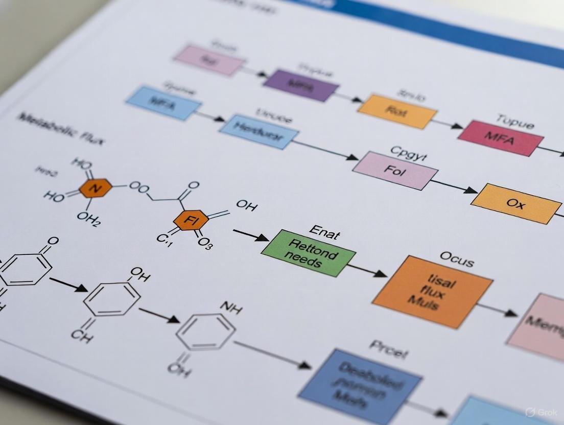
The diagram below illustrates why an underdetermined system lacks a unique solution.
Frequently Asked Questions (FAQs) and Troubleshooting
FAQ 1: How can I determine if my metabolic network is underdetermined?
Answer: Calculate the degrees of freedom in your system using the formula [1]:
If the degrees of freedom are greater than zero, your system is underdetermined. For example, in a network with 8 fluxes and 5 independent metabolites, you have 3 degrees of freedom, indicating an underdetermined system [1].
Troubleshooting Tip: Use software tools like 13CFLUX2 [3] or Metatool [1] to automatically analyze your network structure and identify the number of free fluxes.
FAQ 2: What practical approaches can I use to resolve underdeterminacy?
Answer: Two main strategies exist for tackling underdeterminacy [1]:
Table 1: Strategies for Handling Underdetermined Systems
| Strategy | Description | Common Methods | When to Use |
|---|---|---|---|
| Dealing with Underdeterminacy | Characterizing the range of possible solutions without eliminating ambiguity | Flux Variability Analysis (FVA), Elementary Flux Modes (EFMs), Random Sampling [1] | When seeking to understand possible flux ranges rather than single values |
| Reducing/Eliminating Underdeterminacy | Adding constraints to narrow or uniquely determine the solution | 13C-MFA, Thermodynamic constraints, Optimality principles (FBA), Most Accurate Fluxes [1] [2] | When a single, unique flux distribution is required |
FAQ 3: What are the best practices for designing tracer experiments to reduce underdeterminacy?
Answer: Effective 13C-MFA experimental design should incorporate these key practices [4] [5] [3]:
- Use Parallel Labeling Experiments: Employ multiple 13C-labeled tracers (e.g., [1-13C]glucose, [U-13C]glucose) in parallel cultures to generate complementary labeling information [5].
- Apply Robust Experimental Design (R-ED): When prior flux knowledge is limited, use sampling-based approaches to identify tracer mixtures that are informative across a wide range of possible flux values [3].
- Measure Multiple Labeling Patterns: Analyze protein-bound amino acids, glycogen-bound glucose, and RNA-bound ribose via GC-MS to obtain extensive labeling data for flux constraints [5].
- Validate Tracer Purity: Always measure the isotopic purity of tracers and actual labeling in the medium, as impurities can significantly impact flux results [4].
Troubleshooting Tip: If working with novel organisms or pathways with unknown fluxes, implement the R-ED workflow [3] to avoid the "chicken-and-egg" problem where tracer design requires prior flux knowledge.
FAQ 4: How can I validate the results from my flux analysis in underdetermined systems?
Answer: Implement these validation techniques [4] [1]:
- Goodness-of-fit Testing: Perform statistical analysis (eχ²-test) to determine how well the estimated fluxes explain the experimental labeling data [4].
- Flux Confidence Intervals: Calculate confidence intervals for all estimated fluxes to quantify their precision and identifiability [4] [5].
- Sensitivity Analysis: Use Flux Variability Analysis (FVA) to determine the minimum and maximum possible values for each flux within the solution space [1].
- Cross-Validation: If possible, compare flux results obtained using different tracers or analytical methods.
FAQ 5: What software tools are available for handling underdetermined systems?
Answer: Several specialized software packages support work with underdetermined networks:
Table 2: Software Tools for Underdetermined Metabolic Networks
| Software | Primary Function | Underdeterminacy Features | Reference |
|---|---|---|---|
| 13CFLUX2 | 13C-MFA simulation and flux estimation | Flux confidence intervals, statistical analysis | [3] |
| mfapy | Python-based 13C-MFA package | Customizable flux estimation, support for trial-and-error analysis | [6] |
| EFMtool | Elementary Flux Mode calculation | Pathway analysis for underdetermined systems | [1] |
| Metatool | EFM computation | Decomposition of flux space into minimal pathways | [1] |
Experimental Protocols
Protocol 1: Flux Variability Analysis (FVA) for CharacterUnderdetermined Systems
Purpose: To determine the range of possible values for each flux in an underdetermined metabolic network [1].
Materials:
- Stoichiometric matrix of the metabolic network
- Measured extracellular fluxes (if available)
- Flux analysis software (e.g., COBRA Toolbox, 13CFLUX2)
Procedure:
- Formulate the mass balance constraints: Nv = 0
- Add constraints for measured extracellular fluxes: Nmv = vm
- Apply physiological constraints (e.g., flux reversibility, enzyme capacity)
- For each flux vi in the network: a. Minimize vi subject to all constraints b. Maximize v_i subject to all constraints
- Record the minimum and maximum values for each flux
- Analyze fluxes with large ranges as key targets for additional experimental constraints
Protocol 2: Robust Tracer Design for Systems with Unknown Fluxes
Purpose: To design informative 13C-labeling experiments when prior flux knowledge is limited [3].
Materials:
- Metabolic network model in FluxML format
- 13CFLUX2 software suite
- Custom Python/Matlab scripts for evaluation
Procedure:
- Formulate the 13C-MFA model including atom transitions
- Sample the feasible flux space to represent possible flux distributions
- Evaluate different tracer mixtures using a design criterion that characterizes how informative each mixture is across all possible flux values
- Screen sampled experimental designs for best compromise solutions considering both information content and cost
- Select the tracer mixture that provides the best trade-off between informativeness and practical implementation
The workflow below illustrates this robust experimental design process.
Research Reagent Solutions
Table 3: Essential Materials for 13C-MFA Studies
| Reagent/Resource | Function/Purpose | Application Notes |
|---|---|---|
| 13C-labeled substrates (e.g., [1-13C]glucose, [U-13C]glucose) | Tracers for metabolic labeling experiments | Use multiple tracers in parallel; verify isotopic purity >99% [5] [3] |
| GC-MS instrumentation | Measurement of isotopic labeling in metabolites | Analyze protein-bound amino acids for labeling patterns [5] |
| FluxML model files | Standardized representation of metabolic networks | Use for consistent model specification across software tools [3] |
| Custom Python/Matlab scripts | Automated evaluation of flux identifiability | Implement statistical analysis and confidence interval calculation [3] |
| Cell culture media components | Defined medium for controlled labeling experiments | Avoid complex carbon sources that complicate labeling interpretation [4] |
Advanced Methodologies for Flux Determination
Most Accurate Fluxes (MAF) Approach
For researchers requiring a unique solution from an underdetermined system, the Most Accurate Fluxes (MAF) method provides a systematic algorithm [2]. This approach introduces a measure of flux accuracy and iteratively determines the fluxes with the highest possible accuracy given the available constraints. The MAF distribution has been shown to be similar to the mean values obtained from uniform sampling of admissible solutions, providing a mathematically justified single solution [2].
Dynamic Metabolic Flux Analysis (DMFCA)
When working with time-varying systems such as fed-batch cultures, Dynamic Metabolic Flux Analysis based on convex analysis (DMFCA) can be applied to compute the time evolution of bounded flux intervals [7]. This approach is particularly valuable for bioprocess applications where metabolic activity changes throughout the cultivation process.
Underdeterminacy is not a limitation to be overcome but rather a fundamental property of metabolic systems that must be understood and managed. By applying the troubleshooting guides, experimental protocols, and methodologies outlined in this technical support resource, researchers can design more informative experiments, interpret their flux results with appropriate caution, and make meaningful physiological inferences even in the face of mathematical ambiguity. The continued development of robust experimental design strategies and analytical frameworks will further enhance our ability to extract biological insights from underdetermined metabolic networks.
Frequently Asked Questions (FAQs)
FAQ 1: What is the fundamental role of the stoichiometric matrix in metabolic flux analysis?
The stoichiometric matrix (denoted as N) is the algebraic core of any metabolic model under a steady-state assumption. It mathematically represents the entire metabolic network, where each row corresponds to an intracellular metabolite and each column corresponds to a reaction [1]. The matrix entries describe the stoichiometric coefficients of each metabolite in every reaction. The fundamental equation N · v = 0 encapsulates the mass balance constraint, stating that for every intracellular metabolite, the sum of its production fluxes must equal the sum of its consumption fluxes, leaving no net accumulation [1] [8]. This equation forms the primary set of constraints that defines all possible, feasible flux distributions (the solution space) within the network.
FAQ 2: What are "degrees of freedom" (DOF) in the context of 13C-MFA, and why are they a problem? Degrees of freedom represent the number of independent variables (fluxes) that are not uniquely determined after accounting for all existing constraints (like mass balances and measured external fluxes) [1]. In simple terms, if your system has more unknowns (fluxes) than independent equations (constraints), it is underdetermined. This means there is not a single unique solution but an infinite number of flux distributions that satisfy all the constraints [1]. The number of DOF is calculated as the total number of unknown fluxes minus the number of independent constraints [1] [9]. Underdeterminacy is a central problem because it prevents the precise quantification of intracellular fluxes from external measurements alone.
FAQ 3: What is the minimum number of measurements required to resolve fluxes in an underdetermined system? In theory, you need a number of independent measurements at least equal to the degrees of freedom of the system. However, in practice, 13C-MFA utilizes isotopic labeling data, which provides a wealth of additional information. Each measured mass isotopomer fraction of an intracellular metabolite acts as a non-linear constraint that further narrows the solution space [10] [11]. Often, the rich information from 13C-labeling patterns is sufficient to reduce the feasible solution space to a single, unique flux map, even for networks that are highly underdetermined when only external flux measurements are considered [11].
FAQ 4: How can I check if my 13C-MFA model is well-determined and has a unique solution? A successful 13C-MFA result typically provides not only a set of estimated fluxes but also confidence intervals for each flux. A well-determined model is characterized by small, statistically justified confidence intervals [12] [11]. Furthermore, model validation techniques, such as the χ²-test of goodness-of-fit, are used to check whether the difference between the measured labeling data and the model-predicted labeling is statistically insignificant, indicating that the model and the estimated flux map are a good fit for the experimental data [12].
Troubleshooting Guides
Problem: The model solution space is too large, leading to wide confidence intervals for estimated fluxes.
This is a classic symptom of an underdetermined system where the available data is insufficient to precisely pinpoint the intracellular fluxes.
| Troubleshooting Step | Description and Action |
|---|---|
| 1. Verify External Flux Measurements | Re-check the accuracy of your measured uptake and secretion rates (e.g., glucose, lactate, ammonia). Ensure calculations account for cell growth, evaporation, and spontaneous degradation (e.g., of glutamine) [11] [13]. Inaccurate external fluxes propagate error and enlarge the solution space. |
| 2. Optimize Tracer Selection | Not all tracers are equally informative for all pathways. If fluxes in a specific pathway (e.g., pentose phosphate pathway, reductive TCA cycle) are poorly resolved, consider switching to a tracer that provides better positional labeling information for that pathway, or use parallel labeling experiments with multiple tracers [12] [11]. |
| 3. Increase Labeling Data Points | Incorporate measurements of additional metabolite isotopologues. Using tandem mass spectrometry (MS/MS) to obtain positional (fragment) labeling data can provide more powerful constraints than overall mass isotopomer distributions alone [12]. |
| 4. Apply a Parsimony Principle | If the solution space remains large, use parsimonious 13C-MFA (p13CMFA). This method selects the flux map from the feasible solution space that minimizes the total sum of absolute fluxes, a principle often consistent with cellular economy. This can be weighted by gene expression data to favor fluxes through enzymes with higher expression [14]. |
| 5. Re-evaluate Network Topology | Ensure your metabolic network model includes all relevant reactions for your biological system. An overly simplified model that omits active pathways will be unable to fit the labeling data well, leading to a large residual error and unreliable flux estimates [12]. |
Problem: The model fails the goodness-of-fit test (e.g., high χ² value).
A poor statistical fit indicates a discrepancy between the experimental measurements and the labeling patterns simulated by the model.
| Troubleshooting Step | Description and Action |
|---|---|
| 1. Check for Measurement Outliers | Carefully scrutinize your isotopic labeling data and external flux measurements for outliers or technical errors. Even a single grossly inaccurate data point can significantly degrade the model fit [12]. |
| 2. Inspect Metabolic Steady-State | 13C-MFA assumes the system is at metabolic and isotopic steady state. For INST-MFA, ensure accurate measurement of metabolite pool sizes. For SS-MFA, verify that the labeling pattern has reached equilibrium and that cell physiology (growth, uptake rates) remains constant during the experiment [10] [11]. |
| 3. Review Atom Mapping | Incorrect atom transitions in the model will generate wrong simulated labeling patterns. Double-check the carbon atom mapping for every reaction in your network, paying special attention to complex reactions in the TCA cycle and pentose phosphate pathway [11]. |
| 4. Consider Model Extension | The poor fit may suggest the activity of an alternative pathway not included in your model (e.g., glyoxylate shunt, transketolase-like reactions, futile cycles). Explore and test alternative network architectures to see if they yield a better fit to the data [12]. |
The Scientist's Toolkit: Research Reagent Solutions
Table: Essential reagents and materials for 13C-MFA experiments.
| Item | Function in 13C-MFA |
|---|---|
| 13C-Labeled Tracers (e.g., [1,2-13C]Glucose, [U-13C]Glutamine) | The core reagents that introduce a measurable pattern into metabolism. The specific labeling pattern of the tracer determines which pathways can be elucidated [11]. |
| Quenching Solution (e.g., cold aqueous methanol) | Rapidly halts all metabolic activity at the time of sampling to preserve the in vivo isotopic labeling state of metabolites [11]. |
| Mass Spectrometer (GC-MS, LC-MS) | The primary analytical instrument for quantifying the relative abundances of different mass isotopomers (M+0, M+1, M+2, etc.) in extracted metabolites [10] [11]. |
| Cell Culture Media (custom, tracer-compatible) | A defined medium without unaccounted carbon sources that could dilute the label and complicate the analysis. It serves as the vehicle for the tracer [11]. |
| Software Platforms (e.g., INCA, Metran, Iso2Flux) | User-friendly tools that implement the computational machinery of 13C-MFA, including the EMU framework, non-linear parameter fitting, and statistical analysis [11] [14]. |
| A-385358 | A-385358, MF:C32H41N5O5S2, MW:639.8 g/mol |
| Abarelix Acetate | Abarelix Acetate|GnRH Antagonist |
Workflow Visualization
The following diagram illustrates a logical workflow for diagnosing and resolving the core problem of underdeterminacy in 13C-MFA, linking the stoichiometric matrix, degrees of freedom, and solution strategies.
Diagram: A workflow for diagnosing and resolving underdeterminacy in 13C-MFA.
Experimental Protocols
Protocol: Determining External Fluxes for Exponentially Growing Cells
Accurate external flux measurements are critical constraints that directly reduce the degrees of freedom in the model.
- Cell Culture and Sampling: Culture cells in a defined medium. Record the exact culture volume (V, in mL). At two or more time points (t1, t2), take a sample and perform two actions simultaneously:
- Calculate Growth Rate (µ): Plot the natural logarithm of the cell number (ln Nx) versus time. The growth rate µ (1/h) is the slope of the resulting line. For two time points, use:
- Calculate External Fluxes (ri): For each metabolite i, calculate the external flux using the formula for exponentially growing cells:
- Apply Necessary Corrections:
- Glutamine Degradation: Correct the apparent glutamine uptake rate for spontaneous degradation (using a first-order degradation constant of ~0.003/h) to obtain the true net uptake rate [11] [13].
- Evaporation: For long-term experiments, run a control without cells to estimate evaporation rates and correct concentrations accordingly [11].
Table: Example calculation of a glucose uptake rate.
| Parameter | Value at t1 (24h) | Value at t2 (48h) | Change (Δt=24h) |
|---|---|---|---|
| Cell Number (million) | 1.0 | 3.5 | ΔNx = 2.5 |
| Glucose Concentration (mM) | 25.0 | 10.2 | ΔC = -14.8 mM |
| Culture Volume (mL) | 10 | 10 | V = 10 mL |
| Growth Rate (µ) | (ln(3.5) - ln(1.0)) / 24 = 0.052 /h |
||
| Glucose Uptake Rate (r_glc) | 1000 * (0.052 * 10 * -14.8) / 2.5 ≈ -307 nmol/10^6 cells/h |
FAQ: Understanding the Core Problem
What is underdeterminacy in metabolic flux analysis? Underdeterminacy occurs when a metabolic system has more unknown fluxes than available mass balance equations to constrain them [15] [1]. This means that infinitely many flux distributions can perfectly fit the same experimental data, posing a significant challenge for obtaining a unique, biologically correct solution [15].
Why is underdeterminacy a critical problem for biomedical researchers? A model that appears accurate for one dataset may fail dramatically under different conditions [15]. In drug development or when engineering metabolic pathways, an incorrect flux model can lead to the identification of poor therapeutic targets or inefficient bioproduction strains [12]. Relying on a single, potentially non-unique solution without assessing uncertainty can lead to incorrect biological conclusions and wasted resources.
Troubleshooting Guide: Resolving Underdeterminacy
Problem: My flux solution is not unique. How can I reduce the degrees of freedom? Solution: Systematically add biologically reasonable constraints to reduce the feasible solution space.
- Method 1: Integrate Additional Experimental Data
- 13C Tracer Experiments: Use stable-isotope tracing (e.g., with 13C-labeled glucose or glutamine) to generate intracellular labeling data. This provides additional information that drastically reduces the space of possible fluxes [11] [12].
- Measure External Rates: Precisely quantify nutrient uptake and waste secretion rates, as these provide critical boundary constraints [11] [1]. The methodology is outlined in the table below.
Table: Key External Rates for Constraining Flux Models in Proliferating Cells
| External Rate | Typical Range (nmol/10^6 cells/h) | Function as a Constraint |
|---|---|---|
| Glucose Uptake | 100 - 400 | Constrains upper bound of glycolytic and TCA cycle fluxes |
| Lactate Secretion | 200 - 700 | Indicates glycolytic and Warburg effect activity |
| Glutamine Uptake | 30 - 100 | Constrains nitrogen metabolism and anaplerotic fluxes |
- Method 2: Apply Computational and Theoretical Constraints
- Flux Balance Analysis (FBA): Assume the cell optimizes an objective (e.g., growth rate) to select a single flux distribution from the feasible set [1] [16].
- Thermodynamic Constraints: Incorporate knowledge of reaction reversibility/irreversibility to eliminate thermodynamically infeasible cycles [1].
- Stepwise Inference: Use algorithms that iteratively reduce degrees of freedom by identifying the most likely flux profiles in a statistical sense [15].
Problem: How do I know if my flux estimates are reliable? Solution: Quantify the uncertainty of your flux estimates.
- Perform Flux Variability Analysis (FVA): For each reaction, calculate the minimum and maximum possible flux it can carry while still satisfying all model constraints and optimality objectives. This reveals which fluxes are well-determined and which have large ranges of uncertainty [1].
- Use Statistical Sampling: Sample the space of all possible flux distributions to build a probability distribution for each flux. The standard deviation of this distribution provides a confidence interval for your estimate [17] [18]. The workflow below visualizes this process.
Problem: I have a candidate flux solution. How can I validate it? Solution: Use model validation and selection techniques.
- Goodness-of-fit Test: In 13C-MFA, a χ2-test is commonly used to compare the fit between the model-predicted and experimentally measured isotopic labeling patterns. A good fit suggests the model is consistent with the data [12].
- Incorporate Metabolite Pool Sizes: When using Isotopically Nonstationary MFA (INST-MFA), including measurements of intracellular metabolite concentrations (pool sizes) during the fitting process provides a more robust validation [12].
- Compare Against Independent Data: Validate your flux predictions against data not used in the model fitting, such as enzyme knockout phenotypes or gene expression data [12].
The Scientist's Toolkit: Research Reagent Solutions
Table: Essential Reagents and Software for 13C-MFA
| Item | Function / Explanation | Considerations for Use |
|---|---|---|
| [1,2-13C]Glucose | Tracer to elucidate glycolytic and pentose phosphate pathway fluxes [11]. | Different labeling positions (e.g., [1-13C], [U-13C]) probe different pathways. |
| 13C-Glutamine | Tracer to analyze TCA cycle and reductive metabolism [11]. | Correct for spontaneous degradation in culture medium for accurate uptake rates [11]. |
| Mass Spectrometry (GC-MS, LC-MS) | Measures the Mass Isotopomer Distribution (MID) of metabolites from tracer experiments [11] [12]. | Tandem MS provides higher resolution for positional labeling. |
| User-Friendly 13C-MFA Software (INCA, Metran) | Software tools that convert isotopic labeling data into flux maps using the Elementary Metabolite Unit (EMU) framework [11]. | Designed for accessibility to researchers without deep computational backgrounds. |
| Flux Sampling Software | Generates a distribution of possible fluxes to quantify uncertainty [17] [18]. | Essential for understanding the reliability of predictions in underdetermined systems. |
| Ac-DEVD-pNA | Ac-DEVD-pNA, CAS:189950-66-1, MF:C26H34N6O13, MW:638.6 g/mol | Chemical Reagent |
| Acemetacin | Acemetacin, CAS:53164-05-9, MF:C21H18ClNO6, MW:415.8 g/mol | Chemical Reagent |
Experimental Protocol: Core 13C-MFA Workflow
This protocol provides a detailed methodology for a foundational 13C-MFA experiment to estimate intracellular fluxes.
1. Experimental Design and Setup
- Cell Culture: Use exponentially growing cells to approximate metabolic steady-state [11].
- Tracer Selection: Choose a tracer that best differentiates the pathways of interest (e.g., [1,2-13C]glucose to resolve pentose phosphate pathway activity) [11].
- Labeling Experiment: Replace the standard growth medium with an identical medium containing the 13C-labeled substrate. Ensure adequate duration for the labeling to reach isotopic steady-state in central metabolites (typically 12-24 hours, but must be optimized).
2. Data Collection
- External Rates:
- Sample the culture medium at multiple time points.
- Measure concentrations of key metabolites (glucose, lactate, glutamine, etc.) using standard biochemical assays or HPLC.
- Count cell numbers to determine the growth rate (µ) using the formula:
µ = (ln(Nx,t2) - ln(Nx,t1)) / Δt[11]. - Calculate nutrient uptake and product secretion rates (ri) in nmol/10^6 cells/h using:
ri = 1000 * µ * V * ΔCi / ΔNx[11].
- Isotopic Labeling:
3. Computational Flux Analysis
- Model Construction: Define a stoichiometric model of the metabolic network, including atom transitions.
- Data Integration: Input the measured external rates and MIDs into 13C-MFA software (e.g., INCA) [11].
- Parameter Estimation: Solve the least-squares optimization problem to find the flux map that minimizes the difference between simulated and measured MIDs [11] [12].
- Uncertainty Analysis: Perform statistical sampling or profiling to compute confidence intervals for each estimated flux [17] [12].
The logical flow of this protocol is summarized in the following diagram:
Frequently Asked Questions (FAQs)
FAQ 1: My 13C-MFA solution space is too large to identify a unique flux distribution. How can I reduce it? A large solution space, or underdetermined system, is common when the number of unknown reactions exceeds the available labeling data [14]. You can reduce it by:
- Applying the Parsimony Principle: Run a secondary optimization that selects the flux distribution minimizing the total sum of absolute reaction fluxes within the 13C-MFA solution space. This is the core of the p13CMFA approach [14].
- Integrating Transcriptomic Data: Weight the flux minimization by gene expression evidence. This penalizes fluxes through reactions with low gene expression, steering the solution toward biologically relevant values [14].
- Incorporating Additional Experimental Constraints: Use measured extracellular rates (e.g., nutrient uptake, secretion rates) and growth rates to provide boundary constraints that further narrow the solution space [11].
FAQ 2: My Flux Balance Analysis (FBA) model has become infeasible after integrating measured flux values. What should I do? Infeasibility occurs when the measured fluxes violate model constraints like mass balance or reaction reversibility [19]. To resolve this:
- Identify the Core Conflict: Use linear programming (LP) or quadratic programming (QP) methods to find the minimal corrections required for your measured flux values to make the FBA problem feasible again [19].
- Check Reaction Directionality: Ensure that fixed fluxes for irreversible reactions are not set to a value that violates their thermodynamic constraints [19].
- Review Steady-State Assumptions: Verify that the measured fluxes are consistent with the steady-state mass balance assumption for internal metabolites [19].
FAQ 3: How can I validate if my metabolic model is a good fit for the experimental data? Beyond detecting gross measurement errors, you can assess model fit statistically.
- Employ a Generalized Least Squares (GLS) Framework: Formulate the MFA as a GLS problem. This allows you to use a t-test to check if each calculated flux is significantly different from zero [20].
- Simulate for Significance: Generate ideal flux profiles from your model, perturb them with estimated measurement error, and then re-calculate the fluxes. Comparing the significance of fluxes from this ideal data to your real data helps differentiate between measurement error and a poor model fit [20].
FAQ 4: What are the advantages of using Bayesian methods for 13C-MFA? Bayesian 13C-MFA offers several advantages over conventional best-fit approaches:
- Handles Model Uncertainty: It allows for multi-model inference, so you don't have to rely on a single model. Bayesian Model Averaging (BMA) weights the evidence from multiple plausible models, providing a more robust flux inference [21].
- Quantifies Uncertainty Naturally: It provides full probability distributions for the estimated fluxes, giving a complete picture of the uncertainty associated with each prediction [21].
- Tests Bidirectional Reactions: The framework makes it statistically testable whether a reaction step is bidirectional or unidirectional [21].
FAQ 5: What is the fundamental geometric representation of the flux solution space? The space of all feasible flux distributions that satisfy the mass-balance (steady-state) and thermodynamic constraints forms a convex polytope [22].
- H-representation: The polytope is defined by a set of linear inequalities (e.g., ( A^{\ddagger}v \leq b^{\ddagger} )) derived from stoichiometry and flux bounds [22].
- V-representation: The same polytope can be described by its set of vertices ( ( V^{\ddagger} ) ), which correspond to elementary flux modes. Any feasible flux distribution can be represented as a convex combination of these vertices [22].
Troubleshooting Guides
Problem: Underdetermined System in 13C-MFA Symptoms: A wide range of flux values provide a similarly good fit to your isotopic labeling data, leading to high uncertainty in flux estimates. Solution: Apply Parsimonious 13C-MFA (p13CMFA).
| Step | Action | Technical Details | ||
|---|---|---|---|---|
| 1 | Perform Standard 13C-MFA | Identify the set of all flux distributions that fit the labeling data within a defined statistical threshold [14]. | ||
| 2 | Define the Objective Function | Minimize the total weighted sum of absolute fluxes: ( \sum w_i | v_i | ). If gene expression data is available, use it to set the weights ( w_i ) [14]. |
| 3 | Run Secondary Optimization | Solve the linear programming problem to find the flux distribution within the feasible set that minimizes the objective function [14]. |
Problem: Infeasible FBA Scenario Symptoms: The FBA solver returns an "infeasible" error after constraining reactions with measured flux values. Solution: Find minimal flux corrections to restore feasibility.
| Step | Action | Technical Details | ||
|---|---|---|---|---|
| 1 | Formulate the Infeasible Problem | The problem is: find ( v ) such that ( Nv = 0 ), ( lb \leq v \leq ub ), and ( vi = fi ) for ( i \in F ), which has no solution [19]. | ||
| 2 | Set Up the Correction Model | Introduce a correction variable ( \deltai ) for each fixed flux. The new constraint becomes ( vi = fi + \deltai ) [19]. | ||
| 3 | Solve the Optimization | Minimize ( \sum | \delta_i | ) (LP) or ( \sum \delta_i^2 ) (QP) subject to the mass balance and bound constraints. The solution gives the smallest adjustments to your measurements that make the model feasible [19]. |
Problem: Lack of Model Fit in MFA Symptoms: Calculated fluxes are not statistically significant, or confidence intervals are unreasonably large. Solution: Implement a statistical validation protocol.
| Step | Action | Technical Details |
|---|---|---|
| 1 | Formulate as Regression | Frame the MFA as ( -So vo = Sc vc + \varepsilon ), where ( \varepsilon ) is the residual [20]. |
| 2 | Estimate Covariance | Estimate the variance-covariance matrix ( \text{Cov}(\varepsilon) ) from measurement uncertainties [20]. |
| 3 | Compute t-statistics | For each calculated flux ( v{c,i} ), compute its t-statistic: ( ti = \frac{\hat{v}{c,i}}{\text{SE}(\hat{v}{c,i})} ), where SE is the standard error [20]. |
| 4 | Interpret Results | Fluxes with a t-statistic below the critical value (e.g., < 2) are not statistically significant and may indicate a problem with the model's structure for those pathways [20]. |
The following workflow diagram illustrates the process for validating flux models using the t-test approach:
Workflow for Flux Model Validation
Comparative Analysis of MFA Methods
The table below summarizes key quantitative and methodological aspects of different flux analysis approaches to guide method selection.
| Method | Key Principle | Applicable Scene | Computational Complexity | Key Limitation |
|---|---|---|---|---|
| Qualitative Fluxomics (Isotope Tracing) [10] | Deduce pathway activity by comparing isotopic labeling patterns. | Any system. | Easy. | Provides only local and qualitative flux information. |
| 13C Metabolic Flux Ratios [10] | Calculate relative flux fractions at metabolic branch points from isotopic patterns. | Systems where fluxes and labeling are constant. | Medium. | Provides only local and relative quantitative values. |
| Stationary State 13C-MFA [10] | Optimize fluxes to fit isotopic labeling data at isotopic steady state. | Systems where fluxes, metabolites, and their labeling are constant. | Medium. | Not applicable to dynamic or transient systems. |
| Parsimonious 13C-MFA (p13CMFA) [14] | Selects the flux solution with the minimal sum of absolute fluxes from the 13C-MFA feasible set. | Underdetermined systems where 13C-MFA yields a wide solution space. | Medium. | Relies on the biological assumption of flux parsimony. |
| Bayesian 13C-MFA [21] | Uses Bayesian inference to compute posterior probability distributions of fluxes. | Any 13C-MFA scenario, especially when model uncertainty is high. | High. | Computationally intensive; requires familiarity with Bayesian statistics. |
The Scientist's Toolkit: Research Reagent Solutions
This table lists essential computational and methodological tools for advanced flux analysis.
| Item | Function | Example Use Case |
|---|---|---|
| Elementary Metabolite Unit (EMU) Framework [10] [11] | A computational framework that dramatically simplifies the simulation of isotopic labeling in large metabolic networks. | Essential for efficiently calculating the labeling patterns of metabolites in any arbitrary biochemical network model during 13C-MFA. |
| Stoichiometric Matrix (S) [16] [20] | A mathematical matrix where rows represent metabolites and columns represent reactions. The core of all constraint-based modeling. | Used to enforce mass-balance constraints (( S \cdot v = 0 )) in both FBA and MFA, defining the space of possible flux distributions. |
| Isotopically Instationary MFA (INST-MFA) [10] | A variant of 13C-MFA that analyzes transient labeling patterns before isotopic steady state is reached. | Used for measuring fluxes in systems with very fast metabolic dynamics or for probing fluxes in specific metabolite pools. |
| Coefficients of Importance (CoIs) [23] | Weights that quantify each reaction's contribution to a cellular objective function in FBA. | In the TIObjFind framework, CoIs are used to identify the objective function that best aligns FBA predictions with experimental flux data. |
| Linear & Quadratic Programming (LP/QP) [16] [19] | Optimization techniques used to solve FBA problems and resolve infeasible scenarios. | LP is used for standard FBA (e.g., growth maximization). QP can be used to find minimal least-squares corrections for infeasible flux measurements. |
| Actarit | Actarit, CAS:18699-02-0, MF:C10H11NO3, MW:193.20 g/mol | Chemical Reagent |
| Actinonin | Actinonin, CAS:13434-13-4, MF:C19H35N3O5, MW:385.5 g/mol | Chemical Reagent |
The following diagram illustrates the Bayesian Model Averaging process, a robust approach to flux inference that accounts for model uncertainty.
Bayesian Model Averaging for Flux Inference
Computational and Experimental Strategies to Constrain Flux Solutions
# Frequently Asked Questions (FAQs)
1. What does it mean for a metabolic system to be "underdetermined," and how do extracellular measurements help? An underdetermined system is one where the number of unknown intracellular fluxes exceeds the number of available mass balance equations, leading to a range of possible flux solutions rather than a single, unique answer [1]. Integrating extracellular rate measurements (e.g., substrate uptake or product excretion rates) adds crucial equality constraints to the stoichiometric model. This significantly reduces the space of feasible flux solutions, bringing the analysis closer to a unique determination of the intracellular flux map [1].
2. Which extracellular rates are most critical to measure for constraining central carbon metabolism? The most critical measurements are the uptake rates of carbon sources (e.g., glucose, glutamine) and the production rates of major metabolites such as lactate, ammonia, and carbon dioxide. Additionally, the specific growth rate of the cells is essential, as it defines the drain of metabolites into biomass precursors [4]. Accurate measurement of these rates provides the foundation for applying stoichiometric constraints.
3. My model is still underdetermined after adding all available extracellular measurements. What are my options? This is a common scenario. Your options include:
- Conduct Flux Variability Analysis (FVA): This method computes the minimum and maximum possible flux for each reaction within the constrained solution space, identifying which fluxes are well-determined and which remain uncertain [1].
- Apply a Parsimony Principle: Techniques like parsimonious FBA (pFBA) or parsimonious 13C MFA (p13CMFA) can be used. These methods find the flux distribution that fits the data while minimizing the total sum of absolute fluxes, which often reflects a biologically efficient state [24] [14].
- Integrate 13C Labeling Data: 13C Metabolic Flux Analysis (13C-MFA) uses isotopic tracer data to provide additional, highly specific constraints on intracellular reaction pathways, further narrowing the solution space [24] [12].
4. How can I validate that my extracellular measurements are sufficient and accurate?
- Check Carbon and Electron Balances: Perform a carbon and (if possible) electron balance check. A significant imbalance (e.g., >10%) often indicates missing measurements, inaccurate assays, or the presence of unmeasured byproducts [4].
- Goodness-of-Fit Test: In 13C-MFA, a chi-squared (χ²) goodness-of-fit test is used to evaluate whether the differences between the experimental measurements and the model's predictions are statistically acceptable [12] [4].
- Sensitivity Analysis: Analyze how sensitive your key flux predictions are to small variations in your extracellular measurements. If fluxes change dramatically, those measurements are highly influential and their accuracy is critical [25].
# Troubleshooting Guides
### Problem 1: Implausibly High or Infinite Flux Ranges in FVA
Issue: After integrating extracellular measurements and performing Flux Variability Analysis (FVA), some reactions show extremely wide or even infinite flux ranges, indicating the system is poorly constrained.
Diagnosis and Solution:
| Step | Diagnosis | Solution |
|---|---|---|
| 1. Check Constraints | The stoichiometric constraints and extracellular measurements are insufficient to bound the flux for particular network cycles or pathways. | Add all available exchange flux measurements (e.g., secretion of acetate, succinate, etc.). Review literature for known thermodynamic constraints (irreversible reactions) and apply them as flux bounds [1]. |
| 2. Identify Futile Cycles | The network may contain thermodynamically infeasible cycles (futile cycles) that can carry flux without a net change in metabolites. | Apply additional thermodynamic constraints to prevent these infeasible loops [1]. Tools like NetworkReduce can systematically detect and remove such cycles. |
| 3. Apply Parsimony | The solution space contains flux distributions with unnecessarily high total flux. | Implement a parsimonious constraint. First, find the solution that fits your data with the minimum sum of absolute fluxes (pFBA). Then, perform FVA within a small tolerance of this optimal parsimonious solution [24] [1]. |
### Problem 2: Poor Goodness-of-Fit in 13C-MFA
Issue: The metabolic model, after integration with extracellular fluxes, fails the χ² goodness-of-fit test when fitting 13C-labeling data, indicating a mismatch between the model and experimental measurements [12].
Diagnosis and Solution:
| Step | Diagnosis | Solution |
|---|---|---|
| 1. Verify Measurement Accuracy | Inaccurate extracellular flux or 13C-labeling data can cause a poor fit. | Re-check the standard deviations of all measurements. Ensure raw mass isotopomer distributions are properly corrected for natural isotope abundances [4]. |
| 2. Inspect Model Completeness | The stoichiometric model may be missing key reactions or contain incorrect atom transitions. | Verify the atom mapping for all reactions, especially less common ones. Check if an alternative nutrient (e.g., glutamine) is contributing significantly to biomass and should be included as a labeled input [4]. |
| 3. Evaluate Model Overfitting | The model might be too complex for the available dataset, or a simpler model might be more appropriate. | Use statistical model selection techniques. Compare the fit of different model variants (e.g., with/without a specific pathway) using criteria like the Akaike Information Criterion (AIC) or Bayesian Model Averaging [12] [21]. |
### Problem 3: Failure to Achieve a Carbon Balance
Issue: The measured carbon inputs (from substrates) do not match the measured carbon outputs (in products, biomass, and COâ‚‚), suggesting missing data.
Diagnosis and Solution:
| Step | Diagnosis | Solution |
|---|---|---|
| 1. Identify Major Missing Outputs | Common byproducts like acetate, ethanol, or secreted amino acids are often not measured. | Review the metabolic capabilities of your organism. Implement assays for common fermentation products or use extracellular metabolomics to profile the medium [4]. |
| 2. Account for Biomass | The biomass composition and its associated carbon drain may be inaccurately defined. | Use a detailed, experimentally determined biomass equation specific to your organism and growth conditions. Ensure the growth rate measurement is accurate [4]. |
| 3. Check for Evasive Products | Gaseous products other than COâ‚‚ (e.g., Hâ‚‚) or volatile compounds (e.g., ketones) may be unaccounted for. | Consult literature on the organism's metabolism. If suspected, implement headspace analysis or specific sensors for these volatile compounds. |
# Experimental Protocols
### Protocol 1: Quantifying Key Extracellular Metabolites
Objective: To accurately measure the uptake and secretion rates of major metabolites to constrain the stoichiometric model.
Materials:
- Cell culture supernatant from timed samples.
- HPLC system with UV/RI detector or a similar analytical platform (e.g., GC-MS).
- Standards for metabolites (e.g., glucose, lactate, glutamine, ammonia, amino acids).
Procedure:
- Sample Collection: Collect culture supernatant at multiple time points during steady-state growth (e.g., in chemostat or mid-exponential batch culture). Immediately filter (0.2 µm) and freeze at -80°C.
- Instrument Calibration: Prepare a standard curve for each metabolite of interest using known concentrations.
- Analysis: Run samples and standards on the HPLC/GC-MS. Quantify metabolite concentrations by comparing peak areas to the standard curve.
- Rate Calculation: Plot concentration against time. The specific consumption/production rate (mmol/gDW/h) is calculated from the slope of the linear regression, the culture volume, and the cell dry weight (gDW) [4].
### Protocol 2: Implementing Parsimonious Flux Analysis
Objective: To find the most efficient flux distribution that fits the extracellular measurements by minimizing the total flux.
Methodology: This is a two-step optimization process [24] [14]:
- Primary Optimization (Standard FBA): Solve the FBA problem to find the optimal objective (e.g., growth rate) given the stoichiometric and extracellular flux constraints.
- Mathematical Formulation:
Maximize: c^T * vsubject toS * v = 0andlb ≤ v ≤ ub, wherevis the flux vector,Sis the stoichiometric matrix, andcis the objective vector.
- Mathematical Formulation:
- Secondary Optimization (Flux Minimization): Fix the primary objective to its optimal value and solve a second optimization problem that minimizes the sum of absolute fluxes.
- Mathematical Formulation:
Minimize: Σ |v_i|subject toS * v = 0,lb ≤ v ≤ ub, andc^T * v = Z_opt, whereZ_optis the optimal objective from step 1 [24].
- Mathematical Formulation:
Software Tools: This protocol can be implemented using COBRA Toolbox in MATLAB or with Python packages like COBRApy.
# Workflow Visualization
### Logical Workflow for Constraining Fluxes
# Research Reagent Solutions
The following materials are essential for successfully implementing this strategy.
| Reagent / Material | Function in the Strategy |
|---|---|
| 13C-Labeled Substrates (e.g., [1-13C]glucose) | Serves as isotopic tracers in 13C-MFA; the pattern of label propagation provides extra constraints on internal pathway fluxes [26] [25]. |
| Stoichiometric Model (in SBML/FluxML format) | A computational representation of the metabolic network, defining the S matrix for mass balance constraints [26] [4]. |
| COBRA Toolbox / 13CFLUX2 | Software suites used to set up constraints, perform FVA, FBA, and 13C-MFA simulations, and conduct statistical analysis [24] [25]. |
| HPLC / GC-MS Platform | Essential analytical equipment for accurately quantifying the concentrations of extracellular metabolites in the culture medium to calculate uptake/secretion rates [4]. |
Frequently Asked Questions (FAQs)
FAQ 1: What is the primary cause of underdetermined flux distributions in 13C-MFA? Underdetermined flux distributions occur when the available experimental data and constraints are insufficient to define a unique solution for all intracellular metabolic fluxes. This is common because the system of stoichiometric equations is often larger than the number of measured fluxes and labeling data points. The solution space includes a range of feasible flux values rather than a single, unique set [1].
FAQ 2: How can I reduce underdeterminacy in my 13C-MFA study? Underdeterminacy can be reduced by:
- Increasing measurement constraints: Using parallel labeling experiments with multiple tracers provides more labeling data to constrain the system [14] [1].
- Integrating other data types: Incorporating additional data, such as transcriptomics data, can help constrain the solution space [14].
- Applying advanced statistical methods: Techniques like Bayesian Model Averaging or parsimonious 13C-MFA can help select a biologically relevant solution from the set of possible fluxes [21] [14].
FAQ 3: Why is correcting for natural abundance critical, and what are the best methods? Natural abundance of heavy isotopes (e.g., 1.1% for 13C) contributes to the measured mass isotopomer distribution. If not accurately corrected, this leads to significant errors in the calculated isotopic enrichment and subsequent flux estimates [27]. The "skewed" correction method or using modern software tools like ElemCor, which accounts for high-resolution mass spectrometry data, is recommended over outdated "classical" methods [27] [28].
FAQ 4: What is the difference between metabolic steady state and isotopic steady state?
- Metabolic steady state: Intracellular metabolite levels and metabolic fluxes are constant over time [29].
- Isotopic steady state: The 13C enrichment in metabolites no longer changes over time [29]. For straightforward interpretation of labeling data, the biological system should be at a metabolic pseudo-steady state, and the tracer experiment must be long enough for the relevant metabolites to reach isotopic steady state [29].
Troubleshooting Guides
Problem 1: Non-Unique or Physiologically Implausible Flux Solutions
Symptoms: The flux estimation software returns a wide range of possible values for many fluxes, or the best-fit solution suggests flux through a pathway that is not supported by other biological evidence.
| Troubleshooting Step | Description and Action |
|---|---|
| Verify Data Quality | Ensure the accuracy of measured external rates (e.g., nutrient uptake, by-product secretion) and labeling patterns. Inaccurate data is a major source of ill-constrained solutions [13] [11]. |
| Use Multiple Tracers | Employ a set of complementary tracers (e.g., [1,2-13C]glucose, [U-13C]glutamine). Different tracers illuminate different pathways, collectively providing more comprehensive constraints [14] [1]. |
| Apply Flux Minimization | Use parsimonious 13C-MFA (p13CMFA). This approach selects the flux solution that minimizes the total sum of fluxes from the set of solutions that fit the labeling data equally well, often yielding more physiologically relevant results [14]. |
| Integrate Omics Data | Weigh the flux minimization by gene expression data. This penalizes fluxes through enzymes with low gene expression, further steering the solution toward biological relevance [14]. |
| Adopt Bayesian Methods | Use Bayesian 13C-MFA. This framework does not rely on a single model but performs multi-model inference, providing a robust flux estimation that accounts for model uncertainty [21]. |
The following diagram illustrates the logical workflow for tackling this problem.
Problem 2: Inaccurate Correction for Natural Abundance
Symptoms: Corrected mass isotopomer distributions (MIDs) do not match expected patterns, leading to poor model fits and unreliable flux estimates, even when using high-resolution mass spectrometry data.
| Troubleshooting Step | Description and Action |
|---|---|
| Identify Correction Method | Review the correction method used. Avoid the outdated "classical" method. Use the correct "skewed" method or matrix-based approaches [27]. |
| Account for Derivatization | If using GC-MS, ensure the correction algorithm accounts for all atoms introduced during the chemical derivatization of metabolites [29]. |
| Use High-Resolution Tools | For high-resolution LC-MS data, use specialized software like ElemCor. It uses Mass Difference Theory (MDT) or Unlabeled Sample (ULS) data to perform resolution-dependent corrections, significantly improving accuracy [28]. |
| Validate with Unlabeled Samples | Run unlabeled control samples. The corrected MIDs for these samples should show negligible enrichment (e.g., M+0 ~100%). This serves as a quality control for the correction process [28]. |
Problem 3: Failure to Reach Isotopic Steady State
Symptoms: Labeling patterns for key metabolites (especially amino acids) continue to change over the duration of the tracer experiment, making data interpretation difficult.
| Troubleshooting Step | Description and Action |
|---|---|
| Confirm Metabolic Steady State | Ensure cells are in a metabolic pseudo-steady state (e.g., exponential growth phase with non-limiting nutrients) before and during the tracer experiment [29] [11]. |
| Optimize Experiment Duration | Perform a time-course experiment to track labeling in key metabolites (e.g., TCA cycle intermediates, amino acids). The experiment duration should be based on the time required for the slowest-metabolizing pool of interest to reach isotopic steady state [29]. |
| Check Media Composition | Be aware that amino acids present in the culture media can rapidly exchange with intracellular pools, preventing the intracellular pool from ever reaching full isotopic steady state. For these metabolites, quantitative, formal modeling approaches are required instead of simple intuitive interpretation [29]. |
Research Reagent Solutions
Essential materials and computational tools for conducting robust 13C tracer experiments.
| Item | Function in Experiment |
|---|---|
| 13C-Labeled Tracers | Substrates with specific carbon atoms replaced with 13C (e.g., [1,2-13C]glucose, [U-13C]glutamine) to trace metabolic pathways [13] [29]. |
| Mass Spectrometry Instrumentation | Analytical equipment (e.g., GC-MS, LC-MS) to measure the mass isotopomer distribution (MID) of metabolites, providing the primary data for flux calculation [13] [27]. |
| Natural Abundance Correction Software | Tools like ElemCor to accurately remove the spectral contribution from natural isotopes, which is critical for correct MID determination [28]. |
| 13C-MFA Software Platforms | Computational tools such as INCA, Metran, and Iso2Flux that integrate external rates and labeling data to estimate intracellular metabolic fluxes [13] [14] [28]. |
Experimental Workflow for Robust Flux Determination
The following diagram outlines a comprehensive workflow, from experimental design to flux calculation, incorporating strategies to handle underdeterminacy.
The Challenge of Underdetermined Flux Distributions
A fundamental challenge in 13C Metabolic Flux Analysis (13C-MFA) is system underdeterminacy, where the algebraic system formed by steady-state mass balance equations does not define a unique solution for intracellular flux distributions. Instead, it defines a set of solutions belonging to a convex polytope [1]. This underdeterminacy arises because metabolic networks typically contain more reactions than metabolites, creating degrees of freedom that cannot be resolved with single tracer experiments alone [1] [19].
What are Parallel Labeling Experiments?
Parallel labeling experiments represent an advanced 13C-MFA approach where multiple labeling experiments are conducted under identical biological conditions but with different isotopic tracers [30]. This methodology, termed COMPLETE-MFA (Complementary Parallel Labeling Experiments Technique for Metabolic Flux Analysis), has emerged as a powerful strategy to overcome underdeterminacy by providing complementary information that collectively constrains the flux solution space [31].
Table: Comparison of Single Tracer vs. Parallel Labeling Approaches
| Aspect | Single Tracer Experiment | Parallel Labeling Experiments |
|---|---|---|
| Flux Precision | Limited, especially for exchange fluxes | Significantly improved for overall network |
| Flux Observability | Partial resolution of independent fluxes | More independent fluxes resolved |
| Tracer Coverage | Optimal for specific pathway sections | Comprehensive network coverage |
| Experimental Complexity | Lower | Higher, but with greatly enhanced information |
| Computational Requirements | Standard | Increased, but manageable with modern tools |
Key Methodologies and Experimental Design
COMPLETE-MFA Workflow
The following diagram illustrates the complete workflow for implementing parallel labeling experiments:
Tracer Selection Strategies
Selecting optimal tracers is crucial for successful parallel labeling experiments. Research demonstrates that no single optimal tracer exists for resolving all fluxes in a metabolic network [31] [32]. Tracers that produce well-resolved fluxes in upper metabolism (glycolysis, PPP) often show poor performance for lower metabolism (TCA cycle), and vice versa [31].
Table: Performance of Selected Glucose Tracers in Different Metabolic Regions
| Tracer | Upper Metabolism Performance | Lower Metabolism Performance | Key Characteristics |
|---|---|---|---|
| [1,2-13C]Glucose | High | Moderate | Optimal for parallel experiments, doubly labeled [32] |
| [1,6-13C]Glucose | High | High | Best single tracer, 20x improvement over reference [32] |
| 75% [1-13C]Glucose + 25% [U-13C]Glucose | Best for upper metabolism | Poor | Optimal mixture for glycolysis and PPP [31] |
| [4,5,6-13C]Glucose | Poor | Best for lower metabolism | Optimal for TCA cycle and anaplerotic reactions [31] |
| [5-13C]Glucose | Poor | Best for lower metabolism | Optimal for TCA cycle resolution [31] |
Precision and Synergy Scoring
A key advancement in parallel labeling experimental design is the development of quantitative scoring systems [32]:
Precision Score Formula:
Where:
- P = Overall precision score
- n = Number of fluxes of interest
- UB95,ref and LB95,ref = 95% confidence intervals for reference tracer
- UB95,exp and LB95,exp = 95% confidence intervals for experimental tracer
Synergy Score Formula:
This score quantifies the benefit of conducting tracer experiments in parallel rather than individually [32].
Research Reagent Solutions
Table: Essential Materials for Parallel Labeling Experiments
| Reagent/Category | Specifications | Function in Experimental Workflow |
|---|---|---|
| 13C-Labeled Glucose Tracers | [1,2-13C]glucose (99.8%), [1,6-13C]glucose (99.2%), [4,5,6-13C]glucose (99.9%) [31] [32] | Carbon source with specific labeling patterns to probe different metabolic pathways |
| Culture Medium | M9 minimal medium [31] | Defined growth medium ensuring tracer is sole carbon source |
| Mass Spectrometry Derivatization Agents | TBDMS or BSTFA [33] | Rendering molecules volatile for GC-MS analysis |
| Strain | Escherichia coli K-12 MG1655 [31] | Model organism with well-characterized metabolism |
| Analytical Instruments | GC-MS, LC-MS systems [33] | Measuring isotopic labeling patterns in metabolites |
| Software Tools | 13CFLUX2, Metran, INCA, OpenFLUX2 [33] | Computational flux analysis and data integration |
Troubleshooting Guide: Frequently Asked Questions
FAQ 1: How many parallel experiments are typically needed for sufficient flux resolution?
Answer: The number of parallel experiments depends on network complexity and desired flux precision. Most studies use 2-4 parallel experiments, though comprehensive studies have successfully integrated up to 14 parallel labeling experiments [31]. The key is selecting truly complementary tracers rather than maximizing quantity. Begin with 2-3 optimally chosen tracers targeting different network regions, then assess if additional experiments are needed for specific flux uncertainties.
FAQ 2: How do we address biological variability between parallel cultures?
Answer: Implement strict standardization protocols:
- Use the same seed culture for all parallel experiments [30]
- Maintain identical growth conditions (temperature, pH, aeration)
- Control for equivalent growth phases at sampling time
- For microbial systems, use mini-bioreactors with controlled aeration (e.g., 5 mL/min air flow) [31]
- Monitor growth parameters (OD600) and convert to consistent cell dry weight measurements
FAQ 3: What computational approaches best integrate data from parallel experiments?
Answer: Employ integrated data analysis where labeling data from all experiments are simultaneously fitted to a single metabolic model [31] [30]. This approach:
- Improves both flux precision and observability
- Resolves more independent fluxes with smaller confidence intervals
- Particularly enhances estimation of exchange fluxes, which are notoriously difficult with single tracers
- Uses nonlinear optimization to minimize differences between simulated and measured isotopologue distributions across all experiments
FAQ 4: How do we validate that our parallel labeling strategy has successfully resolved underdeterminacy?
Answer: Implement these validation strategies:
- Calculate nonlinear confidence intervals for all estimated fluxes [32]
- Perform statistical tests (chi-square test) for model goodness-of-fit
- Use precision scores to quantify improvement over single tracer approaches [32]
- Check if previously unobservable fluxes now have finite confidence intervals
- Validate with theoretical simulations before experimental implementation
FAQ 5: What are common pitfalls in tracer selection for parallel experiments?
Answer: Avoid these common mistakes:
- Selecting tracers with redundant information content (poor synergy)
- Neglecting cost-benefit analysis - some moderately performing tracers offer better value
- Over-optimizing for specific fluxes at the expense of global resolution
- Ignoring commercial availability and purity of proposed tracers
- Failing to consider the specific biological system and its dominant metabolic pathways
Advanced Applications and Future Directions
Bayesian Approaches for Flux Inference
Recent advancements include Bayesian 13C-MFA, which provides several advantages for parallel labeling studies:
- Unifies data and model selection uncertainty in a single framework
- Enables multi-model flux inference for robust conclusions
- Enables statistical testing of bidirectional reaction steps [21]
- Bayesian Model Averaging helps overcome model selection uncertainty [21]
Parsimonious 13C-MFA (p13CMFA)
The p13CMFA approach addresses situations where 13C measurements alone cannot fully constrain the flux solution space:
- Runs secondary optimization in the 13C-MFA solution space
- Identifies solutions that minimize total reaction flux while maintaining fit to experimental data
- Can be weighted by gene expression data for biological relevance [24]
- Particularly valuable for large metabolic networks or limited measurement sets
Integration with Other Constraints
The flux resolution from parallel labeling can be further enhanced by incorporating:
- Thermodynamic constraints to eliminate infeasible flux directions [1]
- Enzyme capacity constraints based on proteomic data
- Compartmentalization information in eukaryotic systems
- Gas exchange measurements for additional flux constraints
Conceptual Framework: How Parallel Labeling Resolves Underdeterminacy
The following diagram illustrates how information from complementary tracers constrains the flux solution space:
Frequently Asked Questions (FAQs)
Q1: What is a biological objective function in FBA, and why is it important? In FBA, a biological objective function is a linear combination of metabolic reactions that the model is programmed to maximize or minimize. It represents a hypothesized cellular goal, such as maximizing growth rate or ATP production [34]. Selecting an appropriate objective is crucial because it determines which single flux distribution is predicted from the vast space of possible solutions that are all consistent with the network stoichiometry [34] [35]. An inaccurate objective function will lead to predictions that do not match experimental data.
Q2: My FBA-predicted growth rate does not match the experimentally measured value. How can I troubleshoot this? Discrepancies between predicted and experimental growth rates often point to issues with model constraints or the objective function itself. Follow these troubleshooting steps:
- Verify Biomass Composition: Ensure the biomass reaction in your model accurately reflects the organism's known macromolecular composition (e.g., proteins, lipids, nucleic acids) under your specific experimental conditions.
- Check Exchange Flux Boundaries: Confirm that the upper and lower bounds for substrate uptake (e.g., glucose, oxygen) and product secretion in the model match the measured uptake and secretion rates from your experiment [34].
- Inspect the Objective Function: Ensure the model is correctly set to maximize the flux through the biomass reaction. Use tools like MEMOTE for quality control to check that biomass precursors can be synthesized from the provided substrates [36].
- Evaluate Network Completeness: The model may be missing reactions critical for growth under your conditions. Use gap-filling algorithms to identify and add essential missing reactions [34].
Q3: What advanced methods can help identify the correct objective function for my system? For systems where the true biological objective is unknown, data-driven frameworks can be used. Methods like ObjFind and TIObjFind use experimental flux data (e.g., from 13C-MFA) to calculate "Coefficients of Importance" for different reactions [35]. These coefficients form a weighted objective function that, when maximized, yields flux predictions that best align with the experimental data, thereby inferring the organism's metabolic objectives [35].
Q4: How can I visualize an FBA-calculated pathway to check its biological feasibility? Manually interpreting a list of reactions from an FBA result is challenging. Use dedicated visualization tools that automatically generate pathway maps from the flux distribution. Tools like CAVE (a cloud-based platform) can take your model and FBA solution to create an interactive graph of the pathway, helping you quickly examine mass flow and identify unusual routes or errors [37]. Escher is another tool that allows visualization on pre-drawn metabolic maps [37].
Q5: How does FBA complement 13C-MFA in analyzing underdetermined flux distributions? 13C-MFA and FBA have a synergistic relationship when dealing with underdetermined systems.
- 13C-MFA uses experimental isotopic labeling data to estimate a single, statistically justified flux map, effectively reducing the space of possible solutions [36] [38].
- FBA can use the fluxes determined by 13C-MFA (e.g., substrate uptake or growth rates) as constraints to define its solution space. Conversely, the flux map from 13C-MFA can be used to validate the predictions of an FBA model or to infer its objective function, creating a cycle of model improvement and hypothesis testing [36] [35].
Troubleshooting Guides
Problem: FBA Predicts Non-Viable Growth After Gene Knockout, but the Mutant Grows in the Lab
Potential Causes and Solutions:
- Metabolic Flexibility: The model may not account for alternative pathways or isozymes that can compensate for the knocked-out gene.
- Solution: Perform Flux Variability Analysis (FVA) to assess the range of possible fluxes for each reaction. The existence of feasible alternate pathways in the FVA results suggests redundancy [34].
- Regulatory Misrepresentation: Standard FBA does not include regulatory rules that might activate compensatory pathways in the real organism.
- Solution: Use methods like Regulatory FBA (rFBA) if regulatory information is available, or manually relax constraints on putative bypass reactions [35].
- Incorrect Gene-Protein-Reaction (GPR) Association: The association between the knocked-out gene and its catalyzed reaction(s) in the model may be incomplete or incorrect.
- Solution: Manually curate the GPR rules for the affected reaction to ensure they accurately represent gene essentiality [39].
Problem: FBA Predicts Unrealistic ATP or Reductant (NADPH) Yields
Potential Causes and Solutions:
- Unconstrained Energy Metabolism: The model may be generating ATP without any thermodynamic or kinetic penalties.
- Solution: Apply additional constraints, such as setting a non-growth-associated ATP maintenance (NGAM) demand or using a method like parsimonious FBA (pFBA) to find the flux distribution that minimizes total enzyme usage [37].
- Infinite Sinks for Currency Metabolites: The model might lack necessary demand or exchange reactions for cofactors, allowing them to be created or consumed without cost.
- Solution: Review the model's boundaries for metabolites like ATP, NADH, and NADPH. Tools like CAVE allow for easy checking and modification of these reaction bounds [37].
Experimental Protocols for Key Workflows
Protocol: Validating an FBA Model with Experimental Data
Objective: To assess the predictive accuracy of a genome-scale metabolic model by comparing its growth predictions to experimental data across multiple conditions.
Materials:
- Software: A computational environment with an FBA solver (e.g., COBRA Toolbox [34] or cobrapy [36]).
- Model: A genome-scale metabolic model in SBML format.
- Data: Experimentally measured growth rates and substrate uptake rates for the organism grown on different carbon sources.
Methodology:
- Define the Biological Objective: Set the model's objective function to maximize the flux through the biomass reaction [34].
- Constrain the Model: For each growth condition (e.g., glucose, glycerol):
- Run Simulation: Perform FBA for each condition to predict the optimal growth rate.
- Validate: Compare the predicted growth rates against the experimentally measured rates. A strong positive correlation indicates a well-constrained and accurate model [36].
Protocol: Inferring an Objective Function Using Experimental Fluxes
Objective: To determine the set of metabolic reactions a cell prioritizes by reconciling FBA predictions with 13C-MFA flux data.
Materials:
- Software: Implementation of the TIObjFind framework or similar [35].
- Model: A metabolic network model.
- Data: Experimentally determined internal flux map (
v_exp) from 13C-MFA.
Methodology:
- Formulate the Optimization Problem: The framework solves for a vector of Coefficients of Importance (
c) that defines a weighted objective function (Z = c^T * v). - Align Prediction with Data: The optimization finds the
cthat, when used in FBA, produces a flux distribution (v) that is as close as possible to the experimental data (v_exp) [35]. - Interpret Results: The resulting Coefficients of Importance (
c) quantify the contribution of each reaction to the inferred cellular objective. Reactions with high coefficients are those the metabolism is optimized for under the measured conditions [35].
Quantitative Data Tables
Table 1: Common Biological Objective Functions in FBA
| Objective Function | Mathematical Formulation | Biological Rationale | Typical Use Case | ||
|---|---|---|---|---|---|
| Maximize Biomass | Z = v_biomass |
Simulates natural selection for maximum growth rate. | Predicting wild-type growth phenotypes and nutrient requirements [34]. | ||
| Maximize ATP Yield | Z = v_ATPM |
Assumes energy production is a key driver. | Studying energy metabolism under stress [34]. | ||
| Minimize Total Flux | `Z = ∑ | v_i | ` (or pFBA) | Mimics evolutionary pressure to minimize enzyme investment. | Finding the most efficient pathways; often used with other constraints [37]. |
| Maximize Product Yield | Z = v_product |
A user-defined objective for metabolic engineering. | Optimizing microbial strains for chemical production [35]. |
Table 2: Summary of FBA Software and Tools
| Tool Name | Key Features | User Skill Level | Access/Reference |
|---|---|---|---|
| COBRA Toolbox | Comprehensive suite for constraint-based analysis in MATLAB. | Advanced | [34] |
| cobrapy | Python version of COBRA tools; good for scripting. | Intermediate | [36] |
| CAVE | Cloud-based; no coding required; integrated calculation & visualization. | Beginner/Intermediate | [37] |
| Escher | Web-based tool for visualizing FBA results on pathway maps. | Beginner | [37] |
Pathway and Workflow Visualizations
Research Reagent Solutions
Table 3: Essential Computational Tools for FBA
| Item | Function/Benefit | Example/Note |
|---|---|---|
| COBRA Toolbox | A MATLAB suite providing the core algorithms for FBA and other constraint-based methods. | Essential for implementing advanced methods like OptKnock for metabolic engineering [34]. |
| cobrapy | A Python package that provides similar functionality to the COBRA Toolbox. | Enables FBA integration into larger Python-based data analysis and bioinformatics workflows [36]. |
| SBML Format | Systems Biology Markup Language; a standard format for exchanging and storing metabolic models. | Allows models to be used across different software tools and shared with the community [34] [39]. |
| BiGG Models Database | A repository of high-quality, curated genome-scale metabolic models. | Provides reliable, ready-to-use models for many organisms, facilitating reproducible research [37]. |
| 13C-MFA Flux Data | Experimentally determined internal flux maps. | Serves as the ground-truth data for validating FBA models or inferring objective functions [36] [35]. |
Frequently Asked Questions (FAQs)
1. What is the primary benefit of adding thermodynamic constraints to 13C-MFA? Integrating thermodynamic constraints eliminates thermodynamically infeasible flux solutions by relating reaction directions and fluxes to Gibbs free energy values. This significantly reduces the solution space in underdetermined systems, leading to more accurate and physiologically relevant flux estimates [40].
2. My flux solution is thermodynamically infeasible. What is the first parameter I should check? You should first verify the physicochemical parameters used in the calculations, particularly the ionic strength (I) and temperature (t). Many tools use default values (e.g., I=0.25 M, t=25°C) that may not match your experimental conditions. Using incorrect parameters, especially with adjustment equations that are only valid for I < 0.1 M, can lead to incorrect estimations of Gibbs free energy and thus infeasible fluxes [40].
3. How can I use enzyme capacity as a constraint? Enzyme capacity can be incorporated via the parsimonious 13C MFA (p13CMFA) approach. This method runs a secondary optimization that minimizes the total reaction flux in the network. This flux minimization can be weighted by gene expression data, ensuring that the selected flux solution favors pathways with higher enzymatic evidence and is more biologically relevant [14].
4. What is the key difference between TFA and traditional FBA? While both are constraint-based modeling approaches, Thermodynamics-based Flux Analysis (TFA) directly incorporates thermodynamic laws as constraints (e.g., forcing reactions to proceed in the direction of negative Gibbs free energy) and can simulate metabolite concentrations. In contrast, traditional Flux Balance Analysis (FBA) assumes reaction reversibility based on an optimality principle or other assumptions and does not inherently guarantee thermodynamic feasibility [40] [41].
5. When should I consider using Bayesian 13C-MFA? Bayesian methods are particularly advantageous when dealing with model selection uncertainty. Instead of relying on a single "best" model, Bayesian Model Averaging (BMA) allows for multi-model inference, producing flux estimates that are robust and account for the uncertainty in the network model itself. This is a game-changer for interpreting the fluxes of bidirectional reaction steps [21].
Troubleshooting Guides
Problem 1: The Flux Solution Appears Thermodynamically Infeasible
Symptoms: The estimated flux distribution suggests reactions are proceeding in the direction of a positive Gibbs free energy change, or the confidence intervals for fluxes remain excessively wide despite incorporating labeling data.
Solution:
- Validate Physicochemical Parameters: Ensure that the ionic strength (I), temperature (t), and salinity (S) used in the thermodynamic calculations match your experimental conditions. Do not rely solely on default software values [40].
- Inspect Key Reactions: Focus on the anaplerotic reactions (e.g., phosphoenolpyruvate carboxykinase, malic enzyme) and other bidirectional steps in the central carbon metabolism, as these are often poorly constrained and a common source of thermodynamic violations [40].
- Implement TFA: Use a Thermodynamics-based Flux Analysis (TFA) tool like
matTFA(or a modified version of it) to explicitly constrain the solution space with thermodynamic laws. This ensures that all output flux distributions are thermodynamically feasible [40].
Problem 2: The Flux Solution is Not Unique or is Underdetermined
Symptoms: Multiple, vastly different flux distributions provide a similarly good fit to your isotopic labeling data, making it impossible to identify the true physiological state.
Solution:
- Apply a Parsimony Constraint: Use the p13CMFA method. This approach selects the flux solution from the group of statistically equivalent fits that minimizes the total sum of absolute fluxes. This is based on the principle that the cell minimizes protein investment and metabolic burden [14].
- Integrate Omics Data: Within the p13CMFA framework, weight the flux minimization by transcriptomic or proteomic data. This gives priority to flux solutions that are more heavily supported by the measured levels of enzyme expression [14].
- Employ Bayesian Model Averaging: If the problem stems from uncertainty about the correct network model (e.g., which transport reactions or pathways are active), use a Bayesian framework. BMA provides flux estimates that are averaged over multiple plausible models, making the final inference more robust to model selection errors [21].
Problem 3: Model Selection and Validation are Challenging
Symptoms: You are unsure if your metabolic network model is correct. The traditional χ2-test may pass for several different model structures, or its result is sensitive to your estimates of measurement error.
Solution:
- Use Validation-Based Model Selection: Avoid selecting a model based solely on its fit to a single dataset. Instead, use an independent validation dataset from a different isotopic tracer experiment. The correct model should be the one that best predicts this new, unseen labeling data [42].
- Quantify Prediction Uncertainty: When using validation data, calculate the prediction uncertainty for the mass isotopomer distributions. This helps ensure the validation experiment is sufficiently different from the estimation data to be meaningful, but not so different that the predictions are uninformative [42].
- Test Model Components: Use this validation approach to test the necessity of specific reactions (e.g., pyruvate carboxylase) by comparing models with and without the reaction. The model whose predictions are consistently better across multiple validation datasets is more likely to be correct [42].
Research Reagent Solutions
The following table details key reagents, software, and data types essential for implementing thermodynamic and enzyme capacity constraints.
| Item Name | Type/Category | Primary Function in Constraint Implementation |
|---|---|---|
| Group Contribution Method (GCM) [40] | Computational Method | Estimates standard Gibbs free energy of formation (ΔfG'°) for metabolites, which is a critical input for calculating reaction Gibbs energy. |
| matTFA [40] | Software Toolbox | Performs Thermodynamics-based Flux Analysis (TFA) by integrating thermodynamic constraints into a constraint-based modeling framework, typically using Mixed-Integer Linear Programming (MILP). |
| eQuilibrator [40] | Software/Database | A web-based tool and database for calculating thermodynamic parameters of biochemical reactions, including equilibrium constants and Gibbs energies. |
| Iso2Flux (with p13CMFA) [14] | Software Tool | Performs 13C-MFA and includes the parsimonious 13C MFA (p13CMFA) module for applying flux minimization constraints, optionally weighted by transcriptomic data. |
| RNA-seq or Proteomics Data [14] | Experimental Data | Provides gene expression or protein abundance levels used to weight the flux minimization in p13CMFA, ensuring the solution is enzymatically feasible. |
| Ionic Strength & pH Data [40] | Experimental Parameter | Critical physicochemical parameters that must be accurately measured in the experimental system to correctly adjust standard Gibbs free energies and ensure thermodynamic calculations are realistic. |
| Bayesian 13C-MFA Software (e.g., MCMC-based) [21] | Software/Method | Provides a statistical framework for flux inference that naturally handles model selection uncertainty and allows for multi-model inference through techniques like Bayesian Model Averaging (BMA). |
Workflow and Protocol Diagrams
Diagram 1: Workflow for Constrained 13C-MFA
The diagram below outlines the integrated workflow for implementing thermodynamic and enzyme capacity constraints in 13C-MFA to resolve underdetermined distributions.
Diagram 2: Thermodynamic Constraint Integration Logic
This diagram details the logical decision process and key parameters for incorporating thermodynamic constraints.
In 13C Metabolic Flux Analysis (13C-MFA), researchers often face a fundamental challenge: the problem of underdetermined flux distributions [43]. This occurs when multiple, distinct flux maps can explain the experimental isotopic labeling data with nearly equal statistical goodness-of-fit [21] [44]. Traditional best-fit approaches to 13C-MFA provide a single flux solution, potentially masking this inherent uncertainty and leading to overconfident or biologically implausible conclusions [21]. This underdetermination is particularly pronounced when working with large metabolic networks or when limited experimental measurements are available [14].
Bayesian Model Averaging (BMA) addresses this core problem through a paradigm shift from single-model to multi-model inference [21]. Rather than selecting one "best" model, BMA incorporates uncertainty directly into the flux estimates by averaging over a ensemble of candidate models, weighted by their statistical evidence [21]. This approach provides a more robust statistical framework for flux inference, effectively functioning as a "tempered Ockham's razor" that balances model complexity with explanatory power [21]. For researchers and drug development professionals, this translates to more reliable flux estimates that better capture the true biological uncertainty in metabolic systems.
Core Concepts: Bayesian Model Averaging in Flux Analysis
Theoretical Foundation
Bayesian Model Averaging (BMA) for flux inference is grounded in Bayesian statistics, which treats unknown parameters as probability distributions. In the context of 13C-MFA, BMA accounts for model selection uncertainty—the reality that multiple metabolic models (e.g., with different bidirectional reactions or pathway assumptions) may be consistent with the experimental data [21]. Conventional 13C-MFA uses optimization algorithms to find the single set of fluxes that minimizes the difference between simulated and measured isotopic labeling patterns [43]. In contrast, the Bayesian approach uses Markov Chain Monte Carlo (MCMC) sampling to explore the entire posterior distribution of possible flux values, thereby quantifying the uncertainty for each flux [44].
The key advantage of BMA is its ability to perform multi-model inference. Instead of relying on inferences from a single model, BMA averages the flux distributions across all plausible models, with each model's contribution weighted by its posterior probability [21]. This results in flux estimates that are more robust and less sensitive to the selection of any particular model structure.
Comparative Framework: Traditional vs. Bayesian 13C-MFA
Table 1: Comparison of Traditional and Bayesian 13C-MFA Approaches
| Feature | Traditional 13C-MFA | Bayesian 13C-MFA with BMA |
|---|---|---|
| Statistical Basis | Frequentist; best-fit optimization [43] | Bayesian; posterior inference [21] [44] |
| Primary Output | Single flux map with confidence intervals [43] | Full probability distribution for each flux [44] |
| Model Uncertainty | Typically ignored; single model used [21] | Explicitly quantified and incorporated [21] |
| Handling of Underdetermination | Provides one solution, potentially missing alternatives [14] | Reveals all flux ranges consistent with data [44] |
| Bidirectional Reactions | Often requires pre-specified constraints [21] | Statistically testable within the framework [21] |
| Computational Demand | Lower (optimization) | Higher (MCMC sampling) [44] |
Key Software and Computational Tools
Table 2: Key Research Reagent Solutions for Bayesian Flux Analysis
| Tool Name | Type/Function | Key Features and Applications |
|---|---|---|
| BayFlux | Software library (Python) | Implements Bayesian inference for genome-scale and two-scale 13C-MFA; uses MCMC sampling to quantify flux uncertainty [44]. |
| Open-Source Code | Repository (GitHub) | The code for BayFlux is publicly available at https://github.com/JBEI/bayflux, enabling method replication and application [44]. |
| COBRApy | Software library (Python) | A dependency for BayFlux; provides core functionality for constraint-based reconstruction and analysis [44]. |
| Iso2Flux | Software for 13C-MFA | An isotopic steady-state 13C MFA software that has been extended to implement methods like parsimonious 13C MFA (p13CMFA) [14]. |
| MCMC Algorithms | Computational method | Engine of Bayesian inference (e.g., used in BayFlux); samples the probability distribution of fluxes [44]. |
Experimental Protocol: Implementing Bayesian Flux Analysis
The following diagram illustrates the core workflow for conducting a Bayesian Metabolic Flux Analysis using model averaging:
Step-by-Step Methodology
Experimental Design and Data Acquisition
- Tracer Selection: Choose appropriate 13C-labeled substrates (e.g., [1-13C]-glucose, [U-13C]-glutamine) based on the metabolic pathways of interest. Using multiple tracers in parallel experiments can provide more comprehensive information [43] [14].
- Data Collection: Grow cells in the presence of the chosen tracer until isotopic steady state is reached. Measure:
Model Space Definition
- Construct Candidate Models: Define a set of plausible metabolic network models that may explain the data. Differences between models can include the presence or absence of specific bidirectional reaction steps, alternative pathways, or different assumptions about drain fluxes to biomass or energy maintenance [21] [44].
- Stoichiometric and Atom Mapping: For each model, define the complete stoichiometric matrix and the atom transition mappings for every reaction, which are essential for simulating the isotopic labeling patterns [43].
Computational Inference via MCMC and BMA
- Bayesian Setup: Formulate the statistical model. The posterior probability of a flux vector
vgiven dataDis proportional to the likelihood of the data given the fluxes,P(D|v), multiplied by the prior probability of the fluxes,P(v)[44]. - MCMC Sampling: For each candidate model, run an MCMC algorithm (e.g., the one implemented in BayFlux) to sample from the posterior distribution of its fluxes,
P(v|D, Model)[44]. This generates a chain of flux values that represents the full uncertainty. - Model Averaging: Compute the posterior probability of each model given the data. The final, averaged flux distribution is then the sum of the posterior distributions of each model, weighted by the model's posterior probability [21]. This is the core of BMA, which provides a flux estimate that is robust to model selection uncertainty.
- Bayesian Setup: Formulate the statistical model. The posterior probability of a flux vector
Troubleshooting Guide and FAQs
Common Computational Challenges
Question: My MCMC sampling is slow or fails to converge, especially with a genome-scale model. What can I do?
- Answer: Genome-scale models are highly complex. Consider these strategies:
- Two-Scale Modeling: Use a method like the one supported by BayFlux and the "Limit Flux To Core" (lftc) library. This approach uses a detailed atom-mapping model for central carbon metabolism while representing the larger network with a simplified stoichiometric model, significantly reducing computational cost [44].
- Check Sampler Settings: Ensure you are using an appropriate number of "thinning" steps and chains. The BayFlux implementation uses the OptGP algorithm, which supports parallelization to improve performance [44] [45].
- Validate with a Core Model: First, run the analysis on a well-curated core model of central carbon metabolism to verify your pipeline before scaling up.
Question: The flux distributions for some reactions are very wide. Does this mean the method failed?
- Answer: Not necessarily. Wide flux distributions are a feature, not a bug, of the Bayesian approach. They honestly reflect that the experimental data (labeling and/or exchange fluxes) does not constrain that particular reaction well. This is a quantitative measure of underdetermination [44]. To address this:
- Gather More Informative Data: Consider designing a new tracer experiment that specifically targets the poorly constrained pathway [43] [14].
- Incorporate Additional Constraints: If available, integrate other data types, such as gene expression data, to apply additional constraints. For example, the p13CMFA method uses gene expression to weight a flux minimization principle, which can help reduce the solution space [14].
Interpretation and Biological Validation
Question: How do I know if the BMA result is biologically plausible?
- Answer: Use the following checklist for validation:
- Check Model Probabilities: BMA automatically assigns higher weights to more plausible models. Examine the posterior probabilities of the candidate models you defined [21].
- Compare to Traditional MFA: Validate your findings against a traditional 13C-MFA result for the same system, if it exists. The Bayesian flux means should be consistent, but BMA will provide a more complete picture of the uncertainty [44].
- Literature Consistency: Check if the predicted active pathways and their flux ranges are consistent with independent biological knowledge, such as known enzyme knockouts or pharmacological inhibitions.
Question: We found that using a genome-scale model with BayFlux actually produced narrower flux uncertainties than a core model. Is this expected?
- Answer: Yes, this is a non-intuitive but valid finding. While one might expect a larger model to have more uncertainty, the reverse can happen. A genome-scale model imposes additional stoichiometric and thermodynamic constraints that connect peripheral metabolism to central carbon metabolism. These extra constraints can further reduce the space of feasible flux solutions that are also consistent with the 13C labeling data, leading to narrower, more precise posterior distributions [44].
Methodological and Experimental Pitfalls
Question: What are the most critical steps in the experimental design to ensure successful BMA?
- Answer: The quality of the posterior inference is directly tied to the quality of the input data.
- Tracer Selection: The single most important factor is choosing a tracer whose labeling pattern is sensitive to the fluxes you wish to resolve [43] [14]. For example, [1-13C]-glucose is good for differentiating glycolysis and the oxidative pentose phosphate pathway.
- Accurate Exchange Fluxes: Precise measurement of extracellular uptake and secretion rates is crucial, as these strongly constrain the absolute flux values [43] [45].
- Multiple Tracers: If possible, using multiple tracers from different entry points (e.g., both 13C-glucose and 13C-glutamine) provides the most comprehensive labeling constraints for the network [14].
Question: How do I test the activity of bidirectional (reversible) reaction steps with BMA?
- Answer: This is a key strength of the Bayesian framework. Instead of pre-specifying the net directionality of a reaction, you can include bidirectional steps in your candidate models. The MCMC sampling will then estimate the posterior distribution for the net and gross fluxes of that reaction. You can then statistically test if the net flux is significantly different from zero, providing direct evidence for or against its activity in your experimental conditions [21].
Best Practices, Pitfalls, and Protocol Optimization for Robust Flux Estimation
Troubleshooting Guides and FAQs
Frequently Asked Questions
Q1: What does the "precision score" actually measure and how is it calculated? The precision score (P) is a metric that quantifies the overall improvement in flux precision for a given tracer experiment compared to a reference tracer. It is calculated as the average of individual flux precision scores (p_i) for all fluxes of interest in the network model [46].
The individual flux precision score is determined using the formula: pi = ((UB95,i - LB95,i)ref / (UB95,i - LB95,i)_exp)², where UB95,i and LB95,i represent the upper and lower bounds of the 95% confidence interval for flux i [46]. A precision score greater than 1.0 indicates the tracer experiment delivers narrower confidence intervals (better precision) than the reference experiment.
Q2: When should I consider using parallel labeling experiments instead of single tracers? Parallel labeling experiments are particularly valuable when studying complex metabolic networks where no single tracer provides sufficient information for all fluxes of interest. The decision can be guided by calculating the synergy score (S), which quantifies the additional information gained by combining multiple tracer experiments [46].
A synergy score greater than 1.0 indicates a greater-than-expected gain in flux precision, suggesting the tracers provide complementary information. Research has demonstrated that optimal tracer pairs like [1,6-13C]glucose and [1,2-13C]glucose can improve flux precision by nearly 20-fold compared to commonly used tracer mixtures [46] [32].
Q3: How does the precision and synergy scoring system help with underdetermined flux distributions? Underdetermined flux distributions occur when the experimental data lacks sufficient information to uniquely determine all fluxes in the network model. The precision scoring system directly addresses this by evaluating how different tracers reduce flux confidence intervals, thereby identifying which tracers provide the most constraint information for the network [46] [24].
The synergy scoring system further helps by identifying tracer combinations that collectively constrain a wider range of fluxes, effectively reducing the solution space for underdetermined systems through complementary information provision [46].
Q4: Are pure glucose tracers or tracer mixtures generally more effective? According to systematic evaluations, pure glucose tracers typically outperform tracer mixtures. Specifically, doubly 13C-labeled glucose tracers such as [1,6-13C]glucose, [5,6-13C]glucose, and [1,2-13C]glucose consistently produce the highest flux precision across different metabolic flux maps [46] [32]. The widely used mixture of 80% [1-13C]glucose + 20% [U-13C]glucose was significantly outperformed by optimal pure tracers and tracer pairs [46].
Troubleshooting Common Experimental Issues
Problem: Inadequate flux resolution in specific pathway segments Solution: Implement parallel labeling experiments with complementary tracers specifically targeted to the problematic pathways. For example, if TCA cycle fluxes are poorly resolved, consider adding [2,5-13C]glucose or [3,4-13C]glucose to your experimental design, as these tracers provide complementary information for these metabolic segments [32].
Problem: Inconsistent results between biological replicates Solution: Ensure consistent tracer purity across experiments by verifying isotopic purity from suppliers and measuring actual tracer labeling in the culture medium. Also confirm metabolic and isotopic steady-state has been reached by testing multiple time points [4].
Problem: Large confidence intervals for estimated fluxes Solution: This typically indicates insufficient information content in your labeling data. Consider switching to optimal doubly-labeled tracers like [1,6-13C]glucose or implementing parallel labeling with [1,6-13C]glucose and [1,2-13C]glucose, which provide substantially improved flux precision [46] [32]. Also verify you're measuring comprehensive labeling data including amino acids, glycogen-bound glucose, and RNA-bound ribose [47].
Performance Comparison of Selected Glucose Tracers
Table 1: Relative performance of single glucose tracers for 13C-MFA
| Tracer Type | Examples | Relative Performance | Key Characteristics |
|---|---|---|---|
| Doubly 13C-labeled | [1,6-13C]glucose, [5,6-13C]glucose, [1,2-13C]glucose | Highest flux precision | Consistent performance across different flux maps [46] |
| Tracer mixtures | 80% [1-13C]glucose + 20% [U-13C]glucose | Moderate | Widely used but outperformed by pure tracers [46] |
| Natural abundance | Unlabeled glucose | Reference | Baseline for comparison [46] |
Table 2: Optimal tracer combinations for parallel labeling experiments
| Tracer Combination | Synergy Score | Precision Improvement | Application Context |
|---|---|---|---|
| [1,6-13C]glucose + [1,2-13C]glucose | >1.0 | ~20x vs. reference mixture | Overall network resolution [46] [32] |
| [2,5-13C]glucose + [3,4-13C]glucose | >1.0 (expected) | Complementary information | TCA cycle and related pathways [32] |
Experimental Protocols
Protocol 1: Implementing Parallel Labeling Experiments
Objective: To determine intracellular metabolic fluxes with improved precision and accuracy using parallel labeling experiments [46] [47].
Materials:
- Isotopic tracers: [1,6-13C]glucose (99.2% purity), [1,2-13C]glucose (99.8% purity)
- M9 minimal medium
- GC-MS system for isotopic labeling measurement
- Appropriate cell culture equipment
Procedure:
- Prepare separate culture vessels with identical growth conditions except for the isotopic tracer used.
- For each parallel experiment, use 2 g/L of the respective tracer ([1,2-13C]glucose or [1,6-13C]glucose) in M9 minimal medium [46].
- Inoculate cultures with the same pre-culture at the same cell density.
- Harvest cells during mid-exponential growth phase for isotopic labeling analysis.
- Quench metabolism rapidly and extract intracellular metabolites.
- Derivatize metabolites for GC-MS analysis following established protocols [46] [4].
- Measure mass isotopomer distributions of proteinogenic amino acids, glycogen-bound glucose, and RNA-bound ribose.
- Integrate all labeling datasets from multiple parallel experiments into a single flux model.
- Estimate fluxes by minimizing the variance-weighted sum of squared residuals between measured and simulated labeling data [46].
Protocol 2: Calculating Precision and Synergy Scores
Objective: To quantitatively evaluate and compare different tracer designs using precision and synergy scoring metrics [46].
Procedure:
- Perform flux estimation: For each tracer experiment, estimate metabolic fluxes and determine 95% nonlinear confidence intervals for each flux (UB95,i and LB95,i) [46].
- Select reference experiment: Designate a commonly used tracer as reference (e.g., 80% [1-13C]glucose + 20% [U-13C]glucose).
- Calculate individual precision scores: For each flux i, compute pi = ((UB95,i - LB95,i)ref / (UB95,i - LB95,i)_exp)².
- Calculate overall precision score: Compute the average of all individual precision scores: P = (1/n) × Σp_i from i=1 to n.
- Calculate synergy scores: For parallel experiments, compute the synergy score S = (1/n) × Σsi, where si = pi,1+2 / (pi,1 + p_i,2) [46].
- Interpret results: Precision scores >1.0 indicate better performance than reference; synergy scores >1.0 indicate complementary information from parallel experiments.
Workflow Visualization
Precision and Synergy Scoring Workflow
Parallel Labeling Experiment Flow
The Scientist's Toolkit: Research Reagent Solutions
Table 3: Essential materials for precision and synergy scoring experiments
| Reagent/Resource | Function/Purpose | Specifications |
|---|---|---|
| [1,6-13C]glucose | Primary metabolic tracer for parallel experiments | 99.2% isotopic purity [46] |
| [1,2-13C]glucose | Complementary tracer for parallel experiments | 99.8% isotopic purity [46] |
| M9 minimal medium | Defined culture medium | Eliminates unlabeled carbon sources [46] |
| GC-MS system | Isotopic labeling measurement | Quantifies mass isotopomer distributions [46] [47] |
| EMU-based modeling software | Flux calculation and scoring | Enables precision and synergy score computation [46] [48] |
| Adiphenine | Adiphenine, CAS:64-95-9, MF:C20H25NO2, MW:311.4 g/mol | Chemical Reagent |
| Anticancer agent 211 | Anticancer agent 211, CAS:314022-97-4, MF:C19H21ClN2O2, MW:344.8 g/mol | Chemical Reagent |
A central challenge in 13C Metabolic Flux Analysis (13C-MFA) is solving the underdetermined inverse problem, where multiple flux maps can explain the same experimental data. This fundamental limitation arises because metabolic networks typically contain more reactions than measurable metabolites, creating a situation where the system has more unknowns than equations. Underdetermination is particularly problematic in complex mammalian systems where parallel pathways, substrate cycles, and compartmentalization create significant redundancies. When flux distributions are underdetermined, researchers cannot uniquely quantify the operational rates of metabolic reactions, limiting the biological insights that can be gained from expensive and time-consuming isotopic labeling experiments.
The core thesis of this protocol is that experimental design decisions, rather than just computational analysis techniques, provide the most powerful approach to overcoming underdetermination in 13C-MFA. By strategically designing isotopic labeling experiments, researchers can generate the specific information content needed to resolve previously unidentifiable fluxes. This guide provides a comprehensive step-by-step framework for designing informative labeling experiments, with particular emphasis on troubleshooting the common pitfalls that lead to underdetermined flux distributions.
Theoretical Foundations: How Labeling Data Constrains Flux Space
Core Principles of 13C-MFA
13C-MFA works by introducing 13C-labeled substrates to biological systems and tracing how these labels distribute through metabolic networks. As metabolites are transformed through biochemical reactions, their carbon atom arrangements create unique labeling patterns that serve as fingerprints for the active pathways. The core principle is that different flux distributions produce distinct labeling patterns in intracellular metabolites, allowing researchers to infer reaction rates from measured isotope distributions.
Two critical concepts must be understood for proper experimental design. First, metabolic steady state requires that intracellular metabolite levels and metabolic fluxes remain constant during the experiment. Second, isotopic steady state occurs when the 13C enrichment in metabolites becomes stable over time [29]. Most 13C-MFA protocols assume both conditions are met, though specialized approaches exist for non-steady state conditions. The time to reach isotopic steady state varies significantly between metabolites—glycolytic intermediates may reach steady state within minutes, while TCA cycle intermediates and amino acids may require several hours or never reach steady state due to exchange with large extracellular pools [29].
The Underdetermination Problem in Flux Analysis
Underdetermination manifests when the stoichiometric matrix of a metabolic network has more columns (reactions) than rows (metabolites), creating a solution space with infinitely many flux combinations that satisfy mass balance constraints. Isotopic labeling data provides additional constraints that can reduce this solution space, but the information content varies dramatically depending on how the labeling experiment is designed.
Table: Common Sources of Underdetermination in 13C-MFA
| Source of Underdetermination | Description | Impact on Flux Resolution |
|---|---|---|
| Parallel Pathways | Multiple routes producing the same metabolite (e.g., glycolysis vs. PPP) | High - creates symmetrical solutions |
| Reversible Reactions | Bidirectional flux through thermodynamically favorable reactions | Medium - confounds net flux determination |
| Metabolite Channeling | Direct transfer of intermediates between enzyme active sites | High - violates steady-state assumptions |
| Network Compartmentalization | Separate pools of metabolites in different organelles | High - creates apparent contradictions |
| Measurement Limitations | Insensitive measurement positions in network | Variable - depends on coverage |
The fundamental goal of experimental design is to select tracers and measurement strategies that break these symmetries and create unique, identifiable flux solutions.
Step-by-Step Experimental Design Protocol
Step 1: Define Biological Questions and Target Fluxes
Clearly articulate the specific metabolic questions to be addressed, as this determines which parts of the network require the highest flux resolution. For example:
- Oxidative Pentose Phosphate Pathway: Questions about NADPH production or nucleotide biosynthesis
- Pyruvate Carboxylase vs. Pyruvate Dehydrogenase: Questions about anaplerotic carbon entry into TCA cycle
- Glutaminolysis: Questions about nitrogen metabolism and TCA cycle fueling
Defining target fluxes upfront enables selective optimization of tracer design for the specific pathways of interest, rather than attempting to resolve all network fluxes equally.
Step 2: Select Optimal Tracers Using Rational Design Principles
Traditional trial-and-error approaches to tracer selection often fail to identify optimal tracers for resolving specific fluxes. Instead, use rational design principles based on elementary metabolite unit (EMU) analysis and sensitivity analysis [49].
Table: Optimal Tracer Selection for Target Fluxes
| Target Flux | Optimal Tracer | Alternative Tracers | Rationale |
|---|---|---|---|
| Oxidative PPP | [2,3,4,5,6-13C]Glucose | [1,2-13C]Glucose | Maximizes sensitivity of lactate M+1 mass isotopomer to oxPPP flux |
| Pyruvate Carboxylase | [3,4-13C]Glucose | [1-13C]Glutamine | Generates unique OAA labeling pattern from PC activity |
| TCA Cycle Rate | [U-13C]Glucose + [U-13C]Glutamine | [1,2-13C]Glucose | Provides complementary constraints on mitochondrial fluxes |
| Transhydrogenase | [1,2-13C]Glucose | [3-13C]Glutamine | Resolves NADPH production sources |
For the oxidative pentose phosphate pathway (oxPPP), [2,3,4,5,6-13C]glucose produces optimal resolution by maximizing the sensitivity of key mass isotopomer measurements to oxPPP flux [49]. Similarly, for pyruvate carboxylase (PC) flux, [3,4-13C]glucose generates unique oxaloacetate labeling patterns that distinguish PC activity from other anaplerotic routes [49].
The following diagram illustrates the rational tracer design workflow:
Step 3: Design Parallel Labeling Experiments
Single tracer experiments often lack sufficient information to fully resolve metabolic networks. Parallel labeling experiments, where multiple isotopic tracers are applied to separate cell cultures, dramatically increase flux resolution by providing complementary constraints [12].
A well-designed parallel labeling strategy should combine:
- A glucose tracer optimized for upper glycolysis and pentose phosphate pathway
- A glutamine tracer optimized for TCA cycle and anaplerotic fluxes
- A mixed tracer with complementary labeling patterns to break symmetries
The most powerful parallel labeling designs use optimized tracer mixtures rather than single tracers, though these require custom synthesis [49].
Step 4: Establish Metabolic and Isotopic Steady State
Before beginning isotopic measurements, verify that your system has reached both metabolic and isotopic steady state:
Metabolic Steady State Validation:
- Measure extracellular nutrient concentrations and byproduct secretion at multiple timepoints
- Confirm constant growth rate (for proliferating cells)
- Verify stable intracellular metabolite concentrations (via LC-MS)
Isotopic Steady State Validation:
- Conduct time-course measurements of labeling patterns in key metabolites
- Confirm stable mass isotopomer distributions over time
- Note that amino acids in rapid exchange with media may never reach isotopic steady state [29]
For systems where isotopic steady state cannot be achieved (e.g., primary cells with limited lifespan), consider isotopically non-stationary MFA (INST-MFA), which requires specialized experimental protocols and computational tools.
Step 5: Select Appropriate Analytical Measurements
The choice of which metabolites to measure significantly impacts flux resolution. Strategic measurement selection should prioritize:
- Positionally Informative Metabolites: Fragments that differentiate between alternative pathways
- Network Branch Points: Metabolites at convergence of multiple pathways (e.g., pyruvate, oxaloacetate)
- Secreted Metabolites: Lactate, alanine, glutamate that reflect intracellular labeling
Tandem mass spectrometry provides positional labeling information that significantly enhances flux resolution compared to conventional mass spectrometry [12]. When using GC-MS, select derivative fragments that maximize carbon atom coverage and positional information.
Troubleshooting Guide: Addressing Common Experimental Problems
FAQ: Resolving Underdetermined Flux Distributions
Q: How can I determine if my flux solution is underdetermined? A: Underdetermination manifests in several ways: (1) Wide confidence intervals on key fluxes after 13C-MFA fitting, (2) Multiple local optima with similar goodness-of-fit, (3) Model selection uncertainty where different network structures fit the data equally well [50]. Computational tools can quantify parameter identifiability through sensitivity analysis and profile likelihood approaches.
Q: What should I do if my model cannot resolve the fluxes of interest? A: First, verify that the lack of resolution is not due to model misspecification by testing alternative network architectures [50]. If the true network structure is unknown, use validation-based model selection with independent data [50]. If model structure is correct but fluxes remain underdetermined, design a follow-up experiment with tracers optimized for the target fluxes using rational design principles [49].
Q: How can I improve flux resolution without completely redesigning my experiment? A: Several approaches can enhance resolution: (1) Add complementary measurements of other metabolites, particularly at network branch points, (2) Incorporate quantitative metabolite concentration data to constrain pool sizes, (3) Integrate omics data to eliminate inactive reactions, (4) Apply flux minimization constraints (parsimonious 13C-MFA) to select the simplest solution from the feasible space [14].
FAQ: Technical Issues in Labeling Experiments
Q: How long should I incubate cells with labeled tracers? A: Incubation time depends on metabolic turnover rates of your specific system. For most mammalian cell lines, 24-48 hours is sufficient to reach isotopic steady state in central carbon metabolism. However, slower metabolic systems (e.g., primary cells, tissues) may require longer. Always conduct a time-course experiment to empirically determine the appropriate duration [13].
Q: Why do my labeling patterns show high variance between biological replicates? A: High variance typically indicates inconsistent metabolic states or incomplete isotopic steady state. Ensure consistent: (1) cell passage number, (2) seeding density, (3) nutrient availability, (4) confluency at harvest, and (5) tracer incubation duration. Also verify that your analytical methods have proper quality controls for sample processing and instrument performance.
Q: How should I correct for natural isotope abundance in my data? A: Natural isotope correction is essential for accurate flux estimation. Use established algorithms that account for all atoms in your measured ions, including those from derivatization agents if using GC-MS [29]. Most 13C-MFA software packages (e.g., mfapy, INCA, 13CFLUX) include built-in correction functions [51].
Table: Key Research Reagents and Software for 13C-MFA
| Resource Type | Specific Examples | Function/Purpose |
|---|---|---|
| Isotopic Tracers | [U-13C]Glucose, [1,2-13C]Glucose, [U-13C]Glutamine | Create distinct labeling patterns for flux elucidation |
| Analytical Standards | 13C-labeled amino acids, organic acids | Quantification correction and instrument calibration |
| Software Tools | mfapy (Python), INCA (MATLAB), 13CFLUX2 | Flux estimation from labeling data |
| Model Selection | Validation-based methods, Bayesian model averaging | Identify correct network structure [50] [21] |
| Data Correction | Natural abundance correction algorithms | Account for natural 13C, 2H, 15N, 18O isotopes [29] |
Visualization of the Experimental Design Workflow
The following comprehensive workflow diagram integrates all protocol steps and highlights critical decision points:
Designing informative isotopic labeling experiments requires moving beyond conventional tracer choices and adopting a systematic approach to experimental design. By following this step-by-step protocol—defining clear biological questions, selecting optimal tracers using rational design principles, implementing parallel labeling strategies, and rigorously validating steady state conditions—researchers can overcome the fundamental challenge of underdetermined flux distributions.
The troubleshooting guides and FAQs provided here address the most common pitfalls in 13C-MFA experiments, while the visualization workflows offer clear roadmaps for experimental planning. As the field continues to evolve, emerging approaches like Bayesian model averaging [21] and parsimonious flux analysis [14] will provide additional tools for handling uncertainty in flux estimation. By adopting these rigorous experimental design practices, researchers can maximize the information gained from each labeling experiment and generate more reliable, biologically meaningful flux maps.
FAQs on Essential Data Corrections
Q1: Why is correcting for natural isotopes necessary in 13C-MFA? Raw Mass Spectrometry (MS) data reflects the measured mass isotopomer distributions (MIDs) of metabolites. However, this raw signal contains contributions from naturally occurring isotopes (e.g., 13C, 2H, 17O, 18O) present in all carbon atoms of the metabolite. Failure to correct for this leads to inaccurate MIDs, which directly compromises the precision of the calculated metabolic fluxes. For results to be reproducible and verifiable, publications should ideally provide both the uncorrected and the natural isotope-corrected MS data [4].
Q2: How does uncorrected glutamine degradation affect flux results? Glutamine is an unstable molecule that spontaneously degrades in culture medium to pyroglutamate and ammonium. If this degradation is not accounted for, the calculated glutamine uptake rate will be overestimated, as it will reflect both the cellular consumption and the chemical breakdown. Since uptake rates provide critical boundary constraints for the flux model, this error propagates through the entire analysis, leading to a distorted view of intracellular metabolism [13] [11].
Q3: What are the minimum data standards for publishing a 13C-MFA study? To ensure reproducibility and quality, studies should provide:
- Isotopic Labeling Data: Uncorrected mass isotopomer distributions in tabular form, along with the standard deviations for measurements [4].
- External Flux Data: Measured cell growth rates, nutrient uptake rates, and product secretion rates in tabular form [4] [13].
- Metabolic Network Model: A complete model in tabular form, including atom transitions for all reactions [4].
- Validation: A report on the goodness-of-fit and confidence intervals for the estimated fluxes [4].
The table below summarizes these common data quality issues and their impacts.
| Data Quality Issue | Impact on 13C-MFA | Required Correction |
|---|---|---|
| Natural Isotope Abundance | Inaccurate Mass Isotopomer Distributions (MIDs); biased flux estimates [4]. | Apply computational algorithms to raw MS data to subtract natural abundance contributions. |
| Glutamine Degradation | Overestimation of glutamine uptake rate; incorrect boundary constraints for the model [13] [11]. | Measure degradation rate in cell-free control experiments and apply a first-order kinetic correction. |
Troubleshooting Guides
Protocol 1: Correcting for Natural Isotope Abundance
Objective: To purify the mass isotopomer distribution (MID) data, ensuring it reflects only the labeling from the administered tracer.
Materials & Reagents:
- MS Data Processing Software: Tools like INCA or Metran often have built-in correction functions [13] [11].
- Raw, Uncorrected MID Data: The direct output from the mass spectrometer.
- Molecular Formulas: The exact chemical formula for each measured metabolite fragment.
Step-by-Step Methodology:
- Data Input: Compile the uncorrected MID data for all measured metabolites and their fragments [4].
- Algorithm Selection: Use established algorithms that calculate the expected contribution of natural isotopes to each mass isotopomer based on the metabolite's molecular formula.
- Calculation: Apply the algorithm to subtract the calculated natural abundance component from the raw, measured MID.
- Output: The final output is the corrected MID, which should be used for all subsequent flux fitting procedures. It is good practice to archive the raw data and document the correction method used.
The logical workflow for this correction is outlined below.
Protocol 2: Accounting for Glutamine Degradation
Objective: To determine the true, cell-mediated net uptake rate of glutamine by correcting for non-biological, spontaneous degradation in the culture medium.
Materials & Reagents:
- Cell Culture Medium: The same medium used in your tracer experiment.
- Control Flasks: Culture flasks without cells.
- Analytical Instrument: HPLC or NMR for quantifying glutamine concentration over time.
Step-by-Step Methodology:
- Control Experiment Setup: Prepare culture flasks containing the complete medium and incubate them under the same conditions as your cell culture (e.g., 37°C, 5% CO₂), but without any cells [13] [11].
- Time-Point Sampling: Collect samples from the control flasks at the same time points used in your main experiment.
- Concentration Measurement: Quantify the glutamine concentration in each control sample.
- Degradation Rate Calculation: Model the degradation as a first-order kinetic process. The degradation rate constant (k_deg) is approximately 0.003 / hour [13] [11]. It can be calculated from the control experiment data using the formula:
C_t = C_0 * e^(-k_deg * t), whereC_tis the concentration at timetandC_0is the initial concentration. - Apply Correction: The measured glutamine uptake rate from the cell culture is adjusted by subtracting the degradation rate determined from the control. This yields the net cellular glutamine uptake rate.
This integrated experimental and computational workflow is summarized in the following diagram.
The Scientist's Toolkit: Research Reagent Solutions
| Item | Function in 13C-MFA |
|---|---|
| 13C-Labeled Tracers | Substrates (e.g., [1,2-13C]glucose) introduced to the culture medium to generate unique isotopic patterns in metabolites, enabling flux determination [13]. |
| User-Friendly 13C-MFA Software (INCA, Metran) | Software tools that incorporate computational frameworks like the Elementary Metabolite Unit (EMU) to efficiently simulate isotopic labeling and estimate fluxes from complex labeling data [13] [11]. |
| Glutamine Degradation Control | Cell-free culture medium incubated under the same conditions as the main experiment to quantify and correct for the non-biological decay of glutamine [13] [11]. |
| Mass Spectrometer (GC-MS, LC-MS) | The primary analytical instrument for measuring the mass isotopomer distributions (MIDs) of metabolites, which serve as the primary data for flux calculation [10]. |
| Alclofenac sodium | Alclofenac sodium, CAS:24049-18-1, MF:C11H10ClNaO3, MW:248.64 g/mol |
| Aliskiren | Aliskiren|Direct Renin Inhibitor For Research |
Frequently Asked Questions (FAQs) and Troubleshooting Guide
This technical support resource addresses common challenges researchers face when using high-performance platforms for 13C Metabolic Flux Analysis (13C-MFA), with a special focus on handling underdetermined flux distributions.
FAQ 1: What are the primary licensing options for 13CFLUX2 and METRAN, and how do I obtain them?
The software suites have distinct licensing models tailored for different user groups [52] [53]:
- 13CFLUX2: For academic and non-profit research, a free non-commercial license is available. This requires an active internet connection for license validation. A standalone version is also available under a commercial license [52].
- METRAN: A ready-to-sign license is available at no cost, but it is restricted to academic research and educational purposes only [53].
Troubleshooting: If you encounter connection errors with the academic version of 13CFLUX2, ensure your machine has a stable HTTPS connection to the license server, as this is mandatory [52].
FAQ 2: My flux estimation fails to converge or returns a non-unique solution. How can I improve the identifiability of my flux map?
This is a classic symptom of an underdetermined system. Your model may have more degrees of freedom than your experimental data can constrain [12]. The following strategies can help:
- Refine Your Experimental Design: Use the experimental design programs within 13CFLUX2 (
edscanner,edopt) to determine the most informative carbon labeling substrates before conducting your experiment. This helps in designing tracer experiments that provide maximum information to constrain the model [52] [54]. - Incorporate Additional Data Types: Leverage parallel labeling experiments, where multiple tracers are used simultaneously. This significantly improves flux precision compared to single-tracer experiments [12].
- Perform Sensitivity and Identifiability Analysis: Use tools like
multi-fwdsimin 13CFLUX2 to detect non-identifiable fluxes before parameter estimation. This helps avoid flawed optimization runs [52] [54]. - Validate with Pool Size Data: For Isotopically Nonstationary MFA (INST-MFA), incorporate metabolite pool size measurements into the flux estimation process to provide additional constraints [12].
FAQ 3: How do I statistically validate that my flux model is a good fit for the experimental data?
Robust validation is crucial for reliable flux maps [12].
- Goodness-of-Fit Test: The χ2-test is the most widely used quantitative validation method in 13C-MFA. It assesses whether the differences between the measured labeling data and the model-predicted labeling are statistically significant [12].
- Understand the Limitations: Be aware that the χ2-test has limitations. It might not always detect a poor fit, especially in complex models. It is recommended to use it as part of a combined validation framework rather than as the sole arbiter of model quality [12].
- Characterize Uncertainty: After obtaining a flux map, use statistical methods like
mcbootstrapin 13CFLUX2 to perform flux uncertainty estimation. This quantifies the confidence intervals for your flux estimates, which is especially important in underdetermined parts of the network [52] [12].
FAQ 4: What are the system requirements for 13CFLUX2?
13CFLUX2 is engineered for Linux/Unix environments. It has been tested to run on 64-bit Ubuntu LTS distributions. The suite consists of command-line applications, making it suitable for high-performance computing (HPC) clusters [52].
The Scientist's Toolkit: Essential Research Reagents & Materials
The following table details key computational and experimental components used in a typical 13C-MFA workflow.
| Item Name | Type | Function in 13C-MFA |
|---|---|---|
| 13C-Labeled Substrates | Experimental Reagent | Tracer compounds (e.g., [1-13C]glucose) fed to biological system to generate unique isotopic labeling patterns in intracellular metabolites [12]. |
| FluxML Document | Computational Model | An XML-based file format used in 13CFLUX2 to specify the metabolic network, atom mappings, stoichiometric constraints, and measurement configurations [52] [54]. |
| Elementary Metabolite Units (EMU) Framework | Modeling Algorithm | A breakthrough modeling framework that reduces computational complexity by grouping atoms, enabling efficient simulation of isotopic labeling. It is the foundation of both METRAN and 13CFLUX2 [53] [54]. |
| IPOPT / NAG-C Libraries | Computational Tool | Powerful optimization libraries used by 13CFLUX2 for parameter estimation during flux calculation, enabling the solving of large-scale nonlinear problems [52]. |
| Mass Isotopomer Distribution (MID) | Experimental Data | The relative abundances of different mass isotopomers of a metabolite, typically measured by Mass Spectrometry (MS) or GC-MS, serving as the primary data for flux inference [12]. |
| Omix | Software Tool | An easy-to-use graphical front-end for 13CFLUX2 that aids in visual modeling, network specification, and visualization of resulting flux maps [52]. |
| 10-Propoxydecanoic acid | 10-Propoxydecanoic acid, CAS:119290-00-5, MF:C13H26O3, MW:230.34 g/mol | Chemical Reagent |
| HIV-1 Integrase Inhibitor | HIV-1 Integrase Inhibitor, CAS:544467-07-4, MF:C11H9N3O4, MW:247.21 g/mol | Chemical Reagent |
Experimental Workflow and Protocol for 13C-MFA
The following diagram and protocol outline the core methodology for determining intracellular fluxes using platforms like 13CFLUX2.
Diagram Title: 13C-MFA Workflow for Flux Estimation
Detailed Protocol:
Network Modeling (FluxML Formulation):
- Define all metabolic reactions, stoichiometry, and constraints in a FluxML document. This is the core model file [52] [54].
- Specify the atom mappings for each reaction, describing the fate of individual carbon atoms from substrate to product. This is essential for simulating isotope labeling [54].
- Use the
fmllinttool to validate the syntax and semantics of your FluxML document [52].
Labeling Experiment and Data Collection:
- Tracer Experiment: Feed your biological system (e.g., microbes, cells) with a specifically chosen 13C-labeled substrate (e.g., [1-13C]glucose) [12].
- Measurement: After the system reaches isotopic steady state, quench metabolism and extract metabolites. Measure the Mass Isotopomer Distribution (MID) of intracellular metabolites using techniques like GC-MS or LC-MS/MS [12] [54].
Computational Flux Analysis (13CFLUX2 Suite):
- Initialization & Sampling: Use tools like
sscannerorssamplerto generate constraint-compliant initial values for the free flux parameters [52] [54]. - Flux Estimation: Execute the
multi-fitfluxesmodule. This core function performs parameter estimation by iteratively adjusting flux values to minimize the difference between the measured MIDs and the MIDs simulated by the model [52] [54]. - Addressing Underdetermination: If fluxes are non-identifiable, return to the experimental design stage or use flux sampling (
ssampler) to characterize the solution space [52] [12].
- Initialization & Sampling: Use tools like
Statistical Validation and Quality Control:
- Perform a χ2-test of goodness-of-fit to evaluate the model fit to the data [12].
- Quantify flux uncertainty using methods like
mcbootstrap(a bootstrap analysis) to determine confidence intervals for the estimated fluxes [52] [12]. - Visualize the final, validated flux map using a tool like Omix [52].
Overcoming Computational Bottlenecks in Large-Scale Network Analysis
Frequently Asked Questions (FAQs)
FAQ 1: What are the most common computational bottlenecks in 13C-MFA, and how can I identify them?
The most common computational bottlenecks in 13C-Metabolic Flux Analysis (13C-MFA) typically involve challenges related to model identifiability, extensive computational time, and high costs of labeled substrates [12] [55]. You can identify a potential bottleneck if your flux estimations have very wide confidence intervals, if the optimization process takes an exceptionally long time, or if your models consistently fail statistical validation tests like the χ2-test of goodness-of-fit [12].
FAQ 2: My flux distribution is underdetermined. What practical steps can I take to resolve this?
An underdetermined system, where multiple flux maps fit your data, is a central challenge. You can resolve this by:
- Improving your experimental design: Use optimal tracer mixtures, such as a combination of 1,2-13C2 glucose and uniformly labeled glucose, which can significantly enhance flux resolution [55].
- Applying advanced statistical methods: Consider Bayesian statistical methods, which are gaining popularity for handling model uncertainty and providing more robust flux estimates [21].
- Utilizing parallel labeling experiments: Conducting experiments with multiple tracers and fitting the data to a single model can provide more precise flux estimations than single-tracer experiments [12].
FAQ 3: Which software tools are best suited for handling large-scale metabolic networks and complex flux analysis?
The choice of software often depends on your specific problem. Several efficient software packages have been developed to manage computational load. Table: Software for Metabolic Flux Analysis
| Software Name | Key Feature | Primary Algorithm | Platform |
|---|---|---|---|
| 13CFLUX2 [33] | Steady-state 13C-MFA | EMU (Elementary Metabolite Unit) | UNIX/Linux |
| Metran [33] | Steady-state 13C-MFA | EMU | MATLAB |
| OpenFLUX2 [33] | Efficient flux calculation | EMU | Not Specified |
| INCA [33] | Comprehensive MFA | EMU | MATLAB |
| FiatFLUX [33] | User-friendly analysis | Not Specified | Not Specified |
FAQ 4: How can I validate my flux model to ensure the results are statistically robust?
Robust validation is critical for reliable conclusions. The standard method is the χ2-test of goodness-of-fit, which compares the measured and model-simulated data [12]. However, be aware of its limitations, and consider complementary methods. A powerful approach is to incorporate metabolite pool size information into your validation framework, which can provide additional constraints and increase confidence in your flux predictions [12]. Furthermore, exploring multi-model inference with techniques like Bayesian Model Averaging (BMA) can make your flux inference more robust against the uncertainty of selecting a single model structure [21].
Troubleshooting Guides
Issue 1: High Uncertainty in Flux Estimates
Problem: Your flux estimation results have unacceptably wide confidence intervals, making it difficult to draw definitive biological conclusions.
Solution:
- Diagnose: Check the precision of your flux estimates using the confidence intervals generated by your 13C-MFA software [12].
- Implement Optimal Experimental Design (OED): Before conducting a costly experiment, use computational design to find the most informative tracer mixture. The D-criterion (a linear approach) and S-criterion (a non-linear approach) can identify optimal substrate mixtures. For example, for a carcinoma cell line, a mixture of 1,2-13C2 glucose and uniformly labeled glutamine was found to be highly effective [55].
- Apply Multi-Objective Optimization: Balance information content with experimental cost. This approach can reveal excellent compromise experiments. For instance, combining 100% 1,2-13C2 glucose with 100% position one labeled glutamine may offer similar information to a more expensive mixture, reducing cost by approximately $120 per experiment [55].
Issue 2: Model Fails Statistical Validation
Problem: Your metabolic model fails the χ2-test of goodness-of-fit, indicating a poor match between the experimental data and the model simulation.
Solution:
- Verify Data Quality: Ensure the accuracy of your Mass Isotopomer Distribution (MID) measurements from GC-MS or LC-MS analysis [33] [12].
- Inspect Model Structure: The failure may suggest an incorrect or incomplete network model. Consider if alternative pathways or cycles are active in your biological system that are not captured in your current model [12] [56].
- Adopt a Multi-Model Inference Framework: Instead of relying on a single model, use Bayesian methods to average fluxes over multiple competing models. This approach, known as Bayesian Model Averaging (BMA), acts as a "tempered Ockham's razor," reducing reliance on any single potentially incorrect model structure and providing more robust flux estimates [21].
Experimental Protocols
Protocol: A 13C-MFA Workflow for Identifying Metabolic Bottlenecks
This protocol outlines a standard workflow for using 13C-MFA to pinpoint bottlenecks in a metabolic network, such as in an engineered production strain.
1. Cell Cultivation on Labeled Substrate
- Objective: Grow your microbial cells (e.g., E. coli) on a strictly minimal medium with a selected 13C-labeled substrate as the sole carbon source [33].
- Recommended Tracer: For E. coli, [1,3-13C]glycerol or a mixture of 80% [1-13C] and 20% [U-13C] glucose has been shown to resolve key fluxes with high precision [33] [57].
- Culture Mode: Use either batch or chemostat modes to achieve a metabolic and isotopic steady state, where the concentration and isotopic labeling of intracellular metabolites are constant [33].
2. Isotopic Analysis of Metabolites
- Sample Processing: Quench metabolism rapidly and extract intracellular metabolites.
- Derivatization and Measurement: For GC-MS analysis, derivatize metabolites (e.g., amino acids) using agents like TBDMS to make them volatile. Analyze the samples to obtain the Mass Isotopomer Distribution (MID) [33].
- Data Correction: Systematically correct the raw MID data for naturally occurring isotopes using established algorithms to generate accurate mass distribution vectors (MDVs) for flux fitting [33].
3. 13C-Assisted Pathway and Flux Analysis
- Model Construction: Build a stoichiometric model of the central metabolic network, including atom mappings for each reaction.
- Flux Estimation: Use computational software (see Software Table above) to find the flux map that provides the best fit between the simulated and measured MDVs [33] [57].
- Bottleneck Identification: Compare the flux distributions between a producer strain and a control (non-producer) strain. Key indicators of a bottleneck include:
- Cofactor Imbalance: A reversal or insufficiency in transhydrogenation flux (converting NADH to NADPH) can indicate a shortage of NADPH for biosynthesis [57].
- Rigid Pathways: Parts of the metabolism (e.g., upper glycolysis, TCA cycle) that do not re-route flux toward the desired product despite engineering efforts [57].
- Carbon Partitioning: Analyze key metabolic nodes (e.g., DHAP node) to see if carbon is effectively directed into the product pathway [57].
Diagram Title: 13C-MFA Bottleneck Identification Workflow
Protocol: Implementing a Bayesian Flux Analysis
For researchers facing significant model uncertainty, this protocol provides a high-level overview of implementing a Bayesian approach.
1. Problem Formulation
- Define the set of plausible metabolic network models you wish to evaluate.
2. Prior Elicitation
- Specify prior probability distributions for the fluxes in your model(s), based on existing literature or physiological knowledge [21].
3. Model Fitting via MCMC
- Use Markov Chain Monte Carlo (MCMC) sampling to explore the posterior distribution of fluxes, which combines your prior knowledge with the information from your new isotopic labeling data [21].
4. Multi-Model Inference via BMA
- Instead of selecting one "best" model, use Bayesian Model Averaging (BMA) to compute a weighted average of the flux estimates from all candidate models, weighted by their supporting evidence. This provides a robust inference that accounts for model selection uncertainty [21].
The Scientist's Toolkit: Research Reagent Solutions
Table: Essential Reagents for 13C-MFA Experiments
| Reagent / Material | Function / Role in Experiment | Example Use-Case |
|---|---|---|
| 1,2-13C2 Glucose [55] | Labeled carbon source; optimal for resolving phosphoglucoisomerase flux and other central carbon metabolism fluxes. | Resolving key fluxes in mammalian and bacterial cells [55]. |
| Uniformly Labeled (U-13C) Glucose [33] [55] | Common labeled substrate; provides broad labeling pattern for flux constraint. | Often used in mixtures with other tracers to improve flux identifiability [55]. |
| [1,3-13C] Glycerol [57] | Labeled carbon source for glycerol metabolism studies. | Used in E. coli for high-precision flux resolution when glycerol is the main carbon source [57]. |
| TBDMS / BSTFA [33] | Derivatization agent; renders metabolites volatile for GC-MS analysis. | Preparation of proteinogenic amino acids for isotopic measurement via GC-MS [33]. |
| NAD Kinase (nadK) [57] | Enzyme for cofactor engineering; converts NAD+ to NADP+ to enhance NADPH supply. | Overexpression to overcome NADPH bottleneck in acetol production in E. coli [57]. |
| Membrane-Bound Transhydrogenase (pntAB) [57] | Enzyme for cofactor engineering; converts NADH to NADPH. | Overexpression to improve NADPH regeneration and increase product titers [57]. |
| 5Hpp-33 | 5Hpp-33, CAS:105624-86-0, MF:C20H21NO3, MW:323.4 g/mol | Chemical Reagent |
Core Concepts and Minimum Data Standards
What are minimum data standards and why are they critical for 13C-MFA?
Answer: Minimum data standards are a checklist of essential information that must be included in a 13C-Metabolic Flux Analysis (13C-MFA) study to ensure its reproducibility and verification by other scientists [4]. They are critical because 13C-MFA is a model-based technique where fluxes are not measured directly but inferred from isotopic labeling data using a metabolic network model [10]. Without complete information, the flux results cannot be independently verified or reproduced. One review noted that only about 30% of published 13C-MFA studies provided sufficient information to be considered acceptable, creating confusion and hindering progress [4].
What are the seven categories of minimum information required?
Answer: Based on community-established good practices, the minimum information required for a 13C-MFA study can be divided into seven key categories [4]. The table below summarizes these categories and their essential components.
Table 1: Minimum Data Standards for 13C-MFA Publications
| Category | Minimum Information Required |
|---|---|
| 1. Experiment Description | Source of cells, medium, isotopic tracers; detailed culture conditions and sampling times [4]. |
| 2. Metabolic Network Model | Complete reaction network with atom transitions for all reactions; list of balanced metabolites [4]. |
| 3. External Flux Data | Measured cell growth rate and extracellular substrate uptake/product secretion rates [4] [11]. |
| 4. Isotopic Labeling Data | Raw, uncorrected mass isotopomer distributions (MIDs) or NMR spectra with standard deviations [4]. |
| 5. Flux Estimation | Description of the software used and the methodology for flux calculation [4]. |
| 6. Goodness-of-Fit | Statistical results (e.g., χ² test, residuals) showing how well the model fits the experimental data [4]. |
| 7. Flux Confidence Intervals | Statistical precision (e.g., confidence intervals) for all reported flux values [4]. |
Troubleshooting Common Experimental and Data Reporting Issues
Our model fit is poor (high χ² value). What are the common causes?
Answer: A poor model fit indicates a significant discrepancy between the measured isotopic labeling data and the labeling patterns simulated by the model. Common causes and solutions include:
- Incorrect Metabolic Network Model: The model may be missing key reactions, contain incorrect atom transitions, or lack important pathways (e.g., reversibility, parallel pathways). Solution: Re-examine the network model, particularly for the metabolic system under investigation, and verify all atom mappings [4].
- Low-Quality Labeling Data: The data may have high measurement errors or be based on too few measurements. Solution: Report standard deviations for all labeling measurements and ensure a sufficient number of data points are collected. Using Parallel Labeling Experiments (PLEs) can greatly increase the number of independent measurements and improve flux resolution [47].
- Metabolic Steady-State Violation: The fundamental assumption of metabolic steady-state was not met during the labeling experiment. Solution: Ensure cells are grown in controlled conditions (e.g., chemostat or exponential batch growth) and that isotopic labeling has reached a steady state before sampling [10] [11].
The confidence intervals for our key fluxes are too wide. How can we resolve this?
Answer: Wide confidence intervals mean the fluxes are poorly determined from your data. This is a classic symptom of an underdetermined system, a core challenge in 13C-MFA. Solutions include:
- Optimize Tracer Design: The chosen isotopic tracer may not be informative for the specific pathway of interest. Solution: Use rational tracer design. For instance, to resolve oxidative PPP flux, [2,3,4,5,6-13C]glucose may be optimal, while [3,4-13C]glucose is better for elucidating pyruvate carboxylase flux [49].
- Use Parallel Labeling Experiments (PLEs): Conduct multiple tracer experiments with different labeled substrates (e.g., both 13C-glucose and 13C-glutamine) and integrate all data into a single flux model. This provides more labeling constraints, significantly reducing the solution space and tightening confidence intervals [47].
- Apply Parsimonious 13C-MFA (p13CMFA): This approach performs a secondary optimization on the valid solution space, selecting the flux map with the minimum total flux. This principle can be weighted by gene expression data to select a biologically relevant solution that is consistent with both 13C-data and transcriptomics [14].
How can we ensure our metabolic network model is reproducible?
Answer: To ensure model reproducibility:
- Document Everything: Provide the complete model in tabular form in the publication's supplement, including all reactions, stoichiometry, and atom transitions [4].
- Use a Standardized Modeling Language: Adopt a universal model specification language like FluxML. FluxML digitally codifies all network and data configuration details, preventing undocumented implicit assumptions made by software tools and making models unambiguous and re-usable [58].
Essential Experimental Protocols
Protocol: Determining External Metabolic Rates
Accurate quantification of external fluxes is a critical boundary constraint for 13C-MFA [11].
- Cell Culture and Sampling: Grow cells and take at least two samples at different time points during exponential growth. Record the time, cell count (N_x, in millions of cells), and metabolite concentrations (e.g., glucose, lactate) for each sample.
- Calculate Growth Rate (µ): Plot the natural logarithm of the cell count against time. The growth rate µ (hâ»Â¹) is the slope of the line. Alternatively, for two time points, use: ( \mu = \frac{\ln(N{x,t2}) - \ln(N{x,t1})}{Δt} ) [11].
- Calculate External Rates (ri): For exponentially growing cells, calculate the uptake/secretion rate (nmol/10ⶠcells/h) for each metabolite *i* using: ( ri = 1000 \cdot \mu \cdot V \cdot \frac{\Delta Ci}{\Delta Nx} ) where V is culture volume (mL) and ΔC_i is metabolite concentration change (mM). Uptake rates are negative, secretion rates are positive [11].
- Apply Corrections: Correct for glutamine degradation in the medium and evaporation effects in long-term experiments [11].
Protocol: Conducting a Parallel Labeling Experiment (PLE)
PLEs are a powerful method to resolve underdetermined flux distributions [47].
- Tracer Selection: Choose multiple tracers that are informative for different parts of the network. Common choices include [1,2-13C]glucose, [U-13C]glucose, and [U-13C]glutamine.
- Parallel Cultivation: Set up separate but identical cell cultures, each with a different 13C-labeled tracer as the sole carbon source or as a mixture.
- Sampling and Measurement: Once isotopic steady state is reached, harvest cells and quench metabolism. Extract intracellular metabolites.
- Data Integration: Measure the mass isotopomer distributions (MIDs) of target metabolites (e.g., amino acids, organic acids) from all parallel experiments via GC-MS or LC-MS. Integrate all MIDs and external rate data into a single flux model for estimation.
The following workflow diagram illustrates the key steps and decision points in a robust 13C-MFA study, incorporating strategies to handle underdetermination.
The Scientist's Toolkit: Research Reagent Solutions
Table 2: Essential Reagents and Resources for 13C-MFA
| Item | Function / Application | Key Considerations |
|---|---|---|
| 13C-Labeled Tracers | Serve as the source of isotopic label to trace metabolic pathways. | Select based on pathways of interest. [1,2-13C]glucose, [U-13C]glucose, and [U-13C]glutamine are common starting points [49] [47]. |
| GC-MS / LC-MS Instrumentation | Measures the Mass Isotopomer Distribution (MID) of intracellular metabolites and extracellular pools. | High sensitivity and chromatographic separation are critical. LC-MS is often used for central carbon metabolism intermediates [47]. |
| Flux Estimation Software | Computational tools to estimate metabolic fluxes by fitting the model to the labeling data. | User-friendly software like INCA and Metran has made 13C-MFA more accessible. Iso2Flux implements p13CMFA [11] [14]. |
| FluxML Language | A universal, computer-readable format to unambiguously define 13C-MFA models. | Ensures model reproducibility, re-usability, and exchange between different labs and software tools [58]. |
| Standardized Cell Lines | Provides a consistent and reproducible biological system for metabolic studies. | Well-characterized lines (e.g., HEK-293, HeLa) help in comparing results across different studies [49]. |
Statistical Validation, Uncertainty Quantification, and Method Comparison
Statistical Foundations for Flux Analysis
Frequently Asked Questions
Q: What is the fundamental difference between a Confidence Interval (CI) and a Credible Interval (CrI) for reporting flux uncertainty?
A: The difference is foundational in how they interpret probability:
- Frequentist 95% Confidence Interval (CI): This means that if we were to repeat the experiment an infinite number of times, 95% of the calculated confidence intervals would contain the true (unknown) flux value. It is a statement about the long-run performance of the procedure, not the probability of a specific interval containing the true flux [59].
- Bayesian 95% Credible Interval (CrI): This means that, given the observed data, there is a 95% probability that the true flux value lies within the specified interval. It is a direct probability statement about the parameter itself [60] [59].
Q: In the context of an underdetermined 13C-MFA system, why might I choose a Bayesian approach?
A: A Bayesian approach is particularly powerful for underdetermined systems because it allows you to incorporate prior knowledge (e.g., from enzyme abundance data, thermodynamic constraints, or previous experiments) to constrain the solution space. This prior information is formally updated with your new 13C labeling data to produce a posterior distribution of fluxes, from which credible intervals are derived. This can help guide the solution toward biologically realistic values when the data alone are insufficient to identify a unique flux map [14].
Q: How should I interpret a confidence interval that includes zero for a net flux?
A: If a 95% CI for a net flux includes zero, you cannot be 95% confident that the flux is directionally different from zero (i.e., active in the forward direction) based on your data. This suggests the flux is not statistically significant at the 5% level and may be negligible, reversible, or simply not well-constrained by your experimental measurements [59].
Troubleshooting Guide: Interval Estimation
| Problem | Potential Cause | Solution |
|---|---|---|
| Excessively wide confidence intervals for key fluxes. | The model is underdetermined; insufficient 13C labeling data to constrain fluxes [10] [14]. | - Use multiple, complementary 13C tracers [10].- Increase the number of measured mass isotopomer fragments [14].- Apply additional physiological constraints (e.g., substrate uptake, secretion rates). |
| Credible intervals are highly sensitive to the choice of prior distribution. | The prior information is overly influential, potentially due to weak data or an incorrectly specified prior. | - Perform sensitivity analysis using different, reasonable priors.- Use non-informative or weakly informative priors to let the data dominate.- Ensure the prior is based on solid biological evidence. |
| Intervals for bidirectional fluxes are computationally intractable to estimate. | The optimization problem is too complex for standard methods. | - Use specialized algorithms like parsimonious 13C-MFA (p13CMFA) that minimize total flux to find a unique solution [14].- Employ advanced sampling methods (e.g., Markov Chain Monte Carlo) to explore the solution space. |
Key Experimental Protocols for Reducing Flux Uncertainty
Protocol: Multi-Tracer 13C Labeling Experiment
Objective: To generate rich isotopomer data for constraining complex metabolic networks and reducing flux uncertainty.
Methodology:
- Tracer Selection: Choose at least two 13C-labeled substrates that provide complementary labeling patterns. Common choices for central carbon metabolism include:
- [1,2-13C] Glucose: Excellent for resolving Pentose Phosphate Pathway vs. Glycolysis fluxes [10].
- [U-13C] Glucose: Provides extensive labeling information for TCA cycle and anaplerotic reactions [10].
- [U-13C] Glutamine: Crucial for understanding nitrogen metabolism and TCA cycle activity in cancer cells.
- Cell Cultivation: Cultivate cells in parallel bioreactors or culture flasks, each with a different 13C tracer as the sole carbon source. Ensure metabolic and isotopic steady-state is reached before sampling [10].
- Metabolite Extraction: Quench metabolism rapidly (e.g., cold methanol) and extract intracellular metabolites.
- Mass Spectrometry Analysis: Analyze the mass isotopomer distributions (MIDs) of key intermediate metabolites using GC-MS or LC-MS. Target metabolites from glycolysis, TCA cycle, and amino acid pools [10] [14].
Protocol: Implementing Parsimonious 13C-MFA (p13CMFA)
Objective: To identify a unique, biologically relevant flux solution in underdetermined systems by minimizing the total sum of absolute fluxes, potentially weighted by omics data.
Methodology:
- Standard 13C-MFA: First, perform a conventional 13C-MFA to identify the space of possible flux distributions that are consistent with your measured MIDs [14].
- Secondary Optimization: Within the solution space from step 1, run a secondary optimization to find the flux map that minimizes the total reaction flux (sum of absolute values of all net fluxes) [14].
- Integration of Transcriptomic Data (Optional): Weight the minimization for each reaction by the inverse of its corresponding gene expression level. This ensures that fluxes through enzymes with low expression evidence are penalized more heavily, seamlessly integrating transcriptomics data [14].
- Validation: Compare the parsimonious solution to the standard MFA solution space and check if it aligns with known physiological constraints.
Diagram 1: The p13CMFA workflow for resolving underdetermined systems.
The Scientist's Toolkit: Research Reagent Solutions
Table: Essential Reagents and Software for Advanced 13C-MFA
| Item | Function / Description | Application in Flux Uncertainty |
|---|---|---|
| [1,2-13C] Glucose | A tracer that generates distinct labeling patterns in lower glycolysis depending on pathway activity. | Helps resolve parallel pathways like PPP and glycolysis, reducing correlation between fluxes [10]. |
| [U-13C] Glutamine | A uniformly labeled tracer for probing glutaminolysis and TCA cycle anaplerosis. | Constrains fluxes in the TCA cycle, which are often poorly determined with glucose-only tracers. |
| GC-MS System | Instrumentation for measuring mass isotopomer distributions (MIDs) of proteinogenic amino acids and metabolites. | Provides the primary data for flux estimation. Higher precision measurements lead to tighter confidence/credible intervals [10] [14]. |
| p13CMFA Software | Software extension (e.g., in Iso2Flux) that performs flux minimization on the 13C-MFA solution space. | Directly addresses underdetermination by selecting the simplest flux profile that fits the data [14]. |
| Markov Chain Monte Carlo (MCMC) Sampler | A computational algorithm for sampling probability distributions. | Used in Bayesian 13C-MFA to fully characterize the posterior distribution of fluxes and calculate robust credible intervals. |
Visualizing the Statistical Frameworks
The following diagram illustrates the conceptual difference between the Frequentist and Bayesian interpretations of interval estimation, a critical concept for interpreting flux uncertainty.
Diagram 2: Conceptual difference between Confidence and Credible Intervals.
In 13C-Metabolic Flux Analysis (13C-MFA), researchers aim to estimate intracellular metabolic reaction rates (fluxes) that cannot be measured directly. The chi-square goodness-of-fit test serves as a fundamental statistical tool for validating how well a proposed metabolic model and its flux estimates explain experimental isotopic labeling data [12] [61]. By comparing observed mass isotopomer distributions (MIDs) against those simulated by the model, the test determines if any significant discrepancies exist that might indicate model inadequacy [12]. This application is critical for ensuring confidence in flux predictions used in metabolic engineering and biomedical research.
Key Concepts and Terminology
- Goodness-of-Fit Test: A statistical test used to determine if an observed frequency distribution differs from a theoretical or expected distribution [61].
- Observed Frequencies (Oáµ¢): The actual experimental measurements, such as the measured MIDs from mass spectrometry data [62] [12].
- Expected Frequencies (Eáµ¢): The theoretical frequencies predicted by the metabolic model and its estimated flux map for a given network structure and labeling input [62] [12].
- Chi-Square Statistic (χ²): The test statistic calculated as χ² = Σ[(Oᵢ - Eᵢ)² / Eᵢ], which quantifies the total discrepancy between observed and expected values [62].
- Degrees of Freedom (df): In the context of 13C-MFA, this is typically calculated as the number of independent labeling measurements minus the number of estimated free fluxes [12].
- Null Hypothesis (Hâ‚€): The hypothesis that the proposed metabolic model fits the experimental data adequately, meaning any differences between observed and expected MIDs are due to random chance [61].
Frequently Asked Questions (FAQs) and Troubleshooting
FAQ 1: What does a "significant" chi-square test result (p < 0.05) mean for my 13C-MFA model?
A significant result indicates that the probability of observing the measured data, assuming your model is correct, is very low (less than 5% if α=0.05). This leads to a rejection of the null hypothesis and suggests a statistically significant discrepancy between your model's predictions and the experimental data [12] [61].
- Troubleshooting Steps:
- Investigate Model Structure: The most common cause is an incorrect or incomplete metabolic network. Re-examine the network for missing reactions, incorrect compartmentation, or wrong carbon atom mappings [12].
- Check for Outliers: Examine the residual values (Oáµ¢ - Eáµ¢) for specific mass isotopomers. Large residuals for a particular metabolite can pinpoint where the model fails [12].
- Verify Experimental Constraints: Ensure that all measured external fluxes (e.g., substrate uptake, growth rate) used to constrain the model are accurate and relevant to the experimental conditions.
- Assess Data Quality: Evaluate the possibility of gross errors in the labeling data itself, though this is less common with modern mass spectrometry techniques.
FAQ 2: My chi-square test is not significant (p > 0.05), can I fully trust my flux estimates?
A non-significant result suggests your model is statistically consistent with the data, but this does not guarantee the flux map is unique or completely accurate [12].
- Critical Limitations to Consider:
- Underdetermined Systems: Metabolic networks are often underdetermined, meaning multiple different flux maps might be statistically consistent with the same labeling data, a core challenge in 13C-MFA research [12].
- Low Power: The test may have low statistical power to detect small but biologically important model misspecifications, especially with limited or noisy data [62] [12].
- Flux Uncertainty: Always quantify the confidence intervals of your key fluxes using methods like Monte Carlo sampling or profile likelihoods. A good fit does not preclude wide confidence intervals for certain fluxes [12].
FAQ 3: How many data points do I need for a reliable chi-square test in 13C-MFA?
The required sample size (number of measured MID values) depends on the desired statistical power, effect size, significance level, and model complexity [62].
- Sample Size Guidance: The required sample size can be calculated using power analysis. For a medium effect size (Cohen's w = 0.3), a significance level (α) of 0.05, and desired power of 0.8, a sample size of approximately 88 independent labeling measurements is recommended for a test with 1 degree of freedom [62]. The required sample size increases with the number of degrees of freedom. Online calculators are available to perform this calculation specific to the chi-square test [62].
- Key Factors in Calculation:
- Effect Size (w): The strength of the association or discrepancy you want to detect (small=0.1, medium=0.3, large=0.5) [62].
- Significance Level (α): The probability of a Type I error (false positive), typically set at 0.05 [62].
- Power (1-β): The probability of correctly rejecting a false null hypothesis, typically set at 0.8 or higher [62].
- Degrees of Freedom (df): Determined by your model structure and data [62].
FAQ 4: When should I not use the chi-square test for my 13C-MFA data?
The chi-square test's validity relies on certain assumptions. Be cautious in these scenarios:
- Small Expected Counts: The test can be invalid if expected frequencies (Eáµ¢) are too low (e.g., <5). In 13C-MFA, this can occur for high-mass isotopomers with very low natural abundance. In such cases, consider pooling low-probability isotopomer bins or using statistical methods that do not rely on this assumption [62] [12].
- Complex Model Comparisons: When comparing multiple non-nested models (e.g., different network topologies), the standard chi-square test is not directly applicable. Use model selection criteria like the Akaike Information Criterion (AIC) or Bayesian approaches [21] [12].
- Incorporating Pool Size Data: For Instationary 13C-MFA (INST-MFA), where metabolite pool sizes are also fitted, the standard goodness-of-fit test must be adapted to include both labeling and concentration data [12].
Quantitative Data and Experimental Protocols
Table 1: Interpretation of Chi-Square Test Outcomes in 13C-MFA
| Test Result (p-value) | Statistical Conclusion | Practical Implication in 13C-MFA | Recommended Action |
|---|---|---|---|
| p > 0.05 | Fail to reject Hâ‚€ | No significant evidence of model lack-of-fit. Model is statistically consistent with data. | Proceed to flux uncertainty analysis. Validate fluxes with other methods if possible. |
| p < 0.05 | Reject Hâ‚€ | Significant evidence of model lack-of-fit. Model is inconsistent with data. | Investigate model structure, check data quality, consider alternative models. |
| p << 0.01 (e.g., p < 0.01) | Strongly reject Hâ‚€ | Strong evidence of a fundamental problem with the model or data. | Thoroughly re-examine the network topology, experimental design, and data processing pipeline. |
Table 2: Effect Size Benchmarks (Cohen's w) for Chi-Square Test
| Effect Size | Cohen's w | Interpretation in 13C-MFA Context |
|---|---|---|
| Small | 0.1 | A minor discrepancy between model and data, possibly due to measurement noise. |
| Medium | 0.3 | A noticeable discrepancy, likely indicating a specific flaw in the model. |
| Large | 0.5 | A major discrepancy, suggesting a fundamental error in the network structure or central flux assumptions. |
Standard Protocol for Conducting Chi-Square Goodness-of-Fit Test in 13C-MFA
- Define the Model and Constraints: Establish the metabolic network, including atom mappings, and input the measured external fluxes [12].
- Estimate Fluxes: Use a 13C-MFA software package to find the flux map that minimizes the difference between the simulated and measured MIDs (minimizes the χ² statistic) [12].
- Calculate the Chi-Square Statistic: Once the best-fit fluxes are found, compute χ² = Σ[(Oᵢ - Eᵢ)² / Eᵢ] for all measured isotopomer data points [62] [12].
- Determine Degrees of Freedom: Calculate df = (number of independent labeling measurements) - (number of estimated free fluxes) [12].
- Obtain the P-value: Compare the calculated χ² value to the chi-square distribution with the calculated df to obtain the p-value. Most 13C-MFA software performs this step automatically [12] [61].
- Interpret and Act: Based on the p-value and the guidelines in Table 1, either accept the model as statistically valid or begin troubleshooting.
Visual Workflows and Logical Diagrams
Diagram Title: Chi-Square Test Workflow in 13C-MFA
Diagram Title: Test Limitations & Solutions
Research Reagent and Computational Toolkit
| Category | Item / Tool | Function / Purpose |
|---|---|---|
| Experimental Reagents | ¹³C-Labeled Substrates (e.g., [1-¹³C]-Glucose) | Tracers that introduce a measurable isotopic pattern into metabolism for flux estimation [12]. |
| Quenching Solution (e.g., Cold Methanol) | Rapidly halts metabolic activity to capture intracellular metabolite states. | |
| Extraction Buffers | Releases and stabilizes intracellular metabolites for mass spectrometry analysis. | |
| Computational Tools | 13C-MFA Software (e.g., INCA, OpenFLUX) | Performs flux estimation, simulation of MIDs, and calculates the chi-square goodness-of-fit [12]. |
| Flux Uncertainty Analysis Tools | Quantifies confidence intervals for estimated fluxes, which is crucial even with a good fit [12]. | |
| Sample Size Calculators | Determines the required number of data points for a powerful chi-square test before conducting experiments [62]. | |
| Statistical Framework | Chi-Square Goodness-of-Fit Test | Primary method for validating the consistency between the metabolic model and experimental labeling data [12] [61]. |
| Bayesian Model Averaging (BMA) | An advanced alternative for flux inference and model comparison that helps address model uncertainty [21] [12]. |
In 13C Metabolic Flux Analysis (13C-MFA), determining the correct metabolic network architecture is a fundamental challenge that directly impacts the biological relevance of estimated fluxes. Model selection involves choosing which compartments, metabolites, and reactions to include in the metabolic network model used for flux inference [42]. This process is particularly crucial for handling underdetermined flux distributions, where multiple network architectures might appear consistent with the available data. The statistical justification of the chosen model ensures that flux maps accurately represent the in vivo physiology rather than mathematical artifacts.
The consequences of improper model selection are significant. Overly complex models (overfitting) may capture noise in the data, while overly simplistic models (underfitting) can miss key metabolic pathways, both resulting in biologically implausible flux estimates [42]. As 13C-MFA sees increasing application in metabolic engineering and biomedical research, including cancer metabolism [13], robust model selection frameworks provide the statistical foundation for reliable flux quantification.
Core Model Selection Frameworks
The χ²-Test of Goodness-of-Fit: Applications and Limitations
The χ²-test of goodness-of-fit represents the most widely used quantitative validation approach in 13C-MFA [12]. This method evaluates whether the differences between measured and simulated isotopic labeling patterns are statistically significant.
- Implementation: After fitting fluxes to a model, the χ²-test compares the weighted sum of squared residuals (SSRES) between measured and simulated mass isotopomer distributions (MIDs) to a χ² distribution. The model is considered statistically acceptable if the SSRES falls below a critical threshold based on the degrees of freedom and chosen significance level (typically p < 0.05) [42].
- Role in Model Selection: The iterative model development process often involves testing a sequence of model structures and selecting the first one that passes the χ²-test [42].
However, several critical limitations affect its reliability for model selection:
- Dependence on Accurate Error Estimation: The test requires accurate estimates of measurement errors (σ). When these are underestimated (e.g., using sample standard deviations from replicates that don't capture all error sources), the test becomes too strict and may incorrectly reject true models. Overestimated errors make the test too lenient, risking the acceptance of incorrect models [42].
- Difficulty in Determining Identifiable Parameters: Correctly calculating the degrees of freedom for the χ² distribution requires knowing the number of identifiable parameters, which is challenging for non-linear models like those used in 13C-MFA [42].
- Data-Driven Model Development: When the same dataset is used both to fit the model and to select the model structure (estimation data), it can lead to overfitting, where the model captures noise rather than the underlying biological signal [42].
Validation-Based Model Selection
Validation-based model selection offers a robust alternative that mitigates the limitations of the χ²-test. This method uses an independent dataset (validation data), not used during model fitting, to evaluate model performance [42].
- Core Principle: Candidate model structures are fitted to an "estimation dataset." The best model is selected based on its ability to predict an independent "validation dataset" [42].
- Key Advantage: This approach is robust to uncertainties in measurement error estimates. Simulation studies demonstrate that validation-based selection consistently identifies the correct model structure even when error magnitudes are substantially mis-specified, unlike the χ²-test [42].
- Implementation Workflow:
- Split experimental data into estimation and validation sets.
- Fit all candidate models to the estimation data.
- Use the fitted models to predict the validation data.
- Select the model that achieves the best prediction performance on the validation data.
A critical consideration is the novelty of the validation data. It must be sufficiently different from the estimation data to provide new information for testing the model, yet not so different that it becomes unpredictable [42].
Bayesian Model Selection and Model Averaging
Bayesian statistical methods provide a powerful framework for model selection that naturally handles uncertainty and multi-model inference [21].
- Bayesian Model Averaging (BMA): Instead of selecting a single "best" model, BMA performs flux inference by averaging over multiple competing models, weighted by their posterior model probabilities [21]. This approach acknowledges that often several network architectures may be consistent with the data.
- Advantages:
- Robustness to Model Uncertainty: Flux estimates account for uncertainty in the model structure itself, not just parameter uncertainty within a single model.
- Tempered Ockham's Razor: BMA automatically balances model fit and complexity, assigning low probabilities to both models unsupported by data and models that are overly complex [21].
- Unified Framework: Data and model selection uncertainty are unified within a single, coherent statistical framework [21].
- Application: Bayesian methods are particularly useful for testing the inclusion of specific bidirectional reaction steps, as the framework allows direct statistical comparison of models with and without these features [21].
Parsimonious 13C-MFA (p13CMFA)
Parsimonious 13C-MFA (p13CMFA) applies a principle of flux minimization to select a unique solution from the space of flux maps that are statistically consistent with the 13C labeling data [14].
- Principle: After identifying the solution space of fluxes that fit the experimental data, p13CMFA performs a secondary optimization to find the solution that minimizes the total sum of absolute fluxes (or a weighted version thereof) [14].
- Integration with Omics Data: The flux minimization can be weighted by gene expression data, giving greater penalty to fluxes through enzymes with low gene expression evidence. This ensures the selected flux solution is not only mathematically parsimonious but also biologically relevant [14].
- Use Case: This approach is particularly valuable when 13C-MFA is applied to large metabolic networks or when only a small set of measurements is available, situations that often lead to wide, underdetermined solution spaces [14].
Table 1: Comparison of Model Selection Frameworks in 13C-MFA
| Framework | Core Principle | Key Advantages | Primary Limitations |
|---|---|---|---|
| χ²-Test of Goodness-of-Fit | Tests if difference between model simulations and data is statistically significant [12]. | Widely implemented and understood; provides a clear threshold for acceptance/rejection [12]. | Sensitive to inaccurate measurement error estimates; can promote overfitting when used iteratively on the same data [42]. |
| Validation-Based Selection | Selects model that best predicts an independent validation dataset [42]. | Robust to uncertainties in measurement error specification; reduces overfitting [42]. | Requires additional, independent experimental data; validation data must have the "right" level of novelty [42]. |
| Bayesian Model Averaging | Averages flux inferences across multiple models, weighted by their probability [21]. | Explicitly accounts for model structure uncertainty; provides a principled balance between fit and complexity [21]. | Computationally intensive; requires greater statistical expertise to implement and interpret [21]. |
| Parsimonious 13C-MFA | Selects the flux map with minimal total flux from the statistically acceptable solution space [14]. | Reduces the solution space effectively; allows integration of transcriptomic data for biological relevance [14]. | Assumes cellular metabolism operates to minimize total flux, which may not always hold true [14]. |
Troubleshooting Guides & FAQs
Frequently Asked Questions
Q1: My model fails the χ²-test. What should be my first step? Do not automatically add new reactions or compartments. First, rigorously check the accuracy of your measurement error estimates. The χ²-test is highly sensitive to these values. Consider using validation-based methods if error estimation proves problematic [42].
Q2: How can I design a good validation experiment? The validation experiment should use a tracer that is sufficiently different from the one used for your estimation (training) data to provide new information. However, the labeling pattern should not be so different that it becomes unpredictable. Use tools to quantify the prediction uncertainty of MIDs to check for an appropriate level of novelty [42].
Q3: The Bayesian framework seems complex. When is it most beneficial? Bayesian methods are particularly advantageous when dealing with models of similar complexity or when testing the statistical support for specific, biologically important reactions (e.g., bidirectional steps or alternative pathways) where model uncertainty is high [21].
Q4: My solution space is large and underdetermined. What can I do? Consider applying a parsimonious principle (p13CMFA) to select the flux map that minimizes the total flux. If you have gene expression data, use it to weight the minimization, which helps ensure the selected solution is biologically plausible [14].
Troubleshooting Common Scenarios
Scenario 1: Inconsistent Flux Estimates Between Models
- Problem: Different, equally plausible model structures yield conflicting flux estimates for key reactions.
- Solution: Employ Bayesian Model Averaging (BMA). Instead of choosing one model, use BMA to compute flux estimates that average over all well-supported models. This provides a more robust and honest representation of the uncertainty, indicating that the data may not definitively support a single value [21].
Scenario 2: Unknown Measurement Error Magnitude
- Problem: The standard χ²-test is unreliable because the true magnitude of measurement errors is difficult to estimate and may be influenced by unknown systematic biases.
- Solution: Adopt a validation-based model selection framework. This approach's performance in identifying the correct model is largely independent of the believed measurement uncertainty, making it robust when error estimation is a problem [42].
Scenario 3: Large, Underdetermined Solution Space
- Problem: In large networks or with limited data, many flux distributions fit the data equally well, making it impossible to identify a unique, biologically meaningful solution.
- Solution: Implement parsimonious 13C-MFA (p13CMFA). Run a secondary optimization to find the flux map within the acceptable solution space that minimizes the total sum of absolute fluxes. Integrate transcriptomic data to weight this minimization for more biologically relevant results [14].
The Scientist's Toolkit: Essential Reagents & Materials
Table 2: Key Research Reagent Solutions for 13C-MFA Model Selection
| Reagent / Material | Function in Model Selection | Example Use Case |
|---|---|---|
| 1,2-¹³C₂ Glucose | A highly informative tracer for resolving fluxes in central carbon metabolism, particularly the phosphoglucoisomerase reaction [55] [3]. | Used in optimal mixture designs to improve the identifiability of glycolytic and pentose phosphate pathway fluxes, thereby reducing solution space ambiguity [55]. |
| U-¹³C Glutamine | A labeled essential substrate for mammalian cells; its mixture with glucose tracers helps resolve TCA cycle fluxes and compartment-specific metabolism [55]. | Critical for designing validation experiments in compartmentalized models of cancer cells, providing independent information for testing model predictions [55] [42]. |
| Mass Spectrometry (LC-MS, GC-MS) | The primary analytical technique for measuring Mass Isotopomer Distributions (MIDs), the key data used for flux fitting and model validation [10] [13]. | Generates the estimation and validation datasets required for validation-based model selection. High-quality MS data is fundamental for all statistical testing [42]. |
| Software: INCA, 13CFLUX2, Iso2Flux | User-friendly software platforms that implement flux estimation, uncertainty analysis, and sometimes parsimonious fitting [13] [14] [3]. | Used to perform the computational workflow of fitting multiple candidate models, calculating goodness-of-fit, and applying the p13CMFA algorithm [14] [3]. |
Experimental Design for Optimal Model Identification
A well-designed tracer experiment is the most effective way to reduce model ambiguity from the outset. Optimal Experimental Design (OED) uses mathematical criteria to identify tracer mixtures that maximize the information content of the resulting labeling data for flux estimation [55] [3].
- Multi-Objective Optimization: Modern OED frameworks can simultaneously optimize for both information content and experimental cost. This is crucial, as specialized tracers like 1,2-¹³C₂ glucose are more expensive than uniformly labeled glucose [55]. This approach can reveal excellent compromise experiments that are highly informative yet cost-effective.
- Robustified Experimental Design (R-ED): For research organisms where prior flux knowledge is limited, the R-ED workflow is ideal. Instead of finding a tracer optimal for one assumed flux map, R-ED uses flux space sampling to identify tracer mixtures that are informative across a wide range of possible flux values, thus "robustifying" the design against uncertainty [3].
Workflow Visualization: Model Selection Framework
The following diagram illustrates the integrated workflow for applying model selection frameworks in 13C-MFA, helping to navigate the decision points for handling underdetermined systems.
A fundamental challenge in metabolic research is that intracellular reaction rates, or fluxes, cannot be measured directly. Instead, they must be inferred using computational models that integrate various types of experimental data and constraints. This frequently results in underdetermined systems, where infinite flux maps could satisfy the imposed constraints. Understanding how different flux analysis techniques handle this inherent uncertainty is crucial for selecting the appropriate method and correctly interpreting results.
This technical support guide compares three prominent methods: 13C Metabolic Flux Analysis (13C-MFA), Flux Variability Analysis (FVA), and Kinetic Flux Profiling (KFP). Each method offers distinct strategies for resolving underdetermined systems, with specific strengths, data requirements, and computational challenges that researchers must navigate.
13C Metabolic Flux Analysis (13C-MFA)
13C-MFA is considered the gold standard for quantitatively estimating intracellular metabolic fluxes. It uses data from stable isotope labeling experiments to resolve fluxes at a systems level.
- Principle: Cells are fed a substrate with a defined 13C-labeling pattern (e.g., [1,2-13C]glucose). As the label propagates through the metabolic network, the resulting isotopic patterns in metabolites (measured via GC-MS or LC-MS) become dependent on the intracellular flux distribution. A metabolic network model, including stoichiometry and atom mappings, is used to simulate labeling patterns. Fluxes are estimated by solving a non-linear optimization problem that finds the flux values which best fit the measured labeling data [10] [13] [63].
- Handling Underdetermination: Isotopic labeling data provides a large number of independent measurements (often 50-100), which imposes additional constraints that typically exceed the number of estimated fluxes (10-20). This data redundancy resolves the underdetermination and allows for precise flux estimation [63].
Flux Variability Analysis (FVA)
FVA is a constraint-based modeling technique that explores the range of possible fluxes in an underdetermined system without requiring isotopic labeling data.
- Principle: Given a metabolic network's stoichiometry, FVA computes the minimum and maximum possible flux through each reaction that is consistent with constraints such as measured substrate uptake rates, growth rates, and thermodynamic boundaries (e.g., (v \geq 0) for irreversible reactions). It does not pinpoint a single flux value but defines a solution space of feasible fluxes [36] [64].
- Handling Underdetermination: FVA directly characterizes the uncertainty inherent in underdetermined systems. Instead of finding a single solution, it identifies all fluxes that satisfy (S \cdot v = 0) and other linear constraints. The width of the flux range for a given reaction indicates how well it is constrained by the model and available data [36] [64].
Kinetic Flux Profiling (KFP)
KFP is a local approach that estimates fluxes by monitoring the time-dependent incorporation of an isotopic label into metabolic pools.
- Principle: Following the introduction of a labeled substrate, the unlabeled fraction of a metabolite pool decreases as new, labeled molecules are synthesized. KFP analyzes this kinetic decay curve. Assuming metabolic steady state (constant metabolite concentrations and fluxes), the absolute flux through a metabolite pool can be calculated from the labeling kinetics and the pool size [10] [43] [65].
- Handling Underdetermination: KFP focuses on localized parts of the network, often sequential linear reactions or small subnetworks. By reducing the problem's scope and leveraging dynamic labeling data, it can determine absolute fluxes for specific pathways without needing to solve the entire network simultaneously [10] [65].
The logical relationship and primary data requirements of these methods are summarized in the workflow below.
Comparative Analysis Tables
Table 1: Key Characteristics and Applications
| Feature | 13C-MFA | Flux Variability Analysis (FVA) | Kinetic Flux Profiling (KFP) |
|---|---|---|---|
| Primary Objective | Quantify absolute fluxes in a global network [10] | Characterize the range of possible fluxes in a network [36] [64] | Determine absolute fluxes in local, sequential pathways [10] [43] |
| Core Data Input | Isotopic steady-state labeling patterns (e.g., from GC-MS) [33] [13] | Stoichiometric model, exchange fluxes, constraints [36] [64] | Time-course labeling data (M+0 fraction) and metabolite pool sizes [43] [65] |
| System State Assumption | Metabolic & Isotopic Steady State [10] [43] | Metabolic Steady State | Metabolic Steady State, Isotopic Non-Steady State [10] [65] |
| Network Scope | Core metabolism (~40-100 reactions) [64] | Genome-scale models (>>1000 reactions) [36] [64] | Local, linear pathways or small subnetworks [10] [65] |
| Flux Output | Single set of quantitative fluxes with confidence intervals [13] | Minimum and maximum possible flux for each reaction [64] | Absolute flux through a specific metabolite pool [43] |
| Ideal for | Identifying metabolic rewiring, quantifying pathway activity, metabolic engineering [33] [13] | Exploring network capabilities, identifying flexible/essential reactions, gap-filling [36] [64] | Analyzing fast metabolic dynamics, pathway activity in short-time perturbations [43] |
Table 2: Technical Considerations for Experimental Design
| Consideration | 13C-MFA | Flux Variability Analysis (FVA) | Kinetic Flux Profiling (KFP) |
|---|---|---|---|
| Computational Complexity | High (non-linear optimization) [10] | Medium (linear programming) [36] | Low (analytical solution of ODEs) [65] |
| Tracer Cost | High (specialized 13C substrates) [3] | Not Applicable | High (similar to 13C-MFA) [3] |
| Time to Steady State | Long (hours to days) [43] | Not Applicable | Short (seconds to minutes) [43] |
| Key Limitation | Requires isotopic steady state; limited network scope in core models [10] [64] | Provides ranges, not precise values; relies on assumptions for constraints [36] [64] | Limited to specific network motifs; requires accurate pool size measurement [10] [65] |
| Model Validation | Goodness-of-fit (χ²-test), confidence intervals [36] | Comparison with known physiological capabilities (e.g., growth/no-growth) [36] | Goodness-of-fit of the kinetic curve [65] |
Frequently Asked Questions (FAQs) & Troubleshooting
FAQ 1: My 13C-MFA model fails the goodness-of-fit test. What are the most common causes and solutions?
- Problem: The residual sum of squares (SSR) between your model's predictions and the experimental labeling data is too high, indicating a poor fit [63].
- Troubleshooting Guide:
- Cause A: Incomplete Metabolic Model. The network model may lack an active pathway or an alternative enzyme with different atom transitions [64].
- Solution: Review recent literature for non-canonical pathways in your organism. Consider using genome-scale 13C-MFA (GS-MFA) models if computationally feasible [64].
- Cause B: Incorrect Reaction Reversibility. Assuming a reversible reaction is irreversible, or vice versa, can prevent the model from matching the data [63].
- Solution: Check thermodynamic data for reactions in question. Use database resources (e.g., MetaCyc) to inform reversibility settings.
- Cause C: Poor Quality Isotopic Labeling Data. High measurement noise or low signal-to-noise ratio in the mass spectrometry data [63].
- Solution: Re-examine your sample preparation and instrument calibration. Ensure natural isotope abundance corrections have been applied correctly [33].
- Cause A: Incomplete Metabolic Model. The network model may lack an active pathway or an alternative enzyme with different atom transitions [64].
FAQ 2: FVA predicts wide flux ranges for most reactions, making the results difficult to interpret. How can I tighten these ranges?
- Problem: The solution space is too large, offering little physiological insight.
- Troubleshooting Guide:
- Action 1: Add More Physiological Constraints. Incorporate experimentally measured secretion rates for by-products like acetate or succinate. Add constraints for ATP maintenance costs [36].
- Action 2: Integrate Transcriptomic or Proteomic Data. Use methods like E-Flux or GIM(3E) to integrate omics data and create condition-specific constraints that reduce the feasible solution space [36].
- Action 3: Combine with 13C-Labeling Data. Use 13C-MFA-derived fluxes for core reactions as additional constraints in the FVA model. This hybrid approach can significantly tighten flux ranges in peripheral pathways [64].
FAQ 3: When should I choose INST-MFA over steady-state 13C-MFA or KFP?
- Problem: Uncertainty in selecting the appropriate dynamic flux analysis method.
- Decision Guide:
- Choose Steady-State 13C-MFA if: Your system can reach a true metabolic and isotopic steady state (e.g., continuous culture, stable physiology) and you need a comprehensive, quantitative flux map of the core network [10] [13].
- Choose INST-MFA if: Your system cannot reach isotopic steady state in a reasonable time (e.g., mammalian cells, slow-growing organisms) or you are studying autotrophic systems like plants using CO2 as a sole carbon source, where steady-state is uninformative [64] [65]. It provides a global flux map like 13C-MFA but from time-course data.
- Choose KFP if: You are only interested in the flux through a specific, well-defined pathway (e.g., linear steps in glycolysis) and can rapidly sample during the initial labeling phase. It is simpler to implement than global INST-MFA but offers localized insights [43] [65].
FAQ 4: How can I effectively combine these methods to overcome their individual limitations?
- Problem: Each method has weaknesses, but a synergistic approach is unclear.
- Solution Strategy:
- Use FVA for Initial Scoping: Perform FVA on a genome-scale model to identify all theoretically possible flux distributions and pinpoint network nodes with high flexibility [64].
- Apply 13C-MFA for Precision: Use 13C-MFA with an optimal tracer mixture [3] to obtain precise flux estimates for the central carbon metabolism. This provides "ground truth" data for a subset of the network.
- Constrain the Genome-Scale Model: Integrate the fluxes and ranges obtained from 13C-MFA back as constraints into the genome-scale FVA model. This propagates the high-confidence information from the core to the entire network, dramatically tightening flux ranges and yielding a more reliable, genome-scale flux map [64].
The Scientist's Toolkit: Essential Research Reagents & Materials
Table 3: Key Reagent Solutions for Flux Analysis
| Item | Function in Experiment | Key Considerations |
|---|---|---|
| [1,2-13C] Glucose | A highly informative tracer for 13C-MFA; allows clear distinction between glycolytic and pentose phosphate pathway fluxes [13] [3]. | The gold standard for many mammalian and microbial systems. More expensive than singly-labeled tracers but provides superior flux resolution [63]. |
| Custom Tracer Mixtures (e.g., 80% [1-13C] / 20% [U-13C]) | Designed to maximize isotopic labeling information content for specific flux questions while managing costs [33] [3]. | Requires careful experimental design. Software tools (e.g., 13CFLUX2) can help design optimal mixtures [3]. |
| Strictly Minimal Culture Medium | Serves as the background for tracer experiments; ensures the labeled substrate is the sole carbon source, preventing dilution of the label. | Essential for both steady-state and instationary MFA. The presence of unlabeled carbon sources (e.g., amino acids in serum) complicates data interpretation [33]. |
| Derivatization Reagents (e.g., TBDMS, BSTFA) | Used in GC-MS sample preparation to volatilize polar metabolites (e.g., organic acids, amino acids) for isotopic analysis [33]. | Different reagents are optimal for different metabolite classes. The derivatization process can introduce atoms that must be accounted for in the mass isotopomer distribution (MID) calculation [33]. |
| Internal Standards (13C-labeled amino acids) | Used to correct for natural isotope abundance and quantify absolute metabolite pool sizes in INST-MFA and KFP [65]. | Critical for achieving accurate measurements. Isotopically labeled internal standards should not overlap with the mass isotopomers produced in the tracer experiment. |
Navigating underdetermined flux distributions requires a strategic choice of tools. There is no single best method; rather, the optimal approach depends on the biological question, the system under study, and available resources.
- For a system-wide, quantitative flux map under steady-state conditions, 13C-MFA is the most powerful tool.
- To explore the theoretical capabilities of a genome-scale network and identify potential targets, FVA is indispensable.
- For measuring rapid flux changes in specific pathways or in systems where isotopic steady state is unattainable, KFP or INST-MFA are the appropriate choices.
Ultimately, the most robust strategies often involve a multi-method approach, leveraging the strengths of one technique to compensate for the weaknesses of another. By integrating precise 13C-MFA flux estimates as constraints in genome-scale FVA models, researchers can achieve the most comprehensive and confident view of metabolic function in their experimental system.
Validating FBA Predictions Against Experimental 13C-MFA Flux Maps
Frequently Asked Questions (FAQs)
FAQ 1: Why is validating FBA predictions with 13C-MFA flux maps crucial in metabolic research?
Validating Flux Balance Analysis (FBA) predictions with experimental 13C-Metabolic Flux Analysis (13C-MFA) flux maps is a critical step for enhancing confidence in constraint-based modeling as a whole [36] [12]. FBA predicts metabolic fluxes using an assumed biological objective function, such as growth rate maximization, but these predictions represent hypotheses that require experimental testing [36] [12]. 13C-MFA provides estimated fluxes based on experimental isotopic labeling data, offering a powerful benchmark [36] [66]. This validation is particularly important within the context of underdetermined flux distributions, where multiple flux maps can satisfy the model constraints. Comparing FBA predictions to 13C-MFA helps determine which predictions are physiologically relevant and can reveal limitations in the FBA model's network structure or objective function [12].
FAQ 2: What are the common challenges when comparing FBA and 13C-MFA flux maps?
Several challenges can arise during this comparative analysis:
- Model Scope Mismatch: FBA is often applied to genome-scale models, while 13C-MFA has traditionally been confined to core metabolic models, leading to incomparable flux sets [64].
- Systematic Bias from Core Models: Using simplified core models in 13C-MFA can cause a systematic contraction of estimated flux confidence intervals, potentially making FBA predictions appear more accurate than they are [64].
- Underdetermined Flux Distributions: Both methods can face issues with non-unique solutions. 13C-MFA may find a range of fluxes that fit the labeling data equally well, especially with limited measurements, while FBA can have alternate optimal solutions [36] [14].
FAQ 3: My FBA predictions disagree with 13C-MFA fluxes. What could be wrong?
Disagreements can stem from various sources, primarily related to the FBA model's formulation [36] [12]:
- Incorrect Objective Function: The biological objective (e.g., biomass maximization) may not accurately reflect the cell's priorities under the experimental conditions.
- Missing Network Constraints: The model may lack necessary thermodynamic, regulatory, or enzyme capacity constraints.
- Incomplete Network Structure: Gaps in the metabolic network or incorrect gene-protein-reaction rules can lead to inaccurate predictions.
- Inaccurate Measurement Constraints: Experimentally measured substrate uptake or byproduct secretion rates applied to the FBA model may be incorrect.
FAQ 4: How can I improve the agreement between FBA and 13C-MFA results?
To improve agreement, consider these strategies:
- Incorporate Additional Omics Data: Integrate transcriptomic or proteomic data to constrain the flux solution space in FBA, for instance by limiting fluxes through non-expressed enzymes [12].
- Use Genome-Scale 13C-MFA (GS-MFA): Where possible, employ GS-MFA to eliminate bias from core models and provide a more comprehensive flux map for comparison [64].
- Test Alternative Objective Functions: Evaluate and validate different biological objectives for FBA to identify those that produce flux maps most consistent with experimental 13C-MFA data [12].
- Apply Parsimonious FBA (pFBA): Use pFBA to find the flux distribution that supports the optimal growth rate with the smallest total enzyme usage, which can sometimes better align with 13C-MFA results [67] [14].
Troubleshooting Guides
Guide: Addressing Systematic Errors from Core Metabolic Models
Problem: A systematic error is suspected because the 13C-MFA flux map was generated using a core metabolic model, potentially leading to biased flux estimates and misleading validation outcomes against FBA.
Background: Traditional 13C-MFA using core models (core-MFA) can cause "flux range contraction," where the confidence intervals for fluxes are artificially narrow because alternative metabolic routes present in the full genome are not considered [64]. When this biased flux map is used to validate FBA, it can incorrectly confirm or reject the FBA predictions.
Solution Steps:
- Confirm the Error: Check if the 13C-MFA was performed on a core model (typically 40-100 reactions) [64]. If possible, compare the goodness-of-fit with a larger model; a significantly better fit with a genome-scale model suggests the core model was insufficient [64].
- Upgrade to Genome-Scale 13C-MFA (GS-MFA): The most robust solution is to perform 13C-MFA on a genome-scale atom mapping model (GS-AMM) [64].
- Validate FBA Against GS-MFA: Use the fluxes from GS-MFA as the benchmark for validating your FBA predictions. This provides a more realistic and comprehensive assessment [64].
Table: Core-MFA vs. Genome-Scale MFA (GS-MFA) Comparison
| Feature | Core-MFA | Genome-Scale MFA (GS-MFA) |
|---|---|---|
| Network Size | ~40-100 reactions (Core metabolism) | Genome-scale (All known metabolic reactions) |
| Flux Estimation | Potentially biased; may show flux range contraction | More accurate; accounts for alternative pathways |
| Data Fitting | May have a higher sum of squared residuals (poorer fit) for complex systems | Generally provides a better fit to labeling data |
| Validation Power for FBA | Lower, due to potential systematic bias | Higher, provides a more reliable benchmark |
Guide: Resolving Discrepancies Caused by Underdetermined Systems
Problem: Both the FBA solution space and the 13C-MFA flux estimation are underdetermined, leading to multiple possible flux maps and making direct comparison inconclusive.
Background: An underdetermined system has more unknown variables than constraints, so it lacks a unique solution. In FBA, this can manifest as alternate optimal solutions [36]. In 13C-MFA, it results in a wide range of flux values that all fit the experimental labeling data satisfactorily, a common issue when using small sets of measurements or large networks [14] [64].
Solution Steps:
- Characterize the Solution Space in FBA:
- Characterize the Solution Space in 13C-MFA:
- Compare Solution Spaces:
- Do not just compare a single FBA flux vector to a single 13C-MFA flux vector. Instead, check if the FBA flux range (from FVA) overlaps with the 13C-MFA confidence intervals for key central carbon metabolism reactions.
- Refine 13C-MFA with a Parsimonious Solution:
- If a wide range of fluxes is statistically valid in 13C-MFA, use parsimonious 13C-MFA (p13CMFA). This method selects the flux map from the valid solution space that minimizes the total sum of absolute fluxes, a principle widely used in FBA [14]. This can provide a more biologically relevant single solution for comparison.
- Adopt a Bayesian Framework for 13C-MFA:
- To natively handle uncertainty, consider Bayesian 13C-MFA. This method unifies data and model selection uncertainty, performing multi-model inference and providing a more robust flux estimation through techniques like Bayesian Model Averaging (BMA) [21].
Guide: Troubleshooting FBA Model Formulation Errors
Problem: The FBA model itself is incorrectly formulated, leading to fundamentally inaccurate flux predictions that fail validation against 13C-MFA, regardless of the 13C-MFA's accuracy.
Background: FBA predictions are highly sensitive to the model's construction, including the network stoichiometry, the chosen objective function, and the applied constraints [36] [12]. An error in any of these components will skew the results.
Solution Steps:
- Verify Model Quality:
- Use quality control pipelines like MEMOTE (MEtabolic MOdel TEsts) to check for basic consistency, such as the ability to synthesize all biomass precursors in different media and the inability to generate energy without a substrate [36].
- Interrogate the Objective Function:
- Integrate Additional Experimental Constraints:
- Feed the FBA model with all available experimental data, not just the carbon source. This includes precise measurements of:
- Substrate uptake rates
- Byproduct secretion rates (e.g., acetate, lactate)
- Growth rates
- Applying these constraints will shrink the feasible solution space and lead to more accurate predictions [36].
- Feed the FBA model with all available experimental data, not just the carbon source. This includes precise measurements of:
- Incorporate Omics Data as Constraints:
- Use transcriptomics or proteomics data to further constrain the model. For example, reactions associated with non-expressed genes can have their upper flux bounds set to zero [12]. This approach, sometimes called GEnome-scale Metabolic model with Expression (GEMs) integration, makes the FBA prediction more context-specific and realistic.
Workflow Visualization
The following diagram illustrates the integrated workflow for validating FBA predictions against 13C-MFA, incorporating troubleshooting steps for underdetermined systems.
The Scientist's Toolkit: Research Reagent Solutions
Table: Key Software Tools for Flux Validation
| Tool Name | Primary Function | Role in FBA/13C-MFA Validation |
|---|---|---|
| COBRA Toolbox [68] | A MATLAB suite for constraint-based modeling. | Performs FBA, FVA, and other analysis to generate and characterize FBA predictions. |
| cobrapy [69] [67] | A Python package for constraint-based modeling. | Provides functions for FBA, pFBA, and flux variability analysis, enabling model manipulation and simulation. |
| Metran [70] | Software for 13C-MFA. | Estimates metabolic fluxes from isotopic labeling data and performs comprehensive statistical analysis to determine goodness of fit and confidence intervals. |
| Iso2Flux [14] | Software for steady-state 13C-MFA. | Implements parsimonious 13C-MFA (p13CMFA), allowing flux minimization and integration of transcriptomic data within the 13C-MFA solution space. |
What is the fundamental challenge of underdetermined flux distributions in 13C-MFA?
Answer: Underdeterminacy occurs when the system of stoichiometric and measurement equations does not define a unique solution for intracellular flux distributions. Instead, it defines a set of solutions belonging to a convex polytope [1]. This arises because the number of unknown intracellular fluxes typically exceeds the number of independent mass balance constraints, leaving degrees of freedom in the system [1] [71]. In practice, this means that multiple different flux maps can equally satisfy your experimental data, making it impossible to identify a single, biologically accurate flux distribution without additional constraints.
How does integrating data from multiple experiments resolve this underdeterminacy?
Answer: Integrating data from multiple isotope labeling experiments (ILEs) introduces additional, independent constraints that reduce the feasible solution space [72] [26]. Each unique tracer experiment provides a different "view" of the metabolic network based on how carbon atoms are rearranged through specific pathways. When data from these parallel experiments are combined, they collectively provide more information than any single experiment, effectively reducing the degrees of freedom and narrowing confidence intervals for estimated fluxes [72] [49]. This multi-experiment approach has been shown to enhance information gain and is a cornerstone of modern fluxomics research [72].
Methodologies & Experimental Protocols
What are the primary computational methods for multi-experiment data integration?
Answer: The high-performance simulation platform 13CFLUX(v3) provides native support for multi-experiment integration, treating data from multiple ILEs as a unified dataset during parameter fitting [72]. The software's architecture allows simultaneous analysis of labeling data from multiple analytical platforms and tracer compositions. The core mathematical formulation involves solving a nonlinear least-squares problem where the objective function incorporates measurement residuals from all experiments [72] [24]:
Objective Function for Multi-Experiment 13C-MFA:
min Σ[(η - F(Θ))^T · Σ_η^-1 · (η - F(Θ))] across all experiments
Where η represents measured data (labeling patterns and external rates), F(Θ) is the model simulation, Σ_η is the measurement covariance matrix, and Θ represents the free flux parameters being estimated [71] [24].
What is the detailed protocol for designing and executing a multi-experiment study?
Answer:
Step 1: Tracer Selection and Experimental Design
- Employ Robustified Experimental Design (R-ED) when prior flux knowledge is limited [26]. This sampling-based approach identifies tracer mixtures that remain informative across all possible flux values rather than being optimal for specific flux values.
- Consider using optimal tracers identified through rational design approaches, such as [2,3,4,5,6-13C]glucose for elucidating oxidative pentose phosphate pathway flux or [3,4-13C]glucose for pyruvate carboxylase flux in mammalian systems [49].
- For complex systems, utilize the EMU basis vector methodology to establish rational labeling rules a priori [49].
Step 2: Parallel Labeling Experiments
- Conduct multiple ILEs using different 13C-tracers, either sequentially or as tracer mixtures [49].
- Ensure metabolic and isotopic steady state is reached for each condition [11].
- Precisely quantify external rates (substrate uptake and product secretion) and cell growth rates for each experiment [11].
Step 3: Data Integration and Flux Estimation
- Specify the metabolic network model in a universal format like FluxML [72] [26].
- Use high-performance simulation software (e.g., 13CFLUX2 or v3) to integrate labeling data from all experiments simultaneously [72] [26].
- Perform flux estimation via nonlinear regression to find the single set of intracellular fluxes that best explains all experimental datasets [72] [24].
Step 4: Statistical Analysis and Validation
- Assess flux identifiability using linearized statistics or profile likelihood methods [26].
- Calculate confidence intervals for all estimated fluxes [11] [24].
- Validate flux estimates through consistency checks and comparison with prior knowledge [21].
Table 1: Key Reagent Solutions for Multi-Experiment 13C-MFA
| Research Reagent | Function in Experimental Design | Example Application |
|---|---|---|
| 13C-Glucose Tracers | Primary carbon source for tracing glycolytic, PPP, and TCA fluxes | [1,2-13C]glucose, [U-13C]glucose, [2,3,4,5,6-13C]glucose [49] |
| 13C-Glutamine Tracers | Tracing anaplerotic fluxes, TCA cycle metabolism | [U-13C]glutamine for reductive TCA cycle analysis [11] |
| Custom Tracer Mixtures | Multiple simultaneous labeling perspectives | Optimized glucose-glutamine mixtures for specific pathway resolution [26] [49] |
| FluxML Model Files | Universal format for specifying network stoichiometry and atom transitions | Standardized model representation for 13CFLUX platform [72] [26] |
Performance Benchmarking & Quantitative Gains
What level of precision improvement can be expected from multi-experiment integration?
Answer: Multi-experiment integration typically provides substantial improvements in flux precision, particularly for metabolically cyclic and parallel pathways that are poorly resolved by single-tracer experiments. The table below summarizes quantified performance gains from published studies:
Table 2: Benchmarking Precision Gains from Multi-Experiment Integration
| Study Context | Experimental Approach | Precision Gain | Key Findings |
|---|---|---|---|
| B. subtilis Central Metabolism [71] | Hybrid optimization with parameter compactification | Near-zero deviation in flux re-estimation | Correct identification of non-identifiable fluxes; exact resolution of correlated fluxes |
| S. clavuligerus Antibiotic Production [26] | R-ED workflow for tracer design | Significant reduction in flux solution space | Identification of economically viable, highly informative labeling strategies |
| Mammalian Cell Metabolism [49] | Rational tracer design using EMU framework | High-resolution flux elucidation | Novel optimal tracers identified: [2,3,4,5,6-13C]glucose for oxPPP and [3,4-13C]glucose for PC flux |
| E. coli Flux Analysis [21] | Bayesian multi-model inference | Robust flux estimates against model uncertainty | Probability-weighted flux distributions that account for model selection uncertainty |
| HUVEC Metabolic Phenotyping [24] | Parsimonious 13C-MFA (p13CMFA) | Improved flux prediction with limited measurements | Better flux predictions than standard 13C-MFA when using small measurement sets |
How does multi-experiment integration compare to single-experiment approaches for resolving specific pathway fluxes?
Answer: Multi-experiment integration provides particularly dramatic improvements for resolving parallel and cyclic pathways where carbon atom rearrangements create inherent ambiguities in single-tracer experiments. For example:
- Oxidative Pentose Phosphate (oxPPP) vs. Glycolysis: Single glucose tracers often poorly resolve the contribution of oxPPP, but strategically designed multiple tracers can isolate the unique labeling signatures generated through this pathway [49].
- Anaplerotic/Cataplerotic Reactions: The simultaneous activity of pyruvate carboxylase, phosphoenolpyruvate carboxykinase, and malic enzyme creates metabolic cycles that require multiple labeling perspectives for precise quantification [49].
- Bidirectional Reaction Steps: Multi-experiment data provides the necessary information to simultaneously estimate forward and reverse fluxes through reversible reactions, which is often impossible with single experiments [21].
Troubleshooting Common Implementation Challenges
What should I do if my multi-experiment analysis fails to converge or produces unrealistic flux estimates?
Answer:
Problem: Optimization failure or biologically implausible flux distributions.
Solution Checklist:
- Verify that all external rate measurements (substrate uptake, product secretion, growth rates) are consistent across experiments [11]. Inconsistent physiological states between experiments will invalidate the integrated analysis.
- Check for over-parameterization in your metabolic model. Reduce model complexity by fixing well-known fluxes or applying additional thermodynamic constraints [1] [24].
- Implement a hybrid optimization approach with parameter compactification, which transforms flux variables into a [0,1) range to improve numerical stability and convergence [71].
- Consider using parsimonious 13C-MFA (p13CMFA) as a secondary optimization step to select the most biologically plausible solution from the 13C-MFA solution space [24].
How can I determine if my experimental design provides sufficient information for the fluxes of interest?
Answer:
Problem: Uncertainty about whether planned experiments will resolve target fluxes.
Solution Approach:
- Perform prior identifiability analysis using model linearization to distinguish between identifiable and non-identifiable parameters before conducting experiments [71].
- Utilize the Robustified Experimental Design (R-ED) workflow, which employs flux space sampling to compute design criteria across the entire range of possible fluxes rather than for a single flux guess [26].
- Check the Fisher Information Matrix or covariance matrix for near-singularity, which indicates poor parameter identifiability [71] [26].
- For Bayesian approaches, assess posterior distributions for multi-modality or excessive variance, suggesting insufficient information in the data [21].
What are the best practices for managing computational complexity in multi-experiment analyses?
Answer:
Problem: Prohibitive computational time or memory requirements.
Optimization Strategies:
- Utilize high-performance 13C-MFA platforms like 13CFLUX(v3) that implement dimension-reduced state-space representations (essential cumomers or EMUs) to minimize system size while maintaining accuracy [72].
- Leverage the cascaded system structure of isotopic labeling networks, which allows solving many small systems of equations rather than one large system [72].
- For extremely large networks, consider the p13CMFA approach which can provide reasonable flux estimates with smaller measurement sets, reducing computational burden [24].
- Use the EMU framework, which decomposes metabolites into smaller subunits to dramatically reduce computational complexity compared to full isotopomer models [49].
Advanced Applications & Future Directions
How can I incorporate other data types with multi-experiment 13C-MFA for enhanced flux resolution?
Answer: Emerging approaches enable seamless integration of 13C labeling data with other omics datasets:
- Transcriptomic Integration: Parsimonious 13C-MFA (p13CMFA) can incorporate gene expression data as weights during flux minimization, giving preference to flux distributions that align with enzyme expression levels [24].
- Bayesian Multi-Model Inference: Bayesian 13C-MFA frameworks allow simultaneous consideration of multiple network models, with flux inferences averaged across competing models according to their statistical support [21].
- Thermodynamic Constraints: Incorporating thermodynamic constraints on reaction reversibility can further reduce the solution space and prevent infeasible loops [1].
What are the key differences between classical and Bayesian approaches for multi-experiment flux analysis?
Answer:
Classical (Best-Fit) Approach:
- Seeks a single flux distribution that minimizes the difference between simulated and measured labeling data [24].
- Provides point estimates with confidence intervals based on linear approximation [26].
- Assumes the model structure is correct and does not account for model selection uncertainty [21].
Bayesian Approach:
- Generates probability distributions for fluxes rather than point estimates [21].
- Naturally handles model selection uncertainty through Bayesian Model Averaging (BMA) [21].
- Can incorporate prior knowledge about plausible flux ranges through prior distributions [21].
- Particularly valuable when multiple network models are biologically plausible [21].
Multi-Experiment Data Integration Workflow
Single vs. Multi-Experiment Performance Comparison
Conclusion
Successfully handling underdetermined flux distributions in 13C-MFA requires a multifaceted strategy that combines rigorous experimental design, sophisticated computational methods, and robust statistical validation. The foundational understanding that underdeterminacy is an inherent property of metabolic networks guides the selection of appropriate constraint strategies, ranging from 13C tracer experiments to the integration of thermodynamic and biological principles. Methodologically, the field is moving beyond single-model inference towards more robust frameworks like Bayesian Model Averaging, which gracefully handles model uncertainty. For practitioners, adherence to established best practices in protocol execution and data reporting is non-negotiable for generating reproducible and reliable results. Finally, comprehensive validation through statistical tests and comparative analysis remains the cornerstone for building confidence in estimated fluxes. The ongoing development of high-performance computing tools and advanced statistical frameworks promises to further resolve underdeterminacy, unlocking deeper insights into cellular metabolism that will directly inform therapeutic targeting and metabolic engineering strategies in biomedical research.
References
- 1. How to Tackle Underdeterminacy in Metabolic Flux ... [https://www.mdpi.com/2227-9717/9/9/1577]
- 2. Determining a unique solution to underdetermined ... [https://www.sciencedirect.com/science/article/abs/pii/S2405896319301739]
- 3. Robustifying Experimental Tracer Design for13C-Metabolic ... [https://www.frontiersin.org/journals/bioengineering-and-biotechnology/articles/10.3389/fbioe.2021.685323/full]
- 4. Publishing 13C metabolic flux analysis studies: A review and ... [https://pmc.ncbi.nlm.nih.gov/articles/PMC5866723/]
- 5. High-resolution 13 analysis C metabolic flux [https://pubmed.ncbi.nlm.nih.gov/31471597/]
- 6. mfapy: An open-source Python package for 13C-based ... [https://www.sciencedirect.com/science/article/pii/S2214030121000171]
- 7. Dynamic metabolic flux analysis of underdetermined and ... [https://www.sciencedirect.com/science/article/pii/S2405896316328099]
- 8. 13C Metabolic Flux Analysis [https://www.sciencedirect.com/science/article/pii/S1096717601901879]
- 9. 2.2: Degrees of Freedom- importance, calculation ... [https://eng.libretexts.org/Bookshelves/Industrial_and_Systems_Engineering/Chemical_Process_Dynamics_and_Controls_(Woolf)/02%3A_Modeling_Basics/2.02%3A_Degrees_of_Freedom-_importance_calculation_procedure_and_examples]
- 10. 13C metabolic flux analysis - PubMed Central - NIH [https://pmc.ncbi.nlm.nih.gov/articles/PMC9493264/]
- 11. A guide to 13C metabolic flux analysis for the cancer biologist [https://pmc.ncbi.nlm.nih.gov/articles/PMC5938039/]
- 12. Model Validation and Selection in Metabolic Flux Analysis and ... [https://pmc.ncbi.nlm.nih.gov/articles/PMC10922127/]
- 13. A guide to 13C metabolic flux analysis for the cancer biologist [https://www.nature.com/articles/s12276-018-0060-y]
- 14. p13CMFA: Parsimonious 13C metabolic flux analysis [https://journals.plos.org/ploscompbiol/article?id=10.1371/journal.pcbi.1007310]
- 15. Stepwise inference of likely dynamic flux distributions from ... [https://pmc.ncbi.nlm.nih.gov/articles/PMC5860468/]
- 16. Introduction to flux balance analysis (FBA) [https://www.protocols.io/view/introduction-to-flux-balance-analysis-fba-daz22f8e.html]
- 17. Calculating Uncertainties by Simulating Flux Distributions [https://cxc.cfa.harvard.edu/sherpa/threads/flux_dist/]
- 18. Flux sampling and context-specific genome-scale ... [https://www.sciencedirect.com/science/article/abs/pii/S0167779925002719]
- 19. Analyzing and Resolving Infeasibility in Flux Balance Analysis ... [https://pmc.ncbi.nlm.nih.gov/articles/PMC9317134/]
- 20. Identifying model error in metabolic flux analysis [https://bmcsystbiol.biomedcentral.com/articles/10.1186/s12918-016-0335-7]
- 21. Rethinking 13C-metabolic flux analysis – The Bayesian ... [https://www.sciencedirect.com/science/article/pii/S1096717624000508]
- 22. Flows on convex polytopes [https://arxiv.org/html/2503.10232v1]
- 23. Optimization-based framework with flux balance analysis (FBA ... [https://journals.plos.org/ploscompbiol/article?id=10.1371/journal.pcbi.1013635]
- 24. p13CMFA: Parsimonious 13C metabolic flux analysis - PMC [https://pmc.ncbi.nlm.nih.gov/articles/PMC6750616/]
- 25. OpenFLUX2: 13C-MFA modeling software package adjusted ... [https://microbialcellfactories.biomedcentral.com/articles/10.1186/s12934-014-0152-x]
- 26. Robustifying Experimental Tracer Design for13C-Metabolic ... [https://pmc.ncbi.nlm.nih.gov/articles/PMC8258161/]
- 27. The importance of accurately correcting for the natural ... [https://pmc.ncbi.nlm.nih.gov/articles/PMC5343595/]
- 28. ElemCor: accurate data analysis and enrichment calculation ... [https://bmcbioinformatics.biomedcentral.com/articles/10.1186/s12859-019-2669-9]
- 29. A roadmap for interpreting 13C metabolite labeling patterns ... [https://pmc.ncbi.nlm.nih.gov/articles/PMC4552607/]
- 30. Parallel labeling experiments and metabolic flux analysis [https://pmc.ncbi.nlm.nih.gov/articles/PMC6474253/]
- 31. Integrated 13C-metabolic flux analysis of 14 parallel labeling ... [https://pmc.ncbi.nlm.nih.gov/articles/PMC5845449/]
- 32. Optimal tracers for parallel labeling experiments and 13C ... [https://www.sciencedirect.com/science/article/pii/S1096717616300428]
- 33. 13C-Metabolic Flux Analysis: An Accurate Approach to ... [https://pmc.ncbi.nlm.nih.gov/articles/PMC5597161/]
- 34. What is flux balance analysis? - PMC [https://pmc.ncbi.nlm.nih.gov/articles/PMC3108565/]
- 35. Optimization-based framework with flux balance analysis (FBA ... [https://pmc.ncbi.nlm.nih.gov/articles/PMC12578352/]
- 36. Model Validation and Selection in Metabolic Flux Analysis and ... [https://pmc.ncbi.nlm.nih.gov/articles/PMC10055486/]
- 37. CAVE: a cloud-based platform for analysis and visualization of ... [https://pmc.ncbi.nlm.nih.gov/articles/PMC10320143/]
- 38. Flux analysis in plant metabolic networks: increasing ... [https://www.sciencedirect.com/science/article/abs/pii/S0958166914000299]
- 39. Flux Balance Analysis (FBA) Model [https://docs.kbase.us/data/upload-download-guide/fba-model]
- 40. Physicochemical and metabolic constraints for ... [https://pmc.ncbi.nlm.nih.gov/articles/PMC7861524/]
- 41. Modelling Cell Metabolism: A Review on Constraint-Based ... [https://www.mdpi.com/2227-9717/9/2/322]
- 42. Validation-based model selection for 13C metabolic flux ... [https://journals.plos.org/ploscompbiol/article?id=10.1371/journal.pcbi.1009999]
- 43. Understanding metabolism with flux analysis - PubMed Central [https://pmc.ncbi.nlm.nih.gov/articles/PMC5362364/]
- 44. BayFlux: A Bayesian method to quantify metabolic Fluxes and ... [https://journals.plos.org/ploscompbiol/article?id=10.1371/journal.pcbi.1011111]
- 45. Prediction of Metabolic Flux Distribution by Flux Sampling [https://www.mdpi.com/2306-5354/10/6/636]
- 46. Optimal tracers for parallel labeling experiments and 13C metabolic flux analysis: A new precision and synergy scoring system - PMC [https://www.ncbi.nlm.nih.gov/pmc/articles/PMC5891732/]
- 47. 13C-MFA - Creative Proteomics MFA [https://www.creative-proteomics.com/metabolic-flux/13c-mfa.html]
- 48. Selection of Tracers for 13C-Metabolic Flux Analysis using ... [https://pmc.ncbi.nlm.nih.gov/articles/PMC6474252/]
- 49. Rational design of 13C-labeling experiments for metabolic flux ... [https://bmcsystbiol.biomedcentral.com/articles/10.1186/1752-0509-6-43]
- 50. Validation-based model selection for 13C metabolic flux ... [https://pmc.ncbi.nlm.nih.gov/articles/PMC9022838/]
- 51. mfapy: An open-source Python package for 13C-based ... [https://pmc.ncbi.nlm.nih.gov/articles/PMC8322459/]
- 52. 13CFLUX2: | www.13cflux.net [https://www.13cflux.net/13cflux2/]
- 53. METRAN - Software for 13C-metabolic Flux Analysis [https://tlo.mit.edu/industry-entrepreneurs/available-technologies/metran-software-13c-metabolic-flux-analysis]
- 54. 13CFLUX2—high-performance software suite for 13C ... [https://pmc.ncbi.nlm.nih.gov/articles/PMC3530911/]
- 55. Multi-objective experimental design for 13 C-based ... [https://www.sciencedirect.com/science/article/abs/pii/S0025556415001480]
- 56. Mathematical optimization applications in metabolic networks [https://www.sciencedirect.com/science/article/abs/pii/S1096717612001000]
- 57. 13C metabolic flux analysis-guided metabolic engineering of ... [https://biotechnologyforbiofuels.biomedcentral.com/articles/10.1186/s13068-019-1372-4]
- 58. The Design of FluxML: A Universal Modeling Language for ... [https://www.frontiersin.org/journals/microbiology/articles/10.3389/fmicb.2019.01022/full]
- 59. Understanding and interpreting confidence and credible ... [https://pmc.ncbi.nlm.nih.gov/articles/PMC6630113/]
- 60. What's the difference between a confidence interval and ... [https://stats.stackexchange.com/questions/2272/whats-the-difference-between-a-confidence-interval-and-a-credible-interval]
- 61. Chi-square Goodness of Fit Test [https://link.springer.com/rwe/10.1007/978-0-387-32833-1_55]
- 62. Practical guide to calculate sample size for chi-square test in ... [https://pmc.ncbi.nlm.nih.gov/articles/PMC12107878/]
- 63. Overview of 13c Metabolic Flux Analysis [https://www.creative-proteomics.com/resource/13c-metabolic-flux-analysis.htm]
- 64. Metabolic flux analysis reaching genome wide coverage [https://www.sciencedirect.com/science/article/abs/pii/S2211339820300356]
- 65. Systematic comparison of local approaches for isotopically ... [https://www.frontiersin.org/journals/plant-science/articles/10.3389/fpls.2023.1178239/full]
- 66. 13 C metabolic flux analysis [https://www.frontiersin.org/journals/molecular-neuroscience/articles/10.3389/fnmol.2022.883466/full]
- 67. 17.1.4. cobra.flux_analysis - Documentation for COBRApy [https://cobrapy.readthedocs.io/en/latest/autoapi/cobra/flux_analysis/index.html]
- 68. Tutorials — The COBRA Toolbox - openCOBRA [https://opencobra.github.io/cobratoolbox/stable/tutorials/]
- 69. DD-DeCaF/tutorials: Teaching materials for cobrapy ... [https://github.com/DD-DeCaF/tutorials]
- 70. High-resolution 13 metabolic C analysis | Nature Protocols flux [https://www.nature.com/articles/s41596-019-0204-0?error=cookies_not_supported&code=da3f06f1-18c1-446e-be2e-05a9d6d6580e]
- 71. Hybrid optimization for 13C metabolic flux analysis using ... [https://pmc.ncbi.nlm.nih.gov/articles/PMC2333969/]
- 72. 13CFLUX - Third-generation high-performance engine for ... [https://arxiv.org/html/2509.23847]