A Comprehensive Guide to Measuring Intracellular NADPH and ATP Levels Using HPLC: From Foundational Principles to Advanced Applications
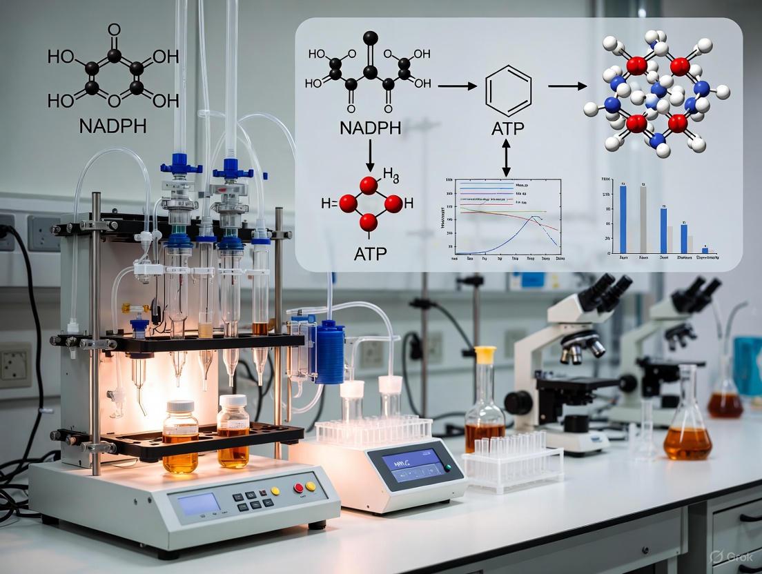
Accurate quantification of intracellular NADPH and ATP is crucial for research in cellular metabolism, redox biology, and drug development.
A Comprehensive Guide to Measuring Intracellular NADPH and ATP Levels Using HPLC: From Foundational Principles to Advanced Applications
Abstract
Accurate quantification of intracellular NADPH and ATP is crucial for research in cellular metabolism, redox biology, and drug development. This article provides a comprehensive guide for researchers and scientists, covering the foundational roles of these metabolites, detailed HPLC and LC-MS/MS methodologies, and common troubleshooting protocols. It further addresses the critical need for method validation in light of significant inter-study variability reported in the literature and explores advanced techniques for resolving compartmentalized NADPH fluxes. The content synthesizes current best practices to enhance data reliability and cross-study comparisons in metabolic research.
The Critical Roles of NADPH and ATP in Cellular Metabolism and Redox Homeostasis
In cellular metabolism, nicotinamide adenine dinucleotide phosphate (NADPH) and adenosine triphosphate (ATP) represent two fundamental currencies with distinct and specialized functions. While both molecules are essential for cellular survival and function, they operate in complementary metabolic domains: ATP serves as the universal energy currency that powers mechanical, transport, and biochemical work, whereas NADPH provides the primary reducing power for biosynthetic processes and antioxidant defense systems [1] [2]. This fundamental distinction arises from their chemical properties and the specialized enzymatic machinery that has evolved to utilize them. Understanding the divergent roles of these essential metabolites is crucial for research areas ranging from cancer metabolism to neurodegenerative diseases and drug development.
The critical functional separation between these molecules is maintained through strict compartmentalization and distinct regulatory mechanisms within cellular metabolic networks. ATP predominantly drives catabolic processes that release energy, while NADPH is preferentially utilized in anabolic reactions that consume energy to build complex molecules [3] [2]. This review comprehensively compares the cellular functions, metabolic pathways, and measurement methodologies for NADPH and ATP, with particular emphasis on their implications for biomedical research and therapeutic development.
Molecular Functions and Metabolic Roles: A Comparative Analysis
Core Biological Functions
Table 1: Comparative Functions of NADPH and ATP in Cellular Metabolism
| Feature | NADPH | ATP |
|---|---|---|
| Primary Role | Reductive biosynthesis & antioxidant defense | Universal energy currency |
| Redox Function | Electron donor (reducing agent) | Not directly involved in redox reactions |
| Energy Transfer | Not primarily for energy transfer | High-energy phosphate bonds store & transfer energy |
| Biosynthetic Role | Essential for fatty acids, cholesterol, nucleotides, amino acids | Provides energy for biosynthetic enzymes |
| Antioxidant Systems | Maintains GSH and thioredoxin in reduced states; reactivates catalase | Provides energy for antioxidant enzyme synthesis & activation |
| Signaling Functions | Substrate for NOX-generated ROS signaling; influences redox-sensitive pathways | Substrate for phosphorylation reactions; energy sensor via AMPK |
NADPH's primary function lies in its capacity as a reducing equivalent, donating electrons in critical cellular processes. It serves as an essential cofactor for reductive biosynthesis, driving the synthesis of fatty acids, cholesterol, amino acids, and nucleotides [1] [3]. Simultaneously, NADPH plays a pivotal role in maintaining redox homeostasis by regenerating reduced glutathione (GSH) and thioredoxin (TRX), which are essential for neutralizing reactive oxygen species (ROS) [1] [3]. Paradoxically, NADPH also serves as a substrate for NADPH oxidases (NOXs), which generate superoxide anions that function as signaling molecules [1] [3].
In contrast, ATP functions as the universal energy currency of the cell, with its high-energy phosphate bonds providing the thermodynamic driving force for countless cellular processes. ATP hydrolysis releases energy that powers mechanical work (e.g., muscle contraction, cell division), transport work (e.g., ion pumping across membranes), and chemical work (e.g., biosynthesis of macromolecules) [4]. The ATP/ADP/AMP system also serves as a critical energy sensing network, with AMP-activated protein kinase (AMPK) monitoring cellular energy status and regulating metabolic pathways accordingly [4].
Metabolic Pathways and Cellular Compartmentalization
Table 2: Metabolic Sources and Cellular Distribution of NADPH and ATP
| Aspect | NADPH | ATP |
|---|---|---|
| Major Production Pathways | Pentose phosphate pathway (PPP), Malic enzymes (ME1/3), Isocitrate dehydrogenases (IDH1/2), Folate metabolism | Glycolysis, Mitochondrial oxidative phosphorylation, TCA cycle, Substrate-level phosphorylation |
| Subcellular Distribution | Cytosol (~3.1 μM), Mitochondrial matrix (~37 μM) [3] | Cytosol, Mitochondria, Nucleus; Total cellular concentration typically 1-10 mM |
| Primary Regulators | NAD kinases (NADK), PPP enzymes (G6PD, PGD), Metabolic flux | AMP/ATP ratio, Oxygen availability, Nutrient status, Mitochondrial function |
NADPH is generated through multiple metabolic routes, with the oxidative pentose phosphate pathway (PPP) serving as a major source, particularly in the cytosol [1] [3]. Additional significant sources include mitochondrial and cytosolic isocitrate dehydrogenases (IDH2 and IDH1), malic enzymes (ME1 and ME3), and folate-mediated one-carbon metabolism [1] [3]. The relative contribution of these pathways varies by cell type, metabolic state, and environmental conditions. A crucial regulatory step in NADPH generation is the phosphorylation of NAD+ to NADP+ by NAD kinases (NADKs), which exists in both cytosolic and mitochondrial isoforms [3] [5].
ATP production occurs primarily through substrate-level phosphorylation in glycolysis and the mitochondrial electron transport chain via oxidative phosphorylation [2]. The tricarboxylic acid (TCA) cycle generates reducing equivalents (NADH and FADH2) that feed into the electron transport chain, where the proton gradient drives ATP synthesis [1] [2]. Unlike NADPH, ATP is produced in large quantities throughout the cell, with mitochondria serving as the primary powerhouse. The adenylate energy charge ([ATP]+1/2[ADP])/([ATP]+[ADP]+[AMP]) represents a key regulatory parameter that reflects the energy status of the cell and influences metabolic flux [4].
The following diagram illustrates the distinct metabolic pathways and cellular compartmentalization of NADPH and ATP:
NADPH and ATP Pathway Compartmentalization
Analytical Methodologies: Measuring Intracellular NADPH and ATP Levels
HPLC-Based Measurement Techniques
Liquid chromatography-coupled mass spectrometry (LC/MS) has emerged as a powerful methodology for the precise quantification of adenine nucleotides, including ATP, ADP, and AMP [4]. This approach offers superior sensitivity compared to traditional HPLC with ultraviolet or fluorescence detection, enabling direct measurement of low-abundance metabolites like AMP without relying on equilibrium assumptions [4]. For NADPH quantification, high-performance liquid chromatography (HPLC) remains a reliable method, though it requires careful sample handling due to NADPH's susceptibility to oxidation and degradation in complete culture media [6].
The experimental workflow for HPLC-based measurement of these metabolites typically involves: (1) rapid metabolite extraction using acidified solvents (e.g., perchloric acid) or organic solvents (e.g., methanol/acetonitrile) to quench metabolic activity; (2) sample neutralization and clarification; (3) chromatographic separation using reverse-phase or ion-pairing columns; and (4) detection via mass spectrometry for ATP or ultraviolet/fluorescence detection for NADPH [6] [4]. For NADPH measurements specifically, samples must be processed on ice and protected from light to prevent degradation [6].
Advanced Biosensors and Fluorescence Techniques
Recent advances in genetically encoded biosensors have revolutionized the real-time monitoring of NADPH dynamics in living cells. The newly developed NAPstar family of biosensors enables specific measurements of NADPH/NADP+ ratios across a broad dynamic range with subcellular resolution [7]. These biosensors are derived from the bacterial transcriptional repressor Rex and feature mutations that favor NADP(H) binding over NAD(H), allowing specific monitoring of NADP redox states [7].
For NAD(P)H detection, fluorescence lifetime imaging microscopy (FLIM) leverages the natural autofluorescence of reduced pyridine nucleotides to investigate mitochondrial redox state [8]. This technique can potentially discriminate between contributions from NADH and NADPH based on their distinct fluorescence lifetimes, providing spatial and temporal information about redox metabolism in intact systems [8].
The following workflow diagram illustrates the key methodological approaches for measuring these metabolites:
Metabolite Measurement Methodologies
Research Reagent Solutions for NADPH/ATP Research
Table 3: Essential Research Tools for NADPH and ATP Studies
| Reagent/Category | Specific Examples | Function/Application |
|---|---|---|
| NADPH Biosensors | NAPstar family [7], iNap sensors [6], SoNar [6] | Real-time monitoring of NADPH/NADP+ ratios in living cells |
| ATP Measurement Kits | Luciferase-based assays, LC/MS standards [4] | Quantification of ATP levels and adenine nucleotide pools |
| Chromatography Standards | NADPH, NADP+, ATP, ADP, AMP reference compounds [6] [4] | HPLC and LC/MS calibration and quantification |
| Pathway Modulators | G6PD inhibitors, NOX inhibitors, NADK targets [3] | Selective manipulation of NADPH production pathways |
| Metabolic Extraction Kits | Acid-based extraction kits, Methanol/acetonitrile kits [4] | Rapid quenching of metabolism and metabolite stabilization |
Research Implications and Therapeutic Applications
The distinct yet interconnected functions of NADPH and ATP have profound implications for understanding disease mechanisms and developing therapeutic interventions. In cancer biology, tumor cells exhibit reprogrammed NADPH metabolism to support rapid proliferation and counteract oxidative stress [3]. Multiple oncogenic signaling pathways converge on NADPH-generating enzymes, making them potential therapeutic targets. Similarly, modulating NADPH homeostasis represents a promising strategy for treating metabolic diseases, neurodegenerative disorders, and aging-related conditions [9] [2].
The development of dynamic regulation strategies for NADPH metabolism, including genetically encoded biosensors that enable real-time monitoring of NADPH/NADP+ ratios, provides powerful tools for metabolic engineering and drug discovery [10] [7]. These approaches allow researchers to move beyond static measurements and capture the dynamic responses of metabolic networks to genetic, environmental, and pharmacological perturbations.
For ATP metabolism, targeting the adenylate energy charge and AMPK signaling pathway offers therapeutic potential for metabolic disorders, including diabetes and obesity [4] [2]. The integration of NADPH and ATP metabolic signatures in disease states provides a more comprehensive understanding of pathophysiology and enables the development of more effective therapeutic strategies that simultaneously address multiple aspects of cellular metabolism.
NADPH and ATP represent fundamental but distinct currencies in cellular metabolism, with NADPH specializing in providing reducing power for biosynthesis and antioxidant defense, while ATP serves as the universal energy currency driving cellular work. This functional specialization is maintained through compartmentalized metabolic pathways, specialized enzyme systems, and distinct regulatory mechanisms. Advanced analytical techniques, including HPLC/MS and genetically encoded biosensors, continue to reveal new dimensions of their complex interplay in health and disease. Understanding the nuanced relationship between these essential metabolites provides critical insights for fundamental biology and therapeutic development across a spectrum of human diseases.
In the evolving landscape of molecular biology, metabolic phenotypes represent the ultimate functional readout of cellular status, precisely reflecting the complex interactions among genetic background, environmental factors, lifestyle, and gut microbiome [11]. These phenotypes serve as key molecular links between healthy homeostasis and disease-related metabolic disruption, offering a dynamic portrait of physiological function that moves beyond static genomic assessments. Particularly for energy metabolism, the quantification of specific metabolites such as NADPH and ATP provides critical insights into cellular bioenergetics, redox balance, and pathological transformations observed in conditions ranging from cancer to neurodegenerative disorders [12] [13]. The intracellular concentrations of these metabolites and their ratios serve as sensitive indicators of the metabolic shifts that characterize disease pathogenesis, enabling researchers to decode the molecular language of cellular dysfunction.
The field has witnessed significant methodological evolution in metabolite quantification, with techniques spanning from traditional spectrophotometric assays to advanced chromatographic separations coupled with mass spectrometry [14] [15] [16]. Each methodology offers distinct advantages in sensitivity, specificity, and applicability to different biological contexts. This guide provides a comprehensive comparison of these analytical approaches, with particular emphasis on HPLC-based platforms that have become central to rigorous NADPH and ATP research. By objectively evaluating the performance characteristics, technical requirements, and practical considerations of each method, we aim to equip researchers with the analytical framework necessary to advance our understanding of how metabolite dynamics shape cellular phenotypes in health and disease.
Metabolic Foundations: NADPH and ATP in Cellular Energy Economy
NADPH: The Master Redox Regulator
Nicotinamide adenine dinucleotide phosphate (NADPH) serves as the primary reducing agent in anabolic biosynthesis and cellular antioxidant defense systems. This crucial cofactor exists in oxidized (NADP+) and reduced (NADPH) forms, with the NADPH:NADP+ ratio representing a critical indicator of the intracellular redox state and reductive capacity [15] [17]. NADPH plays an indispensable role in fundamental redox and metabolic pathways through three major mechanisms: (1) as an essential electron donor to nitric oxide synthase (NOS) for nitric oxide (NO•) generation; (2) as the necessary electron source for reducing thioredoxin reductase (TrxR) to its active form, which subsequently activates peroxiredoxins to neutralize peroxides; and (3) as the cofactor for glutathione reductase (GR), which maintains glutathione in its reduced state (GSH) to support glutathione peroxidase activity [14]. The centrality of NADPH in biosynthetic processes is evidenced by its requirement in approximately 887 distinct enzymatic reactions, far exceeding the dependency of other cofactors [17].
ATP: The Universal Energy Currency
Adenosine triphosphate (ATP) represents the primary energy currency of the cell, synthesized through two major pathways: mitochondrial oxidative phosphorylation (OXPHOS) and glycolytic substrate-level phosphorylation [12] [18]. Mitochondrial ATP synthesis occurs through a sophisticated chemiosmotic process wherein nutrient oxidation generates a proton gradient across the inner mitochondrial membrane, driving ATP synthase (Complex V) [18]. The continuous balance between ATP production and consumption maintains cellular energy homeostasis, with ATP concentrations and synthesis rates providing sensitive readouts of mitochondrial function and overall cellular health [18]. Cancer cells and other pathological states often display reprogrammed ATP generation, preferentially utilizing aerobic glycolysis even under oxygen-sufficient conditions (the Warburg effect), resulting in distinct ATP production profiles that can be leveraged for diagnostic and therapeutic purposes [12].
Integrated Metabolic Sensing
The interplay between NADPH and ATP metabolism extends beyond their individual roles, creating a coordinated network that regulates cellular function. NADPH not only drives biosynthetic processes but also protects catalase from hydrogen peroxide-induced inactivation, thereby regulating the intracellular fate of H2O2, which functions as a key signaling molecule [14]. Similarly, ATP serves not only as an energy transfer molecule but also as a substrate for phosphorylation in kinase cascades and as a critical cofactor for chromatin-modifying enzymes [13]. Metabolites including acetyl-CoA, which exists at the intersection of carbohydrate, fatty acid, and amino acid oxidation, exert tremendous influence on cell signaling through post-translational modifications such as histone acetylation, thereby directly linking metabolic status to epigenetic regulation of gene expression [13].
Figure 1: Metabolic Foundation of Cellular Phenotypes. NADPH and ATP serve as central players in converting nutrient inputs into functional cellular states through multiple interconnected mechanisms. PPP: Pentose Phosphate Pathway; OXPHOS: Oxidative Phosphorylation.
Analytical Methodologies: Quantifying Metabolic Phenotypes
Comparative Performance of Quantification Platforms
Multiple analytical platforms are available for quantifying NADPH, ATP, and related metabolites, each with distinct performance characteristics, advantages, and limitations. The selection of an appropriate methodology depends on factors including required sensitivity, specificity, sample throughput, and the need for absolute versus relative quantification.
Table 1: Method Comparison for NAD(P)H and ATP Quantification
| Method | Sensitivity | Key Advantage | Primary Limitation | Sample Requirements | Throughput |
|---|---|---|---|---|---|
| Enzyme Cycling Assays [15] | Moderate (micromolar) | Cost-effective; Minimal equipment | Limited multiplexing capability; Interference potential | Cell extracts, tissue homogenates | High |
| Spectrophotometric Assays [14] | Moderate (micromolar) | Simple protocol; Wide accessibility | Lower specificity for complex samples | Purified enzymes, cell lysates | Medium |
| HPLC with UV/FLD [19] [15] | High (nanomolar) | Good separation capability | Limited metabolite identification | Tissue extracts, biological fluids | Medium |
| LC-MS [15] [16] | Very High (picomolar) | High specificity; Multiplexing capability | High cost; Technical expertise required | Cell cultures, plasma, tissues | Medium to High |
| HPLC-ICP-MS [20] | Specialized Applications | Exceptional for metal-labeled probes | Limited to appropriate elemental tags | Specialized probe studies | Medium |
HPLC-Based Platforms: Technical Considerations
High-performance liquid chromatography (HPLC) systems coupled with various detection modalities represent a versatile approach for metabolite quantification. Traditional HPLC with ultraviolet (UV) or fluorescence (FLD) detection provides robust separation capabilities with relatively accessible instrumentation [19]. The application of HPLC to metabolite profiling is exemplified by ginsenoside analysis in Panax species, where reverse-phase separation with UV detection enabled discrimination between different plant origins based on distinct chromatographic fingerprints [19]. For NADPH and ATP analysis, HPLC methods typically employ reverse-phase or ion-pairing chromatography to achieve sufficient separation of these hydrophilic compounds, followed by detection at appropriate wavelengths (e.g., 254 nm for adenine nucleotides) [15].
Liquid chromatography coupled to mass spectrometry (LC-MS) has emerged as the gold standard for sensitive and specific metabolite quantification, combining excellent separation power with selective mass detection [15] [16]. Modern LC-MS platforms enable the simultaneous quantification of numerous metabolites, including NADPH, ATP, and related compounds, within complex biological matrices. Automated label-free quantification algorithms, such as the FeatureFinderMetabo implemented within the OpenMS framework, further enhance the robustness and reproducibility of LC-MS data processing by detecting mass traces and aggregating them into features with high precision and recall [16]. The sensitivity of LC-MS methods typically surpasses that of traditional HPLC, with detection limits in the picomolar range, making it particularly suitable for limited sample quantities or low-abundance metabolites [16].
Specialized Methodologies: Real-Time Monitoring and Redox Sensing
Beyond conventional quantification approaches, specialized methodologies have emerged to address specific research questions in energy metabolism. The MitoRAISE assay represents an innovative approach for real-time assessment of mitochondrial ATP synthesis rates in response to various substrates and inhibitors [18]. This technique utilizes plasma membrane-permeabilized cells or isolated mitochondria to directly monitor ATP production dynamics, providing functional insights beyond static metabolite measurements. Similarly, the development of genetically-encoded biosensors has enabled real-time monitoring of NADPH dynamics in living cells, with recent advances including dual-sensing systems capable of simultaneously detecting NADPH and specific metabolites like L-threonine [17]. These tools are particularly valuable for capturing metabolic flux and transient perturbations that might be missed by endpoint measurements.
Experimental Protocols: Methodological Guidelines
Spectrophotometric NADPH Quantification
Principle: This method exploits the unique spectrophotometric properties of reduced pyridine nucleotides, specifically the absorption maximum of NADPH at 340 nm, to quantify its concentration in biological samples [14].
Protocol Details:
- Sample Preparation: Homogenize tissue or cell samples in ice-cold extraction buffer (e.g., 50 mM phosphate buffer, pH 7.4). Precipitate proteins using perchloric acid (PCA) or heat treatment, followed by neutralization for acid-extracted samples. Note that PCA extraction is suitable only for oxidized forms (NADP+), as reduced forms (NADPH) are acid-labile [15].
- Reaction Setup: Prepare assay mixture containing:
- 50 mM Tris-HCl buffer (pH 8.0)
- 0.5 mM EDTA
- 0.1 mM flavin adenine dinucleotide (FAD)
- Sample extract or NADPH standard
- Measurement: Monitor absorbance at 340 nm before and after adding specific enzymes (e.g., glutathione reductase) that utilize NADPH. Calculate NADPH concentration using the extinction coefficient ε340 = 6.22 mM⁻¹cm⁻¹ [14].
- Validation: Include internal standards and blank controls to account for background absorbance. Ensure linearity within the measurement range through appropriate dilution.
Critical Considerations: Sample processing under low-temperature conditions is essential to preserve NADPH stability. The extraction method must be selected based on the specific analytes of interest, as reduced forms require non-acidic conditions [15].
HPLC-MS Based Metabolite Profiling
Principle: This methodology combines chromatographic separation with mass spectrometric detection to achieve high specificity and sensitivity for NADPH, ATP, and related metabolites in complex biological samples [16].
Protocol Details:
- Metabolite Extraction: Use polar organic solvents (e.g., 80% methanol:water) at low temperature to simultaneously quench metabolism and extract metabolites. For comprehensive NAD(P)(H) analysis, avoid acidic conditions that degrade reduced forms [15].
- Chromatographic Separation:
- Column: Reversed-phase C18 column (150 × 2.1 mm, 1.8 μm)
- Mobile Phase: A) Water with 0.1% formic acid; B) Acetonitrile with 0.1% formic acid
- Gradient: 0-5 min: 0% B; 5-15 min: 0-30% B; 15-25 min: 30-95% B; 25-30 min: 95% B
- Flow Rate: 0.2 mL/min; Column Temperature: 40°C [16]
- Mass Spectrometric Detection:
- Ionization: Electrospray ionization (ESI) in negative mode for NADPH and ATP
- Detection: Multiple reaction monitoring (MRM) for enhanced specificity
- Key transitions: NADPH (m/z 744→408), ATP (m/z 506→159)
- Data Processing: Utilize automated algorithms (e.g., FeatureFinderMetabo) for peak detection, retention time alignment, and intensity extraction [16].
Critical Considerations: Incorporate stable isotope-labeled internal standards (e.g., ¹³C-NADPH, ¹⁵N-ATP) for accurate quantification. Maintain consistent sample preparation and injection volumes to minimize technical variability.
Figure 2: HPLC-MS Workflow for Metabolite Quantification. The analytical process from sample preparation to data interpretation, highlighting critical steps for reliable NADPH and ATP measurement.
MitoRAISE Assay for Functional ATP Analysis
Principle: The Mitochondrial ATP Synthesis Response Against Inhibiting and Stimulating Substrates (MitoRAISE) assay measures real-time ATP synthesis dynamics in response to specific mitochondrial substrates and inhibitors, providing functional assessment of mitochondrial capacity [18].
Protocol Details:
- Sample Preparation: Isolate mitochondria or use permeabilized cells (e.g., PBMCs treated with plasma membrane permeabilizer). Confirm permeabilization efficiency (>90%) using trypan blue exclusion.
- Assay Conditions: Resuspend samples in mitochondrial assay solution (MAS: 220 mM mannitol, 70 mM sucrose, 10 mM KH₂PO₄, 5 mM MgCl₂, 2 mM HEPES, 1 mM EGTA, 0.2% fatty acid-free BSA, pH 7.4).
- Substrate-Inhibitor Protocol:
- Basal measurement: Endogenous substrates
- Complex I-driven ATP synthesis: Glutamate + Malate
- Complex I inhibition: Rotenone
- Complex II-driven ATP synthesis: Succinate
- Complex II inhibition: Malonate
- ATP Detection: Utilize luciferase-based ATP detection system with real-time luminescence monitoring.
- Data Analysis: Calculate ATP synthesis rates for each respiratory state and inhibitor responses to assess specific mitochondrial functions.
Critical Considerations: Optimize cell number or mitochondrial protein concentration to ensure signal detection within linear range. Include validation using mitochondrial DNA-depleted (Rho-0) cells as negative controls [18].
Research Reagent Solutions: Essential Materials
Table 2: Essential Research Reagents for Metabolite Studies
| Reagent Category | Specific Examples | Function & Application | Technical Notes |
|---|---|---|---|
| Chromatography Columns | Phenomenex Luna C18 [19]; Capcell Pak C18 MGII [19] | Metabolite separation | 150-250 mm length, 2-3 mm ID, 3-5 μm particle size |
| Mass Spectrometry Standards | Stable isotope-labeled NADPH/ATP [15] | Internal quantification standards | Correct for matrix effects & recovery variations |
| Enzyme Assay Components | Glucose-6-phosphate dehydrogenase [14]; Glutathione reductase [14] | Enzyme cycling assays | Enable specific detection through enzymatic coupling |
| Mitochondrial Assay Reagents | Rotenone, Malonate, FCCP [18] | Modulators of mitochondrial function | Specific inhibitor for Complex I (Rotenone) and Complex II (Malonate) |
| Sample Preparation | Perchloric acid, Methanol, Acetonitrile [15] | Metabolite extraction & protein precipitation | Selection depends on analyte stability (avoid acid for reduced forms) |
Methodological Challenges and Standardization Needs
The quantification of NADPH, ATP, and related metabolites faces significant methodological challenges that impact data comparability across studies. A comprehensive meta-analysis of NAD(P)(H) quantification results revealed substantial inter- and intra-method variability across mammalian tissues, highlighting the relatively poor potential for cross-experimental analyses without rigorous standardization [15]. This variability stems from multiple factors, including differences in sample collection procedures (e.g., pre-mortem vs. post-mortem sampling), extraction methods (with significant implications for acid-labile reduced forms), and detection techniques [15]. Only 7.57% of published studies utilized pre-mortem sampling, while 29.48% relied on post-mortem samples, creating potential confounding factors due to rapid post-mortem metabolic alterations [15].
For HPLC-MS based approaches, additional challenges include ion suppression effects, variable detector response, and the need for appropriate internal standards to achieve truly quantitative results [15] [16]. Without comprehensive quality controls for individual metabolites and matrix effects, even LC-MS methods remain at best semi-quantitative [15]. These technical challenges underscore the critical importance of transparent reporting of methodological details, including sample processing conditions, extraction protocols, and validation parameters, to enable proper interpretation and comparison of metabolic data across studies.
The accurate quantification of intracellular NADPH and ATP levels represents a cornerstone of metabolic phenotyping, providing critical insights into cellular energy status, redox balance, and pathogenic mechanisms. HPLC-based methodologies, particularly when coupled with mass spectrometry, offer the sensitivity, specificity, and multiplexing capability necessary to capture the dynamic nature of metabolic networks in health and disease. The continuing evolution of analytical technologies, including real-time monitoring assays and genetically-encoded biosensors, promises to further enhance our ability to correlate metabolite dynamics with functional phenotypes. As the field progresses, increased methodological standardization and rigorous validation will be essential to translate metabolic measurements into clinically actionable insights and targeted therapeutic interventions for metabolic diseases.
In the field of cellular metabolism research, accurate quantification of intracellular metabolites like ATP and NADPH is not merely a technical exercise—it is the foundation for understanding fundamental biological processes, from energy homeostasis to redox signaling. These molecules are crucial indicators of cellular energy status, with ATP serving as the primary energy currency and NADPH playing an essential role in biosynthetic processes and antioxidant defense [21] [15]. However, their accurate measurement is compromised by a often-overlooked challenge: their inherent instability during sample preparation. Pre-analytical conditions—the procedures from sample collection to metabolite extraction—can introduce significant artifacts that distort the true metabolic picture. This guide examines how these pre-analytical variables impact metabolite stability, compares methodological approaches for preserving ATP and NADPH integrity, and provides researchers with validated protocols to enhance the reliability of their HPLC-based metabolomic data.
The Pre-Analytical Challenge: Metabolite Instability and Interconversion
The journey from living tissue to quantitative data is fraught with potential pitfalls where labile metabolites can degrade or interconvert. Metabolites in tissue homogenates remain vulnerable to enzymatic activity until extraction is complete, with significant changes occurring within minutes [22]. For instance, in metabolically active tissues like the brain, noticeable changes occur "already within the first several minutes during the brain harvest and sample homogenization" [22].
A major challenge in NAD(P)H analysis is the interconversion between oxidized and reduced forms during extraction. When researchers spiked unlabeled NADPH standards into extraction solvents, they observed substantial conversion to NADP+ across most conditions [23]. This interconversion dramatically affects critical ratios like NADPH/NADP+, which serve as important indicators of cellular redox state [23] [15]. Similarly, ATP levels are highly vulnerable, with studies showing concentrations can decrease fivefold in whole blood homogenates during extended incubation [22].
Comparative Analysis of Extraction Method Performance
The choice of extraction method significantly influences metabolite stability and recovery. The table below summarizes the performance of different extraction solvents for preserving NADPH and ATP based on experimental data:
Table 1: Performance Comparison of Extraction Methods for NADPH and ATP Preservation
| Extraction Method | NADPH Recovery | NADPH/NADP+ Ratio Preservation | ATP Preservation | Best Use Cases |
|---|---|---|---|---|
| Acidic ACN/MeOH/H₂O (0.1 M FA) | Highest yield [23] | Superior (least interconversion) [23] | Effective for nucleotides [21] | Gold standard for redox cofactors; LC-MS analysis |
| 80% Methanol (-70°C) | Moderate | Moderate interconversion [23] | Good with rapid processing [22] | General metabolomics; rapid quenching |
| Enzyme Assay Buffer + Detergent | Lower than organic solvents | Moderate (better than buffer alone) [23] | Not specifically reported | Enzyme cycling assays when MS unavailable |
| Hot Aqueous Buffer | Poor | Significant interconversion [23] | Poor (thermal degradation) | Not recommended for labile nucleotides |
The performance variability across methods underscores why published NAD(P)(H) measurements exhibit "important inter- and intra-method variability" across the scientific literature [15]. This methodological inconsistency complicates cross-study comparisons and meta-analyses in NAD+ biology research.
Optimized Experimental Protocols for Metabolite Stabilization
Protocol 1: Acidic Acetonitrile/Methanol/Water Extraction for NAD(P)H
This protocol, adapted from research demonstrating superior performance for redox cofactors, minimizes interconversion while maximizing recovery [23]:
- Preparation: Pre-cool the extraction solvent (40:40:20 acetonitrile:methanol:water with 0.1 M formic acid) to 4°C.
- Rapid Quenching: Immediately add cold extraction solvent to cell pellets or snap-frozen tissue powder (typically 1-2 mL per 50-100 mg tissue).
- Homogenization: Vigorously vortex or homogenize on ice for 1-2 minutes.
- Incubation: Keep the samples at 4°C for exactly 3 minutes to allow complete protein precipitation.
- Neutralization: Add appropriate volume of 10 mM ammonium bicarbonate to neutralize the acid and prevent degradation of acid-labile metabolites.
- Centrifugation: Spin at 16,100 × g for 15 minutes at 4°C to pellet insoluble material.
- Collection: Transfer the supernatant to a new tube for analysis or storage at -80°C.
Critical Note: Neutralization immediately after extraction is essential as "NADPH and NADH are not stable in 0.1 M formic acid in water (pH = 2.4) with a half-life of ∼16 and 20 h, respectively" [23].
Protocol 2: Cold Methanol Extraction for ATP and Adenine Nucleotides
For simultaneous extraction of ATP and related nucleotides, a cold methanol-based method provides excellent results [21] [22]:
- Preparation: Chill HPLC-grade methanol to -20°C.
- Sample Processing: Add cold methanol to samples (typically 3:1 solvent-to-sample ratio).
- Homogenization: Homogenize on ice or using a pre-cooled homogenizer.
- Extraction: Incubate at -20°C for 30-60 minutes to ensure complete metabolite extraction.
- Phase Separation: Add chloroform (if using Folch extraction) or simply centrifuge at 16,100 × g for 15 minutes at 4°C.
- Collection: Collect the upper aqueous phase containing polar metabolites.
- Storage: Flash-freeze in liquid nitrogen and store at -80°C until analysis.
Metabolic Pathways and Experimental Workflow Visualization
Figure 1: Metabolic Pathways and Pre-analytical Challenges. The diagram illustrates the interconnected pathways producing ATP and NADPH, alongside the major pre-analytical factors that can compromise their accurate measurement.
Figure 2: Optimized Workflow for Metabolite Quantification. The experimental workflow highlights critical control points where strict adherence to protocols preserves metabolite stability.
The Scientist's Toolkit: Essential Research Reagents and Materials
Table 2: Essential Research Reagents for ATP/NADPH Analysis
| Reagent/Equipment | Function | Specific Application Notes |
|---|---|---|
| HILIC HPLC Columns | Retention and separation of highly polar metabolites | Superior to reverse-phase for nucleotides; provides better peak shape [21] [24] |
| MS-grade Acetonitrile/Methanol | Organic extraction solvents | Precipitate proteins while extracting metabolites; cold temperature critical [23] |
| Ammonium Acetate Buffer | Mobile phase component for HILIC | 20 mM, pH 9.8 provides optimal separation [21] |
| Formic Acid | Acidification of extraction solvent | 0.1 M in extraction solvent minimizes interconversion [23] |
| Ammonium Bicarbonate | Neutralization agent | Prevents acid-catalyzed degradation after extraction [23] |
| Luna NH2 HPLC Column | HILIC stationary phase | Effective for 26 adenosine nucleotides and NAD metabolites [21] |
| Pre-cooled Homogenizer | Tissue disruption | Maintains low temperature during processing [22] |
Analytical Techniques: HILIC-HPLC-MS as a Superior Approach
For simultaneous quantification of multiple metabolites, hydrophilic interaction liquid chromatography (HILIC) coupled with tandem mass spectrometry (HPLC-MS/MS) has emerged as a powerful approach. This technique effectively retains and separates highly polar compounds like adenine nucleotides that show "weak retention on reversed-phase (RP) HPLC columns and subsequent poor separation and peak shape" [21]. The HILIC-based method has been successfully validated for "26 adenosine nucleotides and precursors as well as metabolic products of nicotinamide adenine dinucleotide (NAD)" across various biological matrices including plasma, liver, adipose tissue, and cell culture supernatants [21].
This methodological advancement is particularly significant given the findings of a comprehensive meta-analysis that examined NAD(P)(H) quantification results across mammalian tissues. The analysis revealed "considerable variability in NAD(P)(H) measures across rodent and human studies," emphasizing the need for standardized sample processing and analytical protocols [15]. The implementation of robust, standardized methods like HILIC-HPLC-MS is essential for generating comparable datasets that allow meaningful interpretation in the field of NAD+ biology.
The quantification of intracellular metabolites like ATP and NADPH demands rigorous attention to pre-analytical conditions. Methodological choices—from extraction solvent composition to processing temperature—directly impact metabolite stability and consequently, data reliability. Through implementation of optimized protocols that emphasize rapid quenching, cold acidic extraction, immediate neutralization, and appropriate analytical techniques like HILIC-HPLC-MS, researchers can significantly reduce artifacts and obtain more accurate representations of cellular metabolic states. As the field advances toward standardized methodologies, the research community will benefit from enhanced data comparability and more robust insights into metabolic regulation in health and disease.
Accurately quantifying intracellular nicotinamide adenine dinucleotide phosphate (NADP(H)) pools represents a fundamental challenge in redox biology research. The physiological ranges of these crucial cofactors vary significantly across cell types, tissues, and subcellular compartments, creating substantial interpretation difficulties for researchers investigating metabolic health, aging, and disease pathogenesis. These measurement complexities arise from multiple analytical obstacles, including the compartmentalized nature of NADPH metabolism within eukaryotic cells, the rapid interconversion between oxidized and reduced forms during extraction, and the technical limitations of traditional bulk measurement approaches [25] [26] [23]. Understanding these methodological constraints is essential for properly evaluating reported NADP(H) levels across scientific studies and designing experiments that generate biologically meaningful data.
The importance of NADPH in maintaining redox homeostasis and supporting reductive biosynthesis extends across numerous physiological and pathophysiological processes. Research has established NADPH as the primary reducing equivalent for antioxidant systems, including glutathione and thioredoxin pathways, while also serving as an essential cofactor for biosynthetic enzymes. Recent investigations have revealed compelling connections between NADPH metabolism and age-related conditions, with studies demonstrating that cytosolic NADPH levels increase during endothelial cell senescence and that pharmacological elevation of NADPH can ameliorate disease progression in models of non-alcoholic steatohepatitis (NASH) [25] [27]. These findings highlight the therapeutic relevance of accurate NADP(H) quantification and the importance of navigating the complex landscape of reported physiological ranges.
Methodological Approaches for NADP(H) Quantification
The quantification of NADP(H) pools has evolved substantially, with researchers now employing diverse methodological approaches ranging from traditional biochemical assays to advanced live-cell imaging techniques. Each method offers distinct advantages and limitations that significantly influence the reported physiological ranges and their biological interpretation.
Comparative Analysis of NADP(H) Measurement Techniques
Table 1: Comparison of Major NADP(H) Quantification Methodologies
| Method | Spatial Resolution | Key Advantages | Reported NADPH/NADP+ Ratios | Technical Limitations |
|---|---|---|---|---|
| Enzymatic Cycling Assays | Bulk cellular measurement | High sensitivity; established protocols; accessible instrumentation | Wide variation (0.04-100+ across tissues/cells) [23] | No subcellular resolution; susceptible to interconversion artifacts [23] |
| LC-MS Approaches | Bulk cellular measurement | High specificity; simultaneous NADP+/NADPH quantification; minimal cross-reactivity | 1.2-15 in cultured mammalian cells [23] | Extensive sample processing; requires specialized equipment; extraction-induced interconversion [23] |
| Genetically Encoded Sensors (iNap, SoNar) | Subcellular compartment resolution | Real-time monitoring in live cells; compartment-specific quantification; dynamic tracking | Increased cytosolic NADPH during endothelial senescence [25] | Requires genetic modification; calibration challenges; potential cellular perturbation |
| Autofluorescence Imaging | Single-cell to subcellular | Label-free; native metabolite detection; temporal dynamics | Oscillatory patterns through cell division in E. coli [28] | UV-induced cellular damage; low quantum yield; complex signal deconvolution [28] |
Critical Methodological Considerations
The extraction process itself represents a significant source of variability in reported NADP(H) levels. A systematic investigation of extraction methods revealed substantial interconversion between oxidized and reduced forms during sample preparation, with certain solvents exacerbating this artifact. The study demonstrated that extraction with 40:40:20 acetonitrile:methanol:water containing 0.1 M formic acid effectively minimized interconversion, while traditional aqueous buffers showed higher rates of NADPH oxidation to NADP+ during processing [23]. This methodological artifact likely contributes to the extraordinarily wide range of reported NADPH/NADP+ ratios in literature, which span from approximately 0.04 in rat heart tissue to over 100 in theoretical textbook calculations [23].
Compartmentalization presents another fundamental challenge, as separate cytosolic and mitochondrial NADPH pools regulate distinct cellular functions. Conventional bulk measurement approaches inevitably mask these subcellular differences, potentially obscuring physiologically important changes. Research using compartment-targeted fluorescent sensors has revealed striking differences in NADPH dynamics, with studies demonstrating that cytosolic NADPH increases during endothelial cell senescence while mitochondrial NADPH remains unchanged [25]. Similarly, metabolic tracing studies using stable isotopes have enabled researchers to resolve NADPH-dependent pathway activities within specific cellular compartments, providing unprecedented insight into compartmentalized metabolic fluxes [26].
Experimental Protocols for NADP(H) Analysis
Optimized Metabolite Extraction for LC-MS Analysis
Accurate quantification of NADP(H) pools requires meticulous extraction protocols that minimize metabolite interconversion and degradation. Based on systematic methodological comparisons, the following procedure has demonstrated efficacy for mammalian cell cultures:
Protocol: Acidic Organic Solvent Extraction for NADP(H) Preservation
- Cell Quenching: Rapidly aspirate culture medium and immediately add 2 mL of pre-chilled (-20°C) extraction solvent (40:40:20 acetonitrile:methanol:water with 0.1 M formic acid) to each 10 cm culture dish.
- Cell Harvesting: Scrape cells on dry ice and transfer the suspension to a pre-cooled microcentrifuge tube.
- Incubation: Maintain samples at -20°C for 15 minutes with occasional vortexing to ensure complete metabolite extraction.
- Protein Removal: Centrifuge at 16,000 × g for 15 minutes at 4°C to pellet insoluble material.
- Supernatant Neutralization: Transfer clarified supernatant to a new tube containing 50 μL of 10 mM ammonium bicarbonate per 100 μL extract to adjust pH approximately to 7.4.
- Sample Storage: Flash-freeze neutralized extracts in liquid nitrogen and store at -80°C until analysis [23].
This protocol significantly reduces the interconversion between NADPH and NADP+ during extraction compared to traditional methods. Validation experiments using isotope-labeled cell extracts demonstrated less than 5% conversion of NADPH to NADP+, whereas conventional hot aqueous buffer extraction resulted in over 25% conversion [23].
Live-Cell NADPH Imaging with Genetically Encoded Sensors
For compartment-specific NADPH monitoring in live cells, genetically encoded sensors such as iNap1 provide real-time spatial and temporal resolution:
Protocol: Subcellular NADPH Monitoring with Targeted iNap1 Expression
- Sensor Expression: Transfect primary human aortic endothelial cells (HAECs) with plasmids encoding either cyto-iNap1 or mito-iNap3 for cytosolic or mitochondrial localization, respectively.
- Confocal Imaging: 48 hours post-transfection, image cells using confocal microscopy with excitation at 405 nm and 488 nm, collecting emission signals appropriate for the sensor.
- Ratio Calculation: Calculate the fluorescence ratio (405/488 nm) to determine NADPH concentration, normalized to the non-responsive iNapc variant.
- In Situ Calibration: Permeabilize cells with 0.001% (plasma membrane) or 0.3% (mitochondrial membrane) digitonin and expose to known NADPH concentrations for standard curve generation.
- Experimental Treatment: Apply experimental treatments (e.g., 2 μM angiotensin II for 72 hours to induce senescence) and monitor NADPH dynamics [25].
This approach enabled researchers to discover that cytosolic NADPH specifically increases during endothelial cell senescence, while mitochondrial NADPH remains stable—a critical compartment-specific insight that bulk measurement methods would have obscured [25].
NADPH Metabolic Pathways and Compartmentalization
NADPH Production Pathways and Cellular Localization
Table 2: Major NADPH-Generating Enzymes and Their Compartmentalization
| Enzyme | Subcellular Location | Reaction Catalyzed | Relative Contribution to NADPH Pool |
|---|---|---|---|
| Glucose-6-Phosphate Dehydrogenase (G6PD) | Cytosol | Glucose-6-phosphate + NADP+ → 6-phosphoglucono-δ-lactone + NADPH | Primary source in most cell types; rate-limiting for oxidative PPP [25] |
| 6-Phosphogluconate Dehydrogenase (PGD) | Cytosol | 6-Phosphogluconate + NADP+ → ribulose-5-phosphate + CO2 + NADPH | Secondary source in oxidative PPP [25] |
| Malic Enzyme 1 (ME1) | Cytosol | Malate + NADP+ → pyruvate + CO2 + NADPH | Minor source; connects TCA cycle with cytosolic NADPH production [29] |
| Isocitrate Dehydrogenase 1 (IDH1) | Cytosol | Isocitrate + NADP+ → α-ketoglutarate + CO2 + NADPH | Significant source in some tissues; cytosolic NADPH generation [29] |
| Methylenetetrahydrofolate Dehydrogenase (MTHFD) | Cytosol | Methylenetetrahydrofolate + NADP+ → methenyltetrahydrofolate + NADPH | Folate metabolism pathway; connects one-carbon metabolism with NADPH production [25] |
| Mitochondrial NADP+-Dependent Dehydrogenases | Mitochondria | Various dehydrogenase reactions | Maintains separate mitochondrial NADPH pool for mitochondrial antioxidant defense [26] |
Diagram 1: NADPH Production Pathways and Functional Compartmentalization. This diagram illustrates the major NADPH-generating pathways in cytosolic and mitochondrial compartments, highlighting their contributions to cellular redox defense and biosynthetic processes.
Metabolic Pathway Regulation in Physiological Contexts
The relative contribution of different NADPH-producing pathways varies significantly across cell types and physiological conditions. In endothelial cells, the oxidative pentose phosphate pathway (oxPPP) serves as the dominant NADPH source, with G6PD activity critically regulating vascular function [25]. Research has demonstrated that G6PD activity decreases during high-glucose-induced endothelial senescence, while G6PD overexpression protects against vascular aging through mechanisms involving enhanced glutathione reduction and HDAC3 inhibition [25].
Under conditions of G6PD deficiency, alternative NADPH-producing enzymes become increasingly important for maintaining redox homeostasis. Studies in microglia have shown that G6PD deficiency reduces NADPH levels and disrupts redox balance, but supplementation with metabolites such as citric acid and malic acid—substrates for IDH1 and ME1 respectively—can restore NADPH pools and lysosomal function [29]. This metabolic flexibility highlights the interconnected nature of NADPH-producing pathways and their collective importance in cellular stress adaptation.
Essential Research Tools for NADP(H) Investigation
Key Reagents and Methodological Solutions
Table 3: Essential Research Reagents for NADP(H) Investigation
| Research Tool | Specific Application | Key Features | Example Use Cases |
|---|---|---|---|
| iNap1 Genetically Encoded Sensor | Live-cell NADPH monitoring | Compartment-specific targeting; ratiometric measurement; real-time dynamics | Tracking cytosolic vs. mitochondrial NADPH during senescence [25] |
| NADP/NADPH Assay Kit-WST | Biochemical quantification | Specificity for NADP(H); colorimetric readout; compatibility with plate readers | Drug screening in cell cultures; metabolic phenotype assessment [30] |
| mBFP (Metagenomic Blue Fluorescent Protein) | NADPH visualization in live cells | Fluorescence enhancement upon NADPH binding; two-photon compatibility | Single-cell NADPH flux monitoring; organelle-specific imaging [31] |
| Acidic Organic Extraction Solvent | Metabolite preservation for LC-MS | Minimal NADPH/NADP+ interconversion; broad metabolite compatibility | Accurate redox ratio determination; metabolomics studies [23] |
| Compartment-Specific Isotope Tracing | Metabolic pathway flux analysis | Resolution of cytosolic vs. mitochondrial NADPH metabolism; pathway contribution quantification | Determining pentose phosphate pathway contribution to NADPH pools [26] |
Experimental Design Considerations
When planning NADP(H) measurement experiments, researchers should consider several critical factors to ensure biologically relevant results. First, the selection of extraction method is paramount—for LC-MS analyses, acidic organic solvents provide superior preservation of in vivo NADPH/NADP+ ratios, while detergent-containing aqueous buffers may be preferable for enzymatic assays [23]. Second, the temporal dynamics of NADPH metabolism necessitate appropriate time-resolution, with continuous monitoring possible using genetically encoded sensors but requiring careful optimization of exposure settings to minimize photodamage [28].
For studies investigating compartment-specific phenomena, the use of targeted sensors or stable isotope tracing approaches is essential, as bulk measurements may completely mask physiologically relevant changes occurring in specific subcellular locations [25] [26]. Additionally, researchers should consider cell-type specific metabolic features, as the relative importance of different NADPH-producing pathways varies significantly across tissues—oxPPP dominates in endothelial cells, while alternative pathways may be more significant in other cell types [25] [29].
The extensive variability in reported NADP(H) physiological ranges largely reflects methodological differences rather than purely biological variation. Researchers must critically evaluate extraction protocols, analytical techniques, and compartmentalization issues when interpreting NADP(H) measurements across studies. The emergence of genetically encoded sensors and improved extraction methods now enables more accurate assessment of these crucial redox cofactors, revealing compartment-specific changes during physiological and pathophysiological processes such as cellular senescence and metabolic disease.
Future advances in NADP(H) quantification will likely focus on enhancing spatial and temporal resolution, further minimizing measurement artifacts, and integrating NADPH dynamics with broader metabolic analyses. As these methodological refinements continue to evolve, researchers will be better equipped to establish definitive physiological ranges and understand how NADPH metabolism contributes to health and disease across different biological contexts.
Optimized Protocols for NADPH and ATP Extraction and HPLC/LC-MS/MS Analysis
The accurate quantification of intracellular metabolites, such as NADPH and ATP, is a cornerstone of metabolic research and drug discovery. Selecting the appropriate analytical technique is paramount, as it directly impacts the reliability, sensitivity, and depth of the acquired data. Researchers are often faced with a choice between established enzymatic methods, workhorse chromatographic systems, and advanced hyphenated mass spectrometry techniques. This guide provides an objective comparison of three principal methodologies—Enzyme Cycling Assays, High-Performance Liquid Chromatography (HPLC), and Liquid Chromatography-Tandem Mass Spectrometry (LC-MS/MS). By examining their fundamental principles, performance metrics, and experimental applications, we aim to equip scientists with the data necessary to select the optimal tool for their specific research context in measuring intracellular metabolites.
Methodological Principles at a Glance
The following diagram outlines the core workflows for each of the three techniques compared in this guide.
Comparative Performance Data
The selection of an analytical technique involves balancing sensitivity, specificity, throughput, and cost. The following table summarizes the key performance characteristics of Enzyme Cycling Assays, HPLC, and LC-MS/MS, drawing from direct comparative studies.
Table 1: Quantitative Performance Comparison of Analytical Techniques
| Performance Characteristic | Enzyme Cycling Assay | HPLC with Conventional Detection | LC-MS/MS |
|---|---|---|---|
| Typical Sensitivity | ~1.00 μmol/L (for creatinine) [32] | Inadequate for trace lactose in "lactose-free" milk [33] | 0.002 - 0.008 pmol/L (for thyroid hormones) [34] |
| Specificity/Selectivity | Moderate (can be affected by interfering substances) [35] | Moderate (depends on separation and detector) [33] | High (mass-to-charge ratio and fragmentation pattern) [36] [34] |
| Analysis Time | ~10 minutes [32] | Varies; longer runs for complex separations | 2-5 minutes (UHPLC-MS) [36] |
| Multiplexing Capability | Low (typically single analyte) | Moderate (with array detectors) | High (simultaneous multi-analyte quantification) [37] [34] |
| Sample Preparation | Often minimal | Required (e.g., protein precipitation, filtration) | Required (can be complex for difficult matrices) |
| Throughput | High (amenable to automated analyzers) [32] | Moderate | High (especially with modern systems) [36] |
| Capital Cost | Low | Moderate | High |
Detailed Experimental Protocols
To ensure reproducibility and provide insight into laboratory practice, here are detailed protocols for key experiments highlighting each method.
Protocol: Enzymatic Cycling Assay for Creatinine
This protocol is adapted from a recent study that developed a high-sensitivity enzymatic cycling method for serum creatinine, demonstrating the principles of catalytic amplification [32].
- Sample Preparation: Dilute serum or cell lysate samples with a compatible buffer (e.g., phosphate buffer). For intracellular NADPH/ATP measurement, a common first step is cell lysis using a suitable lysis buffer, followed by centrifugation to remove debris.
- Reaction Setup:
- Ammonia Elimination Step: Incubate the sample with enzymes like glutamine synthetase (GS) and NAD synthetase (NADS) to eliminate endogenous ammonia, which can interfere with the assay.
- Cycling Reaction: Add the main reaction mixture containing:
- Creatinine deiminase (CD) to convert creatinine.
- Water-soluble tetrazolium salt (WST-8) as the chromogenic probe.
- 1-Methoxy-5-methylphenazinium methylsulfate as an electron carrier.
- Data Acquisition and Analysis: Measure the absorbance of the formed formazan dye at 450 nm using a microplate reader or automated clinical analyzer. The rate of color development is proportional to the creatinine concentration. Calculate concentrations using a standard curve prepared with known creatinine standards [32].
Protocol: HPLC Analysis with Refractive Index Detection
This protocol is based on a comparative study of residual lactose analysis in milk, illustrating a standard HPLC application with universal detection [33].
- Sample Preparation: For milk samples, defatting and protein precipitation are typically required. For cell lysates targeting metabolites like NADPH, protein precipitation using cold acetonitrile or methanol is standard, followed by centrifugation and filtration (0.2 μm) before injection.
- Chromatographic Conditions:
- Column: Reversed-phase C18 or a specialized column (e.g., amino-silica for sugars).
- Mobile Phase: Isocratic or gradient elution using a mixture of water and acetonitrile.
- Flow Rate: 1.0 mL/min.
- Detector: Refractive Index (RI) Detector.
- Injection Volume: 10-20 μL.
- Data Acquisition and Analysis: Inject standards and samples. Identify analytes based on retention time comparison with authentic standards. Quantify by integrating the peak areas and interpolating from a calibration curve [33].
Protocol: LC-MS/MS for Multi-Analyte Quantification
This protocol is derived from methods developed for the simultaneous analysis of thyroid hormones and antibiotics in complex matrices, showcasing the power of MS detection [38] [34].
- Sample Preparation: Use solid-phase extraction (SPE) or protein precipitation. For serum thyroid hormones, a simple protein precipitation with acetonitrile is effective [34]. For intracellular metabolites, similar precipitation is used, often with internal standards added for quantification.
- Chromatographic Conditions:
- Mass Spectrometry Conditions:
- Ionization: Electrospray Ionization (ESI) in negative or positive mode, depending on the analyte.
- Data Acquisition Mode: Selected Reaction Monitoring (SRM) or Multiple Reaction Monitoring (MRM).
- MS/MS Transitions: Monitor specific precursor ion > product ion transitions for each analyte and its internal standard [34].
- Data Analysis: Use dedicated software (e.g., Sciex OS, Xcalibur) to process data. Quantify analytes by comparing the peak area ratio of the analyte to the internal standard against a linear calibration curve.
Essential Research Reagent Solutions
Successful execution of these analytical methods relies on a suite of key reagents and materials.
Table 2: Key Reagents and Materials for Metabolite Analysis
| Category | Specific Examples | Function in Analysis |
|---|---|---|
| Enzymes & Substrates | Creatinine deiminase, 3α-HSD, WST-8, Thio-NAD+ [32] [35] | Catalyze specific reactions and generate detectable signals in enzymatic assays. |
| Chromatography Columns | Phenomenex Kinetex PFP, Pentafluorophenyl (F5), C18 reversed-phase [37] [34] | Separate complex mixtures of analytes prior to detection. |
| Mass Spec Standards | Isotopically-labeled internal standards (e.g., d3-T3, ¹³C-analyte) [37] [34] | Correct for sample loss and ionization variability in LC-MS/MS, enabling precise quantification. |
| Ionization Sources | Electrospray Ionization (ESI), Atmospheric Pressure Chemical Ionization (APCI) [36] | Convert liquid-phase analytes into gas-phase ions for mass analysis. |
| Sample Prep Consumables | Solid-Phase Extraction (SPE) cartridges, 0.2 μm nylon filters, 96-deep-well plates [37] [34] | Purify and concentrate samples, remove interfering matrix components, and enable high-throughput processing. |
The "best" analytical technique is entirely contingent on the research question and operational constraints. Enzyme cycling assays are unparalleled for rapid, cost-effective, and high-throughput analysis of a single, specific metabolite in a large number of samples. HPLC with conventional detection offers a robust middle ground for separating and quantifying multiple analytes, though it may lack the sensitivity for trace-level compounds. LC-MS/MS stands as the most powerful tool, providing unmatched sensitivity, specificity, and the ability to multiplex the analysis of dozens of compounds simultaneously, making it ideal for targeted metabolomics and advanced pharmacokinetic studies.
For research focused specifically on intracellular NADPH and ATP, the choice narrows. While enzymatic kits are available and convenient for single-point measurements, the growing demand for comprehensive metabolic profiling in drug development makes LC-MS/MS the emerging gold standard. Its ability to not only quantify NADPH and ATP with high precision but also simultaneously monitor related metabolites in central carbon pathways provides a systems-level view that is invaluable for understanding complex biological mechanisms and developing novel therapeutics.
The accurate measurement of intracellular metabolites like NADPH, NADP+, and ATP is a cornerstone of modern cellular metabolism research, particularly in cancer biology and drug development. The choice of extraction solvent is not merely a preliminary step but a critical analytical variable that directly determines the reliability of experimental outcomes. Solvents function as an extraction crucible, influencing everything from the yield of bioactive compounds to the preservation of labile redox states. For the critical dinucleotide pair NADPH and NADP+, the solvent choice is especially paramount, as inappropriate extraction can lead to significant interconversion between the reduced and oxidized forms, thereby distorting the true cellular redox potential. This guide objectively compares the efficacy of various solvents and extraction techniques, providing researchers with the experimental data and protocols necessary to navigate this complex analytical landscape and generate reliable, reproducible data for HPLC-based research.
Comparative Solvent Efficacy for Bioactive Compound Extraction
The efficiency of a solvent in extracting intracellular metabolites and bioactive compounds is influenced by its polarity, the extraction method employed, and the specific chemical nature of the target analyte. The following data, derived from comparative studies, provides a quantitative foundation for solvent selection.
Table 1: Comparison of Extraction Solvent Efficacy on Phytochemical Yields
| Solvent | Extraction Method | Total Phenolics (mg GAE/g) | Total Flavonoids (mg QE/g) | Key Findings |
|---|---|---|---|---|
| Ethanol | Microwave-Assisted (MAE) | 69.6 | 44.5 | Highest recorded yield for phenolics, flavonoids, tannins, alkaloids, and saponins from Matthiola ovatifolia [39]. |
| Methanol | Maceration | 99.5 | 22.9 | Effective for phenolic compounds from Caulerpa prolifera; higher toxicity requires caution [40]. |
| Water | Maceration | 402.3 | 196.7 | Highest yield for polar compounds in Caulerpa prolifera; safest solvent but may co-extract interfering compounds [40]. |
| Ethyl Acetate | Maceration | 179.9 | 154.6 | Effective for medium-polarity compounds; shown to be a preferred green solvent in industry guides [40] [41]. |
| 40:40:20 ACM* + 0.1 M FA | Cold Quenching | N/A | N/A | Superior for NADPH/NADP+ extraction from mammalian cells; minimal interconversion [23]. |
ACM: Acetonitrile:Methanol:Water; FA: Formic Acid [23].
The data in Table 1 demonstrates that there is no universal "best" solvent; rather, optimal selection is target-dependent. For instance, ethanol combined with Microwave-Assisted Extraction (MAE) has been shown to be highly effective for a broad spectrum of phytochemicals, offering a favorable efficiency and safety profile [39]. In contrast, for the specific challenge of preserving redox cofactors, a specialized organic mixture with acid is required to inactivate enzymes and prevent artifact generation [23].
Table 2: Suitability of Biobased Solvents for Extractions from Aqueous Solutions
| Solvent | Log P | Recommended Solute Log P | Key Application Note |
|---|---|---|---|
| Cyclopentanol | Low (< 0.5) | < 0.5 | Most hydrophilic; suitable for very polar molecules [41]. |
| 1-Butanol | Low-Mid | < 0.5 | Recommended for hydrophilic solutes; classified as a preferred green solvent [41]. |
| Ethyl Acetate | Mid | 0.5 - 2.6 | Suitable for mid-range solutes; preferred green solvent [41]. |
| 1-Pentanol | Mid | 0.5 - 2.6 | Suitable for mid-range solutes [41]. |
| Cyclopentyl Methyl Ether (CPME) | High (> 2.6) | > 2.6 | Recommended for hydrophobic solutes [41]. |
The Critical Challenge of NADPH/NADP+ Interconversion
The reduced-to-oxidized ratio of nicotinamide adenine dinucleotide phosphate (NADPH/NADP+) is a key indicator of cellular redox state. However, a major barrier to its accurate measurement is the artificial interconversion between these forms during the extraction process itself, caused by residual enzyme activity [23].
Evaluating Extraction Solvents for NADPH Preservation
A landmark study systematically evaluated seven different extraction solvents for their ability to quench metabolism and prevent interconversion in mammalian cell cultures [23]. The researchers employed a robust methodology, growing cells in 13C6-glucose to fully label endogenous metabolites, then spiking unlabeled NAD(P)(H) standards into the extraction solvents. This allowed for precise tracking of conversion via LC-MS.
Table 3: Efficacy of Extraction Methods in Preventing NADPH/NADP+ Interconversion
| Extraction Method | NADPH Yield | NADP+ Yield | Observed Interconversion | Recommendation |
|---|---|---|---|---|
| 40:40:20 ACM + 0.1 M Formic Acid | Highest | Low (indicating minimal oxidation) | Least | Highly Recommended for accurate ratio determination [23]. |
| 80% Methanol (-70°C) | Moderate | Moderate | Substantial | Commonly used but suboptimal for redox ratios [23]. |
| Cold Enzyme Assay Buffer + Detergent | Moderate | Moderate | Substantial but reduced vs. buffer alone | Aqueous method option if LC-MS unavailable [23]. |
| Hot Aqueous Buffer | Low | High | Substantial | Not recommended [23]. |
The key finding was that 40:40:20 acetonitrile:methanol:water with 0.1 M formic acid, followed by rapid neutralization, provided the highest yield of NADPH with the least interconversion to NADP+ [23]. The acidic environment effectively denatures enzymes, while the cold organic solvents rapidly quench metabolic activity.
Experimental Protocol for NADP(H) Extraction and Analysis
The following detailed protocol is adapted from the study that identified the optimal solvent for NADPH preservation [23].
Aim: To extract NADPH, NADP+, NADH, and NAD+ from cultured mammalian cells with minimal interconversion for accurate LC-MS analysis.
Materials:
- Extraction Solvent: 40:40:20 Acetonitrile:Methanol:Water with 0.1 M Formic Acid (chilled to 4°C)
- Neutralization Buffer: 1 M Ammonium Bicarbonate (NH₄HCO₃)
- Cell Culture: HCT116 or other mammalian cells of interest
- PBS (Phosphate Buffered Saline), chilled
- LC-MS System with appropriate columns (e.g., reversed-phase ion-pairing or HILIC)
Procedure:
- Cell Quenching: Grow cells to desired confluency. Rapidly aspirate culture medium and wash cells twice with ice-cold PBS. Immediately add the chilled extraction solvent (e.g., 1 mL per 10⁶ cells).
- Rapid Neutralization: After 3 minutes of incubation with the solvent, add neutralization buffer to return the pH to near-neutral (pH ~7.4). This step is critical to prevent acid-catalyzed degradation of NADPH during storage.
- Sample Processing: Scrape the cells and transfer the extract to a microcentrifuge tube. Centrifuge at >15,000 × g for 10 minutes at 4°C to pellet cell debris.
- Storage: Collect the supernatant and store at -80°C until analysis. It is recommended to analyze samples within 24 hours for optimal stability.
- LC-MS Analysis: Analyze the extracts using a validated LC-MS method. The use of two complementary methods (e.g., reversed-phase and HILIC) is advised to confirm key results.
Workflow Visualization for Extraction and Analysis
The following diagram illustrates the logical workflow and critical decision points for designing an extraction protocol for NADPH/NADP+ and ATP analysis, based on the cited experimental data.
Decision Workflow for Metabolite Extraction
The Scientist's Toolkit: Essential Research Reagent Solutions
Successful execution of these protocols relies on the use of specific, high-quality reagents. The following table details key materials and their critical functions in the extraction and analysis process.
Table 4: Essential Research Reagents for Metabolite Extraction and Assay
| Research Reagent | Function / Application | Key Consideration |
|---|---|---|
| Acetonitrile, Methanol, Formic Acid | Composition of optimized solvent for NADP(H) extraction [23]. | High purity (HPLC/MS grade) is essential to avoid interfering peaks in chromatograms. |
| Ammonium Bicarbonate (NH₄HCO₃) | Neutralization buffer to stabilize NADPH post-extraction [23]. | Prevents acid-catalyzed degradation, ensuring stability for up to 24h in the autosampler. |
| Luminescent ATP Detection Assay Kit | Quantifying intracellular ATP levels in 96-well microplates [42] [43]. | Provides high-throughput, sensitive measurement; results should be normalized to cell number or protein. |
| Triton X-100 Detergent | Non-ionic surfactant for cell lysis in one-step extraction protocols [43]. | Effectively permeabilizes membranes and can help stabilize intracellular components upon release. |
| 2-NBDG (Fluorescent Glucose Analog) | Flow cytometric measurement of glucose uptake in live cells [43]. | Serves as a non-radioactive probe for dynamic glucose uptake studies, complementing static metabolite levels. |
| Oligomycin A | ATP synthase inhibitor; used in metabolic pathway dependency tests [42]. | A tool compound for probing mitochondrial contribution to cellular ATP production. |
| 2-Deoxy-D-Glucose | Glycolysis inhibitor; used in metabolic pathway dependency tests [42]. | A tool compound for probing glycolytic contribution to cellular ATP production. |
The extraction process is a fundamental crucible that defines the success of subsequent analytical efforts in metabolic research. Objective data demonstrates that solvent selection is a deliberate choice that balances efficiency, specificity, and preservation of molecular integrity. For broad-spectrum bioactive compound extraction, ethanol-based MAE offers a powerful and relatively green option. However, for the precise measurement of labile redox cofactors like NADPH and NADP+, the evidence strongly supports the use of a cold, acidic acetonitrile:methanol:water mixture to inactivate enzymes and prevent artifactual interconversion. By adhering to these rigorously compared protocols and utilizing the appropriate research toolkit, scientists can ensure that their data accurately reflects the true biochemical state of the cell, thereby strengthening research in drug development and cellular metabolism.
Within the field of cellular metabolism research, the precise quantification of key metabolites is fundamental to understanding energy regulation, redox homeostasis, and cellular communication. Among these, adenine nucleotides like ATP and NAD(P)H serve as crucial cofactors in energy transfer and redox reactions, with disturbances in their balance linked to various diseases including diabetes, cancer, and neurological disorders [21] [44]. A significant analytical challenge in this domain has been the simultaneous quantification of these highly polar metabolites alongside their precursors and degradation products using a single, robust method. This guide objectively compares a featured hydrophilic interaction liquid chromatography-tandem mass spectrometry (HILIC-HPLC-MS/MS) method against other chromatographic approaches, providing researchers with the experimental data needed to select the optimal platform for their investigations into intracellular NADPH and ATP levels.
Method Comparison: HILIC vs. Alternative Chromatographic Platforms
The core challenge in quantifying polar metabolites like ATP and NADPH lies in their poor retention on traditional reversed-phase (RP) HPLC columns, leading to inadequate separation and peak shape [21]. The following table provides a direct comparison of the primary chromatographic strategies employed to address this issue.
Table 1: Comparison of HPLC Methods for Quantifying Polar Metabolites like ATP and NADPH
| Method Type | Key Principle | Advantages | Limitations | Suitability for ATP/NADPH Analysis |
|---|---|---|---|---|
| HILIC-HPLC-MS/MS [21] | Uses a polar stationary phase with a hydrophobic organic-rich mobile phase. | Excellent retention & separation of polar analytes High MS compatibility No ion-pairing agents needed | Can require careful mobile phase pH optimization [45] Longer column equilibration times | Excellent. Specifically developed for simultaneous analysis of 26 AN and NAD metabolites [21]. |
| Ion-Pair Reversed-Phase HPLC [46] | Adds an ion-pairing reagent to the mobile phase to increase hydrophobicity of ionic analytes. | Good retention of polar compounds on standard RP columns | Ion-pairing agents can suppress MS signal & contaminate the system Complex method development | Good but less ideal for MS. A "very fast" method exists, but ion-pairing can hinder MS performance [21] [46]. |
| Reversed-Phase (RP) HPLC-MS [21] | Relies on partitioning between a non-polar stationary phase and a polar mobile phase. | Simple, widely available methodology | Weak retention of highly polar nucleotides (ATP, NADPH) Poor peak shape Limited in number of analytes | Poor. Limited retention of the most significant, highly polar nucleotides, leading to poor separation [21]. |
The featured HILIC-HPLC-MS/MS method was specifically developed to overcome these limitations, enabling the simultaneous quantification of 26 adenosine nucleotides, NAD metabolites, and their substrates and breakdown products in complex biological matrices [21].
Featured Method: A Validated HILIC-HPLC-MS/MS Workflow
Experimental Protocol
The following is a detailed, step-by-step protocol for the simultaneous quantification of the 26 metabolites as described in the foundational research [21].
Table 2: Key Research Reagent Solutions for the HILIC-HPLC-MS/MS Method
| Reagent / Material | Specification / Function | Vendor Example |
|---|---|---|
| HPLC Column | Luna 3µm NH2 100 Å, 150 mm x 2 mm | Phenomenex |
| Mass Spectrometer | Triple quadrupole (QqQ) MS/MS system (e.g., QTRAP 5500) | Sciex |
| Reference Standards | 26 metabolites (ATP, ADP, AMP, NAD, NADH, NADP, etc.) | Sigma-Aldrich, Axxora, Tocris |
| Internal Standard | C13-labeled Nicotinamide (C13 NAM) | Sigma-Aldrich |
| Mobile Phase A | 20 mM ammonium acetate in MS-grade water, pH 9.8 | Prepared in-lab |
| Mobile Phase B | 100% Acetonitrile (MS-grade) | Supelco |
| Sample Types | Plasma, liver & adipose tissue, cell culture supernatants, cells | N/A |
Step 1: Sample Preparation
- Cell Culture Supernatants: Centrifuge to remove cellular debris.
- Tissues (e.g., Liver): Homogenize the tissue using a TissueLyser or similar device in an appropriate extraction solvent.
- Note on Extraction: The method highlights that extraction conditions are critical. While organic extraction works for NAD+ and NADH, it yields poor results for breakdown products like adenosine and inosine. A single, optimized extraction protocol for all 26 analytes across different matrices is a key feature of this method [21].
Step 2: Standard and Internal Standard Preparation
- Prepare individual stock solutions of each pure analyte at 1 mg/mL in MS-grade water on ice.
- Combine them to create an 80 μM multistandard solution.
- Serially dilute the multistandard in acetonitrile to create 13 calibration levels with concentrations ranging from 0.002 to 20 μM.
- Prepare the internal standard (IS), C13 NAM, at a concentration of 200 μM in acetonitrile [21].
Step 3: Instrumental Configuration and Analysis
- HPLC System: Utilize a UHPLC system (e.g., Shimadzu Nexera X2) with the column specified in Table 2.
- Mobile Phase: Employ a gradient elution with Eluent A (20 mM ammonium acetate in water, pH 9.8) and Eluent B (100% acetonitrile).
- Mass Spectrometry: Operate the triple quadrupole mass spectrometer in multiple reaction monitoring (MRM) mode for optimal sensitivity and specificity. The specific MRM transitions for each of the 26 metabolites must be established during method optimization [21].
Step 4: Data Analysis
- Use analytical software (e.g., Sciex Analyst 1.7) to process the data.
- Use the internal standard (C13 NAM) for quantification to account for sample loss and matrix effects.
- Generate calibration curves for each analyte using the 13-point serial dilution and quantify metabolites in unknown samples based on these curves [21].
Metabolic Pathway Context
The 26 metabolites quantified by this method are integral components of cellular energy and signaling pathways. The diagram below illustrates their metabolic relationships.
Key Validation Data
The featured HILIC method was rigorously validated to ensure reliability for research applications [21]. The quantitative performance characteristics are summarized below.
Table 3: Validation Parameters of the HILIC-HPLC-MS/MS Method
| Validation Parameter | Experimental Detail | Outcome/Value |
|---|---|---|
| Linearity | 13 calibration levels | Correlation coefficient (R²) meeting acceptance criteria (e.g., >0.99) across 0.002 - 20 μM range [21]. |
| Accuracy & Precision | Analysis of QC samples at multiple concentrations | Met pre-defined criteria for both intra-day and inter-day variations [21]. |
| Matrix Effects | Evaluation of ion suppression/enhancement in different matrices (plasma, tissue) | Quantified and accounted for, ensuring accurate quantification [21]. |
| Carryover | Injection of blank samples after high-concentration standards | Confirmed to be negligible, preventing false positives [21]. |
| Application | Analysis of cell culture supernatants from intestinal organoids and RAW 264.7 macrophages | Successfully detected ATP release and extracellular breakdown, proving practical utility [21]. |
Analytical Performance in Real-World Research
Case Study: Monitoring Extracellular ATP Metabolism
The utility of this method was demonstrated in a cell culture model. When applied to supernatants from intestinal organoids and RAW 264.7 macrophages, the method successfully captured the dynamic extracellular breakdown of ATP. It traced the conversion of ATP → ADP → AMP → adenosine, providing a comprehensive view of purinergic signaling events. Furthermore, it detected rapid changes in ATP release from macrophages following inflammatory stimuli, showcasing its sensitivity and applicability in live cell studies [21].
Cross-Platform Verification in Biomarker Research
The reliability of targeted HILIC-MS/MS methods is highlighted by a cross-validation study in breast cancer biomarker discovery. In this study, 26 proposed metabolite biomarkers were analyzed. The targeted HILIC-MS/MS method confirmed that only 9 out of the 26 metabolites changed significantly in breast cancer patients. This underscores the critical importance of using a validated, targeted method to confirm findings from untargeted discovery studies before proposing clinical biomarkers [47].
The validated HILIC-HPLC-MS/MS method detailed in this guide represents a significant advancement for researchers investigating cellular energy metabolism. Its ability to simultaneously and robustly quantify 26 key polar metabolites, including the critical NADPH and ATP, in diverse biological matrices addresses a long-standing analytical challenge. By providing excellent retention without ion-pairing agents, this method offers a reliable and MS-compatible solution that is demonstrably superior to traditional reversed-phase and ion-pairing approaches for this specific application. Its successful application in monitoring dynamic metabolic processes in cell cultures makes it an indispensable tool for drug development professionals and scientists exploring the intricacies of intracellular NADPH and ATP levels.
Accurate measurement of labile intracellular metabolites like ATP and NADPH is a cornerstone of metabolic research, directly influencing our understanding of cellular energy status and redox balance. The integrity of this data, however, is profoundly dependent on the initial steps of sample collection and preparation. Inadequate protocols can lead to rapid metabolite degradation, enzymatic conversion, or incomplete extraction, thereby compromising the entire analytical process [48] [49]. This guide provides a objective comparison of sample preparation methodologies across various biological matrices—liver, brain, muscle, and plasma—framed within the context of measuring intracellular ATP levels. We present standardized protocols, compare the performance of different extraction techniques, and provide supporting experimental data to aid researchers in selecting the most appropriate and robust methods for their work.
Comparative Analysis of Sample Preparation Protocols
The optimal sample preparation strategy varies significantly depending on the tissue type due to differences in matrix composition, metabolite stability, and the presence of interfering substances. The following table summarizes key protocols for different biological samples, with a focus on ATP analysis.
Table 1: Comparison of Sample Preparation Protocols for Different Tissues
| Tissue Type | Key Preparation Steps | Extraction Solvent/Medium | Target Analytes | Reported Performance Data |
|---|---|---|---|---|
| Skeletal Muscle [50] [51] | Homogenization, centrifugation, deproteinization | 0.01 N HCl, or Acetonitrile (for HPLC) | General metabolites, ATP (compatible) | Protocol completed within one week; high sensitivity and reproducibility for proteomics [50]. |
| Liver [52] | Evaluation of four different extraction protocols | Two-step extraction: Isopropanol-Methanol followed by Methanol-Water | Untargeted metabolomics (medium-polar and nonpolar) | Protocol D superior: more extracted features, annotated metabolites, and better reproducibility [52]. |
| Brain [53] | Snap-freezing, manual grinding, sonication, centrifugation | Acetonitrile | Vitamin D metabolites | Recovery: 99.09-106.92%; Intra-/inter-day precision: 0.12-11.53% [53]. |
| Plasma [54] | Liquid-liquid extraction, back-extraction with acid | Heptane:Isoamyl alcohol (95:5), orthophosphoric acid | Clomipramine, metabolites | Linear range 2.5-120 ng/mL; Coefficient of variation <18.3% [54]. |
| Microorganisms (e.g., C. reinhardtii) [49] | Direct sampling, phenol-chloroform extraction, ethanol precipitation | Neutral phenol-chloroform | ATP, Polyphosphates | Full recovery of ATP and long-chain polyPs; polyP/ATP ratio ~70 [49]. |
Specialized Focus: Protocol for ATP and Adenine Nucleotides
Quantifying ATP and its degradation products requires particular attention to extraction efficiency and compatibility with detection methods. A specialized UPLC method was developed to separate ATP and its nine degradation products using a volatile ammonium acetate buffer, making it compatible with both UV-Vis and mass spectrometry detection [48]. Key optimizations included the use of a reverse-phase HSS-T3 column, the addition of the iron chelator deferoxamine to the extraction solvent to limit peak tailing, and a phosphoric acid wash of the system to improve peak shape for phosphorylated analytes [48]. This method achieved baseline resolution of all 10 metabolites and a lower limit of quantification down to 1 pmol per injection for most analytes using UV-Vis, and was successfully applied to the complex matrix of contracted skeletal muscle extracts [48].
Detailed Experimental Methodologies
- Homogenization: Weigh the muscle biopsy sample and homogenize it with 3 volumes (w/v) of ice-cold 0.01 N hydrochloric acid.
- Centrifugation: Centrifuge the homogenate at 4°C for 20 minutes at 10,000 rpm.
- Deproteinization: Mix the resulting supernatant with 3 volumes of ice-cold HPLC-grade acetonitrile to precipitate proteins.
- Incubation: Allow the mixture to stand at 4°C for 20 minutes.
- Final Clarification: Centrifuge again at 4°C for 10 minutes at 10,000 rpm.
- Analysis: Collect the clear supernatant for subsequent HPLC analysis.
- Two-Step Extraction:
- First Step: Perform a two-step extraction using isopropanol-methanol.
- Second Step: Follow with an extraction using methanol-water.
- Sequence: The specific sequence of isopropanol-methanol followed by methanol-water was found to be the optimum preparation sequence.
- Performance: This protocol offered higher extraction efficiency for medium-polar and nonpolar metabolites, satisfactory repeatability, and acceptable stability, making it suitable for high-throughput metabolomics analysis.
- Collection: Euthanize the subject and carefully extract the whole brain. Immediately snap-freeze the tissue in liquid nitrogen and store at -80°C until analysis.
- Thawing and Weighing: Thaw the brain sample and record its weight.
- Homogenization: Manually grind the tissue with a pre-cooled pestle in a calculated volume of LC-MS grade acetonitrile (optimized volume: brain weight (g) / 0.15). Add internal standards at this stage.
- Sonication: Sonicate the homogenate in a water bath at 4°C (using 30-second cycles of 10 seconds on and 20 seconds off) for a total of 15 minutes.
- Incubation: Incubate the sample at 4°C for 10 minutes to facilitate protein precipitation.
- Vortexing and Centrifugation: Vigorously vortex the sample for 5 minutes at room temperature, then centrifuge at 1,500 x g for 10 minutes at 4°C.
- Analysis: Collect the supernatant for LC-MS/MS analysis.
- Alkalinization: To 1 mL of plasma in a test tube, add 0.5 mL of 1 M sodium hydroxide and 100 μL of internal standard solution.
- Liquid-Liquid Extraction: Extract the mixture twice with 3 mL of an organic solvent mixture, specifically heptane:isoamyl alcohol (95:5). Vortex for 1 minute and centrifuge at 2000 x g for 5 minutes.
- Back-Extraction: Combine the separated upper organic layers and back-extract them with 200 μL of 0.3% (v/v) orthophosphoric acid.
- Final Preparation: Aspirate and discard the organic layer. The remaining acidic aqueous phase (100 μL) is injected into the HPLC system.
The Scientist's Toolkit: Essential Research Reagent Solutions
Table 2: Key Reagents and Their Functions in Sample Preparation
| Reagent / Material | Function / Application | Key Considerations |
|---|---|---|
| Acetonitrile (HPLC/MS Grade) [51] [53] | Protein precipitation; extraction solvent for metabolites. | High purity reduces interfering impurities; excellent for HPLC and MS compatibility. |
| Acetone (HPLC Grade) [50] | Protein precipitation and washing steps. | Effective for delipidation and cleaning samples. |
| Formic Acid (HPLC Grade) [50] | Mobile phase additive for LC-MS; aids ionization. | Volatile, ideal for ESI-MS; typically used at 0.1% concentration. |
| Protease Inhibitor Cocktail [50] [55] | Prevents protein degradation during tissue processing. | Critical for preserving the integrity of the proteome and metabolome. |
| Triethylammonium Bicarbonate (TEAB) [50] | Buffer for protein digestion and solubilization in proteomics. | Volatile and MS-compatible. |
| Trichloroacetic Acid (TCA) [50] [49] | Protein precipitation; effective for metabolite extraction. | Denatures proteins effectively; can be used for ATP extraction. |
| Phenol-Chloroform [49] | Joint extraction of ATP and stable metabolites like polyphosphates. | Allows for relative quantification of metabolites with different stabilities from the same extract. |
| Ammonium Acetate [48] | Volatile buffer for LC-MS mobile phases, especially for nucleotides. | Replaces non-volatile phosphate buffers, preventing ion suppression in ESI-MS. |
| C18 Solid Phase Extraction (SPE) [56] | Purification and concentration of analytes from complex matrices. | Automated systems increase throughput, robustness, and reduce human error. |
Experimental Workflow and Logical Relationships
The following diagram illustrates the general decision-making workflow for selecting and executing a sample preparation protocol, from tissue collection to data analysis.
Diagram 1: Sample Preparation Workflow
The logical relationship between the choice of biological matrix and the subsequent sample preparation strategy is critical for experimental success.
Diagram 2: Matrix-Strategy-Goal Relationship
The fidelity of intracellular ATP measurement and related metabolic studies is fundamentally rooted in the sample preparation phase. As demonstrated, a one-size-fits-all approach is not feasible; the optimal protocol is dictated by the specific biological matrix and the analytical objectives. Methodologies range from simple acid homogenization for muscle tissue to sophisticated multi-solvent extraction for liver, and gentle grinding with organic solvents for lipid-rich brain tissue. The consistent themes across all protocols are speed, the maintenance of low temperatures to quench metabolism, and the use of high-purity, MS-compatible reagents. The supporting data on recovery, precision, and quantification limits provided in this guide serve as a benchmark for researchers to evaluate and implement these protocols, thereby ensuring the generation of reliable and reproducible data in the field of metabolic research.
Eukaryotic cells compartmentalize biochemical processes in different organelles, creating distinct metabolic pools that require specialized measurement approaches. The accurate quantification of metabolites like NADPH and ATP within specific subcellular locations—particularly the cytosol and mitochondria—is crucial for understanding cellular bioenergetics, redox regulation, and signaling pathways. This compartmentalization is critical for numerous cellular functions but significantly complicates metabolic analysis using conventional methods. Separate cytosolic and mitochondrial NADPH pools provide reducing power in each respective location, while ATP generated through oxidative phosphorylation in mitochondria must be appropriately measured and distinguished from glycolytically-derived ATP in the cytosol. Understanding these compartmentalized dynamics provides unique challenges and opportunities for researchers investigating metabolic diseases, aging, and drug development.
The fundamental challenge in measuring these pools stems from the fact that traditional biochemical methods requiring cell homogenization inevitably mix these distinct compartments, thereby losing critical spatial information. As noted in studies of NADPH metabolism, this cellular organization "is critical for numerous functions but complicates analysis of metabolic pathways using available methods" [26]. Similarly, research on ATP dynamics has revealed significant "heterogeneity between axonal and somatic compartments, with lower relative levels of ATP in axons" [57], highlighting the importance of subcellular resolution. This guide systematically compares the leading technologies and methodologies for resolving these compartmentalized pools, providing experimental data and protocols to inform research decisions in pharmaceutical development and basic science.
Methodological Comparison for Compartmentalized Metabolite Measurement
Genetically Encoded Fluorescent Biosensors
Genetically encoded biosensors represent a revolutionary advancement for real-time monitoring of metabolite dynamics in living cells with subcellular resolution. These molecular tools typically combine a sensing domain specific to a target metabolite with fluorescent proteins, enabling quantification through fluorescence intensity, ratio, or lifetime measurements.
NAPstars are a recently developed family of NADPH/NADP+ redox state biosensors that offer significant advantages over previous generations. Derived from the Peredox-mCherry scaffold, NAPstars incorporate mutated bacterial Rex domains that favor NADP(H) binding over NAD(H). These sensors exhibit a broad dynamic range, covering NADPH/NADP+ ratios from approximately 0.001 to 5, with Kr(NADPH/NADP+) values ranging from 1.3 to 15.5 across different variants [7]. The specificity profile reveals minimal cross-reactivity, with affinity for NADH being "one to two orders of magnitude lower than the affinity for NADPH" [7]. This specificity is crucial for accurate compartmentalized measurements, as NADH and NADPH play distinct metabolic roles despite their structural similarity.
The iNap sensor family provides another option for NADPH monitoring, utilizing a circularly permuted fluorescent protein and Rex domains engineered for NADPH specificity. In practice, iNap sensors have been successfully targeted to both cytosol (cyto-iNap1) and mitochondria (mito-iNap3) in primary human aortic endothelial cells, enabling compartment-specific measurements. Experimental data reveals that "diamide (100 μM), a strong oxidant, lowered cyto-iNap1, but not mito-iNap3, fluorescence (420/485 ratio), indicating a stronger antioxidant capacity of mitochondrial NADPH redox system" [58]. This differential response highlights the distinct regulatory mechanisms and metabolic functions of NADPH in different compartments.
For ATP measurements, fluorescence lifetime imaging (FLIM) with ATP sensors like QUEEN-2μ has enabled compartment-specific analysis in challenging cellular models such as motoneurons. This approach revealed that "differentiation and maturation of motoneurons are accompanied by an overall upregulation of mitochondrial components and a significant increase in metabolic rates" and that "FUS-ALS mutations" disrupt these normal metabolic patterns [57]. The ability to perform these measurements in subcellular compartments like soma and axons provides critical insights into neuronal metabolism and disease mechanisms.
Table 1: Comparison of Genetically Encoded Biosensors for NADPH/ATP Measurement
| Sensor Name | Target | Compartmentalization | Dynamic Range | Key Advantages | Limitations |
|---|---|---|---|---|---|
| NAPstar family | NADPH/NADP+ ratio | Cytosol, mitochondria, nuclei | Kr 1.3-15.5 | pH stability, FLIM compatibility, specific for NADP(H) | Requires genetic manipulation |
| iNap1 | NADPH concentration | Cytosol, mitochondria | Not specified | Good responsiveness | Potential pH sensitivity |
| SoNar | NADH/NAD+ ratio | Cytosol, mitochondria | Not specified | Responsive to NADH/NAD+ | Not NADPH-specific |
| QUEEN-2μ | ATP | Multiple compartments | Not specified | FLIM compatibility, quantitative | Requires specialized FLIM equipment |
Chromatographic and Mass Spectrometry Methods
Chromatographic approaches coupled with mass spectrometry provide powerful alternatives to biosensors, offering absolute quantification of metabolites without genetic manipulation. These methods typically involve rapid cell extraction followed by sophisticated separation and detection.
Liquid chromatography-tandem mass spectrometry (LC-MS/MS) has been refined for accurate quantification of redox-active metabolites while addressing the challenge of post-collection artifacts. A novel application for quantifying coenzyme Q (CoQ) redox pools exemplifies this advancement, where researchers found that "a reduced stable isotope-labeled internal standard (IS) can correct for oxidation of extracted CoQ9 and CoQ10" [59]. The reduced IS oxidizes at the same rate as endogenous CoQ isoforms, enabling mathematical correction for artifactual oxidation during sample processing. This approach revealed that "PON2-deficient murine peritoneal macrophages contain a partially oxidized CoQ9 redox pool" [59], demonstrating the method's utility in identifying physiologically relevant redox changes.
High-performance liquid chromatography (HPLC) methods have been adapted for nucleotide pool analysis using hydrophilic interaction liquid chromatography (HILIC) mode with zwitterionic columns. This approach enables "determination of the intracellular concentrations of >25 target molecular species" in bacterial systems [24], though similar principles apply to eukaryotic cells. The method employs rapid filtration and cold acidic extraction to preserve labile metabolites, followed by separation and detection using UV and MS detectors.
A critical consideration when applying these bulk methods to compartmentalized questions is the need for subcellular fractionation. While not explicitly detailed in the search results, standard mitochondrial isolation protocols using differential centrifugation can be combined with these analytical methods, though cross-contamination remains a concern. The integration of stable isotope tracing significantly enhances these approaches, as discussed in the following section.
Table 2: Comparison of Chromatographic Methods for Metabolite Measurement
| Method | Target Analytes | Sensitivity | Quantification Capability | Throughput | Artifact Control |
|---|---|---|---|---|---|
| LC-MS/MS with isotope correction | CoQ redox pools, nucleotides | High (μM range) | Absolute quantification with internal standards | Medium | Excellent with reduced internal standard |
| HPLC-HILIC with MS detection | >25 nucleotide species | High | Absolute quantification | Low to medium | Good with rapid cold extraction |
| HPLC with coulometric/UV detection | Various metabolites | Medium to high | Relative or absolute | Medium | Variable, requires careful validation |
Stable Isotope Tracing and Metabolic Flux Analysis
Stable isotope tracing provides a dynamic perspective on metabolic pathway activities within different cellular compartments. By tracking the incorporation of heavy atoms (e.g., ^2H, ^13C) into metabolites, researchers can infer flux through specific pathways and distinguish between cytosolic and mitochondrial contributions.
A groundbreaking approach for compartmentalized NADPH metabolism utilized ^2H stable isotopes to trace NADP(H)-dependent pathways in both cytosol and mitochondria of mammalian cells. This method enabled "quantification of pentose phosphate pathway contribution to cytosolic NADPH" and provided a "reporter system to distinguish cytosolic and mitochondrial NADPH" [26]. The power of this technique was demonstrated by its ability to "determine the direction of serine/glycine interconversion within the mitochondria and cytosol, highlighting the ability of this approach to resolve compartmentalized reactions in intact cells" [26].
13C metabolic flux analysis (MFA) has been applied to investigate proteome efficiency of ATP generation pathways across different organisms and cell types. Surprisingly, this approach revealed that "mitochondrial respiration is actually more proteome-efficient than aerobic glycolysis" across yeasts, T cells, cancer cells, and tissues and tumors in vivo [60]. This finding challenges the long-standing assumption that glycolysis is preferentially utilized for faster ATP production per unit enzyme mass.
The integration of flux analysis with proteomics enables sophisticated assessments of metabolic efficiency. In activated T cells, despite significant glycolytic activation, "the overall nature of the metabolic proteome was largely unchanged, and did not explain the induction of aerobic glycolysis during T cell activation" [60]. Instead, specific "gating glycolytic proteins, however, selectively increased upon T cell activation: GLUT1 (glucose transporter) and HK2 (hexokinase)" [60], demonstrating how compartmentalized regulation of metabolic enzymes can drive physiological shifts.
Experimental Protocols and Methodologies
Protocol for Compartmentalized NADPH Measurement Using NAPstars
Principle: Genetically encoded NAPstar sensors enable real-time monitoring of NADPH/NADP+ ratios in specific subcellular compartments through ratiometric fluorescence measurements.
Procedure:
- Sensor Expression: Transfect cells with NAPstar constructs targeted to specific compartments (e.g., cytosol, mitochondria) using appropriate targeting sequences.
- Calibration: Perform in situ calibration by permeabilizing cellular membranes with digitonin (0.001% for plasma membrane, 0.3% for mitochondrial membrane) and exposing to defined NADPH/NADP+ ratios.
- Image Acquisition: Acquire fluorescence images using confocal microscopy with excitation at 400-420 nm and 485-488 nm, collecting emission at 515 nm for the TS component and >580 nm for mCherry.
- Ratio Calculation: Calculate the TS/mCherry fluorescence ratio for each compartment of interest.
- Data Conversion: Convert ratio values to NADPH/NADP+ ratios using the predetermined calibration curve based on the specific NAPstar variant used (Kr values range from 1.3 for NAPstar1 to 15.5 for NAPstar7) [7].
Validation: Verify sensor specificity by confirming minimal response to NADH/NAD+ changes, which should be "one to two orders of magnitude lower than the affinity for NADPH" [7].
Protocol for LC-MS/MS Quantification of Redox Metabolite Pools
Principle: Liquid chromatography coupled with tandem mass spectrometry enables precise quantification of metabolite concentrations and redox states, with isotope-labeled internal standards correcting for post-collection artifacts.
Procedure:
- Rapid Metabolite Extraction: Use rapid filtration and cold acidic extraction (e.g., acidified methanol) to quench metabolism and extract metabolites.
- Internal Standard Addition: Add reduced stable isotope-labeled internal standards (e.g., CoQ10H2-D6) immediately upon extraction to correct for oxidation artifacts.
- Two-Phase Extraction: Implement a two-phase extraction system (e.g., acidic methanol and hexane) to separate metabolites from proteins and other cellular components.
- Chromatographic Separation: Employ LC separation using appropriate columns (C18 for CoQ analysis, HILIC for nucleotides) with gradient elution.
- MS Detection and Quantification: Use multiple reaction monitoring (MRM) for sensitive detection and quantification of target metabolites relative to internal standards.
- Artifact Correction: Calculate the degree of oxidation of the reduced internal standard and apply this correction factor to endogenous metabolite measurements [59].
Application Note: This method has been validated to show that "the reduced IS oxidizes at the same rate as both CoQ isoforms" [59], enabling accurate determination of physiologically relevant redox states that would otherwise be obscured by extraction artifacts.
Protocol for Stable Isotope Tracing of Compartmentalized NADPH Metabolism
Principle: Deuterium (^2H) labeling from glucose and other substrates enables tracing of NADPH metabolism in specific cellular compartments through mass isotopomer analysis.
Procedure:
- Isotope Labeling: Incubate cells with [2-^2H]glucose or other deuterated substrates in appropriately formulated media.
- Compartment-Specific Metabolite Analysis: Implement fractionation procedures or utilize compartment-specific reporter reactions (e.g., 2-hydroxyglutarate production by mutant isocitrate dehydrogenase enzymes) to distinguish cytosolic and mitochondrial NADPH metabolism.
- Mass Spectrometry Analysis: Analyze metabolite labeling patterns using LC-MS to determine deuterium incorporation.
- Flux Calculation: Apply metabolic flux analysis to quantify contributions of different pathways (e.g., pentose phosphate pathway) to compartmentalized NADPH pools [26].
Key Insight: This approach has enabled researchers to "resolve direction of serine/glycine interconversion within the mitochondria and cytosol" [26], highlighting its power for elucidating compartment-specific metabolic pathways.
Visualization of Methodologies and Metabolic Pathways
Diagram 1: Methodologies for Measuring Compartmentalized Metabolite Pools. This diagram illustrates the three primary methodological approaches for measuring metabolites in different subcellular compartments, highlighting their specific applications and targets.
Diagram 2: LC-MS/MS Workflow for Redox Metabolite Quantification. This diagram outlines the critical steps in the LC-MS/MS method for accurate measurement of redox metabolites, highlighting the use of reduced internal standards to correct for post-collection oxidation artifacts.
Research Reagent Solutions Toolkit
Table 3: Essential Research Reagents for Compartmentalized Metabolite Studies
| Reagent Category | Specific Examples | Function/Application | Key Features |
|---|---|---|---|
| Genetically Encoded Biosensors | NAPstar variants (1-7), iNap1, SoNar, QUEEN-2μ | Real-time monitoring of metabolites in specific compartments | Target specificity, subcellular targeting, rationetric measurement |
| Stable Isotopes | [2-^2H]glucose, ^13C-labeled substrates | Metabolic flux analysis, pathway tracing | Enables tracking of metabolic pathways and contributions |
| Internal Standards for MS | CoQ10H2-D6, other deuterated metabolites | Correction for analytical artifacts in LC-MS/MS | Co-oxidizes with endogenous metabolites for accurate redox state determination |
| Chromatography Columns | HILIC (zwitterionic), C18 reverse phase | Separation of metabolites prior to detection | Compatible with MS detection, preserves labile metabolites |
| Mitochondrial Dyes and Probes | MitoTracker, TMRM, JC-1 | Validation of mitochondrial localization and function | Confirms compartmental specificity and integrity |
Comparative Performance Analysis and Applications
Method Selection Guide
The optimal method for measuring compartmentalized pools depends on research goals, available equipment, and specific biological questions. Genetically encoded biosensors provide unparalleled temporal resolution and subcellular targeting but require genetic manipulation and may have dynamic range limitations. LC-MS/MS approaches offer absolute quantification of multiple metabolites simultaneously but typically require cell lysis, losing spatial information unless combined with fractionation techniques. Stable isotope tracing provides unique insights into metabolic fluxes but requires sophisticated data analysis and modeling.
Critical considerations for method selection include:
- Temporal resolution requirements (real-time monitoring vs. endpoint measurements)
- Spatial resolution needs (subcellular vs. bulk cellular analysis)
- Quantification precision (relative changes vs. absolute concentrations)
- Multiplexing capability (single metabolite vs. multiple analytes simultaneously)
- Artifact control (particularly important for labile redox metabolites)
Applications in Disease Research
The methodologies described have revealed critical insights into disease mechanisms. In studies of amyotrophic lateral sclerosis (ALS), compartment-specific ATP measurements in motoneurons revealed "significantly lower levels of ATP in the somas of cells carrying FUS-ALS mutations" [57]. In vascular aging research, compartmentalized NADPH measurements demonstrated that "cytosolic, but not mitochondrial, NADPH level increases during EC senescence" [58], highlighting the differential regulation of NADPH metabolism in aging. For metabolic engineering applications, the strategic manipulation of NADPH pools through "redox imbalance forces drive (RIFD)" has enabled significant improvements in L-threonine production [17].
These examples illustrate how advanced methodologies for measuring compartmentalized metabolite pools are providing novel insights into physiological and pathological processes, enabling both basic scientific discoveries and applied biotechnology applications.
Solving Common HPLC Problems and Optimizing Your NADPH/ATP Assay
In the precise world of analytical chemistry, particularly in research focused on intracellular energy status via NADPH and ATP levels, high-performance liquid chromatography (HPLC) is an indispensable tool. The accuracy of this analysis hinges on the quality of the chromatographic data. Peak tailing, broad peaks, and retention time drift are three common pitfalls that can severely compromise data integrity, leading to inaccurate quantification, misidentification of compounds, and erroneous biological conclusions. For researchers measuring labile intracellular nucleotides, which are often present in low concentrations within complex biological matrices, understanding and mitigating these issues is paramount. This guide objectively compares the performance of different analytical approaches and columns in preventing these pitfalls, supported by experimental data from the field.
Understanding the Pitfalls and Their Impact on Nucleotide Analysis
Peak Tailing
Peak tailing occurs when the trailing edge of a chromatographic peak extends noticeably, resulting in an asymmetrical shape. The peak tailing factor (Tf) or asymmetry factor (As) is used to quantify this phenomenon, with a value of 1.0 representing a perfectly symmetrical peak. In practice, values between 0.8 and 1.8 are often acceptable, though stricter methods may require 1.2 or 1.5 as the upper limit [61] [62]. The primary cause, especially for basic analytes, is secondary interaction with ionized residual silanol groups on the silica-based stationary phase [61] [62]. For polar nucleotides like ATP, these interactions can be significant, leading to poor peak shape, reduced resolution, and inaccurate integration, which directly impacts the precision of concentration measurements [63].
Broad Peaks
Broad peaks lack sharpness and can result from a variety of factors, including column overload, excessive system dead volume, or a degraded column [62]. Broader peaks reduce the signal-to-noise ratio, raising the limit of detection and making it harder to quantify low-abundance metabolites. They can also lead to co-elution, where two compounds are not fully separated, preventing accurate individual quantification.
Retention Time Drift
Retention time (RT) drift refers to the consistent, gradual shift in a compound's retention time over a series of injections. This is distinct from a sudden step-change. Key drivers include inadequate column equilibration, mobile phase composition changes (e.g., due to evaporation or inadequate mixing), and temperature fluctuations in the laboratory environment [64]. For targeted analysis of nucleotides, where identification is primarily based on retention time matching, even minor drifts can cause misidentification or require continuous updating of reference libraries, complicating automated processing and introducing errors [65].
Comparative Analysis of Chromatographic Solutions
The following tables summarize the root causes and performance of different solutions for addressing these chromatographic challenges in the context of nucleotide analysis.
Table 1: Troubleshooting Peak Tailing and Broad Peaks in Nucleotide Analysis
| Cause of Pitfall | Impact on Data | Recommended Solution | Performance & Experimental Data |
|---|---|---|---|
| Silanol Interactions [61] [62] | Tailing peaks for basic compounds; inaccurate integration. | Use Low-pH Mobile Phase (< pH 3) or Highly Deactivated Columns | At pH 7.0, methamphetamine As = 2.35; at pH 3.0, As = 1.33 [61]. Highly end-capped columns (e.g., Agilent ZORBAX Eclipse Plus) show symmetrical peaks with basic analytes [61]. |
| Mass/Column Overload [61] | Tailing and broadening of all peaks; reduced retention time. | Sample Dilution or High-Capacity Stationary Phase | Diluting sample 10-fold improves peak shape; columns with increased % carbon or larger diameter offer higher capacity [61]. |
| Column Bed Deformation [61] [62] | Tailing and peak splitting. | Use In-line/Guard Columns and Column Reversal | Replacing a blocked inlet frit or reversing the column to wash out contamination can restore peak shape [61]. |
| Excessive Dead Volume [62] | General peak broadening, especially for early eluters. | Minimize Tubing Length/ Diameter and Ensure Proper Fittings | Using shorter tubing with narrower internal diameters reduces band broadening before and after the column, sharpening peaks [64]. |
Table 2: Addressing Retention Time Drift in Nucleotide Analysis
| Cause of Pitfall | Impact on Data | Recommended Solution | Performance & Experimental Data |
|---|---|---|---|
| Temperature Fluctuation [64] | Drifting retention times (≈ -2% per 1°C increase). | Use a Thermostatted Column Oven | Placing the column in an oven eliminates diurnal RT variations caused by lab temperature cycles, ensuring stable retention times [64]. |
| Inadequate Equilibration [64] | Gradual RT shift in initial injections of a sequence. | Perform Multiple Conditioning Injections | Saturating active sites on a new column with several rapid, high-concentration injections accelerates equilibration and stabilizes RT [64]. |
| Mobile Phase Instability | Drifting RT due to solvent evaporation or pH change. | Use Fresh Buffers & Seal Reservoirs | Buffers at 5-10 mM concentration are typically sufficient for reversed-phase, but HILIC and ion-exchange are more sensitive [66]. |
| Retention Time Trajectory (RTT) Matching [65] | Enables reliable peak identification despite RT drift. | Use RTT algorithm for peak matching | Method validated with 3.15x10^5 tests; successfully identifies peaks from any subset of targets, filtering interferents without MS [65]. |
Experimental Protocols for Reliable Intracellular Nucleotide Measurement
Hydrophilic Interaction Liquid Chromatography (HILIC) for Polar Nucleotides
The extreme hydrophilicity of adenosine nucleotides, particularly ATP, results in poor retention and peak shape on standard reversed-phase (C18) columns [63]. HILIC is a powerful alternative that provides excellent retention and separation for these polar metabolites.
Detailed Methodology:
- Column: Luna 3 µm NH2 100 Å (150 mm x 2 mm) [63].
- Mobile Phase: Eluent A: 20 mM ammonium acetate in MS-grade water (pH 9.8); Eluent B: 100% acetonitrile [63].
- Gradient: Start at 85% B for 1.5 min, ramp to 50% B over 13.5 min, hold for 3 min, then re-equilibrate at 85% B for 12 min [63].
- Flow Rate & Temperature: 0.25 mL/min, autosampler maintained at 15°C [63].
- Detection: Tandem mass spectrometry (MS/MS) in negative ionization mode [63].
Supporting Data: This HILIC-MS/MS method has been validated for the quantification of 26 adenosine nucleotides and NAD metabolites in various biological samples, including cell culture supernatants and tissues. It demonstrates excellent linearity (R² > 0.999) and precision (CV% < 10%), enabling the tracking of rapid changes in ATP release from macrophages after inflammatory stimuli [63].
Optimized Reversed-Phase with Phosphate Buffer
While HILIC is highly effective, a well-optimized reversed-phase method can also successfully separate nucleotides, offering a simpler isocratic approach.
- Detailed Methodology:
- Supporting Data: This method successfully separated ATP, ADP, and AMP from human bronchial epithelial cells. The calibration curves were linear from 0.2 to 10 µM for all analytes (R² > 0.999), with LODs ranging from 0.051 to 0.060 µM. The within-day precision (CV%) was excellent, ranging from 0.2% to 2.6% [67]. Notably, initial tests with HILIC and graphite carbon columns for this application resulted in peak tailing or inadequate separation, underscoring the importance of column selection [67].
Visualizing the Troubleshooting Workflow
The following diagram outlines a systematic approach to diagnosing and resolving the discussed chromatography pitfalls.
The Scientist's Toolkit: Essential Research Reagents and Materials
The following table lists key materials and their functions for reliable intracellular nucleotide analysis via HPLC.
Table 3: Essential Research Reagents and Materials for Nucleotide HPLC
| Item | Function & Application | Example from Literature |
|---|---|---|
| Highly Deactivated / End-capped C18 Column | Reduces secondary silanol interactions, minimizing peak tailing for basic and polar compounds. | Agilent ZORBAX Eclipse Plus columns provide symmetrical peaks for acidic, basic, and polar analytes [61]. |
| HILIC Column (e.g., NH2) | Provides strong retention and separation of highly polar metabolites like nucleotides, overcoming the limitations of standard reversed-phase. | Luna 3 µm NH2 100 Å column for HILIC separation of 26 AN and NAD metabolites [63]. |
| Stable Bonded Phase for Low pH | Allows operation at low pH (<3) to protonate silanols without damaging the silica support. | Agilent ZORBAX Stable Bond (SB) columns are designed for low pH operation [61]. |
| MS-Grade Water & Buffers | Ensures high purity of mobile phase to prevent baseline noise, ghost peaks, and column contamination. | Used in preparation of 20 mM ammonium acetate buffer for HILIC-MS/MS analysis [63]. |
| Ammonium Acetate Buffer | A volatile buffer suitable for MS detection, used to control pH in HILIC and some reversed-phase methods. | Mobile phase component at pH 9.8 for HILIC separation [63]. |
| Potassium Phosphate Buffer | A common UV-transparent buffer for isocratic reversed-phase separation of nucleotides. | Used at 50 mM, pH 6.8, for isocratic separation of ATP, ADP, and AMP [67]. |
| Cold Acidic Extraction Solvent | Efficiently quenches metabolism and extracts labile nucleotides from cells and tissues. | Cold acetonitrile/water (80/20, v/v) used for extraction from cells and tissues [63]. |
| In-line Filters & Guard Columns | Protects the analytical column from particulate matter and contaminants, extending its lifespan. | Regular use prevents blockage of column frits, avoiding bed deformation and peak tailing [61] [62]. |
Successfully navigating the pitfalls of peak tailing, broad peaks, and retention time drift is non-negotiable for generating reliable data in intracellular NADPH and ATP research. The comparative data and protocols presented here demonstrate that there is no single solution; the optimal approach depends on the specific analytical challenge. For superior separation of polar nucleotides, HILIC-MS/MS presents a powerful and validated methodology. For simpler, isocratic UV-based analysis, a carefully optimized reversed-phase method on a high-quality column can be equally effective. Ultimately, a systematic troubleshooting strategy—combined with a deep understanding of the chemical principles involved—enables researchers to achieve the robust, high-fidelity chromatographic data required to advance our understanding of cellular energy metabolism.
In high-performance liquid chromatography (HPLC), the baseline serves as a critical diagnostic tool, providing real-time feedback on system stability and analytical performance. For researchers measuring intracellular nucleotides like ATP and NADPH, baseline anomalies pose significant challenges, potentially obscuring low-abundance analytes and compromising quantitative accuracy. A stable, noise-free baseline is especially crucial in metabolite research where detecting small peaks against the background signal determines the success of experiments.
Baseline irregularities manifest in three primary forms: noise (random high-frequency signal variation), drift (gradual upward or downward trends), and ghost peaks (unexpected, unexplained signals not originating from known sample components). Each anomaly type signals different underlying issues within the HPLC system or method. For scientists pursuing accurate quantification of energy metabolites, mastering the identification and resolution of these baseline disturbances is not merely troubleshooting—it's fundamental to generating reliable, reproducible data that advances understanding of cellular energy dynamics.
Understanding and Identifying Baseline Problems
Characterizing Common Baseline Anomalies
Noise represents random, high-frequency fluctuations in the baseline signal. It can range from fine, high-frequency variations to broader, more irregular oscillations. Electronic noise from detector components, improper mixing of mobile phases, or air bubbles in the system often cause these patterns. The impact on data quality is particularly significant when measuring low-concentration analytes, as noise elevates detection limits and compromises reproducible integration [68].
Drift manifests as a gradual upward or downward trend in the baseline over the course of a chromatographic run. Unlike noise, drift represents a systematic shift rather than random variation. In UV detection, significant drift is uncommon unless specific issues exist, though refractive index (RI) detectors are inherently prone to drift. Common causes include temperature fluctuations, mobile phase composition changes during gradients, or contaminated detector flow cells [69] [70].
Ghost peaks (also called system peaks, artifact peaks, or pseudo peaks) are unexpected chromatographic signals that do not originate from known sample components. These anomalies can appear during both sample analysis and blank runs, making them particularly challenging to trace and eliminate. Ghost peaks frequently arise in gradient methods and may originate from mobile phase impurities, contaminants leaching from system components, or carryover from previous injections [71] [72]. Their unpredictable nature complicates method development and can trigger false positive findings in analytical research.
Diagnostic Guide for Baseline Issues
Table: Troubleshooting Common HPLC Baseline Problems
| Problem Type | Common Causes | Diagnostic Steps | Immediate Corrective Actions |
|---|---|---|---|
| Baseline Noise | - Mobile phase contaminants [73]- Air bubbles (especially with buffer-organic mixtures) [69]- Failing pump seals or check valves [70]- Detector lamp degradation [68] | - Run blank gradient without column [73]- Check for pulsations coinciding with pump strokes [73]- Test different wavelength settings [68] | - Degas mobile phase thoroughly [69]- Flush system with strong solvent [70]- Replace HPLC-grade solvents [69] |
| Baseline Drift | - Temperature fluctuations [69]- Mobile phase equilibration issues [70]- Refractive index changes in gradients [74]- Contaminated flow cell [70] | - Monitor column and detector temperatures [69]- Run blank to establish baseline behavior [69]- Check for retained peaks from previous runs [70] | - Use thermostat column oven [70]- Prepare fresh mobile phase daily [69]- Align column/detector temperatures [69] |
| Ghost Peaks | - Mobile phase impurities (water is common source) [73] [72]- Contaminated system components [71]- Sample carryover [72]- Leaching from injector seals or tubing [71] | - Compare blank vs. sample injections [75]- Test different solvent brands [72]- Check cleanliness of injector needle [72] | - Clean injector and needle seat [72]- Use high-purity solvents and additives [72]- Install guard column [70] |
Experimental Protocols for Systematic Troubleshooting
Comprehensive System Cleanup Procedure
When facing persistent baseline issues, a systematic cleaning protocol can resolve contamination-related problems. Begin by removing the analytical column and replacing it with a zero-dead-volume union. Prepare a cleaning solution of water, isopropanol, acetonitrile, and methanol in equal parts (25:25:25:25), which covers a wide polarity range for extracting various contaminants [72]. Flush the system at a flow rate of 1 mL/min for 1-2 hours, monitoring the baseline throughout the process. For UHPLC systems, adjust the flow rate according to manufacturer recommendations to avoid exceeding pressure limits.
After the initial cleaning, replace the union with a new guard column or in-line filter if applicable. This ensures any contaminants deposited on filtration components are eliminated. Continue flushing with the cleaning solution for an additional 30 minutes. Finally, revert to your original mobile phase and condition the system until a stable baseline is achieved. This thorough cleaning process addresses potential contamination sources throughout the HPLC flow path, from pump to detector, establishing a clean foundation for subsequent analyses [72].
Ghost Peak Isolation Methodology
Isolating the source of ghost peaks requires a systematic elimination approach. First, perform a blank injection (mobile phase or sample diluent only) to confirm the ghost peaks are system-related rather than sample-derived [75] [72]. If ghost peaks persist, methodically investigate each potential source:
Mobile phase evaluation: Test each mobile phase component individually by replacing suspicious solvents with high-purity alternatives from different manufacturers [72]. Prepare mobile phases with different water sources (e.g., Milli-Q purified vs. HPLC-grade bottled water) to identify water-borne contaminants. For buffered mobile phases, evaluate different lots and brands of buffer salts and pH modifiers, as demonstrated in a case study where specific brands of acetic acid and ammonium hydroxide were identified as contamination sources [72].
System component inspection: Clean the injector needle and needle seat using a needle wash solution of water and organic solvent [72]. For autosamplers, implement or optimize wash programs to minimize carryover. Inspect and replace worn seals, tubing, and guard columns that may leach contaminants. If ghost peaks persist after these steps, consider adding a post-column static mixer, which can help eliminate mixing-related artifacts, particularly in methods employing additives like TFA [68].
Research Reagent Solutions for ATP/NADPH Analysis
Essential Materials for Nucleotide Analysis
Table: Key Reagents and Materials for ATP-related Compound Analysis
| Reagent/Material | Function in Analysis | Considerations for Nucleotide Research |
|---|---|---|
| HPLC-grade Water | Mobile phase preparation; sample reconstitution | Often a source of ghost peaks; use consistently high-purity source [72] |
| Phosphate Buffer Salts | Mobile phase buffer for pH control | Can precipitate at high organic concentrations; filter and use fresh [69] |
| Ion-Pairing Reagents | Improve retention of polar nucleotides | TFA absorbs at low UV; optimize wavelength to minimize noise [69] |
| ATP, ADP, AMP Standards | System calibration; quantification reference | Use high-purity standards; prepare fresh solutions for calibration [76] |
| Guard Column | Protect analytical column from matrix effects | Essential for biological extracts; retains contaminants that cause ghost peaks [70] |
| In-line Filter | Prevents particulate matter from entering system | Reduces baseline noise and protects expensive analytical columns [73] |
Systematic Troubleshooting Workflow
The following workflow provides a logical pathway for diagnosing and resolving persistent baseline issues in HPLC systems, particularly those used for sensitive nucleotide analysis.
Systematic Troubleshooting Workflow for HPLC Baseline Issues
This workflow emphasizes a logical, step-by-step approach to identifying and resolving the most common baseline problems in HPLC systems. The pathway begins with categorizing the observed issue before proceeding through component-specific checks and corrections. For noise-related issues, the sequence progresses from mobile phase problems (bubbles) to pump components and finally to detection system elements [73] [68]. For drift, the workflow addresses temperature control first, followed by mobile phase stability and flow cell integrity [69] [70]. Ghost peak investigation follows a contamination-focused path, beginning with confirmation of system (rather than sample) origin, then methodical testing of mobile phase components, and finally addressing potential leaching from system components [71] [72].
Advanced Applications: ATP Analysis in Research Context
The principles of baseline management find critical application in the precise quantification of adenosine triphosphate (ATP) and its related compounds. In a 2023 study investigating the effect of antibacterial packaging on sliced dry-cured ham, researchers developed a specialized HPLC method to analyze five ATP-related compounds: ATP, ADP, AMP, hypoxanthine, and inosine [76]. This methodology offers a template for intracellular energy metabolite analysis with direct relevance to NADPH/ATP research.
The experimental protocol utilized a phosphate buffer mobile phase with optimization of concentration and flow rate (0.05 mol/L at 0.7 mL/min) to achieve optimal separation of ATP metabolites [76]. The method successfully demonstrated a positive correlation between ATP content and microbial load in ham samples during storage, validating ATP-related compounds as chemical markers of contamination and spoilage [76]. This application underscores the importance of baseline stability when monitoring multiple metabolites with similar retention characteristics.
For researchers studying intracellular NADPH and ATP dynamics, this approach can be adapted using a synthetic NAD(P)(H) cycle for ATP regeneration. A 2023 study demonstrated a membraneless ATP-regenerating enzymatic cascade that achieved ATP regeneration rates up to 0.74 mmol/L/h with >90% conversion of ADP to ATP [77]. When implementing such systems, maintaining baseline stability becomes paramount for accurately quantifying reaction kinetics and metabolic fluxes. The troubleshooting principles outlined in this guide directly support these advanced applications by ensuring data reliability in complex analytical environments.
The accurate detection and localization of leaks caused by pressure fluctuations is a critical challenge across diverse systems, from industrial pipelines to biological cells. This guide provides a systematic comparison of modern troubleshooting methodologies, framing the analysis within the broader context of measuring intracellular NADPH and ATP levels—a cornerstone of cellular metabolic research. For researchers and drug development professionals, the parallels are clear: just as a hidden pipe leak can cause systemic failure, disruptions in redox balance or energy charge can compromise cellular function. The fundamental principle uniting these fields is the reliance on precise pressure and metabolic monitoring to identify anomalies invisible to the naked eye.
Advancements in real-time sensing and data analytics have transformed leak detection from a reactive to a predictive practice. In industrial settings, this involves smart sensors and hydraulic modeling, while in the laboratory, it employs sophisticated chromatographic techniques to "detect leaks" in metabolic pathways. This guide objectively compares the performance of these technologies, supported by experimental data and detailed protocols, to provide a definitive resource for system troubleshooting.
Comparative Analysis of Leak Detection Methods
The performance of leak detection methods varies significantly based on the system's complexity, the nature of the pressure fluctuations, and the available measurement data. The following table summarizes a comparative analysis of modern techniques.
Table 1: Performance Comparison of Leak Detection and Analytical Methods
| Method Category | Specific Method/Technology | Key Performance Metrics | Best Suited Application Context |
|---|---|---|---|
| Pressure-Based Monitoring | Sensitivity Matrix + Hydraulic Modeling | Accuracy: 73.3%–96.7% (leak size dependent); Effective in noisy, low-pressure systems [78]. | Rural/urban-rural water networks with scarce measurement data [79]. |
| Smart Sensor & AI | AI-Powered Smart Sensors (IoT) | Reduces false alarms; Enables predictive maintenance; 24/7 real-time monitoring [80]. | Industrial pipelines (oil, gas, water); Smart buildings; Remote/hazardous areas [80]. |
| Flow-Based Monitoring | Ultrasonic Water Monitors (e.g., Bluebot) | Monitors 43,200 data points daily; No pipe modification required; DIY installation [81]. | Residential and commercial whole-house water systems; Cost-effective monitoring [81]. |
| Hydraulic Measurement | Hydrant Outflow Measurement | Provides an alternative data source to pressure measurements; Utilizes existing infrastructure [79]. | Water supply systems where periodic hydrant testing data is available [79]. |
| Metabolite Analysis | HILIC-HPLC-MS/MS | Quantifies >25 nucleotide species; LOD: ~0.003 μM; Linear range: 0.003–3 μM [23] [63]. | Absolute quantification of NAD+, NADH, NADP+, NADPH, ATP, ADP, AMP in cells and tissues [23] [63]. |
| Metabolite Analysis | Ion-Pairing RPLC-MS | Suitable for polar metabolites; Requires ion-pairing reagents [63]. | Analysis of adenine nucleotides and NAD metabolites when HILIC is unavailable [63]. |
The data reveals a trade-off between the spatial precision of methods like hydraulic modeling and the broader, system-wide surveillance of flow-based monitoring. For laboratory scientists, the analytical techniques for quantifying intracellular nucleotides represent the gold standard for detecting "metabolic leaks," with HILIC-LC-MS/MS offering superior performance for simultaneous quantification of multiple redox cofactors and energy nucleotides.
Experimental Protocols for System Integrity Assessment
Protocol for Pressure-Based Leak Localization in Pipelines
This protocol, adapted from research on low-pressure gas pipelines, uses a Robust Kalman Filter and improved Variational Mode Decomposition (VMD) to accurately estimate pressure fluctuation trends [78].
- Step 1: Signal Acquisition and Pre-processing: Collect pressure signal data from sensors installed on the pipeline. Use a Robust Kalman Filter based on M-estimation to process the initial signal, removing gross errors and random observation noise. This step provides an optimal estimation of the system state by reconciling past state knowledge with new measurements [78].
- Step 2: Signal Decomposition and Trend Estimation: Apply an improved Variational Mode Decomposition (VMD) to the filtered signal. The modal parameter (K) and penalty parameter (α) for VMD should be adaptively calculated based on the discrete scale space and complex signal period extraction method to avoid presetting errors. Decompose the signal into its intrinsic mode functions (IMFs) [78].
- Step 3: Signal Reconstruction: Separate the trend components from the noise-containing IMFs. Reconstruct the baseline signal using a Fourier series-based reconstruction method to accurately describe the fluctuation trend of the negative pressure wave after a pressure drop, mitigating boundary effects from the VMD process [78].
- Step 4: Leak Identification and Validation: Quantitatively express the characteristic difference between the leakage condition and normal usage condition using the reconstructed baseline signal. Validate the leak location against simulated or controlled leak scenarios. The reported accuracy for this method is 96.7% for small leaks and 73.3% for large leaks in experimental settings [78].
Protocol for Quantifying Intracellular NADPH and ATP via HILIC-MS
This method provides a reliable approach for extracting and quantifying redox cofactors and adenine nucleotides, critical for assessing metabolic "leaks" or dysregulation [23] [63].
- Step 1: Quenching and Metabolite Extraction: Rapidly quench cell metabolism. For mammalian cells, immediately add cold (-70°C) extraction solvent. The recommended solvent is 40:40:20 acetonitrile:methanol:water with 0.1 M formic acid [23]. This acidic organic solvent rapidly inactivates enzymes, minimizing interconversion between oxidized and reduced forms (e.g., NADPH to NADP+), which is a major source of analytical error.
- Step 2: Sample Homogenization and Processing: Vortex the sample vigorously for 1 minute or homogenize tissue samples using a tissue lyser (e.g., 3 min at 20 Hz). Incubate the sample on ice for 10 minutes, then centrifuge at 16,000 × g for 10 minutes at 4°C [63].
- Step 3: Neutralization and Preparation: Carefully pipette the supernatant and neutralize it immediately with ammonium bicarbonate (NH₄HCO₃) to avoid acid-catalyzed degradation of labile compounds like NADPH and NADH. Transfer the neutralized extract to an HPLC vial for analysis [23].
- Step 4: HILIC Chromatography and MS Analysis:
- Column: Use a HILIC column (e.g., Luna NH₂, 150 mm x 2 mm, 3 μm) [63].
- Mobile Phase: A) 20 mM ammonium acetate in water, pH 9.8; B) 100% acetonitrile [63].
- Gradient: Start at 85% B for 1.5 min, ramp to 50% B over 10 min, hold for 3 min, then re-equilibrate [63].
- MS Detection: Use a triple quadrupole mass spectrometer in negative ionization mode with Multiple Reaction Monitoring (MRM). Key transitions include NADPH (744.1→272.1) and ATP (506.0→158.9) [63].
- Step 5: Data Analysis: Quantify metabolites using a calibration curve from authentic standards (e.g., 0.002-20 µM). The limit of detection (LOD) is typically around 0.003 μM [23].
Visualization of Workflows and Pathways
Pressure Fluctuation and Leak Detection Logic
The following diagram illustrates the logical workflow for diagnosing and responding to a system leak, integrating concepts from both engineering and molecular biology.
NADPH/ATP Extraction and Analysis Workflow
This diagram outlines the critical laboratory workflow for the accurate quantification of labile metabolic cofactors, a process prone to "leaks" if not meticulously controlled.
The Scientist's Toolkit: Essential Research Reagents and Materials
Successful troubleshooting and analysis, whether in a pipeline or a cell, depend on the right tools. The following table details essential items for the experimental protocols cited in this guide.
Table 2: Essential Research Reagents and Materials for Metabolic Analysis
| Item Name | Function/Application | Key Characteristics & Notes |
|---|---|---|
| Cold Acidic Extraction Solvent | Quenching metabolism and extracting labile nucleotides (NADPH, ATP) [23]. | 40:40:20 Acetonitrile:MeOH:Water + 0.1 M Formic Acid. Minimizes metabolite interconversion. |
| HILIC Column | Chromatographic separation of highly polar metabolites [63]. | e.g., Luna NH₂, 3 μm, 150 x 2 mm. Provides retention for nucleotides that RPLC cannot. |
| Ammonium Acetate Buffer | Mobile phase for HILIC separation [63]. | 20 mM in water, pH 9.8. Critical for maintaining stable pH and ionization in MS. |
| Authentic Standards | Calibration and quantitative accuracy for NADPH, NADP+, ATP, etc. [23] [63]. | High-purity, unlabeled or isotope-labeled. Used to create a multi-standard calibration curve (e.g., 0.002-20 µM). |
| Triple Quadrupole Mass Spectrometer | Detection and quantification of target analytes [63]. | Operated in MRM mode for high sensitivity and specificity. |
| Pressure Sensors/Data Loggers | Monitoring pressure fluctuations in physical systems [78] [79]. | Measuring range 0–10 bar, accuracy ±0.5% FS. Used for collecting initial pressure signals. |
| Acoustic Listening Devices | Non-invasive location of hidden leaks in pipes [82]. | Detects high-frequency sounds from escaping water. |
| Automatic Shut-Off Valves | Mitigating damage by stopping water flow upon leak detection [83]. | Can be installed at the main line or zone-specific lines. |
The accurate measurement of intracellular nucleotides like ATP and NADPH is fundamental to research in metabolism, bioenergetics, and drug development. High-Performance Liquid Chromatography (HPLC) is a cornerstone technique for these analyses. Achieving optimal sensitivity and resolution is paramount for separating and quantifying these often low-abundance, hydrophilic compounds in complex biological matrices. This guide objectively compares the performance of different HPLC approaches—reverse phase with ion pairing, hydrophilic interaction liquid chromatography (HILIC), and their coupling with mass spectrometry—to provide researchers with a clear path for method selection and optimization.
Table 1. Comparison of HPLC Method Performance for Nucleotide Analysis
| Method Parameter | Reverse Phase Ion-Pair (Isocratic) | HILIC-UV/FLD | HILIC-MS/MS |
|---|---|---|---|
| Best For | Simultaneous dNTP/rNTP/ADP analysis; UV detection [84] | Polar analyte retention; non-MS labs [24] | Highest sensitivity & specificity; complex samples [21] |
| Key Strength | Single-run analysis; no shifting baseline [84] | Excellent retention of hydrophilic compounds [21] | Picogram sensitivity; unambiguous peak identification [21] |
| Reported Sensitivity | Matches mass spectrometry for dNTPs [84] | Varies with UV/FLD detector | High (e.g., 0.002 - 20 μM calibration range) [21] |
| Analyte Scope | dNTPs, rNTPs, ADP, dUTP [84] | >25 nucleotide species [24] | 26+ AN and NAD metabolites [21] |
| Separation Mechanism | Hydrophobicity with ion-pairing [84] | Compound polarity [21] | Compound polarity with mass detection [21] |
| Critical Mobile Phase | Tetrabutylammonium (TBA) bromide [84] | High-ACN with ammonium acetate [21] | High-ACN with volatile salts (ammonium acetate) [21] |
| Gradient Compatibility | Limited (isocratic preferred) [84] | Excellent | Excellent |
| Throughput | High (Fast Protocol: ~20% shorter run time) [84] | Moderate | Lower (due to MS equilibration) |
Column Chemistry and Selection
The stationary phase is the primary determinant of separation in HPLC.
Reverse Phase with Ion Pairing: Standard C18 columns struggle to retain highly polar nucleotides. The solution is to use an ion-pairing reagent, such as tetrabutylammonium (TBA) bromide, in the mobile phase. This reagent masks the phosphate groups' charge, allowing nucleotides to interact with the C18 phase [84]. Columns with smaller (2-2.6 μm), solid-core particles can further increase efficiency and resolution [84] [85]. A 150 mm length provides a good balance between resolution and run time [84].
HILIC Mode: HILIC columns (e.g., zwitterionic or NH2 phases) are ideal for polar compounds. They operate with a high organic (typically >70% acetonitrile) mobile phase, retaining nucleotides through hydrophilic and ionic interactions [24] [21]. A method using a Luna NH2 column (150 x 2 mm, 3 μm) successfully separated 26 adenosine and NAD metabolites, demonstrating HILIC's broad applicability for nucleotide pools [21].
Mobile Phase Optimization
The mobile phase controls selectivity, efficiency, and compatibility with detection.
Ion-Pair Reverse Phase Protocol: A robust isocratic method for dNTPs and rNTPs uses a mobile phase of 5.8% acetonitrile, 0.7 g/L TBA-Br, and ~8.7 g/L potassium phosphate (pH 5.6) at a flow rate of 1.2 mL/min [84]. The pH is critical as it affects the ionization of nucleotides and their interaction with the ion-pair reagent.
HILIC-MS/MS Protocol: For mass spectrometry detection, volatile buffers are essential. A validated method uses 20 mM ammonium acetate in water (pH 9.8) as Eluent A and 100% acetonitrile as Eluent B [21]. A gradient from high to low organic content is typically used to elute the nucleotides. The high pH improves peak shape and separation.
Detector Configuration for Maximum Sensitivity
Detector settings are the final step in maximizing data quality.
UV/Vis and DAD: For UV detection, wavelength selection is paramount. Analyzing the absorption spectrum of your analytes to choose a wavelength that provides the highest absorption minimizes interference [85] [86]. For adenine nucleotides, 270 nm is commonly used [84]. To ensure accurate peak integration, the detector response time should be set to approximately one-third of the narrowest peak's width at half-height, and the data acquisition rate should be high enough to capture at least 20-40 data points across each peak [85].
Mass Spectrometry (MS): MS detection offers superior sensitivity and specificity. When using HILIC-MS/MS, electrospray ionization (ESI) in negative mode is typically used for nucleotides. Detection is based on Multiple Reaction Monitoring (MRM), where the mass spectrometer selects the precursor ion and a specific fragment ion for each compound, providing definitive quantification even in complex samples [21].
This protocol is designed for the simultaneous measurement of deoxy- and ribonucleoside triphosphates and ADP.
- Column: Sunshell C18-WP, 4.6 mm x 150 mm, 2.6 μm.
- Mobile Phase: Combine 38-39% Solution A (23 g/L KH₂PO₄, 5.8% ACN, pH 5.6), 42-43% Solution B (5.8% ACN), and 20% Solution C (3.5 g/L TBA-Br in 5.8% ACN).
- Flow Rate: 1.2 mL/min.
- Temperature: Ambient.
- Detection: UV at 270 nm.
- Sample Preparation: Cells are extracted using a Freon-free trichloroacetic acid-based method. Before injection, samples are mixed with a 10x loading solution that mimics the mobile phase's pH and TBA concentration but has lower KPi and no ACN.
- Analysis: Inject sample. The isocratic run provides baseline separation of dNTPs, rNTPs, and ADP in about 30 minutes. A DNA polymerase-based assay can be used post-run to confirm dNTP peak identity.
This protocol enables the comprehensive analysis of a wide range of nucleotides and related metabolites.
- Column: Luna 3μm NH2 100 Å, 150 mm x 2 mm.
- Mobile Phase: Eluent A: 20 mM ammonium acetate in MS-grade water, pH 9.8; Eluent B: 100% acetonitrile.
- Gradient: A linear gradient from high to low percentage of B is used (specific profile detailed in source).
- Flow Rate: ~0.4 mL/min (typical for 2 mm column MS applications).
- Detection: Tandem Mass Spectrometry (MS/MS) with ESI source in MRM mode.
- Sample Preparation: Tissues or cells are homogenized and extracted with MS-grade solvents. The extraction is optimized for both organic and aqueous soluble metabolites to efficiently recover compounds from ATP to adenosine.
- Analysis: The extracted samples are injected, and metabolites are quantified against a 13-point calibration curve (0.002 - 20 μM) using an internal standard (e.g., C13-labelled nicotinamide).
Visualizing Method Workflows
Figure 1: A generalized workflow for nucleotide analysis, highlighting the two primary chromatographic paths.
Figure 2: A logical guide to the three pillars of HPLC method optimization for nucleotides.
Research Reagent Solutions
Table 2: Essential Reagents and Materials for Nucleotide HPLC Analysis
| Item | Function / Application | Example from Literature |
|---|---|---|
| Tetrabutylammonium (TBA) Bromide | Ion-pairing reagent for retaining polar nucleotides on RP columns [84]. | Used in isocratic RP method for dNTP/rNTP separation [84]. |
| Ammonium Acetate (MS-grade) | Volatile buffer salt for HILIC mobile phases; essential for MS compatibility [21]. | Eluent component in HILIC-MS/MS method for 26 AN metabolites [21]. |
| Zwitterionic / NH2 HILIC Column | Stationary phase for retaining highly polar compounds via hydrophilic interactions [24] [21]. | Luna NH2 150 x 2 mm, 3 μm column [21]. |
| Solid-Core C18 Column | High-efficiency column for ion-pair RP methods; provides high resolution at lower backpressure [84] [85]. | Sunshell C18-WP, 2.6 μm [84]. |
| Trichloroacetic Acid (TCA) | Effective agent for rapid cold acid extraction of nucleotides from cells, denaturing nucleases [84]. | Used in Freon-free extraction protocol for cellular dNTPs/rNTPs [84]. |
Accurate measurement of intracellular metabolites, particularly unstable redox cofactors like NADPH and ATP, is a cornerstone of modern biological research, drug development, and metabolic engineering. The central challenge lies in the rapid turnover of these compounds; metabolites involved in primary metabolism have a particularly fast turnover rate and can be degraded or metabolized by enzymatic reactions in less than a millisecond upon cell disruption [87]. The fundamental goal of sample preparation is to instantly "freeze" the metabolic state of cells at the moment of sampling, a process known as quenching, and then efficiently extract intracellular metabolites without altering their concentrations. This guide objectively compares established and emerging methodologies for ensuring metabolite stability, with a specific focus on techniques validated for NADPH and ATP analysis via HPLC, providing researchers with evidence-based protocols for obtaining reliable analytical data.
Comparative Analysis of Quenching and Extraction Methods
The initial steps of sample preparation are the most critical in determining the accuracy of your final metabolic measurements. Different extraction solvents vary dramatically in their ability to inactivate cellular enzymes and prevent metabolite interconversion.
Table 1: Comparison of Metabolite Extraction Methods for Redox Cofactors
| Extraction Method | Reported NADPH Recovery | Reported NADP+ Recovery | Interconversion (NADPH→NADP+) | Key Advantages | Major Limitations |
|---|---|---|---|---|---|
| Cold Aqueous Buffer (pH 11) | Moderate | Moderate | Substantial [23] | Compatible with enzymatic assays [23] | High metabolite interconversion [23] |
| Cold Aqueous Buffer + Detergent | Moderate | Moderate | Reduced [23] | Reduced interconversion vs. buffer alone [23] | May not be suitable for all analytical techniques |
| 80% Methanol (-70°C) | Good | Good | Moderate [23] | Rapid enzyme inactivation [23] | Requires very low temperatures |
| Buffered 75% Acetonitrile | Moderate | Moderate | Not Specified | - | Less effective for NADPH [23] |
| 40:40:20 ACN:MeOH:Water + 0.1 M Formic Acid | Highest | Lower (prevents conversion) | Least [23] | Best for preserving NADPH/NADP+ ratio [23] | Requires immediate neutralization post-extraction [23] |
The data reveals a clear performance hierarchy. While traditional cold aqueous buffers show significant drawbacks, the 40:40:20 acetonitrile:methanol:water mixture with 0.1 M formic acid has been demonstrated as superior, providing the highest yield of NADPH and the least interconversion between reduced and oxidized forms [23]. This solvent is also widely applicable for general metabolomics studies. A key innovation in validating these methods involves spiking experiments; by growing cells in a 13C-glucose medium and adding unlabeled standards during extraction, researchers can use LC-MS to track and quantify the interconversion artifacts that have historically plagued redox cofactor measurement [23].
Extract Storage and Analysis: Maintaining Stability
After a successful extraction, the stability of metabolites in the extract becomes the next critical point. The choice of storage conditions and the analytical technique must be compatible with the chemical stability of the target metabolites.
Metabolite Stability in Solution
NADPH and NADH are notably unstable under acidic conditions, with studies showing a half-life of approximately 16 and 20 hours, respectively, when stored in 0.1 M formic acid (pH 2.4) at 5°C [23]. This degradation can be mitigated by neutralizing the extract immediately after preparation. For instance, extracts in the acidic 40:40:20 solvent can be neutralized with ammonium bicarbonate, resulting in no significant loss of NADPH or NADH over 24 hours in the autosampler [23]. In contrast, the oxidized forms (NADP+ and NAD+) are generally stable across a wider range of pH conditions [23].
For longer-term storage, temperature is the dominant factor. While ~80°C is the gold standard, studies on dried blood spots (DBS) provide insights for scenarios with limited storage options. Research shows that after 21 days, the number of metabolites retaining stability (RSD < 15%) drops significantly as storage temperature increases: 188 metabolites at 4°C, 130 at 25°C, and only 81 at 40°C [88]. Although this data is for a different matrix, it underscores the universal principle that lower storage temperatures are crucial for preserving metabolic profiles.
Analytical Techniques for Separation and Detection
Liquid Chromatography-Mass Spectrometry (LC-MS) is the predominant platform for comprehensive metabolome analysis. Two primary chromatographic approaches are used:
- Reversed-Phase Liquid Chromatography (RPLC): This method relies on hydrophobic interactions and is robust and reproducible. However, it fails to retain very polar compounds, making it less ideal for certain primary metabolites without derivatization or ion-pairing reagents [89].
- Hydrophilic Interaction Liquid Chromatography (HILIC): This technique is better suited for polar compounds, including many central carbon metabolites, but often requires lengthy separation times [23] [89].
Innovations in chromatography are addressing these trade-offs. Mixed-mode liquid chromatography methods, which integrate multiple separation mechanisms (e.g., anion exchange and hydrophobic interactions) within a single run, have been developed. These methods can achieve comprehensive separation of diverse metabolite classes in as little as 4 minutes per run, offering a robust, high-throughput alternative to running sequential RPLC and HILIC methods [89].
Table 2: Key Research Reagent Solutions for NADPH/ATP Metabolomics
| Reagent/Solution | Function in Workflow | Key Application Note |
|---|---|---|
| 13C6-Glucose Labeling | Tracks metabolite interconversion during extraction by creating a distinct isotopic signature for endogenous metabolites [23]. | Enables quantitative assessment of extraction artifact severity. |
| 40:40:20 ACN:MeOH:Water + 0.1 M Formic Acid | A superior quenching and extraction solvent for redox cofactors [23]. | Must be followed by immediate neutralization to prevent acid-catalyzed degradation of NADPH. |
| Ammonium Bicarbonate (NH₄HCO₃) | Neutralizing agent for acidic extracts [23]. | Critical for stabilizing acid-labile metabolites like NADPH during storage prior to analysis. |
| Atlantis BEH C18 AX Column | A mixed-mode stationary phase for LC-MS [89]. | Enables rapid, simultaneous analysis of metabolites across a wide polarity range. |
| P2X7 Receptor Antagonists (e.g., OX-ATP, BBG) | Pharmacological tools to study NADPH transport mechanisms [6]. | Used in research to validate P2X7R-mediated transmembrane transport of NADPH. |
Integrated Workflow for Optimal Metabolite Stability
The following diagram synthesizes the key steps from sample collection to analysis, integrating the best practices and comparisons outlined above to ensure maximal metabolite stability for accurate NADPH and ATP measurement.
Ensuring metabolite stability from quenching to analysis is a multi-faceted challenge that requires a meticulously optimized and integrated workflow. The experimental data compellingly demonstrates that the choice of extraction solvent is the most critical factor for accurately measuring labile redox cofactors like NADPH. The 40:40:20 acetonitrile:methanol:water with 0.1 M formic acid consistently outperforms traditional aqueous and purely organic solvents by minimizing enzymatic interconversion, provided it is followed by immediate neutralization. For analysis, modern mixed-mode LC-MS methods offer a robust, high-throughput solution for comprehensive metabolic profiling. By adopting these validated protocols and understanding the underlying stability data, researchers in drug development and related fields can significantly enhance the reliability of their intracellular NADPH and ATP measurements, leading to more confident biological conclusions.
Ensuring Accuracy: Method Validation, Data Interpretation, and Cross-Technique Comparisons
Mitochondrial health is a critical determinant of cellular viability and function, playing a central role in numerous physiological processes and disease pathologies. For decades, researchers have relied on adenosine triphosphate (ATP) quantification as a primary metric for assessing mitochondrial function. However, emerging evidence demonstrates that cellular ATP levels alone provide a limited and potentially misleading snapshot of mitochondrial status. This review examines the technical and biological limitations of ATP-centric assessments and explores advanced multidimensional approaches that capture the dynamic complexity of mitochondrial bioenergetics, with particular emphasis on integrated NADPH and ATP analysis via high-performance liquid chromatography (HPLC) methodologies.
The Limitations of ATP Measurement
Technical Challenges in ATP Quantification
ATP quantification is subject to several methodological limitations that compromise its reliability as a standalone metric of mitochondrial health.
Table 1: Technical Limitations of Common ATP Assays
| Method | Key Limitations | Impact on Data Reliability |
|---|---|---|
| Bioluminescence Assays | Vulnerable to ATP degradation if samples are not rapidly quenched | Overestimation of ATP depletion; false negatives for dysfunction [90] |
| Colorimetric Assays | Influenced by pH, salts, and other metabolites | Compromised accuracy in complex biological samples [90] |
| Standard Total ATP Measurements | Cannot distinguish between mitochondrial and cytosolic pools | Masks compartment-specific deficits [90] |
| Single Timepoint Snapshots | Lack information on metabolic flux | Cannot distinguish high vs. low ATP turnover states [90] |
The static nature of conventional ATP measurements presents a fundamental constraint. Cells can maintain stable ATP levels through compensatory mechanisms even while experiencing significant mitochondrial impairment, a phenomenon particularly evident in Barth syndrome research where normal ATP production coexists with abnormal mitochondrial proliferation and membrane potential [90].
Biological Reasons ATP Levels Don't Reflect Mitochondrial Status
Cellular ATP concentrations represent a balance between production and consumption, influenced by multiple factors beyond mitochondrial integrity:
- Adenine Nucleotide Pool Fluctuations: The total cellular adenine nucleotide pool size varies independently of bioenergetic status through mechanisms involving AMP deaminase conversion of AMP to inosine monophosphate (IMP) [90]
- Compartmentalization Challenges: Mitochondrial and cytosolic ATP pools are functionally distinct but difficult to separate analytically [90]
- Cellular Adaptation Mechanisms: Cells may compensate for mitochondrial defects by upregulating glycolysis or altering energy consumption patterns
Beyond ATP: Essential Mitochondrial Metrics
A comprehensive assessment of mitochondrial health requires integration of multiple complementary parameters that capture different aspects of mitochondrial physiology.
Table 2: Multidimensional Assessment of Mitochondrial Health
| Parameter | Measurement Approach | Biological Significance | Relationship to ATP |
|---|---|---|---|
| Oxygen Consumption Rate (OCR) | Seahorse XF Analyzer, O2k Respirometer | Direct measure of electron transport chain activity | Precedes ATP production changes [90] |
| Mitochondrial Membrane Potential (ΔΨm) | Fluorometric dyes (JC-1, TMRM) | Driver of ATP synthesis; indicator of coupling efficiency | May be disrupted before ATP depletion [90] |
| NAD+/NADH Redox State | HPLC, LC-MS, FLIM | Central metabolic cofactor linking TCA cycle to ETC | Regulates ATP production flux [15] [91] |
| NADP+/NADPH Ratio | Enzyme cycling assays, LC-MS | Indicator of antioxidant capacity and reductive biosynthesis | Protects ATP production from oxidative stress [92] [8] |
| Mitochondrial DNA Copy Number | qPCR, AutoMitoC method | Surrogate marker of mitochondrial biogenesis and abundance | Correlates with capacity for ATP production [93] |
The Critical Role of NADPH in Mitochondrial Assessment
NADPH serves as a crucial redox cofactor that protects mitochondrial function through multiple mechanisms:
- Antioxidant Defense Regeneration: NADPH is required to recycle glutathione and thioredoxin, maintaining the primary mitochondrial antioxidant systems [92]
- Redox Signaling Integration: The NADP+/NADPH ratio reflects the balance between oxidative challenge and defensive capacity [8]
- Biosynthetic Support: NADPH enables lipid and nucleotide synthesis essential for mitochondrial maintenance and turnover [92] [91]
Declining NADPH levels with aging and disease contribute to mitochondrial dysfunction independently of ATP status, creating a vulnerability to oxidative stress even when energy production appears normal [92].
Advanced Methodologies: HPLC-Based NADPH and ATP Profiling
Integrated HPLC Workflow for Comprehensive Assessment
High-performance liquid chromatography offers a robust platform for simultaneous quantification of multiple mitochondrial parameters, overcoming key limitations of single-analyte approaches.
Critical Methodological Considerations
Proper sample handling and processing are essential for accurate NADPH and ATP quantification:
- Rapid Quenching: Immediate freezing in liquid nitrogen or acid extraction to preserve labile metabolites [15]
- Redox State Preservation: Specialized extraction buffers without surfactants or using polar organic solvents (acetonitrile, methanol) to prevent artificial redox conversions [15]
- Compartmentalization Analysis: Subcellular fractionation before extraction to distinguish mitochondrial and cytosolic pools [94]
Recent studies employing these methodologies have revealed that the NAD+/NADH ratio declines with aging in model organisms, primarily driven by decreased NAD+ levels, while ATP concentrations may remain stable due to compensatory mechanisms [92].
Experimental Protocols for Comprehensive Mitochondrial Assessment
Protocol 1: Simultaneous NADPH/ATP Quantification via HPLC
This protocol enables coupled assessment of redox and energy states from the same biological sample.
Materials and Reagents:
- Mobile phase: Potassium phosphate buffer (pH 6.0) with ion-pairing reagents
- Stationary phase: C18 reverse-phase column (4.6 × 150 mm, 3.5 μm)
- Extraction solvent: Methanol/acetonitrile/water (5:3:2) mixture
- Internal standards: Stable isotope-labeled NADPH and ATP
Procedure:
- Rapid Sample Collection: Transfer cells or tissue immediately to pre-chilled extraction solvent (-20°C)
- Metabolite Extraction: Homogenize on ice, centrifuge at 14,000 × g for 15 minutes at 4°C
- Supernatant Collection: Transfer supernatant to HPLC vials, maintain at 4°C during analysis
- Chromatographic Separation: Inject 10-20 μL sample, run gradient elution over 25 minutes
- Detection: Monitor NADPH at 340 nm, ATP at 259 nm with photodiode array detection
- Quantification: Compare peak areas to calibration curves with internal standard normalization
Data Interpretation: Calculate NADP+/NADPH and ATP/ADP ratios; significant discrepancies between these ratios often indicate mitochondrial stress preceding ATP depletion.
Protocol 2: Functional Integration with Respiratory Parameters
For comprehensive assessment, combine HPLC-based metabolite quantification with functional respiratory assays.
Sequential Workflow:
- Real-time Respiration: Measure OCR and extracellular acidification rate (ECAR) using Seahorse XF or O2k systems
- Rapid Metabolite Extraction: Immediately following respiratory measurements
- HPLC Analysis: Quantify NADPH, ATP, and related metabolites
- Data Correlation: Integrate functional and metabolite data to calculate ATP production rate and coupling efficiency
This integrated approach revealed in Barth syndrome models that despite normal ATP levels, deficient cardiolipin remodeling causes abnormal membrane architecture and partial uncoupling, demonstrating the critical need for multidimensional assessment [90].
The Scientist's Toolkit: Essential Research Reagents
Table 3: Research Reagent Solutions for Mitochondrial Assessment
| Reagent/Category | Specific Examples | Function/Application |
|---|---|---|
| NAD+ Boosting Compounds | Nicotinamide Riboside (NR), Nicotinamide Mononucleotide (NMN) | Increase NAD+ availability for mitochondrial energy production [91] |
| Redox Sensors | roGFP, MitoTracker Red/Orange | Monitor mitochondrial redox state and membrane potential [90] |
| Metabolic Modulators | MitoQ, ME-344 | Targeted mitochondrial interventions; ROS modulation [95] |
| Sample Stabilization | Perchloric acid, Acetonitrile-Methanol mixtures | Metabolite preservation during extraction [15] |
| Analytical Standards | Stable isotope-labeled NADPH, ATP, ADP, AMP | Internal standards for accurate quantification [15] |
| Respiratory Chain Inhibitors | Oligomycin, Rotenone, Antimycin A | Diagnostic tools for ETC functional assessment [90] |
Mitochondrial-Nuclear Communication: Emerging Paradigms
Recent research has revealed fascinating spatial reorganization of mitochondria under stress conditions that further complicates ATP-centric assessments. Studies demonstrate that mechanical cell confinement induces rapid mitochondrial relocation to the nuclear periphery, creating local nuclear ATP surges that support chromatin remodeling and DNA damage repair without necessarily altering global cellular ATP concentrations [94].
This spatial metabolic adaptation demonstrates how cells can maintain critical functions through mitochondrial redistribution even when global energy metrics appear unchanged, highlighting another limitation of bulk ATP measurements.
Mitochondrial health assessment requires moving beyond the oversimplified paradigm of ATP quantification toward integrated, multidimensional approaches. The combined analysis of NADPH redox state, metabolic flux parameters, and functional respiratory measurements provides a comprehensive picture of mitochondrial status that anticipates dysfunction before catastrophic energy failure. HPLC-based methodologies enabling simultaneous quantification of ATP, NADPH, and related metabolites represent a powerful approach for researchers and drug development professionals seeking to accurately evaluate mitochondrial function in health and disease. As mitochondrial biology continues to reveal increasing complexity, embracing these sophisticated assessment strategies will be essential for developing effective therapeutic interventions targeting mitochondrial dysfunction.
In the field of cellular metabolism research, particularly in studies investigating intracellular NADPH and ATP levels, the reliability of analytical data is paramount. High-Performance Liquid Chromatography (HPLC) has emerged as a powerful technique for quantifying these crucial nucleotides, providing insights into cellular energy status, redox balance, and metabolic flux. However, the biological significance of findings directly depends on the analytical validity of the methods employed. For researchers and drug development professionals, establishing a rigorously validated HPLC method is not merely a regulatory formality but a scientific necessity to generate trustworthy, reproducible data. This guide examines the core validation parameters required for robust nucleotide analysis, focusing specifically on the challenges and considerations unique to quantifying NADPH and ATP in complex biological matrices.
The measurement of intracellular nucleotides presents distinct analytical challenges. These molecules exist in dynamic pools with rapid turnover rates, requiring careful sample preparation to preserve in vivo concentrations. Additionally, the similar chemical properties of NADPH and NADH, along with the complex cellular matrix, demand high method specificity. As highlighted in metabolic studies, NADPH serves as a key reducing equivalent for biosynthetic pathways and antioxidant defense, while ATP functions as the primary energy currency—making their accurate quantification essential for understanding cellular physiology [1]. This guide will provide a comprehensive framework for establishing and validating HPLC methods fit for this purpose, with particular emphasis on the parameters of linearity, accuracy, precision, and detection limits.
Core Validation Parameters: Definitions and Methodologies
Regulatory Framework and Essential Parameters
Method validation for pharmaceutical analysis is required by law and regulatory guidelines, with the objective to demonstrate that a procedure is suitable for its intended purpose [96]. The International Council for Harmonisation (ICH) Q2(R1) guideline serves as the primary reference for validation-related definitions and requirements, complemented by FDA guidance specific to chromatographic methods [97]. For HPLC methods used in critical quality assessments, validation data must be included in regulatory filings such as Investigational New Drug (IND) applications and New Drug Applications (NDAs) [96].
The following table summarizes the fundamental validation parameters required for a stability-indicating HPLC method, as defined by ICH guidelines:
Table 1: Essential HPLC Method Validation Parameters and Requirements
| Parameter | Definition | Typical Methodology & Acceptance Criteria |
|---|---|---|
| Specificity | Ability to unequivocally assess the analyte in the presence of expected components | • Resolution ≥ 2.0 between critical pairs• Peak purity verified by PDA or MS• No interference from blank/placebo |
| Linearity | Ability to obtain test results proportional to analyte concentration within a specified range | • Minimum of 5 concentration levels• Correlation coefficient (r) ≥ 0.998• Visual inspection of residual plot |
| Accuracy | Closeness of agreement between accepted reference value and found value | • Recovery of 98–102% for API• 9 determinations across 3 concentration levels• Sliding scale for impurities (e.g., 90–107% at QL) |
| Precision | Degree of scatter among a series of measurements from multiple sampling of the same homogeneous sample | • Repeatability: RSD ≤ 1.0% for assay• Intermediate precision: RSD ≤ 2.0% for assay• Different days, analysts, instruments |
| LOD/LOQ | Lowest amount of analyte that can be detected/quantitated with acceptable accuracy and precision | • Signal-to-noise ratio: 3:1 for LOD, 10:1 for LOQ• Based on standard deviation of response and slope |
Detailed Experimental Protocols
Establishing Linearity and Range
Linearity demonstrates that an analytical method produces results directly proportional to analyte concentration within a specified range. For nucleotide analysis using HPLC-UV or HPLC-MS, the experimental protocol involves:
Preparation of Standard Solutions: Prepare a minimum of five concentrations covering the expected range (e.g., 50–150% of target concentration) from independent weighings or stock solutions. For NADPH analysis, this typically corresponds to 0.1–100 µM, while ATP calibration might span 1–500 µM, depending on the biological system [24].
Instrumental Analysis: Inject each concentration in triplicate using the proposed chromatographic conditions. For nucleotide separation, HILIC (Hydrophilic Interaction Liquid Chromatography) mode with zwitterionic columns has proven effective for resolving NADPH, ATP, and related nucleotides [24].
Data Analysis: Plot peak area versus concentration and perform linear regression analysis. Calculate the correlation coefficient (r), y-intercept, slope, and residual sum of squares. Acceptance criteria typically include r ≥ 0.998 and visual inspection of residual plots for random distribution.
Range Determination: The validated range is established as the interval between the upper and lower concentration levels where linearity, accuracy, and precision have been demonstrated.
Determining Accuracy through Recovery Studies
Accuracy is typically evaluated through recovery studies using spiked samples, as authentic reference materials for many nucleotides are commercially available:
Sample Preparation: For intracellular nucleotide analysis, rapidly filter and extract cells using cold acidic extraction (e.g., perchloric acid) to preserve labile nucleotides [24]. Split samples into three groups: unspiked, low-spike, and high-spike.
Spiking Protocol: Spike samples with known concentrations of NADPH and ATP reference standards at levels covering the specified range (e.g., 80%, 100%, 120% of target). Use a minimum of nine determinations across three concentration levels.
Analysis and Calculation: Process all samples through the complete analytical procedure. Calculate percent recovery for each spike level using the formula: % Recovery = (Measured Concentration / Expected Concentration) × 100.
Acceptance Criteria: For assay methods, recovery of 98–102% is typically expected, while wider ranges (e.g., 90–107%) may be acceptable for low-level impurities or metabolites [96].
Evaluating Precision: Repeatability and Intermediate Precision
Precision measures the random variation in a series of measurements and includes multiple components:
Repeatability (System Precision): Inject a single reference solution a minimum of five times. Calculate the relative standard deviation (RSD) of peak areas. Acceptance criterion is typically RSD ≤ 1.0% for assay methods.
Repeatability (Method Precision): Prepare six independent samples from a homogeneous lot and analyze using the complete method. Calculate RSD for the measured concentrations. Acceptance criterion is typically RSD ≤ 1.5% for assay methods.
Intermediate Precision: Demonstrate precision under different conditions (different days, different analysts, different instruments) while using the same samples and method. The combined RSD from all intermediate precision studies should not exceed 2.0%.
Reproducibility: Assess precision between different laboratories, typically required for method transfer studies.
Establishing LOD and LOQ
The Limit of Detection (LOD) and Limit of Quantitation (LOQ) define the sensitivity of the method:
Signal-to-Noise Approach: Inject a series of diluted standards and measure the signal-to-noise ratio. LOD is typically defined as S/N ≥ 3:1, while LOQ requires S/N ≥ 10:1.
Standard Deviation Method: Based on the standard deviation of the response (σ) and the slope of the calibration curve (S): LOD = 3.3σ/S and LOQ = 10σ/S.
Visual Method: For chromatographic methods, LOD and LOQ can be determined by visual evaluation by analyzing samples with known concentrations of analytes.
For nucleotide analysis, reported LOQ values for ATP and NADPH typically fall in the low micromolar to nanomolar range using HPLC-UV, with significantly improved sensitivity achievable with MS detection [24].
Application to Intracellular NADPH/ATP Research
Special Considerations for Nucleotide Analysis
Quantifying intracellular nucleotides presents unique challenges that must be addressed during method validation:
Sample Complexity: Cellular extracts contain numerous compounds that can interfere with analysis, including other nucleotides, proteins, and metabolites. Specificity must be rigorously demonstrated through resolution of NADPH from NADH, which have identical spectral properties but distinct biological roles [98]. Using mass spectrometry can provide the necessary specificity.
Analyte Instability: NADPH and ATP are labile compounds that can degrade during sample preparation and analysis. Stability studies should be included in the validation protocol, assessing short-term bench top stability, autosampler stability, and freeze-thaw stability.
Matrix Effects: Cellular components can cause ion suppression/enhancement in MS detection or interfere with UV detection. Evaluate matrix effects by comparing standards in neat solution versus spiked matrix. Use standard addition or stable isotope-labeled internal standards to compensate for matrix effects.
Dynamic Range: Intracellular concentrations of NADPH and ATP can vary significantly under different physiological conditions. The validated range should cover expected physiological variations, typically 1–500 µM for ATP and 0.1–100 µM for NADPH in most mammalian cells.
Experimental Workflow for Intracellular Nucleotide Quantification
The following diagram illustrates a validated workflow for measuring intracellular NADPH and ATP levels, incorporating critical quality control measures:
Research Reagent Solutions for Nucleotide Analysis
Table 2: Essential Materials and Reagents for NADPH/ATP HPLC Analysis
| Reagent/ Material | Function/Purpose | Technical Considerations |
|---|---|---|
| Zwitterionic HILIC Columns | Separation of polar nucleotides | Provides excellent retention and resolution of NADPH, ATP, and related compounds [24] |
| Authentic Nucleotide Standards | Calibration and identification | Enables accurate quantification; essential for method validation |
| Stable Isotope-Labeled Internal Standards | Compensation for matrix effects and recovery variations | Improves accuracy and precision, particularly in MS detection |
| Perchloric Acid | Protein precipitation and extraction | Effectively preserves labile nucleotides during extraction [24] |
| Mobile Phase Additives | Modifying selectivity and improving peak shape | Ammonium acetate/formate buffers compatible with MS detection |
| Quality Control Materials | Monitoring method performance | Pooled quality control samples at low, mid, and high concentrations |
Comparative Analysis of Validation Approaches Across Phases
The extent of method validation should be appropriate to the stage of research or development. Regulatory agencies advocate phase-appropriate method validation, where early-phase methods require cursory validation to verify "scientific soundness," while late-phase methods require full validation compliant with ICH guidelines [96].
For basic research applications, the focus should be on demonstrating specificity, linearity over the expected concentration range, and sufficient precision to detect biologically relevant changes. As research transitions toward diagnostic or therapeutic applications, more comprehensive validation including rigorous accuracy assessment, intermediate precision, and robustness testing becomes necessary.
When comparing HPLC methods across laboratories or instruments, ensuring transferability requires careful consideration. Even identical HPLC systems can produce slightly different results, highlighting the importance of standardized procedures and quality control measures [99]. Implementing real-time flow monitoring can help identify and correct instrumental variations, improving reproducibility across platforms.
Establishing properly validated HPLC methods for intracellular NADPH and ATP quantification requires careful attention to fundamental parameters including linearity, accuracy, precision, and detection limits. By implementing the protocols and considerations outlined in this guide, researchers can generate reliable, reproducible data that advances our understanding of cellular metabolism. The validation approaches discussed provide a framework that can be adapted to various research contexts, from basic biological investigations to applied pharmaceutical development. As analytical technologies continue to evolve, maintaining rigorous validation practices remains essential for producing scientifically defensible results in the study of cellular energy and redox metabolism.
Nicotinamide adenine dinucleotide phosphate (NADPH) serves as an essential electron donor in all living cells, fueling reductive biosynthesis and antioxidant defense systems. A fundamental characteristic of eukaryotic cells is the compartmentalization of biochemical processes, with separate NADPH pools maintained in the cytosol and mitochondria. The impermeability of the inner mitochondrial membrane to pyridine nucleotides means these pools cannot directly mix, presenting a significant challenge for researchers attempting to quantify NADPH production and consumption within specific subcellular locations. Traditional methods like HPLC or enzymatic assays performed on whole-cell lysates provide only a bulk measurement, obscuring critical compartment-specific dynamics. This limitation has hindered our understanding of how cells manage redox homeostasis in different locations. Recently, deuterium tracing techniques have emerged as a powerful approach to resolve these compartmentalized NADPH fluxes, enabling unprecedented insight into subcellular metabolic regulation.
Principle of Deuterium Tracer Analysis for NADPH Metabolism
Fundamental Mechanism
Deuterated tracer analysis operates on the principle of tracking the transfer of hydride ions (H-) accompanied by electrons during redox reactions. When cells are fed substrates with specific hydrogen atoms replaced by deuterium (2H), the deuterium is incorporated into NADPH during enzymatic reactions that utilize NADP+ as a cofactor. This incorporation creates a detectable mass shift in NADPH and its reduction products, which can be precisely measured using mass spectrometry. Unlike 13C tracing, which tracks carbon skeleton rearrangements, 2H tracing directly monitors the electron transfer events central to redox metabolism, providing a more direct route to quantify NADPH turnover.
Compartment-Specific Resolution
The power of deuterium tracing is greatly enhanced when combined with reporter systems that can distinguish between cytosolic and mitochondrial NADPH pools. This is achieved by exploiting enzymes that are localized to specific compartments and produce unique, detectable metabolites. A prominent example uses mutant isocitrate dehydrogenase enzymes (IDH1 in cytosol; IDH2 in mitochondria) that gain the ability to produce 2-hydroxyglutarate (2HG) using NADPH [100] [26]. By measuring deuterium incorporation into 2HG in cells expressing these compartment-targeted mutants, researchers can specifically track the NADPH redox state in each location, effectively creating a window into subcellular redox dynamics.
Comparative Experimental Data from Key Studies
Table 1: Quantitative Contributions of NADPH-Producing Pathways Across Cell Types
| Cell Type / Condition | oxPPP Contribution | Malic Enzyme Contribution | Other Pathways | Tracer Used | Citation |
|---|---|---|---|---|---|
| 3T3-L1 Adipocytes (Normoxia) | ~20% | ~60% (Primary source) | ~20% | [4-2H]glucose, [2,2,3,3-2H]dimethyl-succinate | [101] |
| 3T3-L1 Adipocytes (Hypoxia) | Primary source | Minimal | Variable | [4-2H]glucose, [2,2,3,3-2H]dimethyl-succinate | [101] |
| HCT116 Colorectal Carcinoma (WT) | Significant contribution | Not specified | Folate metabolism, IDH1/2 | [3-2H]glucose, [4-2H]glucose | [102] |
| Transformed Growing Cells | Largest cytosolic source | Roughly comparable collective contribution | Folate metabolism, IDH1/2 | [1-2H]glucose, [3-2H]glucose | [101] [26] |
Table 2: Compartmentalized NADPH Flux Responses to Metabolic Challenges
| NADPH Challenge Intervention | Compartment Targeted | Effect on Cytosolic NADPH Flux | Effect on Mitochondrial NADPH Flux | Evidence for NADPH Shuttling? | Citation |
|---|---|---|---|---|---|
| IDH1 R132H Mutation | Cytosol | Significant alterations | No measurable change | No | [102] |
| IDH2 R172K Mutation | Mitochondria | No measurable change | Significant alterations | No | [102] |
| Genetically Encoded NADPH Oxidase | Specific compartments | Localized changes only | Localized changes only | No | [102] |
Detailed Experimental Protocols for Deuterium Tracing
Protocol 1: Direct Hydride Transfer Tracing with 2H-Glucose
This protocol enables direct measurement of NADPH production from specific enzymatic pathways [101]:
- Cell Culture and Tracer Application: Culture mammalian cells in appropriate medium. Replace medium with fresh medium containing 10 mM [1-2H]glucose, [3-2H]glucose, or [4-2H]glucose depending on pathway of interest. Incubate for 4-48 hours based on experimental needs.
- Metabolite Extraction: Rapidly wash cells with cold 0.9% NaCl. Quench metabolism with 1 mL of -20°C 80% methanol and scrape cells. Alternatively, use 40:40:20 acetonitrile:methanol:water with 0.1 M formic acid to minimize metabolite interconversion [23]. Centrifuge at 16,000 × g for 15 minutes at 4°C. Transfer supernatant and dry under nitrogen or vacuum.
- LC-MS Analysis: Reconstitute samples in solvent compatible with LC-MS. Analyze using reversed-phase ion-pairing chromatography or hydrophilic interaction chromatography (HILIC) coupled to high-resolution mass spectrometry. Monitor mass shifts in NADPH (M+1) and relevant metabolites.
- Data Interpretation: Calculate fractional enrichment of deuterium in NADPH by comparing M+1 peak area to total NADPH peak areas. For [1-2H]glucose tracing, enrichment primarily reflects G6PDH activity, while [3-2H]glucose reports on 6PGDH activity.
Protocol 2: Compartment-Specific Tracing Using IDH Reporter Systems
This protocol utilizes engineered cell lines to distinguish cytosolic versus mitochondrial NADPH metabolism [100] [26]:
- Reporter Cell Line Generation: Stably transfect cells with doxycycline-inducible vectors expressing epitope-tagged R132H-IDH1 (cytosolic reporter) or R172K-IDH2 (mitochondrial reporter). Generate clonal lines and validate expression and localization.
- Tracer Experiments and 2HG Analysis: Induce reporter expression with doxycycline. Feed cells with [3-2H]glucose (preferentially labels cytosolic NADPH) or [4-2H]glucose (preferentially labels mitochondrial NADPH) for 24-48 hours. Extract metabolites as in Protocol 1.
- LC-MS Analysis of 2HG: Analyze 2-hydroxyglutarate deuterium enrichment using LC-MS. Monitor mass isotopomer distributions.
- Data Interpretation and Flux Calculation: Calculate compartmental NADPH fluxes using isotopomer spectral analysis. Higher 2HG deuterium enrichment from [3-2H]glucose in IDH1-expressing cells indicates cytosolic NADPH production, while higher enrichment from [4-2H]glucose in IDH2-expressing cells indicates mitochondrial NADPH production.
Protocol 3: Proline Biosynthesis-Based Compartmental Flux Analysis
This newer approach exploits compartment-specific differences in proline biosynthesis cofactor usage [102]:
- Cell Labeling: Culture wild-type or genetically modified cells with either [3-2H]glucose or [4-2H]glucose for 48 hours to reach isotopic steady state in proline pathway metabolites.
- Targeted Metabolite Extraction and Analysis: Extract metabolites using optimized organic solvents. Specifically analyze proline, pyrroline-5-carboxylate (P5C), and glucose-6-phosphate using LC-MS.
- Flux Calculation: Apply metabolic flux analysis models that incorporate the differential labeling patterns. Use equation: Fraction of NADPH from pathway = (2H labeling of proline)/(2H labeling of G6P) × (1/CKIE), where CKIE is the deuterium kinetic isotope effect.
Diagram 1: Compartmentalized NADPH production pathways traced using position-specific deuterated glucose. [3-2H]glucose predominantly labels cytosolic NADPH via the oxidative pentose phosphate pathway, while [4-2H]glucose labels mitochondrial NADPH via malic enzyme and other pathways.
The Scientist's Toolkit: Essential Research Reagents
Table 3: Key Reagents for Deuterium Tracing of NADPH Fluxes
| Reagent / Tool | Function / Application | Specific Examples | Experimental Considerations |
|---|---|---|---|
| Deuterated Tracers | Source of detectable hydride ions for NADPH labeling | [1-2H]glucose (G6PDH activity), [3-2H]glucose (oxPPP), [4-2H]glucose (mitochondrial pathways), [2,2,3,3-2H]dimethyl-succinate (malic enzyme) | Position specificity determines pathway targeted; >98% isotopic purity recommended |
| Reporter Cell Lines | Compartment-specific NADPH monitoring | Doxycycline-inducible R132H-IDH1 (cytosolic), R172K-IDH2 (mitochondrial) | Require validation of proper subcellular localization and inducible expression |
| Mass Spectrometry | Detection of deuterium incorporation | LC-MS with HILIC or reversed-phase chromatography; high-resolution mass analyzers | Must resolve NADPH from NADH; address instability of reduced phosphonucleotides |
| Extraction Solvents | Metabolite preservation with minimal interconversion | 40:40:20 acetonitrile:methanol:water with 0.1 M formic acid; cold 80% methanol | Acidic solvents require immediate neutralization post-extraction to prevent NADPH degradation |
| Genetically Encoded Sensors | Real-time monitoring of NADPH dynamics | iNAP sensors (NADPH-specific), SoNar (NADH/NAD+ ratio), NAPstars (NADPH/NADP+ ratio) | Provide complementary, real-time data but limited temporal resolution and potential pH sensitivity |
Advanced Applications and Integration with Other Technologies
Resolving Long-Standing Metabolic Questions
Deuterium tracer approaches have enabled resolution of previously intractable metabolic questions. A seminal application demonstrated that cytosolic and mitochondrial NADPH pools are independently regulated with no evidence for NADPH shuttle activity between compartments [102]. When researchers introduced NADPH challenges specifically to either the cytosol (via IDH1 mutation) or mitochondria (via IDH2 mutation), the perturbations affected NADPH fluxes only in the targeted compartment, with no crossover effect observed. This finding fundamentally changes our understanding of cellular redox regulation and suggests each compartment must independently meet its NADPH demands.
Environmental Modulation of NADPH Metabolism
These techniques have revealed how cells dynamically reprogram NADPH metabolism in response to environmental challenges. In differentiating 3T3-L1 adipocytes under normoxia, malic enzyme contributes approximately 60% of total NADPH production, making it the dominant source to support lipid synthesis [101]. However, under hypoxic conditions, adipocytes perform a metabolic switch where the oxidative pentose phosphate pathway becomes the primary NADPH source while malic enzyme's contribution diminishes to minimal levels. This plasticity demonstrates how cells maintain redox homeostasis despite environmental perturbations.
Diagram 2: Hypoxia-induced metabolic reprogramming of NADPH sources in adipocytes. Under normal oxygen conditions, malic enzyme is the dominant NADPH source for lipid synthesis, while hypoxia triggers a switch to the oxidative pentose phosphate pathway as the primary NADPH producer.
Integration with Genetically Encoded Biosensors
The latest research integrates deuterium tracing with genetically encoded biosensors like the recently developed NAPstars family [7] and iNAP sensors [25] [103]. These protein-based sensors enable real-time monitoring of NADPH/NADP+ ratios in specific subcellular compartments of live cells. When combined with deuterium tracing, they provide both dynamic spatial information (from sensors) and quantitative flux data (from tracing), creating a comprehensive picture of NADPH metabolism. This powerful combination has been applied across diverse models including yeast, plants, and mammalian cells, revealing conserved principles of NADPH regulation.
Deuterated tracer methods have fundamentally transformed our ability to investigate compartmentalized NADPH metabolism with unprecedented precision. By tracking hydride transfer into pathway-specific metabolites and exploiting compartment-targeted reporter systems, these approaches have revealed that NADPH metabolism is independently regulated between cytosol and mitochondria, demonstrated dynamic pathway reprogramming in response to environmental cues like hypoxia, and provided quantitative flux measurements across diverse biological systems. The continuing development of new deuterated tracers, enhanced analytical methods, and integration with complementary technologies promises to further illuminate the complex regulation of redox metabolism in health and disease.
The conventional understanding of reduced nicotinamide adenine dinucleotide phosphate (NADPH) has centered on its indispensable role as an intracellular cofactor for redox homeostasis and reductive biosynthesis. Similarly, the P2X7 receptor (P2X7R), a ligand-gated cation channel activated by high extracellular adenosine triphosphate (ATP) concentrations, has been predominantly characterized for its pro-inflammatory functions in immune cells, including microglia. The discovery that these two distinct biochemical entities interact represents a paradigm shift in cellular metabolism and signaling [104] [6]. Emerging evidence now conclusively demonstrates that the P2X7 receptor functions as a conduit for the transmembrane transport of NADPH, establishing a previously unrecognized pathway for modulating intracellular NADPH levels and expanding the functional repertoire of purinergic signaling beyond ionic flux and inflammasome activation [104] [6] [105]. This comparative guide synthesizes experimental data validating this novel transport mechanism, details the methodologies for its investigation, and contextualizes its significance within the broader framework of intracellular NADPH and ATP research.
Core Discovery: P2X7R as a NADPH Transporter
Key Experimental Evidence
The pivotal study by Mou et al. (2024) provided the first direct evidence that exogenous NADPH can traverse the plasma membrane via a P2X7R-mediated pathway [104] [6]. The researchers observed a significant increase in intracellular NADPH levels in cultured microglia upon supplementation with exogenous NADPH, but only in the presence of ATP, the canonical agonist for P2X7R [6]. This transport was not a passive process but was demonstrably dependent on the activated P2X7 receptor.
The specificity of this mechanism was rigorously validated through multiple experimental approaches, summarized in the table below.
Table 1: Experimental Evidence Validating P2X7R-Mediated NADPH Transport
| Experimental Approach | Key Findings | Implications |
|---|---|---|
| Pharmacological Inhibition | P2X7R antagonists (OX-ATP, BBG, A-438079) impeded NADPH influx [104] [6]. | NADPH transport is specifically dependent on P2X7R activation. |
| Genetic Knockdown | P2X7R knockdown in microglia resulted in decreased NADPH transport [104] [6]. | Confirms the receptor's essential role, independent of pharmacological artifacts. |
| Receptor Overexpression | P2X7R overexpression enhanced cellular NADPH transport capacity [104] [6]. | Demonstrates that transport capacity is directly related to receptor expression levels. |
| Heterologous Expression | Transfection of human P2X7R (hP2X7) conferred NADPH uptake capability to naive HEK293 cells [104] [6]. | Provides definitive proof that P2X7R is sufficient to mediate transport. |
This discovery fundamentally alters the understanding of NADPH biology, suggesting that cells can directly acquire this coenzyme from the extracellular environment under specific conditions, bypassing canonical metabolic pathways.
A Dual-Role Mechanism: Transporter and Modulator
Intriguingly, subsequent research has revealed a more complex, dual role for NADPH in relation to the P2X7 receptor. While NADPH is a substrate for P2X7R-mediated transport, it also acts as an endogenous negative modulator of the receptor's channel activity [105]. Drug affinity responsive target stability (DARTS) assays showed that NADPH (but not NADP+ or NADH) binds directly to the extracellular domain of the P2X7R. This binding suppresses ATP-induced P2X7R activation, as evidenced by a reduction in inward currents in whole-cell patch-clamp recordings of BV2 microglial cells [105].
This creates a sophisticated feedback loop: ATP activation of P2X7R allows NADPH to enter the cell, and the incoming NADPH, in turn, can inhibit further P2X7R over-activation. This mechanism is believed to contribute to the neuroprotective and anti-inflammatory effects observed with NADPH supplementation in models of neuroinflammation and depression [105] [106].
Figure 1: The Dual Role of NADPH and P2X7R. ATP activates P2X7R, facilitating NADPH influx. Concurrently, NADPH binds to the receptor's extracellular domain, suppressing its ion channel activity and contributing to anti-inflammatory effects.
Methodological Framework: Detecting NADPH Transport and Quantifying Intracellular Pools
Experimental Workflow for Transport Assays
Establishing the P2X7R-mediated NADPH transport phenomenon required a robust methodological pipeline. The core workflow, as implemented by Mou et al., involves cell preparation, controlled stimulation, and sensitive detection of intracellular NADPH [104] [6].
Figure 2: General Workflow for P2X7R-Mediated NADPH Transport Assays.
Quantifying Intracellular NADPH: HPLC and Genetically Encoded Sensors
A critical component of this research is the accurate quantification of intracellular NADPH. Two primary methodologies are employed, each with distinct advantages.
1. High-Performance Liquid Chromatography (HPLC): This method provides an absolute quantification of NADPH concentration. In the key studies, samples were deproteinized, and the supernatant was filtered and analyzed using HPLC. The method allowed researchers to confirm the stability of NADPH in different culture media and precisely measure its accumulation inside cells [6]. The broader field of nucleotide measurement increasingly uses HPLC coupled with tandem mass spectrometry (HPLC-MS/MS) in hydrophilic interaction liquid chromatography (HILIC) mode for superior separation and sensitivity of polar metabolites like NADPH [21] [24].
2. Genetically Encoded Fluorescence Sensors (e.g., iNap Sensor): For real-time, dynamic monitoring of NADPH levels in living cells, researchers used the iNap sensor [104] [6]. This sensor changes its fluorescence properties upon binding NADPH, allowing for high-throughput screening of NADPH dynamics in response to various stimuli without the need for cell lysis.
Table 2: Comparison of Key NADPH Quantification Methods
| Method | Principle | Key Advantages | Key Limitations | Suitability for Transport Studies |
|---|---|---|---|---|
| HPLC/HPLC-MS/MS | Physical separation and quantification of analytes [6] [21]. | High specificity and accuracy; absolute quantification; can multiplex with other nucleotides [21]. | Requires cell lysis (endpoint measurement); technically demanding; lower throughput. | High - Provides definitive, quantitative proof of NADPH accumulation. |
| Fluorescent Sensors (iNap) | FRET-based or single-fluorophore biosensors [6]. | Real-time kinetics in live cells; high spatial and temporal resolution; high throughput. | Relative quantification (ratio-metric); potential for phototoxicity/bleaching. | High - Ideal for validating transport dynamics and screening modulators. |
Research in this niche field relies on a specific set of pharmacological and biological tools to manipulate and measure the P2X7R-NADPH axis.
Table 3: Key Research Reagent Solutions for P2X7R-NADPH Studies
| Reagent / Resource | Function & Role | Specific Examples |
|---|---|---|
| P2X7R Agonists | Activates the receptor to induce the open state permeable to NADPH. | ATP (natural agonist), BzATP (potent synthetic agonist) [6] [107]. |
| P2X7R Antagonists | Inhibits receptor function; used to validate specificity of transport. | A-438079, A-740003 (selective); Brilliant Blue G (BBG), OX-ATP (less selective) [104] [6] [107]. |
| Genetic Tools | To knock down, knock out, or overexpress P2X7R. | siRNA/shRNA for knockdown; P2X7R plasmid for overexpression; P2X7R knockout mice [104] [6] [108]. |
| NADPH Detection Kits | To quantify intracellular NADPH levels. | HPLC-based kits [6]; commercial enzymatic cycling assays. |
| Genetically Encoded Sensors | For live-cell imaging of NADPH dynamics. | iNap sensor (NADPH-specific), SoNar sensor (responsive to both NADH and NADPH) [6]. |
| Cell Models | Relevant cellular systems for studying the pathway. | Microglial cells (BV2, primary), HEK293 for heterologous expression, neuronal cell lines (SH-SY5Y, HT22) [104] [6]. |
The identification of the P2X7 receptor as a transporter for NADPH represents a significant conceptual advance, bridging the fields of purinergic signaling and cellular metabolism. This mechanism provides an "innovative avenue for modulating intracellular NADPH levels" [104], with profound implications for understanding and treating conditions characterized by redox imbalance and neuroinflammation, such as ischemic stroke, Parkinson's disease, and depression [105] [106]. The experimental data, derived from a combination of pharmacological, genetic, and advanced analytical techniques, provides a compelling and reproducible framework for the scientific community.
Future research will likely focus on elucidating the precise structural features of the P2X7R that permit NADPH passage, the physiological and pathophysiological contexts in which this transport is most relevant, and the therapeutic potential of targeting this axis with novel pharmaceuticals. The tools and comparative data outlined in this guide provide a solid foundation for these forthcoming investigations.
Conclusion
The accurate measurement of intracellular NADPH and ATP is fraught with technical challenges, as evidenced by significant variability in reported physiological levels across studies. Success hinges on rigorous method validation, careful attention to pre-analytical conditions—especially during metabolite extraction to prevent interconversion—and a clear understanding of each quantification technique's limitations. Future directions will likely involve greater adoption of LC-MS/MS for its specificity, the development of more sophisticated tools like deuterium tracing to map compartmentalized metabolism, and the exploration of novel biological phenomena such as extracellular NADPH transport. Standardizing these approaches is paramount for generating reliable, comparable data that can drive discoveries in basic metabolism and the development of therapies targeting metabolic pathways.
References
- 1. NADPH—The Forgotten Reducing Equivalent - PMC [https://pmc.ncbi.nlm.nih.gov/articles/PMC8168431/]
- 2. NAD(H) and NADP(H) Redox Couples and Cellular Energy ... [https://pmc.ncbi.nlm.nih.gov/articles/PMC5737637/]
- 3. NADPH homeostasis in cancer: functions, mechanisms ... [https://www.nature.com/articles/s41392-020-00326-0]
- 4. Measuring Cellular Adenine Nucleotides by Liquid ... [https://pubmed.ncbi.nlm.nih.gov/39992502/]
- 5. Homeostatic regulation of NAD(H) and NADP(H) in cells - PMC [https://pmc.ncbi.nlm.nih.gov/articles/PMC11233901/]
- 6. The P2X7 receptor mediates NADPH transport across ... [https://www.sciencedirect.com/science/article/abs/pii/S0006291X24010362]
- 7. A family of NADPH/NADP+ biosensors reveals in vivo ... [https://www.nature.com/articles/s41467-024-55302-x]
- 8. Investigating mitochondrial redox state using NADH and ... [https://www.sciencedirect.com/science/article/pii/S0891584916303926]
- 9. NAD+ metabolism: pathophysiologic mechanisms and ... [https://www.nature.com/articles/s41392-020-00311-7]
- 10. Dynamic and Static Regulation of Nicotinamide Adenine ... [https://pmc.ncbi.nlm.nih.gov/articles/PMC11314019/]
- 11. Metabolic phenotypes: Molecular bridges between health ... [https://pmc.ncbi.nlm.nih.gov/articles/PMC12615335/]
- 12. Energy metabolism in health and diseases [https://www.nature.com/articles/s41392-025-02141-x]
- 13. Cellular metabolism and disease: what do metabolic outliers ... [https://pmc.ncbi.nlm.nih.gov/articles/PMC3337773/]
- 14. Spectrophotometric assays for measuring redox biomarkers in ... [https://pmc.ncbi.nlm.nih.gov/articles/PMC6748689/]
- 15. Meta-analysis of NAD(P)(H) quantification results exhibits ... [https://www.nature.com/articles/s41598-023-29607-8]
- 16. Automated Label-free Quantification of Metabolites from ... [https://pmc.ncbi.nlm.nih.gov/articles/PMC3879626/]
- 17. Developing a Redox Imbalance Forces Drive (RIFD) ... [https://www.sciencedirect.com/science/article/abs/pii/S1096717625001181]
- 18. Real-time assessment of relative mitochondrial ATP synthesis ... [https://cancerandmetabolism.biomedcentral.com/articles/10.1186/s40170-024-00353-3]
- 19. HPLC-based metabolic profiling and quality control of leaves ... [https://pmc.ncbi.nlm.nih.gov/articles/PMC3659636/]
- 20. Metabolite profiling with HPLC-ICP-MS as a tool for in vivo ... [https://ejnmmipharmchem.springeropen.com/articles/10.1186/s41181-017-0037-5]
- 21. A liquid chromatography-tandem mass spectrometry based ... [https://pmc.ncbi.nlm.nih.gov/articles/PMC10548204/]
- 22. Stability of Metabolomic Content during Sample Preparation [https://pmc.ncbi.nlm.nih.gov/articles/PMC9505456/]
- 23. Extraction and Quantitation of Nicotinamide Adenine ... [https://pmc.ncbi.nlm.nih.gov/articles/PMC5737638/]
- 24. Analysis of nucleotide pools in bacteria using HPLC-MS ... [https://www.sciencedirect.com/science/article/pii/S0039914019307878]
- 25. Elevating cytosolic NADPH metabolism in endothelial cells ... [https://www.nature.com/articles/s41467-025-64652-z]
- 26. Tracing Compartmentalized NADPH Metabolism in the ... [https://www.sciencedirect.com/science/article/pii/S109727651400402X]
- 27. Pharmacological and genetic increases in liver NADPH ... [https://www.sciencedirect.com/science/article/pii/S0891584923011206]
- 28. Dynamic single-cell NAD(P)H measurement reveals ... [https://www.nature.com/articles/s41598-018-20550-7]
- 29. Enhancing NADPH to restore redox homeostasis and ... [https://pmc.ncbi.nlm.nih.gov/articles/PMC11903804/]
- 30. NADP/NADPH Assay Kit-WST [https://www.dojindo.com/products/N510/]
- 31. Real-time monitoring of NADPH levels in living mammalian ... [https://pubmed.ncbi.nlm.nih.gov/31600627/]
- 32. Development of a high-sensitivity double kinetic assay for ... [https://www.sciencedirect.com/science/article/pii/S0003269725001150]
- 33. Comparison of HPLC-RI, LC/MS-MS and enzymatic assays ... [https://pubmed.ncbi.nlm.nih.gov/28530589/]
- 34. A direct comparison of liquid chromatography-mass ... [https://pmc.ncbi.nlm.nih.gov/articles/PMC6522465/]
- 35. Performance evaluation of enzymatic total bile acid (TBA)... [https://www.degruyterbrill.com/document/doi/10.1515/cclm-2024-1029/html?lang=en&srsltid=AfmBOopEMpNJSiA5BX0HWa-Wb0JVw2fA4BTSwdV2ImHRm2N0e3_MaufX]
- 36. Recent Advances in Liquid Chromatography–Mass ... [https://pmc.ncbi.nlm.nih.gov/articles/PMC12204688/]
- 37. Screening by LC–MS-MS and Fast Chromatography [https://www.spectroscopyonline.com/view/screening-lc-ms-ms-and-fast-chromatography-alternative-approach-traditional-immunoassays]
- 38. Comparative performance evaluation of triple quadrupole ... [https://pubs.rsc.org/en/content/articlelanding/2025/an/d5an00482a]
- 39. Comparative analysis of solvent and advanced extraction ... [https://www.nature.com/articles/s41598-025-23565-z]
- 40. A Comparative Study of the Phytochemical Composition ... [https://pmc.ncbi.nlm.nih.gov/articles/PMC12298174/]
- 41. Finding suitable biobased solvents for extractions from water [https://pubs.rsc.org/en/content/articlehtml/2025/su/d4su00628c]
- 42. Protocol for analyzing energy metabolic pathway ... [https://pmc.ncbi.nlm.nih.gov/articles/PMC10960080/]
- 43. A One-Step Extraction and Luminescence Assay for ... [https://www.mdpi.com/1422-0067/19/9/2670]
- 44. Simultaneous quantification of 26 NAD-related metabolites ... [https://pubmed.ncbi.nlm.nih.gov/34648806/]
- 45. Development and validation of an HILIC/MS/MS method for ... [https://www.sciencedirect.com/science/article/pii/S240584402407244X]
- 46. A very fast ion-pair reversed-phase HPLC method for the ... [https://pubmed.ncbi.nlm.nih.gov/2829656/]
- 47. Optimization and validation of a HILIC-LC-ESI-MS ... [https://www.sciencedirect.com/science/article/abs/pii/S0026265X20317264]
- 48. Liquid chromatography method for simultaneous ... [https://www.sciencedirect.com/science/article/abs/pii/S1570023222002550]
- 49. An optimised protocol for the relative quantification of ATP and ... [https://bmcmethods.biomedcentral.com/articles/10.1186/s44330-025-00028-5]
- 50. Muscle Biopsy Sample Preparation and Proteomics ... [https://pmc.ncbi.nlm.nih.gov/articles/PMC11669853/]
- 51. 2.6. Preparation of Skeletal Muscle Tissue for High- ... [https://bio-protocol.org/exchange/minidetail?id=2992279&type=30]
- 52. Evaluation of liver extraction tissue for untargeted... protocol [https://pubmed.ncbi.nlm.nih.gov/34129724/]
- 53. A highly sensitive LC-MS/MS method for quantitative ... [https://pmc.ncbi.nlm.nih.gov/articles/PMC9928823/]
- 54. A Simple Sample Preparation with HPLC-UV Method for ... [https://pmc.ncbi.nlm.nih.gov/articles/PMC3863438/]
- 55. Rat Liver , Lung Perfusion... | Thermo Fisher Scientific - US Preparation [https://www.thermofisher.com/us/en/home/references/protocols/cell-and-tissue-analysis/elisa-protocol/elisa-sample-preparation-protocols/rat-elisa-protocols.html]
- 56. Simple implementation of muscle tissue into routine ... [https://www.sciencedirect.com/science/article/pii/S0379073821002218]
- 57. Live Cell Imaging of ATP Levels Reveals Metabolic ... [https://www.mdpi.com/2073-4409/12/10/1352]
- 58. Elevating cytosolic NADPH metabolism in endothelial cells ... [https://pmc.ncbi.nlm.nih.gov/articles/PMC12583496/]
- 59. A novel application of LC-MS/MS accurately quantifies the ... [https://www.sciencedirect.com/science/article/abs/pii/S0891584925008913]
- 60. Mitochondrial ATP generation is more proteome efficient than ... [https://pmc.ncbi.nlm.nih.gov/articles/PMC11925356/]
- 61. Peak Tailing in HPLC [https://www.elementlabsolutions.com/uk/chromatography-blog/post/peak-tailing-in-hplc]
- 62. Peak Tailing In Chromatography: Troubleshooting Basics | GMP Insiders [https://gmpinsiders.com/peak-tailing-in-chromatography/]
- 63. A liquid chromatography-tandem mass spectrometry based ... [https://www.frontiersin.org/journals/immunology/articles/10.3389/fimmu.2023.1250762/full]
- 64. Factors Impacting Chromatography Retention Time | Separation Science [https://www.sepscience.com/factors-impacting-chromatography-retention-time-10282]
- 65. Retention Time Trajectory Matching for Peak Identification in Chromatographic Analysis - PMC [https://pmc.ncbi.nlm.nih.gov/articles/PMC10347159/]
- 66. Troubleshooting Basics, Part IV: Peak Shape Problems | LCGC International [https://www.chromatographyonline.com/view/troubleshooting-basics-part-iv-peak-shape-problems]
- 67. Validation of a Fast and Simple HPLC-UV Method for the ... [https://pmc.ncbi.nlm.nih.gov/articles/PMC8540776/]
- 68. The LCGC Blog: HPLC Diagnostic Skills–Noisy Baselines [https://www.chromatographyonline.com/view/lcgc-blog-hplc-diagnostic-skills-noisy-baselines]
- 69. Why Your HPLC Baseline Drifts—And How to Stop It [https://www.sepscience.com/why-your-hplc-baseline-drifts-and-how-to-stop-it-11018]
- 70. HPLC Troubleshooting Guide [https://scioninstruments.com/us/blog/hplc-troubleshooting-guide-2/]
- 71. How to Identify Ghost Peaks in U/HPLC [https://www.phenomenex.com/knowledge-center/hplc-knowledge-center/ghost-peaks-uhplc-hplc-causes-preventions?srsltid=AfmBOooCWbzAl9aob2SpMNH8-oSvaJ9odxnc3AJBXAhaiWBcRITGGOXq]
- 72. Ghost Peak Investigation in a Reversed-Phase Gradient ... [https://www.chromatographyonline.com/view/ghost-peak-investigation-reversed-phase-gradient-lc-system]
- 73. HPLC Repair Services: Common Causes of Baseline Noise [https://www.theoverbrookgroup.com/blog/hplc-repair-services-common-causes-of-baseline-noise]
- 74. HPLC - Negative Peaks and Baseline Drift - Axion Labs [https://axionlabs.com/chromatography-training/hplc-negative-peaks-and-baseline-drift/]
- 75. What causes a “ghost” peak and what can I do to prevent ... [https://www.wyatt.com/ask-the-expert/ask-the-expert-what-causes-a-ghost-peak-and-what-can-i-do-to-prevent-this.html]
- 76. Determination of ATP-related compounds by HPLC to ... [https://www.sciencedirect.com/science/article/abs/pii/S2212429223006351]
- 77. Synthetic NAD(P)(H) Cycle for ATP Regeneration [https://pubmed.ncbi.nlm.nih.gov/37369039/]
- 78. Research on Leak Detection of Low-Pressure Gas Pipelines ... [https://pmc.ncbi.nlm.nih.gov/articles/PMC11280783/]
- 79. Comparative Analysis of Leak Detection Methods Using ... [https://www.mdpi.com/2071-1050/16/17/7405]
- 80. How AI and Smart Sensors Are Changing Leak Detection ... [https://magnificentplumbing.com/how-ai-and-smart-sensors-are-changing-leak-detection/]
- 81. Complete Whole-House Water Leak Detection Guide 2025 [https://www.bluebot.com/house-water-leak-detection-system/]
- 82. The Best Repair Methods for Leaks Caused by Pipe Pressure ... [https://www.leakdoctor.com/blog/the-best-repair-methods-for-leaks-caused-by-pipe-pressure-fluctuations/]
- 83. Why Every Property Needs a Water Leak Control Device in ... [https://www.waterautomation.com/why-every-property-needs-a-water-leak-control-device-in-2025/?srsltid=AfmBOopOG8r3PlhFwUgcsvNkFTlVJRCGkDXI0yvxkODUeXpcYL8fuNF5]
- 84. Isocratic HPLC analysis for the simultaneous determination ... [https://pmc.ncbi.nlm.nih.gov/articles/PMC8860589/]
- 85. Real Solutions to Improve Your HPLC Peak Resolution [https://www.thermofisher.com/blog/analyteguru/real-solutions-to-improve-your-hplc-peak-resolution/]
- 86. How to Optimize HPLC Detector Settings for Best Results [https://eureka.patsnap.com/report-how-to-optimize-hplc-detector-settings-for-best-results]
- 87. Analysis of Intracellular Metabolites from Microorganisms [https://pmc.ncbi.nlm.nih.gov/articles/PMC5746733/]
- 88. Evaluation of metabolite stability in dried blood spot stored ... [https://www.nature.com/articles/s41598-024-82041-2]
- 89. Fast, general-purpose metabolome analysis by mixed-mode ... [https://pubs.rsc.org/en/content/articlehtml/2025/an/d5an00641d]
- 90. Cellular ATP levels alone do not reliably reflect overall ... [https://www.oaepublish.com/articles/jtgg.2025.02]
- 91. The role of NAD+ metabolism and its modulation ... [https://www.nature.com/articles/s44324-025-00067-0]
- 92. Cytoplasmic and Mitochondrial NADPH-Coupled Redox ... [https://pmc.ncbi.nlm.nih.gov/articles/PMC6471790/]
- 93. Mitochondrial dysfunction, a new marker warning of ... [https://pmc.ncbi.nlm.nih.gov/articles/PMC12542384/]
- 94. Mitochondria-derived nuclear ATP surge protects against ... [https://pmc.ncbi.nlm.nih.gov/articles/PMC12310956/]
- 95. Mitochondrial dysfunction: mechanisms and advances in ... [https://www.nature.com/articles/s41392-024-01839-8]
- 96. Validation of Stability-Indicating HPLC Methods for ... [https://www.chromatographyonline.com/view/validation-of-stability-indicating-hplc-methods-for-pharmaceuticals-overview-methodologies-and-case-studies]
- 97. Best Practices For Successful Method Validation [https://lgmpharma.com/blog/best-practices-for-successful-method-validation/]
- 98. Separating NADH and NADPH fluorescence in live cells ... [https://www.nature.com/articles/ncomms4936]
- 99. Direct comparison and transferability of HPLC methods ... [https://www.labbulletin.com/articles/direct-comparison-and-transferability-hplc-methods-and]
- 100. Deuterium Tracing to Interrogate Compartment-Specific NAD ... [https://pubmed.ncbi.nlm.nih.gov/31893370/]
- 101. Malic enzyme tracers reveal hypoxia-induced switch in ... [https://pmc.ncbi.nlm.nih.gov/articles/PMC4891982/]
- 102. Cytosolic and Mitochondrial NADPH Fluxes Are ... [https://pmc.ncbi.nlm.nih.gov/articles/PMC12175224/]
- 103. In planta study of photosynthesis and photorespiration ... [https://www.nature.com/articles/s41467-020-17056-0]
- 104. The P mediates 2 across the plasma... X 7 receptor NADPH transport [https://pubmed.ncbi.nlm.nih.gov/39142135/]
- 105. acts as an endogenous NADPH modulator to gate... P 2 X 7 receptor [https://www.nature.com/articles/s41401-025-01638-z?error=cookies_not_supported&code=dcdfb21c-6cba-412c-9a3d-d97577a714a3]
- 106. NADPH alleviates LPS-induced neuropathology and ... [https://www.nature.com/articles/s41398-025-03761-1]
- 107. Frontiers | The P : Central Hub of Brain Diseases 2 X 7 Receptor [https://www.frontiersin.org/journals/molecular-neuroscience/articles/10.3389/fnmol.2020.00124/full]
- 108. Pharmacological evidence for the stimulation of NADPH oxidase by... [https://link.springer.com/article/10.1007/s11302-008-9118-y]