Advanced Strategies for Quantifying Glycolytic and Pentose Phosphate Pathway Fluxes in Biomedical Research
This article provides a comprehensive guide for researchers and drug development professionals on improving the precision of glycolytic and pentose phosphate pathway (PPP) flux measurements.
Advanced Strategies for Quantifying Glycolytic and Pentose Phosphate Pathway Fluxes in Biomedical Research
Abstract
This article provides a comprehensive guide for researchers and drug development professionals on improving the precision of glycolytic and pentose phosphate pathway (PPP) flux measurements. Covering foundational principles to advanced applications, we explore the critical role of these pathways in immunometabolism, cancer, and neurodegeneration. The content details cutting-edge methodological frameworks, including stable isotope tracing with Crabtree-positive yeasts, Flux Balance Analysis (FBA), and topology-informed computational models like TIObjFind. We address common troubleshooting and optimization challenges, from increasing PPP flux in engineered E. coli to inhibiting it in autoimmune T-cells. Finally, we present validation techniques and comparative analyses of tools such as MetaDAG, synthesizing key takeaways for enhancing therapeutic targeting and biomarker discovery in precision medicine.
Understanding Core Pathways: The Critical Roles of Glycolysis and PPP in Health and Disease
Defining Glycolytic and Pentose Phosphate Pathway Flux in Cellular Metabolism
Frequently Asked Questions (FAQs)
FAQ 1: What do "glycolytic flux" and "pentose phosphate pathway flux" mean? A: Glycolytic flux refers to the rate at which glucose is converted to pyruvate through the glycolytic pathway, determining the cellular production rate of ATP and metabolic intermediates [1]. Pentose phosphate pathway (PPP) flux describes the flow of carbon through the PPP, a parallel pathway to glycolysis that generates NADPH and ribose-5-phosphate for nucleotide synthesis [2]. In most healthy mammalian cells under unstressed conditions, PPP flux is substantially lower (10–100 fold) than glycolytic flux, but it can be rapidly activated during oxidative stress or in proliferating cells [2].
FAQ 2: Why is precise measurement of these fluxes important in drug development? A: Many diseases, including cancer, involve reprogrammed cellular metabolism. For instance, cancer cells often exhibit elevated glycolytic flux even in the presence of oxygen (the Warburg effect) to support rapid growth [1]. The PPP flux is crucial for maintaining redox balance and providing precursors for biosynthesis [2]. Accurately quantifying these fluxes enables the identification of new drug targets and assessment of therapeutic efficacy aimed at disrupting cancer metabolism or modulating oxidative stress responses.
FAQ 3: My flux measurements are inconsistent with my experimental conditions. What could be wrong? A: Inconsistent flux measurements often stem from unaccounted-for regulatory mechanisms. Key factors to check include:
- Cellular Energetic State: Glycolytic flux is allosterically regulated by energy molecules. High ATP/ADP ratios can inhibit key enzymes like phosphofructokinase-1 (PFK-1), reducing flux [1].
- Oxidative Stress: Exposure to reactive oxygen species (ROS) can rapidly inhibit glycolytic enzymes like glyceraldehyde-3-phosphate dehydrogenase (GAPD) and activate the PPP, significantly rerouting carbon flux [3] [4].
- Enzyme Complex Formation: Glycolytic enzymes can form transient functional complexes (metabolons) that influence local substrate concentrations and overall flux, particularly under stress conditions like hypoxia [5]. The formation of these complexes is often dependent on cell type and specific experimental conditions.
FAQ 4: What are the main technical methods for quantifying metabolic flux? A: The primary methods are summarized in the table below.
| Method | Core Principle | Key Application | Primary Output |
|---|---|---|---|
| 13C Metabolic Flux Analysis (13C-MFA) [6] [3] | Uses stable isotope (e.g., 13C-glucose) tracing and computational modeling to determine intracellular reaction rates. | Provides high-precision, absolute quantification of fluxes in central carbon metabolism. | A complete map of intracellular reaction rates. |
| Flux Balance Analysis (FBA) [6] | A constraint-based modeling approach that predicts flux by assuming the cell optimizes an objective (e.g., growth). | Useful for predicting theoretical maximum yields and growth rates. | A predicted flux distribution based on stoichiometry and optimization. |
| Metabolic Flux Analysis (MFA) [6] [7] | Estimates fluxes based on measured extracellular uptake/secretion rates and a stoichiometric model, without isotope labeling. | Quantifies flux when isotopic labeling is not feasible, though it may yield less resolution than 13C-MFA. | A set of flux distributions consistent with measured extracellular rates. |
Troubleshooting Guides
Issue 1: Low or Unexpected PPP Flux
Problem: Measured PPP flux is low even under conditions where it is expected to be high (e.g., oxidative stress, high biosynthetic demand).
| Possible Cause | Diagnostic Experiments | Potential Solutions |
|---|---|---|
| Insufficient Oxidative Stress | Measure intracellular NADPH/NADP+ ratios and glutathione redox state before and after stress induction. | Titrate the stressor (e.g., H2O2) concentration and exposure time. Ensure the stressor is fresh and active [4]. |
| Limited G6PD Activity | Measure the activity of Glucose-6-Phosphate Dehydrogenase (G6PD), the committed step enzyme. Check for genetic modifications or inhibitors. | Overexpress zwf (bacterial G6PD) to force flux into the PPP [8]. Ensure adequate NADP+ availability, as it allosterically activates G6PD [2] [3]. |
| Competition for Substrate | Quantify glucose-6-phosphate levels and glycolytic flux simultaneously. | Experimentally reduce glycolytic flux (e.g., by inhibiting downstream enzymes) or use carbon source mixtures (e.g., glucose + glycerol) to alter metabolic partitioning [8] [3]. |
Issue 2: Inconsistent 13C-MFA Results
Problem: 13C-MFA results have high uncertainty or are inconsistent with other metabolic readouts.
| Possible Cause | Diagnostic Experiments | Potential Solutions |
|---|---|---|
| Insufficient Labeling Data | Check the number and quality of measured mass isotopomer distributions. | Increase the number of measured fragments and use multiple tracer substrates (e.g., [1,2-13C]glucose, [U-13C]glucose) to improve network resolution [3]. |
| Failure to Reach Isotopic Steady State | Perform a time-course analysis of labeling patterns to confirm they have stabilized. | Extend the labeling time before sampling. For rapid processes, consider non-stationary 13C-MFA (INST-MFA) [3]. |
| Incorrect Network Model | Validate the stoichiometric model for the specific organism and cell type. Check for missing reactions or incorrect constraints. | Incorporate all relevant reversible reactions and regulatory constraints (e.g., enzyme irreversibility) into the model. Use tools like the PFA Toolbox for MATLAB to handle uncertainty [7]. |
Experimental Protocols & Data
Protocol 1: Quantifying Flux Redistribution in Response to Oxidative Stress using 13C-MFA
This protocol details how to measure the rerouting of glucose carbon from glycolysis to the PPP upon hydrogen peroxide (H2O2) exposure [3] [4].
Workflow Diagram: Oxidative Stress Flux Analysis
Materials:
- Neonatal human skin fibroblasts (or relevant cell line) [3].
- Stable isotope: [1,2-13C]Glucose.
- Oxidant: Freshly prepared H2O2 solution.
- Quenching solution: Cold methanol-acetonitrile-water.
- LC-MS/MS system.
Step-by-Step Method:
- Culture and Pre-equilibrate: Grow cells to mid-log phase in standard medium. Pre-incubate cells in a physiological buffer or medium for 1 hour to stabilize metabolism.
- Tracer Pulse and Stress: Rapidly introduce medium containing 500 µM H2O2 and the [1,2-13C]glucose tracer simultaneously [3]. For controls, use tracer without H2O2.
- Rapid Metabolite Extraction: At specific time points (e.g., 0, 5, 15, 30 minutes) after treatment, quickly quench metabolism using a cold (-20°C) 40:40:20 methanol:acetonitrile:water solution.
- Sample Analysis: Centrifuge the extracts, collect the supernatant, and analyze using LC-MS/MS to determine the mass isotopomer distributions of glycolytic and PPP intermediates (e.g., G6P, F6P, 3PG, R5P).
- Computational Flux Estimation: Input the measured mass isotopomer data and extracellular fluxes into a 13C-MFA software platform (e.g., PFA Toolbox, INCA). Use a Monte Carlo sampling algorithm to determine the posterior distribution of all metabolic fluxes that best fit the isotopic labeling data [3].
Expected Flux Redistribution: The table below summarizes typical flux changes in response to acute oxidative stress, as quantified by 13C-MFA [3].
| Metabolic Branch | Unstressed Condition (Relative Flux) | Oxidative Stress (500 µM H₂O₂) (Relative Flux) | Fold Change |
|---|---|---|---|
| Glucose Uptake | 1.00 | ~1.20 | ~1.2x |
| Oxidative PPP | ~0.20 | ~0.70 | ~3.5x |
| Lower Glycolysis (below GAP) | ~1.00 | ~0.33 | ~0.33x |
| Nucleotide Production | Varies by cell type | Increases | Dependent on demand |
Protocol 2: Validating PPP Dependency via Genetic Modulation
This protocol uses overexpression of a key PPP enzyme to confirm the pathway's role and increase its flux [8].
Materials:
- Engineered E. coli strain (e.g., VH34, PTS- GalP+ ΔpykA) or relevant mammalian cell line [8].
- Plasmid vector for overexpression (e.g., pUC57mini-zwf for zwf / G6PD expression) [8].
- Transfection/transformation reagents.
- Selective antibiotic (e.g., Ampicillin).
- Methods to quantify output (e.g., pDNA yield, LC-MS for metabolites).
Step-by-Step Method:
- Genetic Modification: Transform/transfect your cell system with a plasmid overexpressing the gene for Glucose-6-Phosphate Dehydrogenase (zwf in bacteria, G6PD in mammals). Include an empty vector control.
- Culture and Selection: Grow the transformed cells under appropriate selective pressure to maintain the plasmid.
- Induction (if applicable): Induce gene expression according to your plasmid system (e.g., with IPTG for Ptrc promoters).
- Output Measurement: Harvest cells and quantify the desired downstream output. In an E. coli pDNA production model, this includes measuring plasmid DNA (pDNA) yield, specific production rate (qpDNA), and the supercoiled fraction (SCF) [8].
- Flux Confirmation: Use enzymatic assays to confirm increased G6PD activity and/or 13C-MFA to verify increased PPP flux.
Expected Outcomes: Overexpression of G6PD is expected to create a direct linear relationship between enzyme activity and PPP-dependent products. In one study, this led to a strong improvement in the supercoiled fraction of pDNA, a critical quality attribute, and increased biomass and pDNA production rates [8].
The Scientist's Toolkit: Research Reagent Solutions
| Item | Function in Flux Analysis | Example Application |
|---|---|---|
| [1,2-13C]Glucose Tracer | Allows tracing of carbon atoms through glycolysis and PPP. The specific labeling pattern enables accurate flux estimation at branch points like G6P. | Distinguishing oxidative PPP flux from glycolysis in 13C-MFA [3]. |
| Genetically Encoded Fluorescent Biosensors | Enable real-time, live-cell monitoring of metabolites (e.g., NADPH, glucose, pyruvate) with high temporal resolution. | Capturing rapid, sub-minute metabolic dynamics in response to H2O2 without disrupting cells [4]. |
| PFA Toolbox for MATLAB | An open-source software package for Interval and Possibilistic Metabolic Flux Analysis. It is designed to handle scenarios with imprecise measurements or limited data [7]. | Estimating reliable flux intervals when only a few extracellular rates are measured. |
| Glucose-6-Phosphate Dehydrogenase (G6PD) Assay Kit | Measures the activity of the rate-limiting enzyme of the oxidative PPP. | Diagnosing whether low PPP flux is due to limited enzymatic capacity [2]. |
| Seahorse XF Glycolytic Rate Assay | A commercial platform for real-time, label-free measurement of glycolytic flux and proton efflux in live cells. | Rapidly profiling basal glycolytic capacity and stress response in different cell lines or under drug treatment [1]. |
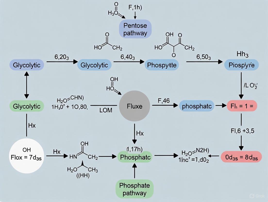
Metabolic Pathway and Regulation Diagram
The following diagram illustrates the interconnection between Glycolysis and the Pentose Phosphate Pathway, highlighting key regulatory nodes that control flux partitioning.
Core Concepts: The PPP in MS Neurodegeneration
Why is the Pentose Phosphate Pathway (PPP) a research focus in Multiple Sclerosis? In Multiple Sclerosis (MS), CD8+ T cells infiltrate the central nervous system (CNS) and contribute to axonal and neuronal injury, which underlies irreversible clinical progression [9]. Research shows that CD8+ T cells from patients with MS exhibit increased engagement of the PPP [9] [10]. This metabolic shift is crucial because it supports the cells' biosynthetic and redox demands, enabling their pro-inflammatory and cytotoxic functions within the CNS. Targeting this pathway therapeutically has been shown to disrupt these harmful effector functions and ameliorate autoimmunity in experimental models [9].
What is the metabolic relationship between neurons and immune cells in MS? A recent study highlights that in neurons, interferon-gamma (IFNγ) signaling induces the immunoproteasome subunit PSMB8. This leads to the accumulation of the metabolic regulator PFKFB3, which forces a shift in neuronal glucose metabolism from the PPP towards glycolysis [11]. This reprogramming depletes antioxidants like NADPH and glutathione, increasing neuronal vulnerability to ferroptosis and damage [11]. This creates a damaging interplay: inflamed neurons become metabolically susceptible, while autoreactive CD8+ T cells, dependent on the PPP, are tasked with immune surveillance but ultimately cause harm.
Experimental Protocols & Methodologies
Protocol 1: Quantitative Assessment of Glycolytic and PPP Fluxes using Stable Isotope Labeling
For researchers investigating dynamic metabolic fluxes, particularly in systems with rapid glucose consumption like activated immune cells, stable isotope labeling coupled with LC-MS/MS is a powerful method [12].
- Summary: This protocol uses a pulse of 13C-labeled glucose to trace carbon flow through glycolysis, the PPP, and the TCA cycle. The key is to define very short, precise time windows to capture label incorporation into metabolic intermediates before isotopic saturation occurs, enabling quantitative flux comparisons [12].
- Detailed Workflow:
- Cell Preparation and Labeling: Grow cells in standard media (e.g., YP with 2% glucose). For the experiment, rapidly introduce a pulse of 50% 13C6-glucose solution to the cell culture [12].
- Rapid Quenching and Metabolite Extraction: At defined short time intervals (e.g., 10 seconds for glycolytic intermediates), quench metabolism instantly using a pre-chilled (-40°C) 60% methanol solution (Quenching Buffer). Metabolites are then extracted from the quenched cells using 75% ethanol (Extraction Buffer) at 80°C [12].
- LC-MS/MS Analysis and Flux Quantification: Analyze the extracted metabolites via Liquid Chromatography-tandem Mass Spectrometry (LC-MS/MS). The rate and pattern of 13C-label incorporation into different intermediates are used to calculate the flux through glycolysis, PPP, and related pathways [12].
Protocol 2: Investigating the Functional Impact of PPP Inhibition on CD8+ T Cells
To directly test the functional role of the PPP in CD8+ T cell-mediated neurotoxicity, a combination of in vitro and in vivo approaches can be used [9].
- Summary: This approach involves pharmacologically inhibiting the PPP in CD8+ T cells and measuring subsequent changes in their metabolism, effector functions, and ability to cause neuronal damage.
- Detailed Workflow:
- T Cell Activation and Treatment: Isolate CD8+ T cells (e.g., from murine spleen or human blood) and activate them via CD3 and CD28 ligation. Treat the cells with a PPP inhibitor versus a vehicle control [9].
- Metabolic and Functional Assays:
- Metabolism: Measure reductions in glycolysis, glucose uptake, NADPH/ATP production using standard assays.
- Proliferation: Quantify via CFSE dilution or similar method.
- Cytokine Secretion: Assess proinflammatory cytokine (e.g., IFN-γ) levels by flow cytometry or ELISA [9].
- Neuronal Injury Models:
- In Vitro: Co-culture activated, antigen-specific CD8+ T cells with neuronal targets and quantify injury.
- In Vivo: Adoptively transfer these T cells into models like the cuprizone-induced demyelination model or experimental autoimmune encephalomyelitis (EAE) and assess disease progression and axonal injury [9].
Troubleshooting Common Experimental Challenges
FAQ: How can I accurately measure rapid glycolytic and PPP fluxes in cells with high metabolic rates? Challenge: In Crabtree-positive cells (including activated immune cells), glycolysis operates at near-saturation, causing 13C-label from glucose to incorporate into intermediates extremely rapidly (within seconds), leading to immediate isotopic saturation. This makes it difficult to track and compare flux rates [12]. Solution: It is critical to define and use very short, linear time windows for sampling before label saturation occurs. For instance, one protocol establishes that a 13C-glucose pulse saturates glycolytic intermediates in yeast within 10 seconds. Sampling at earlier time points (e.g., 5-10 seconds) is essential to capture the linear increase in label incorporation and enable quantitative flux comparisons between different cell states [12].
FAQ: What should I do if I encounter signal drift or batch effects in large-scale LC-MS metabolomics studies? Challenge: In large-scale studies where samples are run in multiple batches, instrumental drift and between-batch variation can introduce systematic errors, compromising data integrity [13]. Solution:
- Quality Controls (QCs): Inject a pooled QC sample repeatedly throughout the batch and between batches. These QCs are used for post-acquisition data normalization (e.g., using QC-SVRC or similar algorithms) to correct for instrumental drift [13].
- Internal Standards (IS): Use a cocktail of isotopically labeled internal standards (e.g., deuterated or 13C-labeled metabolites) covering different chemical classes. These help monitor instrument performance, though their intensity in untargeted studies should not be used for direct normalization between batches due to potential matrix effects [13].
- Sample Randomization and Replication: Randomize experimental samples across batches and include a subset of identical case samples in all batches to assess and correct for inter-batch variation [13].
FAQ: My model shows inconsistent flux rerouting upon oxidative stress. What regulatory mechanisms should I consider? Challenge: The redistribution of flux from glycolysis to the PPP during oxidative stress is complex and involves multiple, coordinated regulatory steps. An incomplete model may fail to capture the true dynamics [3]. Solution: Kinetic modeling studies suggest that efficient rerouting relies on a complementary set of regulations:
- Upregulation of PPP: The consumption of NADPH by the antioxidant machinery lowers the NADPH/NADP+ ratio, which relieves the inhibition of G6PD (the first and rate-limiting enzyme of the oxidative PPP) [3].
- Inhibition of Glycolysis: Simultaneously, key glycolytic enzymes are inhibited. This includes allosteric inhibition of Phosphoglucose Isomerase (PGI) by 6-phosphogluconate (6PG) and oxidative inhibition of Glyceraldehyde-3-phosphate dehydrogenase (GAPD) [3]. Ensuring your experimental or computational model accounts for this joint regulation is key.
Research Reagent Solutions
Table: Key reagents for investigating PPP in immunometabolism.
| Reagent / Tool | Primary Function / Application | Example & Notes |
|---|---|---|
| 13C6-Glucose | Stable isotope tracer for quantifying glycolytic and PPP fluxes via LC-MS/MS [12]. | Used in pulse-labeling experiments to track carbon fate. |
| PPP Inhibitors | Pharmacologic inhibition to probe PPP function in T cell effector responses [9]. | Example: 6-AN (6-aminonicotinamide). Validated to reduce CD8+ T cell glycolysis, NADPH production, and cytotoxicity [9]. |
| LC-MS/MS System | High-sensitivity platform for identifying and quantifying metabolites and isotopic labeling patterns [12] [13]. | Critical for fluxomics and metabolomics. |
| Labeled Internal Standards | Monitor instrument performance and aid in metabolite quantification in large-scale metabolomic studies [13]. | Cocktail of deuterated/13C-labeled compounds (e.g., LPC-D7, carnitine-D3, amino acid-13C,15N). |
| Anti-PD-L1/PD-L2 Blocking Antibodies | Investigate the role of co-inhibitory signals in T cell migration and function at the BBB [14]. | Used in vitro to block PD-1/PD-L interactions on brain endothelial cells. |
Pathway Diagrams
Neuronal metabolic vulnerability in MS.
PPP's role in CD8+ T cell-mediated damage.
Table: Key quantitative findings from foundational studies.
| Experimental Observation | Quantitative Result / Change | Context / Model |
|---|---|---|
| CD8+ T Cell Effector Functions after PPP Inhibition | Reduced glycolysis, glucose uptake, NADPH/ATP production, proliferation, and proinflammatory cytokine secretion [9]. | In vitro human/murine CD8+ T cell activation. |
| Neuronal PSMB8 & PFKFB3 Link | IFNγ-induced PSMB8 stabilizes PFKFB3, shifting metabolism from PPP to glycolysis, depleting GSH and promoting ferroptosis [11]. | Neuronal cultures, EAE models, and post-mortem MS brain tissue. |
| PD-L1/PD-L2 Blockade on T Cell Migration | Increased transmigration of CD8+ and CD4+ T cells across a human brain endothelial cell (HBEC) barrier [14]. | In vitro model of the blood-brain barrier (BBB). |
| Metabolic Flux Redistribution under Oxidative Stress | oxPPP flux increased to ~60% of glucose import flux; lower glycolytic flux (below GAP) reduced ~3-fold [3]. | 13C-fluxomics in human fibroblasts exposed to H2O2. |
Troubleshooting Guides and FAQs
Frequently Asked Questions
FAQ 1: What are the expected functional consequences of PKM2 deletion in CD8+ TILs? PKM2 deletion disrupts the canonical Warburg effect, forcing a metabolic switch. Cells may compensate by enhancing mitochondrial oxidative phosphorylation and potentially increasing flux through alternative anabolic pathways like the pentose phosphate pathway to maintain redox balance and nucleotide synthesis. This can alter T cell differentiation and function, potentially enhancing memory-like phenotypes but possibly at the cost of immediate effector function [15] [16].
FAQ 2: Why might my measurements of PPP flux be inconsistent after PKM2 modulation? Inconsistent PPP flux measurements can arise from several factors:
- Failure to Reach Isotopic Steady State: Mammalian cells can take 4 hours to a full day to fully incorporate isotopic tracers. Measurements taken before this point reflect a transient, non-stationary state [17].
- Dynamic Metabolic Compensation: The cell's metabolic network is interconnected. Inhibiting or deleting PKM2 can trigger immediate compensatory changes in glutamine metabolism or fatty acid oxidation, which can indirectly influence PPP flux. Your measurement might be capturing one moment in a dynamic adaptive process [15] [18].
- Background Nutrient Conditions: The concentrations of glucose and other nutrients in the medium during the experiment critically influence carbon allocation. Even short-term glucose restriction (TGR) has been shown to rewire T cell metabolism, leading to enhanced carbon allocation to the PPP upon glucose re-exposure [16].
FAQ 3: How can I confirm that observed metabolic changes are specific to PKM2 deletion and not general toxicity? It is crucial to implement robust experimental controls:
- Viability and Proliferation Assays: Continuously monitor cell count, viability, and apoptosis markers.
- Rescue Experiments: Re-express a catalytically active PKM2 construct in the knockout cells to see if it restores the wild-type metabolic phenotype.
- Metabolic Phenotyping: Use extracellular flux analyzers to measure real-time glycolysis (ECAR) and mitochondrial respiration (OCR). A toxic insult often causes a global depression of both pathways, whereas a specific metabolic switch might show a decrease in glycolysis coupled with an increase in oxidative phosphorylation [18].
Troubleshooting Common Experimental Issues
Problem 1: Low Efficiency of PKM2 Deletion in Primary CD8+ T Cells
- Potential Cause: Low transduction efficiency with CRISPR/Cas9 vectors or poor siRNA transfection efficiency in non-dividing or slowly dividing T cells.
- Solutions:
- Optimize activation protocol prior to transduction using anti-CD3/CD28 beads.
- Utilize high-titer lentiviral vectors rather than adenoviral vectors.
- Consider using Cas9 protein pre-complexed with guide RNA (ribonucleoprotein) for electroporation, which can increase knockout efficiency.
- Implement a fluorescence-based sorting strategy to isolate successfully transduced cells.
Problem 2: High Variation in 13C-MFA Data from TIL Cultures
- Potential Cause: Underlying heterogeneity in T cell activation states or failure to culture cells in a metabolic steady state before tracer introduction.
- Solutions:
- Standardize Activation: Use a consistent, defined protocol for T cell activation and expansion.
- Ensure Metabolic Steady State: Culture cells for at least 24-48 hours in the experimental medium before adding the isotopic tracer. Ensure cell growth rates and metabolite concentrations (e.g., glucose, lactate) are stable [17] [19].
- Use Purified T Cell Populations: Isolate CD8+ T cells from tumors using magnetic or fluorescence-activated cell sorting to reduce contamination by other cell types.
- Increase Biological Replicates: The inherent variability of primary cell cultures, especially TILs, necessitates a higher number of replicates (n ≥ 5-6) for robust 13C-MFA [17].
Problem 3: Difficulty in Distinguishing Direct vs. Indirect Effects on PPP Flux
- Potential Cause: PKM2 has both metabolic and non-metabolic (e.g., transcriptional) functions. Altered PPP flux might be a secondary consequence of changes in gene expression or overall cellular growth rate.
- Solutions:
- Use a Tetramer-Destabilizing PKM2 Mutant: Employ a PKM2 mutant that is constitutively locked in the high-activity tetrameric form. This tests if the effects are due to loss of enzymatic activity versus loss of its protein kinase or transcriptional co-regulator functions [15].
- Short-Term Pharmacological Inhibition: Compare the acute effects (within hours) of a small-molecule PKM2 inhibitor with the long-term effects of genetic deletion.
- Measure Key Metabolites: Use targeted metabolomics to measure the levels of PPP intermediates (e.g., glucose-6-phosphate, 6-phosphogluconate, ribose-5-phosphate) and nucleotides to build a more complete picture [16].
Experimental Protocols for Key Techniques
Protocol 1: Measuring PPP Flux Using [1,2-13C]Glucose Tracer and LC-MS
Principle: [1,2-13C]Glucose enters the oxidative PPP, where the first carbon is lost as CO2. The resulting ribulose-5-phosphate is labeled in carbons 1-2. Through non-oxidative PPP reactions, this labeling pattern is redistributed to glycolytic intermediates like fructose-6-phosphate (F6P) and glyceraldehyde-3-phosphate (G3P), producing unique mass isotopomer patterns that allow quantification of PPP flux relative to glycolysis [17].
Procedure:
- Cell Preparation: Isolate and activate CD8+ T cells or culture CD8+ TILs. For PKM2-deleted cells, perform genetic manipulation 3-4 days before the experiment.
- Tracer Introduction: Wash cells and replace standard culture medium with a physiologically buffered medium (e.g., RPMI 1640 without glucose and glutamine) supplemented with 10-15 mM [1,2-13C]glucose and 2-4 mM unlabeled glutamine. Ensure cells are in metabolic steady state.
- Incubation & Quenching: Incubate cells for 4-24 hours (time must be determined empirically to reach isotopic steady state). At time points, rapidly quench metabolism by adding cold (-20°C) methanol and subsequently extract intracellular metabolites with a methanol/water/chloroform mixture [17] [19].
- LC-MS Analysis:
- Chromatography: Use a hydrophilic interaction liquid chromatography (HILIC) column to separate sugar phosphates and other polar metabolites.
- Mass Spectrometry: Analyze extracts using a high-resolution mass spectrometer. Key metabolites to monitor include glucose-6-phosphate (G6P), 6-phosphogluconate (6PG), ribose-5-phosphate (R5P), F6P, G3P, and lactate.
- Data Analysis and Flux Calculation:
- Determine the mass isotopologue distributions (MIDs) for each metabolite.
- Input the MIDs, extracellular fluxes (glucose consumption, lactate production), and a metabolic network model into specialized software (e.g., INCA, 13CFLUX2) to compute the absolute fluxes through glycolysis and the PPP [17] [19].
Protocol 2: Assessing CD8+ T Cell Functional Capacity Post-PKM2 Deletion
Principle: This multi-parametric assay evaluates whether metabolic reprogramming induced by PKM2 deletion enhances or impairs critical anti-tumor functions like cytokine production and tumor cell killing.
Procedure:
- Metabolic Conditioning:
- Control TE: Culture activated, PKM2-deleted or control CD8+ T cells in complete medium (e.g., 10mM glucose).
- TGR TE (Transient Glucose Restriction): Culture an aliquot of the same cells for 20 hours in medium containing low glucose (e.g., 1mM) to mimic nutrient stress and trigger metabolic adaptation [16].
- Functional Co-culture:
- Re-plate both Control TE and TGR TE cells in fresh, high-glucose medium.
- Co-culture them with target tumor cells (e.g., B16-OVA spheroids or adherent cells) at a defined effector-to-target ratio (e.g., 1:1).
- Output Measurement (at 24 hours post-co-culture):
- Intracellular Cytokine Staining: Use brefeldin A to block protein secretion, then fix, permeabilize, and stain for IFN-γ and Granzyme B. Analyze via flow cytometry. TGR-conditioned T cells often show increased MFI and percentage of positive cells for these effector molecules [16].
- Cytotoxicity Assay: Use a real-time cell killing assay (e.g., Incucyte with labeled targets) or a standard 51Cr-release assay to quantify specific lysis of tumor cells.
- Metabolic Phenotyping: Simultaneously, analyze the metabolic state of the T cells after co-culture using a Seahorse Analyzer to measure OCR and ECAR [18].
The Scientist's Toolkit: Research Reagent Solutions
Table 1: Essential Reagents for Investigating PKM2 and PPP in T Cells
| Reagent Name | Function/Brief Explanation | Example Application |
|---|---|---|
| [1,2-13C]Glucose | Tracer for quantifying PPP flux; generates unique labeling pattern in glycolysis intermediates. | 13C-MFA to determine fractional contribution of PPP to NADPH and ribose production [16] [17]. |
| CB-839 (Telaglenastat) | Potent and selective inhibitor of glutaminase 1 (GLS1). | Blocks glutamine-to-glutamate conversion; tests T cell reliance on glutaminolysis when glycolysis is impaired [18]. |
| 2-Deoxy-D-glucose (2-DG) | Glucose analog that inhibits hexokinase and glycolysis. | Induces transient glucose restriction to study metabolic adaptation and its functional impact on T cells [16]. |
| Recombinant IL-15 | Cytokine that promotes development of memory-like T cells with enhanced mitochondrial metabolism. | Culture cytokine to generate T cells with high spare respiratory capacity for migration and persistence studies [18]. |
| Etomoxir | Irreversible inhibitor of carnitine palmitoyltransferase 1A (CPT1A), blocking fatty acid oxidation (FAO). | Tests the role of FAO in supporting T cell function when PKM2 is deleted; studies suggest it may have off-target effects [18]. |
| 6,8-Bis(benzylthio)-octanoic acid | Inhibitor of key TCA cycle enzymes (PDH & OGDH). | Collapses mitochondrial respiration; used to demonstrate the essential role of the TCA cycle in supporting T cell 3D migration [18]. |
| Anti-CD3/CD28 Dynabeads | Artificial antigen-presenting cell system for robust and consistent polyclonal T cell activation. | Standardized activation of primary CD8+ T cells prior to genetic modification or metabolic assays. |
Table 2: Key Quantitative Findings from Relevant Studies
| Cell Type / Condition | Metabolic Intervention | Key Quantitative Findings | Experimental Method | Citation |
|---|---|---|---|---|
| Mouse CD8+ TE Cells | Transient Glucose Restriction (TGR: 1mM glucose for 20h) | Upon glucose re-exposure: ↑ Glucose uptake; ↑ Carbon allocation to PPP; ↑ IFN-γ production (MFI); ↑ Granzyme B (MFI & %+ cells). | LC-MS metabolomics, flow cytometry | [16] |
| Human CD8+ T Cells | TCA cycle inhibition (6,8bOA) | Strong decrease in 3D motility in collagen gels. Combination inhibition (Glycolysis + TCA) reduced motility to <10% of initial. | Extracellular flux analysis, live-cell imaging | [18] |
| Human CD8+ T Cells | Acute (1h) glucose vs. glutamine deprivation | Glucose deprivation reduced 3D motility, glutamine deprivation did not. With matched concentrations, both were similarly required. | Live-cell imaging in 3D collagen | [18] |
| Cancer Cells (General) | PKM2 expression & function | PKM2 dimers promote Warburg effect (aerobic glycolysis); PKM2 tetramers favor oxidative metabolism. Nuclear PKM2 acts as a protein kinase and transcriptional co-activator. | Various biochemical and omics assays | [15] [20] |
Signaling Pathway and Metabolic Cross-Talk Visualization
Diagram 1: Metabolic Adaptation to PKM2 Deletion in CD8+ T Cells. This diagram illustrates the key metabolic shifts and functional consequences resulting from PKM2 deletion, highlighting the potential rerouting of glycolytic intermediates into the PPP and the increased reliance on mitochondrial metabolism.
Diagram 2: Experimental Workflow for 13C Metabolic Flux Analysis. This diagram outlines the key steps in a stable isotope-based flux experiment, from introducing the labeled tracer to computational modeling of the resulting data to extract quantitative flux values.
The pentose phosphate pathway (PPP), a fundamental metabolic pathway branching from glycolysis, has emerged as a critical regulator of immune cell function. In immune cells, the PPP serves two primary functions: it generates nicotinamide adenine dinucleotide phosphate (NADPH) for redox balance and biosynthetic reactions, and produces ribose-5-phosphate (R5P) for nucleotide synthesis [21] [22]. These outputs are particularly vital for activated immune cells, which require substantial biosynthetic precursors to support proliferation and effector functions. Recent research has revealed that proinflammatory immune cells, including autoreactive CD8+ T cells in autoimmune diseases, undergo metabolic reprogramming that increases their reliance on the PPP [23] [24]. This dependency creates a therapeutic opportunity—by selectively inhibiting the PPP, it may be possible to disrupt the pathogenic functions of these cells while sparing other immune populations.
Key Quantitative Findings on PPP Inhibition
The table below summarizes core quantitative findings from recent studies investigating PPP inhibition in disease models, providing a consolidated reference for researchers assessing the potential efficacy of this approach.
Table 1: Quantitative Effects of PPP Inhibition in Experimental Models
| Study Context | PPP Inhibitor Used | Key Quantitative Findings | Biological Outcome |
|---|---|---|---|
| Multiple Sclerosis (CD8+ T cells) [23] | 6-Aminonicotinamide (6AN) | - ~50% reduction in NADPH production- Glycolytic capacity suppressed to unstimulated levels- Significant decrease in proinflammatory cytokine secretion | Reduced CD8+ T cell-mediated neuronal injury in vitro and in mouse models |
| Xenopus Tail Regeneration [25] | Not Specified | Increased glucose directed toward PPP, not glycolysis; PPP essential for proliferation | PPP inhibition decreased cell division in regenerating tissue |
| Sepsis (PBMCs) [26] | Not Specified | Proteins related to PPP were upregulated in septic patients vs. healthy controls | Inhibition impaired phagocytosis and cytokine production |
Essential Experimental Protocols
Protocol 1: Assessing PPP Inhibition in Activated CD8+ T CellsIn Vitro
This protocol is adapted from studies investigating metabolic reprogramming of autoreactive T cells in multiple sclerosis [23].
Research Objective: To evaluate the functional and metabolic consequences of PPP inhibition in CD8+ T cells.
Key Reagents & Materials:
- Immunomagnetically isolated CD8+ T cells (murine or human)
- PPP inhibitor: 6-Aminonicotinamide (6AN, 100-200 µM) or Polydatin
- Plate-bound anti-CD3 and soluble anti-CD28 antibodies (for activation)
- Glucose analog: 2-NBDG (for uptake assays)
- LC-MS equipment and stable isotopes (D-glucose-1,2-13C2) for flux analysis
Methodology:
- Cell Isolation & Culture: Isolate CD8+ T cells from spleen or peripheral blood using negative selection magnetic sorting.
- Activation & Inhibition: Stimulate cells with plate-bound anti-CD3 (e.g., 5 µg/mL) and soluble anti-CD28 (e.g., 2 µg/mL). Co-treat with 6AN (100 µM) or vehicle control.
- Metabolic Phenotyping:
- Glycolytic Flux: Measure the Extracellular Acidification Rate (ECAR) using a Seahorse Analyzer.
- Glucose Uptake: Quantify uptake via flow cytometry using 2-NBDG.
- PPP Flux: Use stable isotope tracing with D-glucose-1,2-13C2 and LC-MS to track incorporation into metabolites, calculating Pentose Cycle Activity (PCA).
- Functional Assays:
- Proliferation: Measure via CFSE dilution or Ki67 staining.
- Cytokine Production: Quantify IFN-γ and TNF-α levels in supernatant by ELISA.
- NADPH Production: Use a bioluminescent NADP/NADPH-Glo Assay on cell lysates.
Technical Notes: Cell viability must be monitored concurrently, as 6AN at concentrations up to 200 µM did not significantly affect T cell survival in referenced studies [23]. A dose-response curve for NADPH production inhibition is recommended for new experimental systems.
Protocol 2: Validating PPP Inhibition Efficacy in anIn VivoAutoimmunity Model
This protocol outlines the use of the Experimental Autoimmune Encephalomyelitis (EAE) model, a standard for studying multiple sclerosis [23] [22].
Research Objective: To determine the therapeutic potential of PPP inhibition in suppressing CNS autoimmunity in vivo.
Key Reagents & Materials:
- C57BL/6 mice
- PPP inhibitor: 6AN
- MOG35-55 peptide for EAE induction
- Cuprizone for demyelination studies (optional)
Methodology:
- Disease Induction: Induce EAE in mice by subcutaneous immunization with MOG35-55 peptide emulsified in Complete Freund's Adjuvant, followed by pertussis toxin injections.
- Therapeutic Intervention: Administer 6AN (e.g., 20 mg/kg, i.p.) or vehicle control daily, starting at disease onset or prophylactically.
- Disease Monitoring: Score mice daily for clinical signs of EAE (0: no symptoms, 5: moribund).
- Endpoint Analysis:
- Histopathology: Analyze spinal cord sections for immune cell infiltration (e.g., CD8+ T cells) and demyelination (e.g., LFB staining).
- Flow Cytometry: Isolate CNS-infiltrating cells and profile T cell populations and activation states.
- Metabolic Analysis: Examine T cells ex vivo for glucose uptake and effector functions.
Technical Notes: The adoptive transfer of autoreactive CD8+ T cells can be combined with this model to specifically track the impact of PPP inhibition on pathogenic T cells [23].
The Scientist's Toolkit: Essential Research Reagents
The table below catalogs key reagents crucial for conducting research on PPP inhibition in immunometabolism.
Table 2: Key Research Reagents for PPP Flux and Inhibition Studies
| Reagent Name | Primary Function / Target | Key Application in Research |
|---|---|---|
| 6-Aminonicotinamide (6AN) [23] | Inhibits G6PD (PPP enzyme) | Reduces NADPH production and nucleotide synthesis; used to test T cell dependency on PPP. |
| Polydatin [23] | Inhibits G6PD | Suppresses glycolytic flux and T cell activation; an alternative to 6AN. |
| 2-NBDG [23] | Fluorescent glucose analog | Measures cellular glucose uptake via flow cytometry. |
| D-glucose-1,2-13C2 [23] | Stable isotope tracer | Enables precise measurement of PPP flux via LC-MS. |
| UK5099 [27] | Mitochondrial pyruvate carrier (MPC) inhibitor | Blocks pyruvate entry into mitochondria; useful for studying metabolic crosstalk. |
Troubleshooting Common Experimental Challenges
FAQ 1: Why does PPP inhibition in my T cell cultures show minimal effect on proliferation, contrary to published findings?
- Potential Cause 1: Incomplete Pathway Inhibition. Confirm that your inhibitor concentration is sufficient. Validate efficacy by measuring NADPH/NADPH ratios or incorporating stable isotope tracing (e.g., D-glucose-1,2-13C2) to directly quantify PPP flux reduction [23].
- Potential Cause 2: Metabolic Compensation. T cells might upregulate alternative pathways, such as mitochondrial metabolism or amino acid catabolism, to bypass the PPP block. Assess oxygen consumption rates (OCR) and experiment with combination treatments, such as adding an OXPHOS inhibitor [21] [27].
- Potential Cause 3: Cell Activation State. The effect of PPP inhibition is most pronounced in strongly activated, proinflammatory T cells. Verify your activation protocol (e.g., CD3/CD28 ligation strength) and characterize the resulting T cell phenotype (e.g., effector vs. memory) [23] [24].
FAQ 2: How can I specifically measure PPP flux without relying on indirect proxies like NADPH levels?
- Recommended Solution: Employ Stable Isotope Tracing. This is the gold-standard method. Feed cells labeled D-glucose-1,2-13C2. The PPP-specific cleavage of the first two carbon atoms leads to a unique labeling pattern in downstream metabolites. By using LC-MS, you can quantify the fraction of, for example, lactate that is m+1 labeled, which directly reports on PPP activity and provides a more precise flux measurement than static NADPH levels [23].
FAQ 3: My in vivo administration of a PPP inhibitor is causing off-target toxicity. How can I improve specificity?
- Strategy 1: Dose Optimization. Perform a detailed dose-response and kinetic study to find a therapeutic window that modulates immune function without general toxicity. Refer to studies using 6AN at 20 mg/kg in mice as a starting point [23].
- Strategy 2: Targeted Delivery Systems. Explore nanoparticle-based encapsulation or antibody-drug conjugates (ADCs) that target surface markers on activated T cells (e.g., CD44) to deliver the inhibitor specifically to pathogenic immune cells.
- Strategy 3: Explore More Selective Inhibitors. While 6AN and Polydatin are well-used, the field is developing newer compounds. Conduct a literature review for recent publications on selective G6PD or transketolase inhibitors that may have improved profiles.
Visualizing the Mechanism of PPP Inhibition
The diagram below illustrates the core metabolic pathway, the site of action for key inhibitors, and the subsequent biological impact on T cells.
Diagram 1: PPP inhibition mechanism and consequences. The diagram shows how inhibitors like 6AN target G6PD in the PPP, reducing the production of R5P and NADPH. This metabolic disruption leads to impaired proliferation and effector functions in proinflammatory CD8+ T cells, resulting in reduced pathogenicity in autoimmune contexts [23] [22] [24].
Metabolomics, the comprehensive study of small-molecule metabolites, relies heavily on three principal analytical technologies: gas chromatography-mass spectrometry (GC-MS), liquid chromatography-mass spectrometry (LC-MS), and nuclear magnetic resonance (NMR) spectroscopy [28]. Each platform offers distinct advantages and limitations, making them uniquely suited for different aspects of metabolic investigation. The choice of platform depends on the specific focus of the study, the nature of the samples, and the analytical information required [28]. While LC-MS and GC-MS account for more than 80% of published metabolomics studies due to their high sensitivity, NMR spectroscopy remains invaluable for its quantitative capabilities, reproducibility, and ability to study intact tissues and living systems [28].
In the specific context of glycolytic and pentose phosphate pathway (PPP) research, these techniques enable precise tracking of metabolic fluxes, particularly when combined with stable isotope labeling. The PPP is fundamental to glucose metabolism, involved in nucleotide biosynthesis and redox homeostasis, with its flux significantly increasing following oxidative stress to generate NADPH required for antioxidant defense [3]. Understanding the precise regulation of these pathways requires analytical approaches capable of capturing both metabolite concentrations and flux dynamics.
Technical Comparison of Analytical Platforms
The selection of an appropriate analytical platform is crucial for successful metabolomic investigation. Each technology offers distinct capabilities with respect to sensitivity, coverage, and analytical information.
Table 1: Technical Comparison of GC-MS, LC-MS, and NMR for Metabolomics
| Parameter | GC-MS | LC-MS | NMR |
|---|---|---|---|
| Sensitivity | ~1 nM (after derivatization) [28] | 10-100 nM [28] | >1 μM [28] |
| Sample Preparation | Requires chemical derivatization for most metabolites [28] | Minimal to moderate; protein precipitation often sufficient [13] | Minimal; transfer to NMR tube with deuterated solvent [28] |
| Reproducibility | Moderate | Less reproducible than NMR [28] | Exceptionally high reproducibility [28] |
| Metabolite Coverage | Volatile compounds, organic acids, sugars, amino acids [28] | Broad range of polar to non-polar metabolites [29] | Typically 50-200 identified metabolites [28] |
| Quantitation | Relative quantitation possible; requires internal standards | Semi-quantitative; ionization efficiency varies [28] | Inherently quantitative; signal proportional to concentration [28] |
| Structural Information | Fragmentation patterns | Fragmentation patterns, accurate mass | Atomic-level structural detail, molecular dynamics |
| Sample Recovery | Destructive [28] | Destructive [28] | Non-destructive; sample can be recovered [28] |
| Flux Analysis Capability | Yes, with isotopic labeling | Yes, with isotopic labeling | Excellent for in vivo and in vitro flux analysis [28] |
| Key Strengths | High sensitivity, robust identification | Broad metabolome coverage, high sensitivity | Quantitative, non-destructive, studies intact tissues [28] |
| Key Limitations | Limited to volatile or derivatizable compounds | Ion suppression effects, matrix effects | Lower sensitivity, spectral overlap [28] |
Table 2: Suitability for Glycolytic and PPP Metabolite Analysis
| Pathway | Key Metabolites | GC-MS | LC-MS | NMR |
|---|---|---|---|---|
| Glycolysis | Glucose-6-phosphate, Fructose-6-phosphate, Glyceraldehyde-3-phosphate, Pyruvate | Moderate (requires derivatization) | Good (ion pairing may be needed) | Good for abundant intermediates |
| Pentose Phosphate Pathway | Ribose-5-phosphate, Sedoheptulose-7-phosphate, Erythrose-4-phosphate | Good for sugar phosphates | Excellent with HILIC chromatography | Good for pathway flux determination |
| Redox Cofactors | NADP+/NADPH, NAD+/NADH | Not suitable | Challenging due to instability | Excellent for ratio determinations |
| Energy Metabolites | ATP, ADP, AMP | Not suitable | Good with reverse-phase chromatography | Excellent for phosphorus-containing metabolites |
Diagram 1: Metabolomics Workflow from Sample to Interpretation
Troubleshooting Guides and FAQs
GC-MS Troubleshooting
Q: My GC-MS analysis shows poor peak shape for sugar phosphates from the PPP. What could be the issue?
A: Poor peak shape in GC-MS analysis of PPP metabolites typically stems from derivatization issues or column degradation. First, ensure complete derivatization by checking that your methoximation and silylation steps are performed under anhydrous conditions. Sugar phosphates are highly polar and require complete derivatization for adequate volatility. Second, check column performance – sugar phosphates can be challenging due to their polarity and may require column trimming or replacement. Third, optimize the temperature ramp – a slower ramp often improves separation of isomeric compounds like ribose-5-phosphate and ribulose-5-phosphate.
Q: I'm observing significant variation in retention times across runs in my GC-MS flux analysis. How can I improve stability?
A: Retention time drift in GC-MS can compromise flux analysis accuracy. Implement these corrective measures: (1) Use retention index markers (alkanes) in every run to enable post-acquisition alignment; (2) Ensure proper inlet maintenance – replace liners and seals regularly; (3) Maintain consistent injection technique and volume; (4) Allow sufficient time for oven temperature equilibration between runs; (5) Consider using a retention time locking method if available on your instrument.
LC-MS Troubleshooting
Q: My LC-MS signal intensity drops significantly during large-scale batch analysis of glycolytic intermediates. What should I do?
A: Signal attenuation in large-scale LC-MS studies is common. Implement these strategies: (1) Incorporate quality control (QC) samples prepared from a pool of all samples and analyze them at regular intervals (every 5-10 samples) to monitor performance [13]; (2) Clean the ionization source between batches to prevent contamination buildup [13]; (3) Use labeled internal standards to correct for signal drift, though be aware that in untargeted studies, these should be carefully selected to avoid interference with unknown metabolites [13]; (4) Consider dividing large studies into multiple batches with proper normalization [13].
Q: How can I improve the chromatographic separation of glycolytic and PPP intermediates that co-elute in my HILIC method?
A: Co-elution of polar intermediates is a common challenge. Optimization approaches include: (1) Fine-tune mobile phase pH – slight adjustments can significantly alter selectivity for compounds like glucose-6-phosphate and fructose-6-phosphate; (2) Adjust buffer concentration – higher ammonium acetate or formate concentrations (e.g., 10-20 mM) can improve peak shape; (3) Extend gradient time or use a shallower gradient to increase separation window; (4) Consider column temperature optimization – some separations benefit from elevated temperatures (35-45°C); (5) Evaluate alternative HILIC stationary phases (silica, amino, amide) as each offers different selectivity.
NMR Troubleshooting
Q: The NMR spectra of my cell extracts show overlapping peaks for PPP intermediates, making quantification difficult. What solutions can I implement?
A: Spectral overlap is a fundamental limitation of NMR. Consider these approaches: (1) Implement 2D NMR experiments such as 1H-13C HSQC which spreads signals into a second dimension, resolving overlapping peaks [28]; (2) Use higher magnetic field strength if available (600 MHz or higher) which increases spectral dispersion [28]; (3) Employ mathematical deconvolution methods that use spectral databases of pure compounds to fit overlapping regions; (4) Adjust sample pH to slightly shift the chemical shifts of acidic protons; (5) For flux studies, use 13C-tracing with 13C-edited experiments which reduces spectral complexity by focusing only on labeled species.
Q: I need to monitor real-time flux through the PPP in response to oxidative stress, but NMR sensitivity is limiting. What are my options?
A: For dynamic flux measurements in stress response studies: (1) Use hyperpolarization techniques such as dynamic nuclear polarization (DNP) to temporarily enhance sensitivity by several orders of magnitude [30]; (2) Focus on 31P-NMR for monitoring nucleotide phosphates and sugar phosphates – this often provides better resolution than 1H-NMR for these key pathway metabolites [28]; (3) Employ cryoprobes if available to improve sensitivity approximately 4-fold; (4) Consider using larger sample volumes or higher cell densities when possible; (5) Implement non-uniform sampling in 2D experiments to reduce acquisition time while maintaining spectral quality.
Research Reagent Solutions for Pathway Flux Studies
Table 3: Essential Research Reagents for Glycolytic and PPP Flux Analysis
| Reagent Category | Specific Examples | Application in Pathway Analysis |
|---|---|---|
| Stable Isotope Tracers | [1,2-13C]glucose, [U-13C]glucose, [1-13C]glucose | Tracing carbon fate through glycolytic and PPP fluxes; determining flux partitioning at the glucose-6-phosphate branch point [3] |
| Internal Standards | Deuterated amino acids, 13C-labeled organic acids, deuterated lipids | Correction for sample preparation variability and instrument drift in MS-based analyses [13] |
| Enzyme Inhibitors/Activators | G6PD inhibitors, oxidative stress inducers (H₂O₂) | Probing pathway regulation and control points; studying stress response mechanisms [3] |
| Quality Control Materials | Pooled quality control samples, standard reference materials | Monitoring analytical performance across batches; ensuring data quality in long-term studies [13] |
| Derivatization Reagents | MSTFA, MOX reagent, methoxyamine hydrochloride | Enabling GC-MS analysis of non-volatile pathway intermediates [28] |
| NMR Solvents & Standards | Deuterated water (D₂O), TSP, DSS | Providing field frequency lock and chemical shift reference for NMR experiments [28] |
Experimental Protocols for Pathway Flux Analysis
Protocol: 13C-Labeling Experiment for PPP Flux Determination in Human Fibroblasts
This protocol outlines the procedure for determining PPP flux in response to oxidative stress using 13C-glucose tracing, adapted from kinetic modeling studies of human fibroblast cells [3].
Materials:
- Human fibroblast cell line
- [1,2-13C]glucose or alternative 13C-glucose isotopologue
- Oxidative stress inducer (H₂O₂)
- Quenching solution (cold methanol:acetonitrile:water, 40:40:20)
- Extraction solvents
- GC-MS or LC-MS system
- Appropriate culture medium
Procedure:
- Culture human fibroblast cells to 70-80% confluence in appropriate growth medium.
- Replace medium with fresh medium containing 13C-labeled glucose (e.g., 10 mM [1,2-13C]glucose).
- For stress conditions, add H₂O₂ to appropriate concentration (e.g., 500 μM) based on preliminary dose-response experiments.
- Incubate cells for specific time intervals (e.g., 0, 15, 30, 60, 120 minutes) to capture flux dynamics.
- At each time point, rapidly remove medium and quench metabolism with cold quenching solution.
- Extract intracellular metabolites using appropriate method (e.g., methanol/chloroform/water extraction).
- Derivatize samples for GC-MS analysis or prepare for direct LC-MS analysis.
- Analyze samples by GC-MS or LC-MS to determine isotopic labeling patterns in glycolytic and PPP intermediates.
- Use computational modeling approaches (e.g., Monte Carlo sampling of flux parameter space) to estimate flux distributions [3].
Key Considerations:
- The metabolic state in unstressed conditions typically shows ~20% of glucose import flux diverted toward oxPPP, increasing significantly under oxidative stress [3].
- Exposure to 500 μM H₂O₂ typically leads to significant reduction (approximately 3-fold) of metabolic flux in the lower glycolytic branch below glyceraldehyde-3-phosphate [3].
- Use appropriate controls with unlabeled glucose to determine natural isotope abundance background.
Protocol: LC-MS Based Large-Scale Metabolomics Study with Multi-Batch Analysis
This protocol provides guidance for large-scale metabolomic studies requiring analysis across multiple batches, particularly relevant for clinical studies of PPP flux in patient cohorts.
Materials:
- Serum or plasma samples
- Quality control (QC) pool sample
- Labeled internal standard mix (covering various metabolite classes)
- LC-QToF-MS system
- Appropriate mobile phases
Procedure:
- Prepare QC samples by pooling a small volume of all samples or a representative subset [13].
- Include a comprehensive internal standard mix covering different metabolite classes (e.g., deuterated lysophosphocholine, sphingolipid, fatty acid, carnitine, amino acid) to monitor system performance [13].
- Randomize sample injection order to avoid batch effects confounding biological groups.
- Begin each batch with system conditioning (e.g., 10 QC injections) [13].
- Analyze samples in sequences with QC injections every 5-10 samples to monitor signal stability.
- Include blank injections (extracting solvent) to identify and exclude signals from contamination.
- Clean ionization source between batches to maintain sensitivity [13].
- Use post-acquisition normalization algorithms to correct for both intra- and inter-batch systematic errors [13].
Key Considerations:
- In large-scale studies, sample preparation in smaller sets (e.g., n=32 per day) helps maintain sample integrity [13].
- Computer reboot at the beginning and between analysis modes can improve system stability [13].
- When equipment stops unexpectedly due to technical issues, careful data treatment is required to enable accurate joint analysis of multi-batch data sets [13].
Diagram 2: 13C-Flux Experiment Workflow for Pathway Analysis
The precision of glycolytic and pentose phosphate pathway flux research fundamentally depends on selecting appropriate analytical technologies and implementing robust experimental protocols. GC-MS, LC-MS, and NMR spectroscopy each contribute unique capabilities to this endeavor, with optimal experimental design often combining elements from multiple platforms. The troubleshooting guidance and standardized protocols provided here address common technical challenges in pathway flux analysis, enabling more reliable and reproducible metabolomic investigations. As fluxomics continues to evolve, the integration of these analytical approaches with computational modeling will further enhance our understanding of the complex regulation governing central carbon metabolism in health and disease.
Cutting-Edge Techniques: From Stable Isotope Tracing to Computational Modeling for Flux Analysis
Troubleshooting Guides
FAQ 1: How can I achieve quantitative accuracy in my flux measurements?
Issue: Measured fluxes and metabolite levels do not align with expected biological values or show poor reproducibility.
| Problem | Possible Cause | Solution | Preventive Measures |
|---|---|---|---|
| Inaccurate Flux Data [31] | Failure to correct for Natural Isotope Abundance (NIA) and tracer impurity. | Use dedicated correction software (e.g., IsoCorrectoR). | Always incorporate data correction into your analysis pipeline. |
| Poor Separation of Isobaric Metabolites [32] | Inadequate chromatographic resolution for sugar phosphates. | Implement a HILIC method with medronic acid and a pH 9.0 buffer [32]. | Validate chromatographic separation for all target metabolites before tracing experiments. |
| Non-Linear Flux Calculations [33] | Rapid glucose consumption in Crabtree-positive yeasts saturates labels. | Define a short, specific time window for sampling before label saturation occurs [33] [34]. | Perform a time-course experiment to establish the linear labeling phase. |
| Low Quantification Precision [35] | Lack of appropriate internal standards. | Use a (^{13}\text{C})-labeled yeast extract as an internal standard for isotope dilution [35]. | Use isotope-coded internal standards for all target metabolites. |
FAQ 2: What are the common pitfalls in sample preparation and how can I avoid them?
Issue: Degradation of metabolites or inconsistent quenching of metabolic activity leads to unreliable data.
| Step | Pitfall | Best Practice | Technical Tip |
|---|---|---|---|
| Quenching | Incomplete or slow halting of metabolism alters metabolite levels. | Use fast cold methanol quenching [35]. | Ensure quenching solution is pre-cooled and use a high sample-to-quenchant ratio. |
| Extraction | Inefficient metabolite recovery, especially for labile intermediates. | Employ boiling ethanol extraction [35]. | Optimize extraction solvent and temperature for your specific yeast strain. |
| Sampling Time | Capturing flux outside the linear dynamic range. | For fast glycolytic fluxes, use rapid pulsing and quenching in the time window before isotopic steady state [33]. | Determine the optimal sampling window through a pilot time-course experiment. |
Essential Methodologies
Core Protocol: Estimating Glycolytic Flux in Crabtree-Positive Yeasts
This protocol outlines a method to quantitatively estimate dynamic glycolytic and related carbon metabolic fluxes in Saccharomyces cerevisiae using stable isotope labeling and LC-MS/MS [33] [34].
Workflow Overview:
Detailed Procedure:
Cell Culture and Tracer Pulse:
- Grow Crabtree-positive yeast (e.g., S. cerevisiae) under defined conditions.
- Rapidly introduce a pulse of uniformly labeled (^{13}\text{C})-glucose (([U)-(^{13}\text{C}])-glucose) to the culture. Ensure rapid and homogenous mixing.
Rapid Sampling and Quenching:
- At defined time points post-pulse (e.g., seconds to a few minutes), rapidly withdraw culture aliquots.
- Immediately quench metabolism by injecting the sample into a large volume of cold methanol (e.g., -30°C to -40°C) [33] [35]. Critical: The time window for sampling must be determined empirically to capture label incorporation before saturation, enabling the observation of flux changes [33] [34].
Metabolite Extraction:
- Centrifuge the quenched samples to pellet cells.
- Perform metabolite extraction using a boiling ethanol (80% v/v) solution [35].
- Dry the extracts under a vacuum centrifuge and reconstitute in a solvent compatible with LC-MS analysis.
LC-MS/MS Analysis with HILIC:
- Chromatography: Use a HILIC column (e.g., Acquity UPLC BEH Amide). The mobile phase should be optimized for separating central carbon metabolites.
- Buffer A: 10 mM ammonium acetate in water, pH 9.0.
- Buffer B: 10 mM ammonium acetate in acetonitrile:water (9:1), pH 9.0.
- Add 5 µM medronic acid to both buffers to improve the separation and peak shape of sugar phosphates [32].
- Run a binary gradient from 95% B to 50% B over 8-10 minutes.
- Mass Spectrometry: Use tandem mass spectrometry (MS/MS) in scheduled multiple reaction monitoring (MRM) mode for high sensitivity and specificity of metabolite detection.
- Chromatography: Use a HILIC column (e.g., Acquity UPLC BEH Amide). The mobile phase should be optimized for separating central carbon metabolites.
Data Processing and Flux Analysis:
- Integrate the peak areas for the labeled isotopologues of glycolytic and PPP intermediates (e.g., G6P, F6P, R5P).
- Critical Correction: Use a tool like IsoCorrectoR to correct the raw MS data for Natural Isotope Abundance (NIA) and tracer impurity [31]. Neglecting this step can lead to severely distorted data and incorrect flux interpretations.
- Calculate labeling enrichments and fractions.
- Input the corrected labeling data, along with external uptake/secretion rates [36], into flux analysis software (e.g., INCA, Metran) to compute quantitative metabolic fluxes.
HILIC Method for Central Carbon Metabolites
This method provides a detailed protocol for the simultaneous separation and analysis of sugar phosphates, organic acids, and amino acids, which is crucial for accurate isotopologue detection [32].
Key Parameters for HILIC-MS/MS:
| Parameter | Specification | Function |
|---|---|---|
| Column | Acquity UPLC BEH Amide (2.1 x 100 mm, 1.7 µm) [32] | Separates polar metabolites. |
| Buffer A | 10 mM Ammonium Acetate in H₂O, pH 9.0 [32] | Aqueous mobile phase. |
| Buffer B | 10 mM Ammonium Acetate in ACN:H₂O (9:1), pH 9.0 [32] | Organic mobile phase. |
| Additive | 5 µM Medronic Acid [32] | Chelating agent that improves separation and peak shape of sugar phosphates. |
| Gradient | 95% B to 50% B over 8 min [32] | Elutes metabolites based on hydrophilicity. |
| MS Detection | Scheduled MRM on Tandem MS [32] | Enables specific and sensitive quantification of metabolites and their isotopologues. |
The Scientist's Toolkit: Research Reagent Solutions
| Reagent / Material | Function / Application | Specification / Note |
|---|---|---|
| [U-¹³C]-Glucose | Tracer substrate for quantifying carbon fate through glycolysis, PPP, and TCA cycle [33] [36]. | >99% isotopic purity; verify chemical and isotopic purity upon receipt. |
| ¹³C-Labeled Yeast Extract | Internal standard for absolute quantification via isotope dilution [35]. | In-vivo synthesized from P. pastoris; provides a biologically relevant matrix. |
| Cold Methanol | Quenching solution to instantaneously halt metabolic activity [35]. | LC-MS grade; pre-cool to -30°C to -40°C. |
| Boiling Ethanol | Extraction solvent for intracellular metabolites [35]. | 80% (v/v) in water. |
| HILIC Column | Chromatographic separation of polar metabolites (sugar phosphates, organic acids) [32]. | e.g., UPLC BEH Amide Column (1.7 µm). |
| Ammonium Acetate | Mobile phase buffer for HILIC separation [32]. | LC-MS grade, prepared at 10 mM and pH 9.0. |
| Medronic Acid | Mobile phase additive [32]. | Improves separation of isobaric metabolites (e.g., G6P/F6P). Use at 5 µM. |
| IsoCorrectoR Software | Corrects MS data for natural isotope abundance and tracer impurity [31]. | R-based, open-source tool. Essential for accurate flux determination. |
Frequently Asked Questions (FAQs)
Q1: What is the core innovation of the TIObjFind framework compared to traditional FBA? Traditional Flux Balance Analysis (FBA) often uses a single objective function, like biomass maximization, which may not accurately capture flux distributions under all conditions [37]. TIObjFind addresses this by integrating Metabolic Pathway Analysis (MPA) with FBA to systematically infer context-specific metabolic objectives from experimental data [37]. Its core innovation is the introduction of Coefficients of Importance (CoIs), which quantify each reaction's contribution to a hypothesized objective function, thereby aligning model predictions with experimental flux data and revealing shifting metabolic priorities in different biological stages [37].
Q2: What are the typical outputs of a TIObjFind analysis, and how are they interpreted? The primary output includes the Coefficients of Importance (CoIs). A higher CoI value for a reaction indicates that its flux aligns closely with its maximum potential, suggesting the experimental data is directed toward optimal values for specific pathways [37]. Furthermore, the framework produces a Mass Flow Graph (MFG), a directed, weighted graph representation of metabolic fluxes that facilitates pathway-based interpretation and analysis [37].
Q3: My TIObjFind model is infeasible. What are the first things I should check? Model infeasibility often stems from gaps in the network or incorrect constraints. The first steps should be:
- Verify Network Completeness: Ensure your draft metabolic model can produce all essential biomass precursors. Use gap-filling algorithms to suggest a minimal set of reactions to add to your model to enable growth on a specified medium [38] [39].
- Check Biomass Composition: Review the set of metabolites in your biomass reaction. A single non-producible metabolite can render the entire model infeasible. Some tools can identify the maximal subset of biomass metabolites that can be produced [39].
- Validate Reaction Boundaries: Confirm that the lower and upper flux bounds (e.g., nutrient uptake rates) are set correctly and do not conflict with the steady-state assumption and the biomass production requirement.
Q4: How can I handle and quantify uncertainty in my TIObjFind models? Uncertainty in metabolic models arises from various sources, including genome annotation, environment specification, and biomass formulation [40]. To address this:
- Probabilistic Annotation: Use pipelines like ProbAnno that assign probabilities to metabolic reactions being present based on homology scores and other evidence, rather than binary presence/absence [40].
- Ensemble Modeling: Consider constructing an ensemble of models that represent plausible alternative network structures or parameters, rather than relying on a single model [40].
Troubleshooting Guides
Issue: Poor Alignment Between Predicted and Experimental Fluxes
This occurs when the model's inherent objective function does not reflect the true cellular objectives under your experimental conditions.
| Potential Cause | Recommended Solution | Underlying Principle |
|---|---|---|
| Incorrect Objective Function | Use TIObjFind's core optimization to infer the objective function from your data. The framework determines Coefficients of Importance (CoIs) that quantify each reaction’s contribution to an objective, aligning predictions with experimental data [37]. | TIObjFind reformulates objective function selection as an optimization problem that minimizes the difference between predicted and experimental fluxes [37]. |
| Insufficient Network Constraints | Incorporate additional experimental data (e.g., transcriptomics) to constrain flux bounds further. Use media conditions that reflect your experiment during model construction and gap-filling [38]. | Applying relevant constraints reduces the solution space, preventing physiologically unrealistic flux distributions and improving prediction accuracy. |
| Network Topology Errors (Gaps) | Perform gap-filling on your model. The KBase Gapfill App, for example, uses a cost function to find a minimal set of reactions to add, allowing the model to produce biomass on a specified media [38]. | Draft models often lack essential reactions due to missing annotations, making them unable to simulate growth or metabolite production [38] [39]. |
Issue: High Computational Cost or Long Run Times
TIObjFind involves solving complex optimization problems and graph analyses, which can be computationally intensive.
| Potential Cause | Recommended Solution | Underlying Principle |
|---|---|---|
| Large, Genome-Scale Models | For initial testing and debugging, create a core reconstruction focused on the pathways of interest (e.g., glycolysis and pentose phosphate pathway). | Reducing the number of reactions and metabolites significantly decreases the computational complexity of the linear programming and graph algorithms. |
| Inefficient Pathway Analysis | Leverage the Boykov-Kolmogorov algorithm for the minimum-cut calculations in the Metabolic Pathway Analysis step, as it is implemented in TIObjFind for its superior computational efficiency [37]. | This algorithm delivers near-linear performance across various graph sizes, outperforming conventional algorithms like Ford-Fulkerson [37]. |
| Complex Graph Visualization | For visualizing large flux graphs, use specialized tools like Fluxer, a web application that can automatically compute and efficiently visualize complete genome-scale metabolic networks as spanning trees or dendrograms [41]. | These tools use optimized graph-theory methods and layout algorithms (e.g., Reingold-Tilford) to render large networks comprehensible [41]. |
Experimental Protocols & Workflows
Core TIObjFind Workflow for Pathway Flux Analysis
The following diagram illustrates the key steps for applying TIObjFind to refine flux predictions in a specific pathway.
TIObjFind Analysis Workflow
Step-by-Step Protocol:
Gather Input Data:
- Experimental Flux Data (
v_j^exp): Obtain fluxes for key reactions in your pathways of interest (e.g., via 13C-Metabolic Flux Analysis). These will be the calibration target for TIObjFind [37] [42]. - Stoichiometric Model: Use a genome-scale or core metabolic model that includes glycolysis and the pentose phosphate pathway. Models can be sourced from databases like BiGG or reconstructed using tools like ModelSEED [38] [40].
- Constraints: Define lower and upper bounds for uptake and secretion reactions based on your experimental medium and measurements.
- Experimental Flux Data (
Construct/Refine the Stoichiometric Model:
- Gapfilling: If using a draft model, run a gapfilling procedure to ensure it can produce biomass precursors on your specified media. The KBase Gapfill App uses linear programming to minimize the sum of flux through added reactions [38].
- Curation: Manually review and curate the model to ensure pathway completeness for glycolysis and PPP.
Perform TIObjFind Optimization:
- Implement the TIObjFind framework, which solves an optimization problem to minimize the difference between FBA-predicted fluxes and your experimental data
v_j^exp[37]. - The output of this step is a set of Coefficients of Importance (CoIs) that define a data-driven objective function.
- Implement the TIObjFind framework, which solves an optimization problem to minimize the difference between FBA-predicted fluxes and your experimental data
Metabolic Pathway Analysis (MPA):
- Map the FBA solution from the previous step onto a Mass Flow Graph (MFG), where nodes are reactions and edges represent metabolite flow [37].
- Apply a path-finding algorithm (e.g., a minimum-cut algorithm like Boykov-Kolmogorov) to this graph to identify critical pathways between start (e.g., glucose uptake) and target reactions (e.g., product secretion) [37].
Interpretation and Validation:
- Analyze CoIs: Reactions with high CoIs are major contributors to the inferred cellular objective. Compare CoIs across different experimental conditions to reveal adaptive metabolic shifts [37].
- Validate: Use the new objective function (weighted by CoIs) to predict fluxes under slightly different conditions and validate against new experimental data.
Diagram: From Stoichiometric Model to Refined Flux Predictions
This diagram details the core computational process within the TIObjFind framework.
TIObjFind Core Computational Process
The Scientist's Toolkit: Research Reagent Solutions
The following table lists key software and computational tools essential for implementing the TIObjFind framework and related analyses.
| Tool/Package Name | Primary Function | Application in TIObjFind Context |
|---|---|---|
| MATLAB with maxflow package | Numerical computing and graph algorithm implementation. | The primary environment for implementing the TIObjFind framework, specifically using the maxflow function for the minimum-cut calculations in Metabolic Pathway Analysis [37]. |
| COBRA Toolbox | A suite of functions for constraint-based reconstruction and analysis in MATLAB. | Used for core FBA operations, model manipulation, and validation checks (e.g., flux variability analysis) before and after TIObjFind analysis [42]. |
| Pathway Tools with MetaFlux | Creates, manages, and analyzes metabolic networks and performs FBA. | An alternative platform for generating and gap-filling FBA models from Pathway/Genome Databases. Its multiple gap-filling method can help create a feasible starting model [39]. |
| Fluxer | A web application for computing, analyzing, and visualizing genome-scale metabolic flux networks. | Useful for visualizing the final Mass Flow Graph and flux distributions resulting from TIObjFind, using layouts like spanning trees and dendrograms [41]. |
| SSKernel | A software package for characterizing the FBA solution space as a bounded, low-dimensional kernel. | Complements TIObjFind by helping researchers understand the full range of feasible fluxes, rather than a single optimal point, which informs the interpretation of CoIs [43]. |
Frequently Asked Questions (FAQs)
Q1: What are "Coefficients of Importance" (CoIs) in the context of FBA? Coefficients of Importance (CoIs) are weighting factors, often denoted as c~j~, that quantify each metabolic reaction's contribution to a cellular objective function [37]. Instead of assuming a single objective like biomass maximization, frameworks like TIObjFind use these coefficients to form a weighted sum of fluxes (c^obj^ · v) as the fitness function to be maximized [37]. A higher c~j~ value suggests that a reaction flux is operating closer to its maximum potential, indicating that the experimental flux data may be directed toward optimizing specific pathways [37].
Q2: How does the incorporation of enzyme concentrations improve FBA? Traditional FBA methods consider reaction fluxes but not the enzyme concentrations that catalyze them [44]. Incorporating weighting coefficients corresponding to enzyme concentrations makes the model closer to real-life situations [44]. This approach allows the method to determine an optimal set of enzymes, expressed at specific levels, required to maximize the production rate of a target metabolite from a given substrate [44].
Q3: My FBA predictions do not align with experimental flux data. How can I correct this? Misalignment often stems from an inappropriate objective function. The TIObjFind framework addresses this by solving an optimization problem that minimizes the difference between predicted FBA fluxes and experimental flux data while simultaneously maximizing an inferred, distributed metabolic goal [37]. This ensures predictions align with observed cellular behavior under specific conditions.
Q4: What is the role of Metabolic Pathway Analysis (MPA) in identifying objective functions? MPA, used within frameworks like TIObjFind, helps interpret FBA solutions from a pathway-centric view. It maps flux distributions onto a Mass Flow Graph (MFG), and algorithms like the minimum-cut method can then identify critical pathways [37]. This allows the assignment of Coefficients of Importance to specific pathways rather than the entire network, greatly enhancing interpretability and focusing on functionally relevant reactions [37].
Troubleshooting Guides
Issue 1: Failure to Identify Biologically Relevant Optimal Pathways
Problem: The algorithm identifies a theoretically optimal pathway that is not used by the organism in vivo, often neglecting key regulatory constraints.
Solution:
- Incorporate Enzyme-Level Constraints: Integrate weighting coefficients for enzyme concentrations. The methodology involves:
- Generating flux vectors under steady-state conditions.
- Formulating constraints with weighting coefficients (c~i~) for each enzyme.
- Defining an objective function to maximize the target metabolite's yield and minimizing it under a regularization method to find the optimal c~i~-values [44].
- Validate with Known Pathways: Compare the algorithm's output against well-established pathways (e.g., Pentose Phosphate or Glycolysis). A valid method should correctly identify these pathways as optimal [44].
Issue 2: Inaccurate Flux Predictions in Multi-Condition Studies
Problem: A static objective function fails to capture flux variations when the organism adapts to different environmental conditions (e.g., aerobic vs. microaerobic).
Solution: Implement the TIObjFind framework.
- Reformulate the Objective: Define the objective function as a weighted combination of fluxes: Z = c^T^v, where c is the vector of Coefficients of Importance [37].
- Single-Stage Optimization: Find the best-fit FBA solutions by minimizing the squared error between predicted fluxes (v) and experimental data (v^exp^) using a KKT formulation [37].
- Pathway-Centric Analysis:
Issue 3: Handling Large and Complex Networks
Problem: As network size increases, computational time rises dramatically, and results become difficult to interpret.
Solution:
- Pathway-Focused Weights: Instead of assigning weights across all metabolites, use MPA to focus Coefficients of Importance on specific pathways of interest (e.g., from a key substrate uptake reaction to a target product formation reaction) [37]. This reduces complexity and overfitting.
- Utilize Efficient Algorithms: For graph-based steps like finding minimum cut sets, use computationally efficient algorithms such as Boykov-Kolmogorov, which offers near-linear performance with graph size [37].
Experimental Protocols
Protocol 1: Determining an Optimal Pathway with Enzyme Weighting
This protocol is based on a method that incorporates enzyme concentration levels to find a pathway that maximizes the yield of a target metabolite [44].
1. Generate Flux Vectors:
- For your metabolic network with stoichiometric matrix S, assume a steady state: Sv = 0 [45].
- Define lower and upper bounds (α and β) for each reaction flux v. For reversible reactions, set α = -∞ [44].
- Generate a set of feasible flux vectors that satisfy these constraints.
2. Incorporate Weighting Coefficients:
- Assign a weighting coefficient c~i~ for each reaction, corresponding to the level of concentration of its catalyzing enzyme [44].
3. Formulate and Solve the Optimization Problem:
- Objective Function: Maximize the rate of yield (z) of your target metabolite. For example, if producing metabolite P through reactions v~9~ and v~10~, while consuming a substrate via v~3~, an objective could be z = c~9~v~9~ + c~10~v~10~ - c~3~v~3~ [44].
- Regularization: Minimize a composite function y = z + λ∑c~i~, where λ is a regularization parameter. Start with a small λ (e.g., 0.1) to stress yield maximization, then increase it (e.g., to 1.0) to balance yield with the enzyme cost constraint [44].
- The set of c~i~-values that minimizes y defines the optimal pathway [44].
Protocol 2: Inferring Coefficients of Importance with TIObjFind
This protocol uses the TIObjFind framework to infer a data-driven objective function from experimental fluxes [37].
1. Perform Topology-Informed FBA:
- Input: Stoichiometric model and experimental flux data (v^exp^).
- Optimization: Solve the problem that minimizes the difference between FBA-predicted fluxes and v^exp^, while maximizing a weighted sum of fluxes c · v [37].
2. Construct a Mass Flow Graph (MFG):
- Map the derived FBA flux distribution onto a directed, weighted graph G(V, E). Nodes (V) represent reactions, and edges (E) represent metabolite flow between them [37].
3. Apply Metabolic Pathway Analysis (MPA):
- Select start (e.g., glucose uptake) and target (e.g., product secretion) reactions.
- Use a minimum-cut algorithm (e.g., Boykov-Kolmogorov) on the MFG to identify the critical pathways connecting them [37].
4. Compute Pathway-Specific Coefficients:
- The results of the MPA are used to compute final Coefficients of Importance (CoIs), which act as pathway-specific weights, quantifying each reaction's contribution to the inferred cellular objective under the tested conditions [37].
Data Presentation
| Method Name | Core Approach | Key Inputs | Key Outputs | Application Context |
|---|---|---|---|---|
| Enzyme-Conscious FBA [44] | Incorporates enzyme concentration weights into flux balance constraints. | Stoichiometric model, enzyme weighting coefficients (c~i~). | Optimal metabolic pathway, optimal enzyme concentration levels. | Maximizing yield of a target metabolite; identifying enzyme sets for metabolic engineering. |
| TIObjFind [37] | Integrates MPA with FBA to infer objective functions from data. | Stoichiometric model, experimental flux data (v^exp^). | Coefficients of Importance (CoIs), data-aligned flux distributions, critical pathways. | Analyzing adaptive metabolic shifts; determining condition-specific objectives in complex networks. |
| ObjFind [37] | Maximizes a weighted sum of fluxes to fit experimental data. | Stoichiometric model, experimental flux data (v^exp^). | A single set of weights for all reactions in the objective function. | Fitting genome-scale models to one condition; can risk overfitting. |
| NEXT-FBA [46] | Uses neural networks to relate exometabolomic data to intracellular flux constraints. | Extracellular metabolomic data, 13C fluxomic data. | Biologically relevant constraints for intracellular fluxes in GEMs. | Improving flux prediction accuracy when intracellular flux data is scarce but exometabolomic data is available. |
| Serial Number | Possible Metabolic Path | Optimal c-values | Average Quantity (z) of P |
|---|---|---|---|
| 1 | R1 → R5 → R9 → R3 | c1 = 0.88, c5 = 0.80, c9 = 0.80, c3 = 0.86 | 51.53 |
| 2 | R1 → R6 → R8 → R9 → R3 | c1 = 0.88, c6 = 0.56, c8 = 0.57, c9 = 0.80, c3 = 0.86 | 12.22 |
| 3 | R1 → R5 → R8 → R10 → R3 | c1 = 0.88, c5 = 0.80, c8 = 0.57, c10 = 0.04, c3 = 0.86 | 24.63 |
| 4 | R1 → R6 → R10 → R3 | c1 = 0.88, c6 = 0.56, c10 = 0.04, c3 = 0.86 | 19.88 |
Mandatory Visualization
Diagram 1: Workflow for Enzyme-Constrained FBA
Diagram 2: TIObjFind Framework for Inferring Metabolic Objectives
The Scientist's Toolkit: Research Reagent Solutions
| Item Name | Type/Format | Function in Research | Example/Reference |
|---|---|---|---|
| Genome-Scale Metabolic Model (GEM) | Computational Model (SBML Format) | Provides the stoichiometric matrix (S) representing all known metabolic reactions in an organism, forming the core constraint set for FBA. | E. coli core model [45]; KEGG Database for pathway information [44] [37]. |
| COBRA Toolbox | Software Toolbox (MATLAB) | A primary tool for performing Constraint-Based Reconstruction and Analysis (COBRA) methods, including FBA and related algorithms. | [45] |
| TIObjFind Framework | Custom Code & Algorithms | Implements the specific workflow of integrating MPA with FBA to infer Coefficients of Importance from data. Implemented in MATLAB [37]. | [37] |
| Stoichiometric Matrix (S) | Numerical Matrix | The mathematical representation of the metabolic network, where rows are metabolites and columns are reactions. The equation Sv = 0 enforces mass balance at steady state [45]. | Core component of any metabolic model. |
| Experimental Flux Data (v_exp) | Quantitative Dataset | Serves as the ground truth for validating and refining FBA predictions. Can be obtained via techniques like 13C metabolic flux analysis [37] [46]. | Used as input for TIObjFind and ObjFind [37]. |
The pentose phosphate pathway (PPP) is fundamental in bioproduction as it generates essential precursors for nucleotide synthesis and provides reducing power in the form of NADPH. For plasmid DNA (pDNA) production in engineered E. coli, increasing the flux through the PPP is a key metabolic engineering strategy to enhance yield and quality. This approach aligns with broader research efforts to improve the precision of controlling glycolytic and pentose phosphate pathway fluxes, directing carbon away from glycolysis toward the synthesis of ribose-5-phosphate and erythrose-4-phosphate, thereby boosting the cellular capacity to produce nucleic acids and other aromatic compounds [47].
Core Strategies and Experimental Data
Several metabolic engineering strategies have been successfully employed to increase PPP flux in E. coli for improved pDNA production. The most effective approaches include the deletion of key genes in competing pathways and the overexpression of rate-limiting enzymes in the PPP.
Table 1: Key Metabolic Engineering Strategies for Increasing PPP Flux
| Strategy | Target Gene/Pathway | Physiological Effect | Impact on pDNA Production |
|---|---|---|---|
| Gene Deletion | pykA (Pyruvate Kinase A) |
Reduces carbon flux to pyruvate, increasing availability of glucose-6-phosphate for the PPP [48]. | Increases pDNA yield and specific production rate [48]. |
| Gene Overexpression | zwf (Glucose-6-Phosphate Dehydrogenase) |
Directly increases the entry point flux into the oxidative branch of the PPP [48]. | Improves growth rate, pDNA production rate, and supercoiled fraction (SCF) [48]. |
| Gene Overexpression | rpiA (Ribose-5-Phosphate Isomerase A) |
Enhances the non-oxidative branch of the PPP, improving the regeneration of glycolytic intermediates and the production of ribose-5-phosphate [49]. | Can increase plasmid copy number [49]. |
| Carbon Source Co-utilization | PTS- deletion with glucose-glycerol co-feeding | Simultaneous consumption of glucose and glycerol can increase growth rate and pDNA production rate without catabolite repression [48]. | Increases growth rate, pDNA production rate, and supercoiled fraction [48]. |
The quantitative impact of these strategies is evident in experimental data. For instance, engineering the PPP flux in a specialized E. coli strain (VH34, a PTS- ΔpykA derivative) has demonstrated significant success.
Table 2: Quantitative Impact of PPP Engineering on pDNA Production in E. coli VH34
| Engineering Condition | Specific pDNA Production Rate (qpDNA) | pDNA Yield from Biomass (YpDNA/X) | Supercoiled Fraction (SCF) | Key Findings |
|---|---|---|---|---|
| Base Strain (VH34) | Increased at dilution rates of 0.16-0.23 h⁻¹ | Higher than wild-type W3110 [48] | N/A | Deleting pykA increases biomass yield and eliminates acetate production [48]. |
| + Glycerol Co-feeding | Increased | Increased | Significantly improved [48] | A simple operational strategy to boost performance. |
+ zwf Overexpression |
Increased | Increased | Significantly improved [48] | A linear relationship between G6PDH activity and pDNA yield was found [48]. |
Troubleshooting Common Experimental Issues
FAQ 1: Why is my engineered high-copy number plasmid strain growing very slowly, and how can I address this?
Issue: Slow growth and potential plasmid instability in strains engineered for high pDNA production.
Solutions:
- Host Strain Selection: The genotype of the host strain is critical. If your plasmid shows structural instability or is lost during large-scale cultivation, switch to a more genetically stable host. The E. coli Stbl3 strain (
recA13), which has a hybrid K12/B genetic background, has proven effective for maintaining instability-prone plasmids that are not stable in otherrecA1strains like Stbl2 [50]. - Carbon Source Adjustment: Slow growth can result from metabolic burden. Co-feeding glycerol alongside glucose is a simple and effective strategy to increase the growth rate and pDNA production rate, especially in strains lacking the phosphotransferase system (PTS-) that are therefore devoid of catabolite repression [48].
- Minimize Metabolic Stress: The presence of plasmid DNA itself perturbs host cell metabolism, increasing glucose uptake and acetate secretion, and decreasing growth rate [49]. Ensure your culture medium and process conditions (e.g., temperature, feeding strategy) are optimized to minimize these stresses.
FAQ 2: I have overexpressed a key PPP enzyme, but I am not seeing the expected increase in pDNA yield. What could be wrong?
Issue: Overexpression of a single gene does not guarantee increased flux to the final product due to complex network regulation.
Solutions:
- Combinatorial Approach: A single genetic modification may be insufficient. Combine strategies, such as overexpressing
zwf(G6PDH) with thepykAdeletion, to synergistically increase flux through the PPP [48]. - Verify Enzyme Activity: The relationship between gene expression and functional enzyme activity can be complex. Directly assay the activity of your overexpressed enzyme (e.g., G6PDH for
zwf) to confirm it has been functionally upregulated. One study established a direct linear relationship between G6PDH activity and pDNA yield [48]. - Check for Bottlenecks: Increasing flux at one point in the pathway can reveal bottlenecks elsewhere. Consider overexpressing other key genes, such as
rpiA(ribose-5-phosphate isomerase), to enhance the non-oxidative branch of the PPP and ensure efficient conversion of metabolites toward ribose-5-phosphate [49].
FAQ 3: My pDNA yield is acceptable, but the supercoiled fraction (SCF) is below the 80% regulatory threshold. How can I improve it?
Issue: The quality of the pDNA, specifically the proportion of the supercoiled isoform, is too low.
Solutions:
- Overexpress
zwf: Engineering the PPP flux by overexpressing glucose-6-phosphate dehydrogenase (Zwf) has been experimentally demonstrated to significantly improve the supercoiled fraction of produced pDNA [48]. - Co-utilize Carbon Sources: Culturing your production strain with a mixture of glucose and glycerol has also been shown to increase the SCF, in addition to improving the production rate [48].
- Consider Additional Genetic Modifications: Deleting the
recAgene can increase the stability of pDNA supercoiling by reducing homologous recombination events that can compromise plasmid integrity [48].
The Scientist's Toolkit: Essential Research Reagents
Table 3: Key Reagents for Engineering and Analyzing PPP Flux in E. coli
| Reagent / Tool | Function / Description | Example Use in PPP/pDNA Research |
|---|---|---|
| E. coli VH34 Strain | A PTS- ΔpykA strain with reduced overflow metabolism and higher inherent pDNA yield [48]. | Base host strain for implementing further PPP engineering strategies. |
Plasmids for zwf/rpiA Overexpression |
Expression vectors (e.g., pUC57mini-zwf, pUC57mini-rpiA) with the target gene under a Ptrc promoter [48]. | Used to increase flux into and through the PPP. |
| Glucose & Glycerol Mixture | Co-feeding carbon sources to boost growth and pDNA quality in PTS- strains [48]. | Simple process-based strategy to enhance PPP flux and pDNA metrics. |
| Flux Balance Analysis (FBA) | A mathematical approach to analyze the flow of metabolites through a genome-scale metabolic network [51]. | Used to predict metabolic fluxes, including PPP flux, in silico following genetic modifications. |
| NADPH/NADP+ Sensors | Genetically encoded fluorescent indicators (e.g., iNap1) for real-time monitoring of NADPH levels [52]. | Enables single-cell resolution monitoring of the redox cofactor output of the PPP. |
Visualizing the Metabolic Engineering Strategy
The following diagram illustrates the core metabolic engineering strategies for increasing PPP flux in E. coli for enhanced pDNA production.
Detailed Experimental Protocol: Increasing PPP Flux viazwfOverexpression and Carbon Co-utilization
This protocol details the steps to engineer an E. coli strain for enhanced pDNA production by overexpressing the zwf gene and using a mixed carbon source.
Principle: Overexpression of glucose-6-phosphate dehydrogenase (Zwf) increases the entry of carbon into the oxidative phase of the PPP. Simultaneously, co-feeding glucose and glycerol in a PTS- strain provides a balanced carbon influx that supports rapid growth and enhances PPP flux, leading to higher pDNA yield and supercoiled fraction [48].
Materials:
- Strain: E. coli VH36 (VH34
recA-), or another suitable PTS-ΔpykAstrain [48]. - Plasmids: pUC57mini (control) and pUC57mini-zwf (test) [48].
- Media:
- Terrific Broth (TB): For precultures.
- Defined Mineral Medium: For main cultures. Composition (per liter): K₂HPO₄ (17 g), KH₂PO₄ (5.3 g), (NH₄)₂SO₄ (2.5 g), NaCl (1.0 g), MgSO₄·7H₂O (1.0 g), thiamine HCl (0.01 g), ampicillin (0.1 g), and trace elements solution (2.5 mL) [48].
- Carbon Sources: 20% (w/v) Glucose stock, 20% (w/v) Glycerol stock, sterile-filtered.
- Inducer: Isopropyl β-d-1-thiogalactopyranoside (IPTG).
Procedure:
- Strain Preparation: Transform the chemically competent E. coli VH36 host with either the pUC57mini (control) or pUC57mini-zwf (test) plasmid. Select transformants on LB agar plates with the appropriate antibiotic.
- First Preculture: Inoculate 8 mL of TB medium containing antibiotic in a 250 mL Erlenmeyer flask with a single colony. Incubate at 37°C with shaking at 350 rpm for 6 hours.
- Second Preculture: Transfer 0.05 mL of the first preculture into 8 mL of defined mineral medium supplemented with 10 g L⁻¹ of the target carbon source (e.g., glucose or a 1:1 glucose-glycerol mix) and antibiotic. Incubate at 30°C with shaking at 350 rpm for 14-16 hours until the mid-exponential phase is reached.
- Main Culture:
a. Inoculate 50 mL of defined mineral medium in a 250 mL baffled shake flask to an initial OD₆₀₀ of 0.3.
b. Carbon Source Conditions: Use one of the following:
- 1.5 g L⁻¹ Glucose only.
- 1.5 g L⁻¹ Glycerol only.
- A mixture of 1.0 g L⁻¹ Glucose and 1.0 g L⁻¹ Glycerol.
c. Induction: Add IPTG to the culture to induce the expression of the
zwfgene from the Ptrc promoter. d. Incubate the culture at the required temperature (e.g., 30°C or 37°C) with vigorous shaking. - Analytics:
- Monitor cell growth by measuring OD₆₀₀.
- Analyze pDNA concentration and quality (supercoiled fraction) using suitable methods like HPLC or capillary gel electrophoresis.
- To confirm the metabolic shift, assay Glucose-6-phosphate dehydrogenase (G6PDH) enzyme activity in cell lysates using a standard spectrophotometric assay that monitors NADPH production at 340 nm.
Frequently Asked Questions (FAQs)
Q1: What is MetaDAG and what are its primary applications in metabolic research? MetaDAG is a web-based tool designed for metabolic network reconstruction and analysis. It constructs two computational models from user queries: a reaction graph (where nodes are reactions and edges represent metabolite flow) and a metabolic Directed Acyclic Graph (m-DAG), which simplifies the reaction graph by collapsing strongly connected components into single nodes called Metabolic Building Blocks (MBBs). This simplifies visualization and topological analysis. Its primary applications include classifying organisms at various taxonomic levels, distinguishing metabolic phenotypes (e.g., from different diets), and identifying core and pan metabolism across groups of organisms or samples [53].
Q2: What types of input does MetaDAG accept for network reconstruction? MetaDAG offers flexible input options, allowing researchers to generate networks from [53]:
- A specific organism or a group of organisms from the KEGG database.
- Specific reactions, enzymes, or KEGG Orthology (KO) identifiers.
- Custom data, enabling the creation of "synthetic metabolisms" independent of taxonomic classification.
Q3: I received an error that my image "lacks flux calibration metadata" when using a related tool. What does this mean and how is it resolved? This error indicates that the software cannot perform flux calibration because essential metadata is missing. The general troubleshooting steps involve [54]:
- Verify Astrometric Solution: Ensure your data has a proper astrometric solution, as this is often a prerequisite for flux calibration.
- Configure External Databases: Confirm that necessary external database files (e.g., Gaia DR3/SP files) are correctly downloaded, installed, and configured within the tool's settings.
- Check Sample Size: Some calibration processes require a minimum number of samples. An error like "Insufficient data: only 1 sample(s) are available; at least 5 are required" means you must provide more data points for a valid calculation [54].
Q4: How can I validate the fit of my metabolic model to the experimental data? Beyond traditional gross error detection, you can frame Metabolic Flux Analysis (MFA) as a Generalized Least Squares (GLS) problem. This allows for the use of a t-test to check if each calculated flux is significantly different from zero. A lack of significance can indicate that the uncertainty in the flux is too high, potentially due to a poor fit between the model and the data, rather than measurement error alone [55].
Troubleshooting Guides
Issue: Handling Long Processing Times for Large-Scale Queries
Problem: Queries involving large datasets, such as the global metabolic network of all prokaryotes and eukaryotes, can take many hours to complete and generate very large output files [53].
Solution:
- Plan Ahead: For very large queries, be prepared for execution times that can exceed 40 hours and result files that may require over 70 GB of storage [53].
- Use the Email Notification System: MetaDAG is designed with a layered architecture. When you submit a query that requires significant calculation time, the system will process it asynchronously. You must provide an email address to receive a notification with a link to your results once they are ready [53].
- Start Small: Begin your analysis with a smaller subset, such as a single pathway or a few organisms, to verify your approach before scaling up.
Issue: Model-Data Fit and Flux Validation
Problem: Traditional MFA might calculate fluxes that appear valid but have high uncertainty, leading to poor reliability in downstream analysis, such as in glycolytic or pentose phosphate pathway flux precision [55].
Solution: Implement a validation method using a t-test for significance [55].
- Perform GLS-based MFA: Calculate fluxes using a GLS framework that incorporates measurement uncertainty.
- Conduct t-tests: For each calculated flux, perform a t-test to determine if it is statistically significant from zero.
- Simulate for Baseline Significance: Generate ideal flux profiles from your model, perturb them with estimated measurement error, and re-calculate. Compare the significance of your real data fluxes against this ideal baseline to differentiate between measurement error and model error.
Experimental Protocols & Data Presentation
Protocol: Comparative Metabolic Analysis of Dietary Interventions using MetaDAG
This protocol outlines how to use MetaDAG to analyze and compare metabolic networks, for example, from gut microbiome samples under different dietary regimes [53].
1. Experimental Design and Data Preparation
- Define Comparison Groups: Clearly define your sample groups (e.g., Western diet vs. Korean diet, high weight loss vs. low weight loss responders).
- Obtain Metabolic Data: Compile the metabolic data for each sample. This could be in the form of:
- KEGG organism identifiers.
- Lists of KEGG Orthology (KO) identifiers identified from metagenomic sequencing.
2. MetaDAG Network Reconstruction
- Access the Tool: Navigate to the MetaDAG web interface at https://bioinfo.uib.es/metadag/.
- Submit Queries: For each group, submit the corresponding list of organisms or KO identifiers as a separate query to MetaDAG.
- Retrieve Results: Download the generated reaction graphs and m-DAGs. For large jobs, use the link provided in the email notification.
3. Core and Pan Metabolism Analysis
- Execute Comparative Function: Use MetaDAG's built-in function to calculate the core metabolism (reactions shared by all samples in a group) and the pan metabolism (the union of all reactions present in a group) for each of your defined groups.
- Generate m-DAG Comparisons: Run the tool's analysis to compare the m-DAGs between your experimental groups. This will highlight topological differences in the metabolic networks.
4. Data Interpretation
- Identify Key MBBs: Analyze the m-DAGs to identify which Metabolic Building Blocks (pathway modules) are unique or enriched in one group versus another.
- Link to Phenotype: Correlate the differences in network topology and core/pan metabolism with the observed phenotypic outcomes (e.g., diet type or weight loss).
Quantitative Performance Data for MetaDAG
The table below summarizes the performance characteristics of different query types in MetaDAG, based on internal testing [53].
Table 1: MetaDAG Query Performance Metrics
| Query Type | Mean Execution Time (s) | Standard Deviation (s) | Number of Tests |
|---|---|---|---|
| Specific organism pathway | 1.07 | 1.00 | 179 |
| Global network (list of organisms) | 12237.17 | 132.00 | 132 |
| m-DAG similarity calculation | 9905.37 | 10169.47 | 51 |
Table 2: Essential Research Reagent Solutions for Metabolic Reconstruction & Analysis
| Item / Resource | Function / Description |
|---|---|
| KEGG Database | A curated knowledge base used as the primary source for metabolic pathways, reactions, enzymes, and orthologs. It provides the standardized data for network reconstruction [53]. |
| KO (KEGG Orthology) Identifiers | Used to link gene families from genomic data to specific functional roles in the KEGG metabolic pathways, serving as a key input for MetaDAG [53]. |
| Gaia DR3/SP Database | In related flux analysis tools, this astrophysical database provides spectrophotometric data essential for flux calibration and astrometric solutions [54]. |
Metabolic Pathway and Workflow Visualizations
MetaDAG Analysis Workflow
MFA Model Validation via t-test
Solving Practical Challenges: Strategies for Enhancing Flux Precision and Overcoming Analytical Hurdles
Why use a mixture of glucose and glycerol? In metabolic engineering, controlling the flux of central carbon metabolism is crucial for efficiently producing target compounds like plasmid DNA (pDNA) or valuable chemicals. The pentose phosphate pathway (PPP) is a key route for generating essential precursors and redox cofactors, particularly NADPH, which is vital for anabolic reactions. Research demonstrates that the simultaneous co-consumption of glucose and glycerol is a simple yet effective strategy to increase the carbon flux through the PPP. This approach enhances the production rate, yield, and quality of target bioproducts by redirecting central carbon metabolism without requiring complex genetic modifications [48] [56].
Frequently Asked Questions (FAQs)
Q1: What are the primary experimental outcomes of using a glucose-glycerol mixture?
Implementing a co-feeding strategy leads to several measurable improvements in fermentation performance, as summarized in the table below.
Table 1: Key Outcomes from Glucose-Glycerol Co-consumption in E. coli
| Performance Metric | Impact of Glucose-Glycerol Co-utilization |
|---|---|
| Growth Rate | Increased [48] |
| pDNA Production Rate | Increased [48] |
| Supercoiled Fraction (SCF) of pDNA | Increased [48] |
| Carbon Flux to PPP | Confirmed to be higher via Flux Balance Analysis [48] |
Q2: How does this mixture specifically increase flux through the PPP?
The precise mechanism is an active area of research, but it is believed that the simultaneous metabolism of two carbon sources alters the internal metabolic network. Co-consumption creates a unique metabolic state that likely reduces the allosteric inhibition of glycolysis and naturally redirects a greater proportion of carbon from glucose-6-phosphate into the PPP instead of proceeding through glycolysis. This flux redistribution provides more precursors for nucleic acid synthesis and generates more NADPH, supporting the high energy demands of processes like pDNA replication [57] [48].
Q3: Are there genetic engineering strategies that can further enhance this effect?
Yes. Overexpressing key PPP enzymes, such as glucose-6-phosphate dehydrogenase (G6PDH, encoded by zwf), is a highly effective complementary strategy. A linear relationship has been observed between G6PDH activity and pDNA yield. In cultures using only glucose as a carbon source, overexpression of zwf or rpiA (ribose-5-phosphate isomerase A) significantly improved pDNA production. However, when a glucose-glycerol mixture is used, the additional benefit of overexpressing these genes may be less pronounced, as the co-feeding strategy itself already robustly increases PPP flux [48].
Q4: My engineered production strain grows poorly after modifying glycolysis/PPP. What could be the cause?
Severe growth defects are a common challenge when central carbon metabolism is perturbed. For instance, knocking out the pfkA gene (encoding phosphofructokinase A) in an E. coli strain to shunt flux toward the PPP can cause significant growth impairment. This is because drastic and static genetic modifications can imbalance the supply of energy (ATP) and building blocks required for normal cellular growth [58].
Q5: How can I overcome growth defects in my engineered strain?
A dynamic regulation strategy is preferable to a static knockout. Instead of deleting a gene like pfkA, consider using a CRISPR interference (CRISPRi) system to fine-tune its expression. This allows for partial down-regulation of glycolysis, redirecting sufficient flux to the PPP to enhance product yield while maintaining enough flux through glycolysis to support cell growth and health [58]. Alternatively, using carbon source mixtures like glucose-glycerol can sometimes help alleviate such growth defects by providing a more balanced metabolic state [48].
Troubleshooting Common Experimental Issues
Problem: Low Product Yield or Slow Growth with Co-feeding Strategy
Table 2: Troubleshooting Guide for Co-consumption Experiments
| Problem | Possible Cause | Suggested Experiment | Potential Solution |
|---|---|---|---|
| Low growth rate/pDNA yield | Suboptimal carbon ratio | Test different ratios of glucose to glycerol (e.g., 1:1, 2:1). | Use a 1:1 mass ratio (e.g., 1.5 g/L each) as a starting point [48]. |
| Low PPP flux enhancement | Inefficient co-consumption | Verify simultaneous uptake of both carbon sources via HPLC. | Use engineered strains lacking catabolite repression (e.g., PTS- mutants) [48]. |
| High byproduct formation (e.g., acetate) | Overflow metabolism | Measure acetate levels in the broth. | Use engineered strains with reduced overflow metabolism (e.g., PTS- GalP+ pykA-) [48]. |
| Inconsistent results | Uncontrolled gene expression | Measure enzyme activity of key PPP genes like zwf. | Fine-tune PPP gene expression with synthetic promoters of defined strength [58]. |
Essential Experimental Protocols
Protocol 1: Cultivating E. coli with Glucose-Glycerol Mixture
This protocol is adapted from methods used to enhance pDNA production in engineered E. coli [48].
- Strain and Plasmids: Use an engineered E. coli strain (e.g., VH34 or VH36) that lacks the phosphotransferase system (PTS-) and possesses the galactose permease (GalP+). The strain may also have a pykA deletion and carry the pDNA of interest.
- Medium Preparation: Prepare a defined mineral medium. For a 1L culture, use:
- K₂HPO₄: 17 g
- KH₂PO₄: 5.3 g
- (NH₄)₂SO₄: 2.5 g
- NaCl: 1.0 g
- MgSO₄·7H₂O: 1.0 g
- Thiamine hydrochloride: 0.01 g
- Ampicillin sodium salt: 0.1 g
- Trace elements solution: 2.5 mL
- Carbon Source Addition: Add filter-sterilized glucose and glycerol to the sterile medium from concentrated stock solutions. A final concentration of 1.5 g/L of each carbon source has been used successfully.
- Culture Conditions:
- Vessel: 250 mL baffled shake flasks.
- Volume: 50 mL of medium.
- Inoculum: Start with an initial OD₆₀₀ of 0.3.
- Conditions: Incubate at 37°C with shaking at 350 rpm in an orbital shaker.
- Monitoring: Track cell growth (OD₆₀₀), glucose/glycerol consumption (HPLC), and product formation (e.g., pDNA concentration, supercoiled fraction).
Protocol 2: Fine-Tuning PPP Flux via Promoter Engineering
This method describes how to systematically adjust the expression of a key PPP gene [58].
- Gene Target: Select a key PPP gene, such as zwf (encodes G6PDH).
- Promoter Library: Choose a set of constitutive promoters with known and varied strengths (e.g., from the Anderson promoter library: BBa-J23100, J23104, J23108, etc.).
- Strain Construction: Replace the native promoter of the zwf gene on the genome with the selected promoters using standard genetic techniques like lambda Red recombineering.
- Validation: Measure the actual strength of each promoter by fusing it to a reporter gene (e.g., gfp) and quantifying fluorescence intensity or by directly assaying G6PDH enzyme activity.
- Evaluation: Cultivate the engineered strains and compare key performance indicators, including growth rate, product titer/yield, and byproduct formation. Use ¹³C metabolic flux analysis (¹³C-MFA) to confirm the changes in PPP flux.
Pathway and Workflow Visualizations
Metabolic Flux Redirection via Co-feeding
This diagram illustrates how co-consuming glucose and glycerol redirects carbon flux from the glycolytic pathway into the Pentose Phosphate Pathway (PPP), enhancing the production of key precursors and cofactors for bioproduct synthesis.
Troubleshooting Logic for Low Yield
This workflow outlines a systematic approach to diagnose and resolve the common issue of low product yield when applying the co-feeding strategy.
Research Reagent Solutions
Table 3: Key Reagents for Pathway Flux Optimization
| Reagent / Tool | Function / Application | Example Use Case |
|---|---|---|
| Engineered E. coli Strains (e.g., PTS- GalP+ pykA-) | Chassis with reduced overflow metabolism and altered carbon flux for enhanced production. | Base strain for pDNA production; allows efficient co-consumption of carbon sources [48]. |
| Constitutive Promoter Library (e.g., Anderson Library) | Provides a range of transcription strengths for fine-tuning gene expression without inducers. | Systematically modulating the expression level of zwf to optimize PPP flux [58]. |
| CRISPRi System | Enables targeted, tunable knockdown of gene expression without full knockout. | Dynamically down-regulating pfkA to redirect flux to PPP while minimizing growth defects [58]. |
| Stable Isotope Labels (e.g., ¹³C-Glucose) | Tracer for quantifying in vivo metabolic flux distribution via ¹³C-MFA and software like VistaFlux. | Experimentally confirming the increase in PPP flux upon implementation of engineering strategies [48] [59]. |
Troubleshooting Guide: Common Experimental Challenges
FAQ: I overexpressed zwf but did not observe a significant increase in pDNA yield. What could be wrong?
- Potential Cause: Inefficient carbon flux into the Pentose Phosphate Pathway (PPP). Overexpressing zwf alone may not be sufficient if the carbon source or strain background leads to high glycolytic flux, diverting glucose-6-phosphate away from the PPP.
- Solutions:
- Co-utilize Carbon Sources: Cultivate your engineered strain in a medium with a mixture of glucose and glycerol. This has been shown to increase the growth rate, pDNA production rate, and supercoiled fraction (SCF), synergizing with zwf overexpression to enhance flux into the PPP [48].
- Engineer Host Metabolism: Use an engineered E. coli host strain with a deleted pyruvate kinase A (pykA) gene. This mutation reduces carbon flow through lower glycolysis and has been demonstrated to increase carbon flux toward the PPP and improve pDNA yields [48].
- Check Induction Conditions: Verify the concentration and timing of your inducer (e.g., IPTG). Suboptimal induction can lead to insufficient G6PDH protein levels.
FAQ: My pDNA yield is good, but the supercoiled fraction (SCF) is below the 80% threshold. How can I improve it?
- Potential Cause: Inadequate NADPH production or redox imbalance, leading to oxidative stress that can damage DNA and affect supercoiling.
- Solutions:
- Overexpress zwf: The gene coding for glucose-6-phosphate dehydrogenase (G6PDH) has been shown to strongly improve the SCF, growth rate, and pDNA production rate. A linear relationship between G6PDH activity and pDNA yield has been observed [48].
- Modulate Pathway Flux: Consider co-expressing zwf with rpiA (ribose-5-phosphate isomerase A) to ensure a balanced flux through the oxidative and non-oxidative branches of the PPP, preventing the accumulation of intermediates that might cause metabolic bottlenecks [48].
- Delete recA: Inactivate the recA gene to increase the stability of pDNA supercoiling by reducing recombination events [48].
FAQ: I am observing slow growth in my engineered strain, which is hampering productivity.
- Potential Cause: Metabolic burden from plasmid maintenance and protein overexpression, or impaired central metabolism due to genetic modifications.
- Solutions:
- Use a PTS- Mutant Strain: Employ an E. coli strain where the phosphotransferase system (PTS) has been replaced with galactose permease (PTS- GalP+). These strains exhibit reduced overflow metabolism (acetate production) and can be cultured to high cell-densities, improving overall yield [48].
- Supplement with Glycerol: As mentioned previously, using a mix of glucose and glycerol can increase the growth rate of strains that are otherwise slow-growing, such as the pykA mutant strain VH34 [48].
Table 1: Impact of Metabolic Engineering and Culture Strategies on pDNA Production in E. coli [48]
| Strategy | Host Strain | Carbon Source | Key Outcome on pDNA Production | Supercoiled Fraction (SCF) |
|---|---|---|---|---|
| G6PDH (zwf) Overexpression | VH34 (pykA deleted) | Glucose | Improved pDNA production rate | Strongly improved |
| R5P Isomerase (rpiA) Overexpression | VH34 (pykA deleted) | Glucose | Improved pDNA production | Not Specified |
| Carbon Source Co-utilization | VH34 (pykA deleted) | Glucose + Glycerol | Increased growth rate and pDNA production rate | Increased |
| pykA Deletion | VH34 | Glucose | Increased biomass yield, eliminated acetate production, higher pDNA yield from biomass (YpDNA/X) | Not Specified |
Detailed Experimental Protocol
Protocol: Assessing the Effect of zwf Overexpression on pDNA Yield and Quality
1. Strain Construction and Preparation [48]
- Host Strain: Use an appropriate E. coli host, such as the engineered strain VH36 (a W3110 derivative with ΔptsI, ptsH, crr::km, ΔPgalP::Ptrc, pykA::cat, recA-) [48].
- Plasmid: Transform the strain with a plasmid carrying the zwf gene under the control of an inducible promoter (e.g., Ptrc with IPTG induction). A control strain should contain an empty vector.
- Preculture: Inoculate cryopreserved cells into rich medium (e.g., Terrific Broth) and grow for ~6 hours at 37°C. Then, subculture into a defined mineral medium with the desired carbon source(s) and appropriate antibiotics, growing to mid-exponential phase.
2. Culture Conditions for pDNA Production [48]
- Medium: Use a defined mineral salts medium with necessary supplements and antibiotics.
- Carbon Source: Supplement with 3 g L⁻¹ glucose, glycerol, or a mixture (e.g., 1.5 g L⁻¹ each).
- Induction: Add IPTG prior to inoculation to induce zwf expression.
- Vessel: Use baffled shake flasks to improve oxygen transfer.
- Conditions: Incubate at 37°C with vigorous shaking (e.g., 350 rpm). Monitor growth by measuring optical density at 600 nm (OD600).
3. Analytical Methods
- pDNA Quantification: Harvest cells and extract pDNA using a standard miniprep kit or alkaline lysis method. Quantify the total pDNA yield.
- Supercoiled Fraction Analysis: Analyze the pDNA samples on an agarose gel to separate supercoiled from relaxed forms. Quantify the bands to determine the SCF.
- Enzyme Activity Assay: Measure G6PDH activity in cell lysates to confirm successful overexpression and establish the correlation with pDNA yield [48].
The diagram below illustrates the logical workflow of this protocol.
The Scientist's Toolkit: Essential Research Reagents
Table 2: Key Reagents for Metabolic Engineering of the PPP [48]
| Item | Function in the Experiment |
|---|---|
| Engineered E. coli Strain (e.g., VH34/VH36) | A host with a deleted pykA gene and/or PTS system to inherently increase carbon flux toward the PPP and reduce acetate production. |
| Expression Plasmid with zwf gene | A vector for overexpressing Glucose-6-phosphate Dehydrogenase (G6PDH), the first and rate-limiting enzyme of the PPP. |
| Glucose and Glycerol Carbon Sources | Used simultaneously to increase the growth rate and pDNA production rate by modulating central carbon metabolism. |
| IPTG (Isopropyl β-D-1-thiogalactopyranoside) | A molecular biology reagent used to induce the expression of genes (like zwf) under the control of the Ptrc promoter. |
| Defined Mineral Medium | A controlled growth medium that allows precise manipulation of carbon sources and avoids undefined components from complex media that can affect metabolism. |
| Plasmid Miniprep Kit | For the small-scale extraction and purification of pDNA from bacterial cultures for yield and quality analysis. |
Metabolic Pathway Context
The pentose phosphate pathway (PPP) is a fundamental glucose-oxidizing pathway that runs parallel to glycolysis. Its primary roles are to generate ribose-5-phosphate (R5P) for nucleotide synthesis and NADPH for redox homeostasis and biosynthetic processes [2]. In the context of pDNA production, R5P is a direct precursor for DNA synthesis, while NADPH helps maintain a reducing environment to protect DNA integrity and support supercoiling.
Overexpression of G6PDH, the first and committed enzyme of the PPP's oxidative branch, directly increases the flux of glucose-6-phosphate into the pathway. This enhances the production of both NADPH and R5P, thereby addressing the two key metabolic demands for high-yield, high-quality pDNA production [48] [2]. The pathway and key engineering strategy are summarized below.
Metabolomics, the comprehensive study of small molecules within biological systems, faces a significant challenge: extracting meaningful biological signals from incredibly complex, noisy datasets. This is particularly true for research focused on quantifying fluxes in central carbon metabolism pathways like glycolysis and the pentose phosphate pathway. Modern high-resolution mass spectrometry can profile hundreds of thousands of metabolic features in a single experiment, generating gigabytes of data and creating substantial computational bottlenecks [60] [61]. In untargeted metabolomics, it is common to detect over 25,000 features, of which a large proportion are non-reproducible signals, adducts, contaminants, and artifacts, ultimately resulting in fewer than 1,000 true metabolites [61]. This complexity is a major barrier to the wider adoption of untargeted approaches for hypothesis generation.
For researchers investigating glycolytic and pentose phosphate pathway fluxes, this data complexity translates into specific analytical challenges, including distinguishing true metabolic signals from noise, accurately identifying and quantifying pathway intermediates, and integrating flux data with other omics layers. The following sections provide a technical support framework to help researchers navigate these challenges through modern bioinformatics and artificial intelligence solutions.
FAQ: Navigating Data Complexity in Flux Analysis
Q1: What are the primary sources of noise in untargeted metabolomics data for flux studies?
Mass spectrometry-based metabolomics data contains inherent noise from multiple sources, including:
- Chemical noise: Contaminants, solvent impurities, and polymer ions
- Instrumental noise: Electronic noise, detector variability, and column bleed
- Biological noise: Non-reproducible features not present across all relevant samples
- Spectral artifacts: In-source fragmentation, adduct formation, and isotope clusters
In practice, more than 25,000 features may be detected, but after removing noise, often fewer than 1,000 true metabolites remain [61]. This noise-to-signal ratio is particularly problematic for detecting low-abundance intermediates in glycolytic and pentose phosphate pathways.
Q2: How can AI and machine learning improve metabolite identification accuracy?
AI enhances metabolite identification through several mechanisms:
- Pattern recognition: Machine learning algorithms can classify detected features as real signals or noise with up to 94% accuracy, dramatically reducing false positives [61]
- Spectral matching: Convolutional Neural Networks (CNNs) enable frequency-based spectral denoising and composite scoring against public-domain libraries (HMDB, GNPS, LipidBlast) [62]
- Retention time prediction: AI models can predict retention behavior, improving alignment across samples
- Automated annotation: Rule-driven and peak-to-peak correlation-based tagging classifies isotopes, adducts, and in-source fragments with minimal manual intervention [62]
Q3: What are the key considerations for ensuring reproducible flux analysis?
Reproducible flux analysis requires:
- Standardized workflows: Use of validated, end-to-end pipelines that track user-defined parameters [60]
- Quality control: Implementation of systematic QC samples to balance analytical platform bias and correct signal noise [63]
- Data standards: Adherence to Metabolomics Standards Initiative (MSI) levels for reporting metabolite annotation [63]
- Version control: Maintenance of code and parameter histories for all analyses
- Public data deposition: Sharing of raw and processed data in repositories like MetaboLights to enable independent verification [64]
Troubleshooting Guide: Common Issues in Metabolic Flux Research
Problem: Inconsistent Peak Detection and Alignment
Symptoms:
- High technical variation between replicate samples
- Missing values for known pathway intermediates
- Poor chromatographic alignment across large sample sets
Solutions:
- AI-powered preprocessing: Implement tools like MSOne that use Convolutional Neural Networks (CNNs) for peak classification and U-Net-based CNNs for peak segmentation [62]
- Advanced alignment: Apply RANSAC-guided nonlinear retention-time alignment and graph-based peak grouping for robust feature definition [62]
- Parameter optimization: Utilize quality assessment modules (e.g., MetaExplorer in MSOne) for automated parameter optimization rather than manual tuning [62]
Problem: Low Statistical Power in Pathway Flux Detection
Symptoms:
- Inability to detect significant changes in pathway intermediates
- High false discovery rates in differential abundance testing
- Poor correlation between metabolite levels and functional flux measurements
Solutions:
- Feature quality filtering: Apply machine learning-based classification (e.g., Polly-PeakML) to remove non-reproducible features before statistical testing [61]
- Batch effect correction: Implement quality control-based normalization using pooled QC samples to reduce technical variance [63]
- Pathway-centric analysis: Use integrated pathway analysis tools that combine statistical testing with topological pathway information [60]
Problem: Integration of Metabolomic and Functional Flux Data
Symptoms:
- Discrepancies between metabolite pool sizes and functional flux measurements
- Difficulty correlating extracellular flux measurements (e.g., Seahorse) with intracellular metabolomic profiles
- Challenges in reconciling steady-state levels with pathway activities
Solutions:
- Multi-modal data integration: Utilize software suites that support both metabolomic and functional flux data within unified statistical frameworks [65]
- Time-series designs: Implement repeated sampling protocols to capture metabolic dynamics rather than single time points
- Flux inference algorithms: Apply computational approaches that use stable isotope tracing and constraint-based modeling to infer intracellular fluxes
Quantitative Benchmarks: AI and Bioinformatics Tool Performance
Table 1: Performance Comparison of Metabolomics Data Analysis Solutions
| Tool/Platform | Primary Function | Automation Level | Key Performance Metric | Applicability to Flux Studies |
|---|---|---|---|---|
| Rodin [60] | End-to-end analysis & visualization | High (web interface & Python library) | Integrated workflow combining preprocessing, statistics, and pathway analysis | Excellent for rapid hypothesis testing in glycolytic pathways |
| Polly-PeakML [61] | Peak classification using ML | High (automated classification) | 94% accuracy in distinguishing real signals from noise; 120x faster than manual curation | Highly applicable for cleaning flux data prior to statistical analysis |
| MSOne [62] | AI-powered end-to-end analysis | High (vendor-agnostic, web-based) | Superior true-feature recovery and quantitation accuracy vs. existing tools | Specifically designed for complex LC-MS data from pathway flux studies |
| XCMS/MZmine [63] | Traditional peak detection & alignment | Medium (requires parameter optimization) | Established benchmarks, but performance varies with parameter tuning | Suitable with expert curation for targeted flux analysis |
Table 2: Metabolic Pathways Frequently Disrupted in Disease States [63]
| Pathway | Related Cancers/Diseases | Key Metabolite Alterations | Relevance to Flux Measurement |
|---|---|---|---|
| Glycolysis | Liver cancer, Diabetes, Obesity | Increased lactate, altered glucose/ intermediates | Directly measurable via extracellular acidification rate (ECAR) [65] |
| Tricarboxylic Acid (TCA) Cycle | Bladder cancer, Obesity, Diabetes | Changes in citrate, succinate, fumarate | Central hub connecting multiple pathways; requires stable isotope tracing |
| Fatty Acid Metabolism | Liver cancer, Diabetes, Alzheimer's | Altered acylcarnitines, phospholipids | Can be probed through oxygen consumption rate (OCR) and metabolomics |
| Amino Acid Metabolism | Liver cancer, Alzheimer's | Changes in glycine, serine, branched-chain amino acids | Gateway to nucleotide synthesis and redox balance in pentose phosphate pathway |
Experimental Protocols for Glycolytic Flux Analysis
Background: This protocol adapts Seahorse extracellular flux technology for measuring glycolytic flux in fresh tissue biopsies, providing a template for other tissue types.
Materials:
- Seahorse XFe24 or XFe96 analyzer (Agilent)
- XF Islet Capture Microplates (Agilent #103518-100)
- Seahorse XF DMEM Medium, pH 7.4 (Agilent #103575-100)
- 2 mM Glutamine solution (Agilent #103579-100)
- Tissue biopsy punch (1.0 mm, 1.5 mm, or 2.0 mm diameter)
- Islet capture screen insert tool (Agilent #101135-100)
Methodology:
- Tumor Preparation: Following euthanasia, immediately excise tissue and meticulously dissect the region of interest under microscopic observation.
- Biopsy Extraction: Extract tissue cores using a biopsy punch with diameters of 1.0 mm, 1.5 mm, or 2.0 mm. Optimal results were achieved with 1.5 mm punches [65].
- Plate Loading: Place each biopsy in a designated well of the islet capture microplate pre-filled with assay solution. Secure tissue using the capture screen insert tool.
- Assay Configuration: Prepare Glycolysis Stress Test reagents according to manufacturer instructions. Optimal oligomycin concentration was determined to be 1.5 μmol/L for corneal tissue [65].
- Instrument Run: Load cartridge into Seahorse analyzer and initiate programmed measurements consisting of mix, wait, and measure cycles.
- Data Analysis: Calculate key glycolytic parameters (glycolysis, glycolytic capacity, glycolytic reserve) from ECAR measurements following injection sequences.
Troubleshooting Notes:
- Tissue orientation (e.g., corneal limbus vs. center) can significantly impact ECAR values [65]
- Partial glycolytic function varies between animal strains (e.g., Balb/c vs. C57BL/6J mice) [65]
- For non-adherent cells or small tissues, the Islet Capture Microplate provides superior retention compared to standard tissue culture plates
Background: This protocol leverages machine learning to overcome noise and reproducibility challenges in untargeted metabolomics, enabling more reliable identification of flux alterations.
Materials:
- Liquid chromatography-mass spectrometry system (high-resolution preferred)
- Quality control samples (pooled from all experimental samples)
- Solvent blanks for contamination monitoring
- AI-powered analysis platform (e.g., MSOne, Polly-PeakML)
Methodology:
- Sample Preparation: Follow standardized extraction protocols appropriate for your metabolite classes of interest. Include randomization to avoid batch effects.
- Data Acquisition: Utilize reversed-phase LC-MS for broad metabolite coverage. Include quality control samples throughout the run to monitor instrument performance.
- AI-Powered Preprocessing:
- Metabolite Annotation:
- Utilize rule-driven and correlation-based tagging for isotopes, adducts, and fragments
- Apply composite scoring against public databases (HMDB, GNPS, LipidBlast)
- Report identification levels according to Metabolomics Standards Initiative guidelines [63]
- Statistical Analysis and Pathway Mapping:
- Perform quality control-based normalization using QC samples [63]
- Conduct differential abundance testing with appropriate multiple testing correction
- Map significant metabolites to glycolytic and pentose phosphate pathways using pathway analysis tools
Validation Approaches:
- Compare AI-curated features with manual expert curation for subset of data
- Spike internal standards to assess quantitative accuracy
- Validate key pathway findings with targeted assays or stable isotope tracing
Essential Research Reagents and Tools
Table 3: Research Reagent Solutions for Metabolic Flux Studies
| Reagent/Tool | Function | Example Application | Considerations for Flux Studies |
|---|---|---|---|
| Seahorse XF Glycolysis Stress Test Kit | Measures extracellular acidification rate (ECAR) | Real-time assessment of glycolytic flux in live cells or fresh tissues [65] | Optimize cell density/tissue size; 1.5 μmol/L oligomycin optimal for some tissues [65] |
| LC-MS Grade Solvents | Sample preparation and chromatography | High-purity solvents for metabolite extraction and separation | Low analyte background is critical for detecting low-abundance pathway intermediates |
| Stable Isotope Tracers (e.g., 13C-Glucose) | Metabolic flux tracing | Mapping carbon fate through glycolytic and pentose phosphate pathways | Requires specialized data analysis tools for flux calculation; consider position of labeled atoms |
| Quality Control Reference Materials | Instrument performance monitoring | Pooled QC samples for signal correction and batch effect removal [63] | Should represent overall study sample composition; run frequently throughout sequence |
| AI-Powered Preprocessing Software | Automated peak detection and curation | Tools like Polly-PeakML for distinguishing true signals from noise [61] | Reduces subjective expert curation; provides consistent, reproducible results |
Workflow Visualization: AI-Enhanced Metabolomic Data Analysis
AI-Enhanced Metabolomics Workflow for Flux Analysis
This workflow illustrates the integrated approach of modern metabolomics analysis, highlighting how AI and machine learning components (green nodes) enhance traditional processing steps to improve accuracy and reproducibility specifically for glycolytic and pentose phosphate pathway flux research.
Addressing data complexity in metabolomics requires a strategic combination of experimental design, appropriate computational tools, and AI-enhanced analytics. For researchers focused on glycolytic and pentose phosphate pathway fluxes, key recommendations include:
- Implement AI-powered preprocessing to overcome noise and reproducibility challenges with demonstrated improvements in accuracy (94% classification accuracy) and speed (120x faster than manual curation) [61]
- Adopt integrated platforms like Rodin or MSOne that combine multiple analysis stages while tracking parameters to ensure reproducibility [60] [62]
- Correlate functional flux measurements (e.g., Seahorse ECAR) with comprehensive metabolomic profiling to validate pathway activities [65]
- Prioritize data quality assessment through systematic QC protocols to distinguish technical artifacts from true biological signals [63]
As the metabolomics field continues to evolve with technological advancements, the integration of AI and bioinformatics will play an increasingly critical role in unlocking the full potential of metabolomic data for understanding metabolic flux regulation in health and disease.
The Pentose Phosphate Pathway (PPP) is a fundamental glucose metabolism pathway with two crucial roles: it generates NADPH for antioxidant defense and biosynthetic reactions, and it produces ribose-5-phosphate for nucleotide synthesis [66] [3]. In immune cells, particularly upon activation, there is a marked increase in PPP engagement to support rapid proliferation and manage oxidative stress [9]. This metabolic reprogramming is especially critical for effector functions in autoaggressive diseases, making PPP inhibition a promising therapeutic strategy [9].
Targeting the PPP with specific inhibitors like 6-Aminonicotinamide (6AN) and Polydatin allows researchers to precisely modulate these metabolic fluxes. 6AN acts as an antimetabolite that is converted into 6-amino-NADP, a competitive inhibitor of the NADP+-dependent enzyme glucose-6-phosphate dehydrogenase (G6PD) with a Ki of 0.46 μM [67]. Polydatin, a natural glucoside of resveratrol, similarly inhibits G6PD but through a different mechanism, causing accumulation of reactive oxygen species and endoplasmic reticulum stress [66]. This technical support document provides detailed methodologies and troubleshooting guidance for employing these inhibitors in immune cell studies to improve the precision of glycolytic and pentose phosphate pathway flux research.
Research Reagent Solutions
The following table details key reagents essential for studying PPP inhibition in immune cell models.
Table 1: Essential Research Reagents for PPP Inhibition Studies
| Reagent Name | Primary Target/Function | Key Experimental Applications |
|---|---|---|
| 6-AN (6-Aminonicotinamide) | Competitive inhibitor of G6PD (Ki = 0.46 μM) [67] | - Inhibit NADPH production [67]- Increase oxidative stress susceptibility [67] [68]- Potentiate effects of radiation/oxidants [69] [68] |
| Polydatin | Natural G6PD inhibitor [66] | - Induce ER stress and apoptosis [66]- Inhibit cancer cell proliferation and invasion [66]- Modulate immune cell function [9] |
| DMSO (Dimethyl Sulfoxide) | Solvent for 6-AN and Polydatin [67] | - Prepare stock solutions of inhibitors [67]- Maintain reagent stability |
| N-Acetylcysteine (NAC) | Antioxidant [66] | - Abrogate ROS-dependent effects [66]- Confirm oxidative stress mechanisms |
| Methylene Blue | PPP stimulator [70] | - Assess maximum PPP capacity [70]- Serve as a positive control in flux experiments |
The table below summarizes critical quantitative information for 6AN and Polydatin, providing essential guidance for experimental design.
Table 2: Key Quantitative Data for PPP Inhibitors
| Parameter | 6-AN (6-Aminonicotinamide) | Polydatin |
|---|---|---|
| Molecular Weight | 137.14 g/mol [67] | 390.38 g/mol (calculated from C20H22O8) |
| CAS Number | 329-89-5 [67] | 27208-80-6 (Not in sources, but known) |
| In Vitro Working Concentration (Immune/Cancer Cells) | 100 nM - 10 μM [67] [68] | 10 - 30 μM (EC50 ~22 μM at 24h in HNSCC) [66] |
| In Vivo Dosage (Mouse Models) | 1 - 15 mg/kg (IP) [67] | 100 mg/kg (induces ~30% tumor reduction) [66] |
| Solubility for Stock Solutions | 27 mg/mL (196.87 mM) in DMSO [67] | Information not available in search results |
| Key Biomarkers Affected | - ↑ 6-phosphogluconate [69]- ↓ NADPH/NADP+ ratio [67]- ↓ PRPP, ↑ nucleobases (in Leishmania) [68] | - ↑ ROS [66]- ↑ NADP+/NADPH ratio [66]- ↑ ER stress markers (XBP1, CHOP) [66] |
Experimental Protocols
In Vitro Protocol: Assessing PPP Inhibition in Cultured Immune Cells
Cell Lines: This protocol has been validated in HeLa cells, murine embryonic stem (mES) cells [67], head and neck squamous cell carcinoma (HNSCC) cells [66], and CD8+ T cells [9].
Inhibitor Preparation:
- 6AN Stock Solution: Dissolve 6AN in DMSO to prepare a 10 mM stock solution (e.g., 1.37 mg of 6AN in 1 mL DMSO). Aliquot and store at -20°C [67].
- Polydatin Stock Solution: Prepare a 20-50 mM stock solution in DMSO based on molecular weight.
Treatment and Analysis:
- Cell Seeding: Seed cells in appropriate culture plates (e.g., 96-well plates at 1,000-10,000 cells/well depending on cell type and assay duration) [67] [66].
- Inhibitor Exposure: After cell attachment, treat cells with 6AN (e.g., 100 nM) [67] or Polydatin (e.g., 10-30 μM) [66] for 24-48 hours. Include vehicle control (DMSO at same dilution).
- Viability Assessment: At endpoint, measure cell viability using CCK-8 [67] or MTT assays [66].
- Metabolic Phenotyping:
- NADPH/NADP+ Ratio: Use commercial kits to quantify NADP+ and NADPH levels. Expect a decreased ratio upon successful inhibition [66].
- ROS Accumulation: Use fluorescent probes (e.g., H2DCFDA) to measure reactive oxygen species. Expect increased fluorescence with inhibitor treatment [66].
- Apoptosis Analysis: Perform Annexin V/PI staining followed by flow cytometry to quantify apoptosis and necrosis [66].
In Vivo Protocol: Evaluating PPP Inhibition in Autoimmune/Inflammatory Models
Animal Models: This protocol is applicable to male CAnN.Cg-Foxn1nu/CrlVr mice [67], orthotopic metastatic tongue cancer models [66], and experimental autoimmune encephalomyelitis (EAE) models for CNS autoimmunity [9].
Dosing and Administration:
- 6AN Formulation: Prepare a homogeneous suspension using 1-5 mg/mL in 0.5-1% CMC-Na [67]. Administer via intraperitoneal (IP) injection at 1-15 mg/kg [67].
- Polydatin Formulation: Prepare a solution in saline or vehicle appropriate for IP injection or oral gavage. Administer at 100 mg/kg [66].
- Treatment Schedule: Administer inhibitors daily for the study duration (e.g., 1-4 weeks) based on the experimental model and research objectives.
Endpoint Analyses:
- Tissue Collection: Harvest target tissues (e.g., tumors, lymph nodes, spleen, CNS tissue).
- Metabolic Analysis: Homogenize tissues and measure metabolites (6-phosphogluconate for 6AN [69], PPP intermediates) or enzyme activities (G6PD activity for Polydatin [66]).
- Immune Profiling: Isolate immune cells from tissues and analyze T cell populations and their proinflammatory cytokine secretion using flow cytometry [9].
- Histopathological Assessment: Process tissues for histology. Stain with H&E or antibodies against markers like phosphorylated AKT/S6 (for 6AN [67]) or ER stress markers (for Polydatin [66]).
Troubleshooting Guides
FAQ: Common Experimental Issues and Solutions
Q1: My 6AN treatment in Leishmania promastigotes is not showing the expected 6-phosphogluconate accumulation that was reported in mammalian cells. What could be wrong?
A: This is a documented species-specific difference. While 6AN's primary target in mammalian cells is the PPP, its mechanism can differ in other organisms. In Leishmania, 6AN primarily interferes with the Preiss-Handler salvage pathway for NAD+ biosynthesis, inhibiting nicotinamidase. This causes a profound decrease in phosphoribosylpyrophosphate (PRPP) and accumulation of nucleobases, rather than 6-phosphogluconate [68]. Always consult literature specific to your model organism before designing experiments.
Q2: I am observing high cytotoxicity in my control cells when using the recommended DMSO concentration for 6AN stock solutions. How can I mitigate this?
A: DMSO cytotoxicity is concentration-dependent. Ensure your final DMSO concentration in cell culture media does not exceed 0.1% (v/v). For a 100 nM 6AN working concentration from a 10 mM stock, the DMSO dilution is 1:100,000, resulting in 0.001% DMSO, which is typically harmless. If you are using higher 6AN concentrations, prepare a more concentrated stock (e.g., 27 mg/mL in DMSO as per manufacturer [67]) to maintain low DMSO percentages. Always include a vehicle control with the same DMSO concentration as your highest treatment group.
Q3: Polydatin treatment in my CD8+ T cell experiment is not showing the expected reduction in proinflammatory cytokine secretion. What might be the issue?
A: Consider the activation status of the T cells. The metabolic effects of PPP inhibition are most pronounced in activated, proliferating cells. Ensure your T cells are properly stimulated (e.g., via CD3/CD28 ligation [9]) before adding Polydatin. Also, verify the potency of your Polydatin stock by testing it on a sensitive cancer cell line (e.g., HNSCC cells) as a positive control, looking for a dose-dependent reduction in viability with an EC50 around 22 µM at 24 hours [66].
Q4: The radiation-enhancing effect of 6AN in my tumor model is inconsistent. Are there metabolic prerequisites for this effect?
A: Yes. The radiosensitization by 6AN is associated with its ability to inhibit glycolysis and reduce high-energy phosphate levels (phosphocreatine), not solely by PPP inhibition. Use (^{31})P-NMR to confirm the accumulation of 6-phosphogluconate and a subsequent reduction in the phosphocreatine/inorganic phosphate ratio in your model. This metabolic state correlates with significant potentiation of radiation [69].
Signaling Pathways and Experimental Workflows
PPP Inhibition Mechanism and Signaling Consequences
The following diagram illustrates the core mechanisms of 6AN and Polydatin action on the Pentose Phosphate Pathway and the subsequent cellular consequences.
Diagram 1: Mechanisms of PPP inhibition by 6AN and Polydatin. 6AN is metabolized to 6ANADP, which competitively inhibits 6-phosphogluconate dehydrogenase (6PGD). Polydatin directly inhibits glucose-6-phosphate dehydrogenase (G6PD). Both inhibitions reduce NADPH production, leading to oxidative stress, endoplasmic reticulum stress, and apoptosis [67] [66] [68].
Experimental Workflow for PPP Modulation Studies
The diagram below outlines a standardized workflow for conducting PPP inhibition studies, from experimental setup to data analysis.
Diagram 2: Experimental workflow for PPP modulation studies. The process begins with experimental design and progresses through reagent preparation, cell treatment, and multiple analytical phases to comprehensively assess the effects of PPP inhibitors on cellular metabolism and function [67] [66] [9].
FAQ: Frequently Asked Questions
Q1: What are the most significant technical barriers in metabolic flux research today? The two most prominent barriers are the high capital costs of analytical instrumentation and the challenges of integrating diverse, complex data sets. High-cost equipment like extracellular flux analyzers, NMR spectrometers, and confocal microscopy systems represent major investments [71] [65]. Furthermore, combining data from these different platforms creates integration challenges including heterogeneous data structures, data quality issues, and establishing a common data understanding across research teams [72] [73].
Q2: How can our lab justify the high initial cost of a Seahorse Analyzer for glycolytic studies? Maximize utilization through shared resource facilities and demonstrate its value through high-impact research. The Seahorse XF Analyzer provides critical real-time functional data on glycolytic flux by measuring the Extracellular Acidification Rate (ECAR) [65]. One study optimized its use for mouse corneal tissues, determining that a 1.5 mm punch size and 1.5 µmol/L oligomycin concentration were ideal for assessment [65]. This standardization allows multiple researchers to use the same instrument efficiently, improving the return on investment.
Q3: What are the practical alternatives to purchasing extremely high-cost equipment? Several cost-saving approaches exist: implementing preventative maintenance on existing equipment can reduce downtime by up to 60% [74]. Purchasing modular solutions rather than all-in-one systems makes repairs more affordable [74]. For rarely used techniques, consider core facility collaborations or utilizing service providers rather than purchasing dedicated instrumentation.
Q4: How can we effectively integrate data from different flux analysis platforms? Successful data integration requires both technical and organizational strategies. Technically, use ETL (Extract, Transform, Load) tools to standardize diverse data formats into a unified structure [72] [73]. Organizationally, implement data governance policies and appoint data stewards to create a common data language across research teams, ensuring consistent interpretation and usage [72] [73].
Q5: What emerging technologies show promise for more accessible metabolic flux measurements? Novel biosensors and advanced NMR techniques are expanding possibilities. The HYlight biosensor enables real-time monitoring of fructose 1,6-bisphosphate (FBP) using standard confocal microscopy, providing single-cell resolution of glycolytic activity without specialized flux equipment [75]. Additionally, hyperpolarized 13C-NMR with labeled substrates like glucose allows real-time tracking of glycolytic pathway kinetics, revealing metabolic plasticity in CAR T cells with a 30-fold flux difference between activation states [76].
Troubleshooting Guides
Problem 1: Inconsistent Results in Glycolytic Flux Assays
Symptoms: High variability in ECAR measurements, inconsistent response to pharmacological modulators, poor replicate agreement.
Solution: Follow this systematic troubleshooting workflow:
Optimize Cell Preparation:
- For tissue samples like cornea, standardize biopsy size (1.5 mm optimal for mouse cornea) [65].
- Ensure consistent cell density across assays; test densities from 50,000-200,000 cells/well for human corneal stromal cells [65].
- Maintain consistent primary cell passage numbers or cell line authentication.
Validate Assay Conditions:
Verify Pharmacological Modulators:
- Use oligomycin at optimized concentrations (1.5 μmol/L for corneal tissue) [65].
- Prepare fresh inhibitors from concentrated DMSO stocks.
- Include DMSO vehicle controls (typically <0.1% final concentration).
Problem 2: Data Silos Across Multiple Analytical Platforms
Symptoms: Inability to correlate NMR metabolomics with Seahorse flux data, manual data transfer errors, conflicting results between techniques.
Solution: Implement a unified data integration strategy:
Establish Common Data Standards:
- Create standardized metadata templates for all experiments.
- Define common units across platforms (e.g., lactate production as pmol/min/cell).
- Appoint data stewards to maintain standards [72].
Technical Integration Solutions:
Cross-Platform Validation:
- Use internal standards across all platforms (e.g., uniform lactate spikes for both NMR and mass spec).
- Schedule correlated measurements temporally (same cell passage, same culture conditions).
Problem 3: Limited Budget for Real-Time Metabolic Flux Measurements
Symptoms: Cannot afford hyperpolarized NMR or specialized flux analyzers, relying on endpoint measurements only.
Solution: Implement cost-effective alternatives that provide kinetic information:
Biosensor Approaches:
- Utilize genetically encoded biosensors like HYlight for fructose 1,6-bisphosphate monitoring [75].
- Implement on standard confocal microscopy systems without specialized equipment.
- Provides single-cell resolution compared to population averages.
Optimize Existing Equipment:
- Adapt standard NMR for metabolic screening:
- Use 1H-NMR to track nutrient depletion in culture media [76].
- Monitor glucose consumption and lactate production kinetics.
- Substantially lower operating cost than hyperpolarized systems.
Collaborative Access Models:
- Partner with core facilities for specific high-end measurements.
- Utilize regional metabolomics centers for hyperpolarized studies [76].
- Share instrument time across multiple research groups.
Comparative Analysis of Flux Analysis Methods
Table 1: Technical Specifications and Cost-Benefit Analysis of Flux Measurement Techniques
| Method | Capital Cost | Throughput | Information Gained | Technical Barriers | Best Application Context |
|---|---|---|---|---|---|
| Seahorse XF Analyzer | High [71] | Medium (12-96 samples/run) | Real-time ECAR & OCR rates; glycolytic capacity & reserve [65] | Cell number optimization; assay medium preparation | Functional glycolytic profiling in live cells & tissues [65] |
| Hyperpolarized 13C-NMR | Very High | Low (1-2 samples/run) | Real-time metabolic kinetics; pathway flux rates [76] | Specialized instrumentation; substrate polarization | Tracking metabolic plasticity in cell therapies [76] |
| HYlight Biosensor | Medium (requires confocal) | Low to Medium | Single-cell FBP dynamics; subcellular localization [75] | Transfection efficiency; sensor calibration | Heterogeneous cell populations; compartmentalized metabolism [75] |
| 13C-Metabolic Flux Analysis (GC/MS) | Medium | High | Comprehensive pathway fluxes; metabolic network modeling [77] | Complex data interpretation; isotopic steady-state | Systems-level metabolic network identification [77] |
Table 2: Quantitative Performance Metrics of Glycolytic Flux Methodologies
| Method Parameter | Seahorse XF24 | Hyperpolarized NMR | HYlight Biosensor | Conventional NMR |
|---|---|---|---|---|
| Temporal Resolution | 5-8 minutes [65] | 1-3 seconds [76] | 30-60 seconds [75] | 5-15 minutes |
| Sample Requirement | 1.5 mm tissue punch or 50,000-200,000 cells [65] | 10 million cells [76] | 10,000-50,000 cells [75] | 1-5 million cells [76] |
| Glycolytic Metric | ECAR (mpH/min) [65] | 13C-glucose to lactate conversion rate [76] | FBP binding kinetics [75] | Nutrient depletion rates [76] |
| Information Depth | Bulk population kinetics | Bulk population pathway flux | Single-cell compartmentalization | Bulk population metabolomics |
| Relative Cost per Sample | Medium | Very High | Low (after setup) | Low to Medium |
The Scientist's Toolkit: Research Reagent Solutions
Table 3: Essential Research Reagents for Advanced Glycolytic Flux Studies
| Reagent / Material | Function in Research | Application Example | Optimization Tips |
|---|---|---|---|
| Oligomycin | ATP synthase inhibitor; reveals glycolytic capacity [65] | Seahorse glycolytic stress tests | Use at 1.5 μmol/L for corneal tissue; test 0.5-2.0 μmol/L for other systems [65] |
| HYlight Biosensor | Genetically encoded FBP sensor for single-cell analysis [75] | Real-time FBP dynamics in live cells | Transfert at 50-70% confluence; image within 24-48 hours post-transfection [75] |
| [U-13C,2H]Glucose | Hyperpolarizable substrate for NMR flux studies [76] | Tracking complete glycolytic pathway kinetics | Enables monitoring of all glycolytic steps versus single steps from pyruvate [76] |
| CD3/CD28 Dynabeads | T-cell activation for immunometabolism studies [76] | CAR T-cell metabolic reprogramming | Activate for 5 days to induce glycolytic switch; monitor metabolic transition points [76] |
| Seahorse XF DMEM Medium | Optimized assay medium for extracellular flux analysis [65] | All Seahorse XF experiments | Supplement with 2 mM glutamine; always adjust to pH 7.4 before use [65] |
| Matrigel/3D Matrices | Physiological culture environment for metabolic studies | In vivo-like condition flux measurements | Test glucose diffusion limitations in dense matrices; adjust assay times accordingly |
Ensuring Accuracy: Benchmarking Techniques and Validating Flux Measurements Across Models
FAQs and Troubleshooting Guides
Errors primarily stem from contaminants, instrument performance, and unmodeled metabolic reactions.
- Contamination: Small organic acids (e.g., succinate, malate, citrate) are common unlabeled contaminants that can distort labeling patterns [78]. Solution: Implement rigorous procedural blank assessments and sample preparation protocols to identify and correct for this background interference [78].
- Spectral Accuracy: Overestimation of the most abundant isotopologue (M0) can occur in high-resolution orbitrap measurements, biasing the data [78]. Solution: Establish a quality control protocol using a metabolite with a known, complex isotopic pattern, such as one containing selenium, to regularly assess and validate instrument performance [78].
- Network Scope: Flux estimates, especially when incorporating pool size measurements, can become sensitive to reactions outside the defined core metabolic network [79]. Solution: Carefully design your network model and consider the trade-offs when adding additional measurements. Validation with in-vivo synthesized standards is recommended [78] [79].
FAQ 2: How can I validate the accuracy of my measured Carbon Isotopologue Distributions (CIDs)?
The most robust method is to use an in-vivo synthesized isotopically enriched reference material.
- Procedure: Ferment an organism like Pichia pastoris on a well-defined mixture of natural and 100% 13C-labeled substrate (e.g., methanol) [78]. The resulting biomass provides a reference material with a predictable, theoretical CID for a wide panel of metabolites.
- Application: Analyze this reference material alongside your experimental samples. Compare your instrument's measured CIDs for amino acids and other central carbon metabolites against the theoretical values to determine the trueness and precision of your method [78]. Excellent precision (<1% deviation) and trueness (bias of 0.01–1%) should be achieved for most compounds [78].
FAQ 3: My 13C-labeling data for glycolytic and PPP intermediates is noisy. What experimental factors should I check?
Noise often arises from suboptimal quenching, extraction, or analytical separation.
- Rapid Quenching: In Crabtree-positive yeasts, glycolytic intermediates saturate with label within 10 seconds. Measurements beyond this window cannot capture flux changes [12]. Solution: For rapid glycolytic systems, define a precise time window (seconds) where label incorporation is linear and has not reached saturation [12].
- Extraction Protocol: Use a validated, cold quenching and extraction method to instantly halt metabolic activity. For example, rapidly transfer cells to 60% methanol at -40°C, followed by metabolite extraction with 75% ethanol [12].
- Chromatographic Separation: The LC method must adequately resolve isomers. HILIC, reversed-phase, and anion-exchange chromatography have different performance characteristics for various metabolite classes. Compare methods to ensure optimal separation for your target PPP intermediates [78].
This is a critical issue, especially given that fragmentation of 13CO₂ in the MS ion source can generate 13CO⁺, which is indistinguishable from 13CO derived from other pathways [80].
- Pitfall: Injecting a mixture of 13CO₂ and vapor into a GC-MS can produce a signal at m/z 29, which could be misidentified as 13CO but is actually a fragment of 13CO₂ [80].
- Solution: Proper chromatographic separation is non-negotiable. A suitable GC column must physically separate CO₂ and CO before they enter the mass spectrometer. This ensures that the signal at m/z 29 at the retention time for CO can be confidently assigned to 13CO, which is a direct product of the oxidative PPP [80].
FAQ 5: What is the minimum change in fractional abundance I can reliably detect?
For a well-validated system using high-resolution orbitrap MS, changes in labeling pattern as low as 1% can be measured for the majority of compounds [78]. Achieving this level of sensitivity requires excellent instrument precision (typically <1%) and trueness, which can be confirmed using the validation schemes described above [78].
The table below summarizes key performance metrics and experimental parameters from the literature for reliable 13C tracing.
Table 1: Key Performance Metrics and Experimental Parameters for 13C Tracing
| Parameter | Typical Value / Range | Context & Importance |
|---|---|---|
| CID Precision | < 1% deviation | For most metabolites using high-resolution orbitrap MS; essential for detecting small flux changes [78]. |
| CID Trueness (Bias) | 0.01 – 1% | Accuracy of isotopologue measurement against theoretical standard; validated with in-vivo reference material [78]. |
| Minimum Detectable Change | ~1% | Smallest significant change in fractional isotopologue abundance [78]. |
| Labeling Time (Zero-Order Kinetics) | < 10 seconds | Time window for linear label incorporation in upper glycolysis in Crabtree-positive yeasts before saturation [12]. |
| T-to-C Substitution Rate (RNA) | 8.40% (mCPBA/TFEA) | Benchmark for efficient chemical conversion in metabolic RNA labeling; higher is better [81]. |
| Fatty Acid Synthesis Flux (Glioma) | ~3-fold increase | Fractional flux increase in glioma tissue vs. healthy brain, measured by spatial INST-MFA [82]. |
Table 2: Comparison of LC-MS Methods for CID Determination
| LC Method | Key Advantages | Considerations |
|---|---|---|
| Reversed-Phase (RP) | Robust, widely available method [78]. | May require ion-pairing for polar anions; potential for source contamination [78]. |
| Hydrophilic Interaction (HILIC) | Excellent retention of polar metabolites (e.g., sugar phosphates) [78]. | Requires high-organic solvents; longer column equilibration times [78]. |
| Anion-Exchange (IC) | Native separation for anions and organic acids [78]. | Often requires metal-free, specialized LC systems and post-column suppression [78]. |
Experimental Protocols
Protocol 1: Validation of Carbon Isotopologue Distribution (CID) Accuracy Using Pichia pastoris Reference Material
This protocol is adapted from the validation scheme proposed in [78].
Principle: Produce a biomass with a predictable binomial carbon isotopologue distribution (CID) by fermenting P. pastoris on a 1:1 mixture of 12C-methanol and 13C-methanol. This material serves as a ground-truth standard for validating analytical instrumentation and methods.
Steps:
- Fermentation: Culture P. pastoris in a defined medium where methanol is the sole carbon source. The methanol feed is a mixture of 50.245% 12C-methanol and 49.755% 13C-methanol (ratios verified by 1H NMR) [78].
- Harvesting & Extraction: Harvest cells (e.g., ~1 billion cells) by centrifugation. Quench metabolism rapidly with cold methanol. Extract intracellular metabolites using a validated method, such as 80% cold methanol [78] [12].
- Sample Preparation: Reconstitute the dried metabolite extract in a solvent compatible with your LC-MS method (e.g., water for RP/IC, 50% ACN for HILIC). Centrifuge to remove insoluble debris [78].
- LC-MS Analysis: Analyze the reconstituted extract using your standard 13C-tracing LC-MS method (e.g., RP, HILIC, or IC coupled to high-resolution MS).
- Data Analysis & Validation: For each metabolite in the panel, calculate the measured CID. Compare this to the theoretical binomial CID, which is calculated using the formula for a molecule with n carbon atoms: Mₖ = [n!/(k!(n-k)!)] * p^ⁱ * (1-p)^(ⁿ⁻ⁱ), where p is the fraction of 13C in the methanol feed (0.49755) [78]. The precision and trueness of your method can be quantified from this comparison.
Protocol 2: Rapid-Pulse Quenching for Measuring Glycolytic and PPP Fluxes in High-Flux Systems
This protocol is adapted from [12] and is designed to capture label incorporation before isotopic steady state is reached in pathways with zero-order kinetics.
Principle: To measure the actual flux through upper glycolysis and the PPP, a very short time-window experiment is necessary where the incorporation of the 13C label into intermediates is linear and has not yet saturated.
Steps:
- Cell Preparation: Grow cells to the desired phase in rich or defined media (e.g., YP with 2% glucose) [12].
- Pulse Labeling: Rapidly introduce a pulse of 50% 13C₆-glucose solution to the culture. The high concentration ensures immediate and maximal uptake [12].
- Rapid Quenching: At precise time points (e.g., 5, 10, 20, 30, 45 seconds), quench metabolism instantly by transferring a known volume of culture into a large volume (e.g., 40 mL) of pre-chilled (-40°C) 60% methanol (Quenching Buffer) [12].
- Metabolite Extraction: Pellet the quenched cells. Extract metabolites by resuspending the cell pellet in 75% ethanol (Extraction Buffer) and incubating at 80°C for 3-5 minutes [12].
- LC-MS Analysis: Analyze the extracts using a targeted LC-MS method optimized for central carbon metabolites, particularly the sugar phosphates of the PPP and glycolysis.
- Flux Determination: Plot the fractional enrichment of M+X isotopologues for each intermediate against time. The initial slope of this curve is proportional to the metabolic flux through that pool. Compare these slopes between different genetic or environmental conditions to assess changes in flux [12].
Pathway and Workflow Visualizations
Diagram 1: 13C Tracing Logic for PPP Engagement
Diagram 2: Experimental & Data Analysis Workflow
The Scientist's Toolkit: Research Reagent Solutions
Table 3: Essential Reagents and Materials for 13C PPP Flux Experiments
| Reagent / Material | Function / Application | Example & Notes |
|---|---|---|
| 13C-Labeled Glucose | Core tracer for PPP/Glycolysis | U-13C₆-glucose: For overall carbon fate mapping. [1,2-13C₂]glucose: Distinguishes PPP from glycolysis based on CO₂ release from C1 [36]. |
| Chromatography Columns | Separation of polar metabolites | HILIC: Ideal for sugar phosphates (e.g., G6P, R5P). RP/T3: Broader metabolome coverage. Anion-Exchange: Native for acids/anions [78]. |
| Quenching Buffer | Halting metabolic activity | 60% Methanol (-40°C): Rapidly quenches metabolism without leaching intracellular metabolites [12]. |
| Extraction Buffer | Metabolite extraction from cells | 75% Ethanol (80°C): Efficient for polar metabolites. 80% Cold Methanol: Common alternative [12]. |
| In-House Reference Material | CID accuracy validation | 13C-Labeled P. pastoris Extract: Provides ground-truth CIDs for method validation [78]. |
| Quality Control Standard | Instrument performance check | Selenomethionine: Unique Se isotope pattern validates spectral accuracy for CIDs [78]. |
| Data Analysis Software | Flux calculation from labeling data | INCA, Metran: Software platforms for 13C Metabolic Flux Analysis (13C-MFA) [36]. |
Troubleshooting Guides
Guide 1: Addressing Discrepancies Between FBA Predictions and Experimental Flux Data
Problem: Flux Balance Analysis (FBA) predictions of metabolic fluxes, particularly in the glycolytic and pentose phosphate pathways (PPP), do not align with experimental (^{13}\mathrm{C}) fluxomic data. This manifests as incorrect predictions of gene essentiality or inaccurate yields of target metabolites.
Explanation: Standard FBA often uses a static, pre-defined objective function (e.g., biomass maximization) that may not reflect the true cellular objective under your specific experimental conditions [37] [83]. Furthermore, the solution space of a genome-scale metabolic model (GEM) has many degrees of freedom, and FBA's single optimal solution may not capture the biologically relevant state, especially in complex mammalian cells or microbial communities [46] [84].
Solution: Implement a hybrid, data-driven framework to infer context-specific objective functions or constraints.
- Step 1: Integrate Experimental Data. Start by incorporating any available exometabolomic data (extracellular uptake and secretion rates) or key intracellular flux measurements from the glycolytic and PPP pathways. This data will constrain the model [46].
- Step 2: Apply a Topology-Informed Framework. Use a method like TIObjFind, which integrates Metabolic Pathway Analysis (MPA) with FBA [37] [83].
- Formulate an optimization problem that minimizes the difference between your FBA-predicted fluxes and the experimental data.
- Map the FBA solution to a Mass Flow Graph to visualize flux distributions.
- Use a path-finding algorithm (e.g., minimum-cut) to identify critical pathways and calculate "Coefficients of Importance" (CoIs), which quantify each reaction's contribution to the inferred cellular objective [37].
- Step 3: Validate and Interpret. Use the CoIs to re-weight the objective function in your FBA model. Re-run the simulation and validate the new flux predictions against a hold-out set of experimental data. The CoIs will also provide insight into which reactions the cell prioritizes under your specific conditions [37] [83].
Guide 2: Managing Prediction Inaccuracies in Gene Essentiality and Metabolic Engineering
Problem: FBA fails to accurately predict gene essentiality, especially for non-model organisms or when designing knock-out strains to redirect flux in central carbon metabolism.
Explanation: FBA's accuracy in predicting gene essentiality drops for higher-order organisms where the optimality objective is unknown or when the model does not fully capture biological redundancy and regulatory constraints [85].
Solution: Supplement or replace traditional FBA with machine learning (ML) approaches trained on the geometry of the metabolic solution space.
- Step 1: Generate Training Data with Flux Sampling. Instead of relying on a single FBA solution, use Markov Chain Monte Carlo (MCMC) methods to randomly sample thousands of feasible flux distributions from your GEM. This explores the entire possible solution space without a pre-defined objective function [85] [84].
- Step 2: Train a Predictive Model. In the Flux Cone Learning (FCL) approach, a supervised ML model (e.g., a random forest classifier) is trained on the flux samples from various gene deletion mutants, using experimental fitness scores as labels [85].
- Step 3: Predict New Phenotypes. Use the trained FCL model to predict the phenotypic outcome (e.g., growth, essentiality, metabolite production) of new gene deletions. This method has been shown to outperform standard FBA in predicting metabolic gene essentiality in E. coli and S. cerevisiae [85].
Frequently Asked Questions (FAQs)
FAQ 1: What are the primary limitations of FBA that lead to flux prediction errors in pathways like glycolysis and the PPP?
The main limitations are:
- Incorrect Objective Function: The assumption that cells always optimize for growth (biomass maximization) is not universally valid, especially in industrial or stress conditions [37] [83].
- Lack of Biological Constraints: Traditional FBA often lacks incorporation of enzyme kinetics, thermodynamic constraints, and regulatory mechanisms, leading to unrealistic flux distributions [86].
- Solution Space Multiplicity: A single optimal flux value may not be unique. Many flux distributions can achieve the same objective, and FBA returns only one [84] [87].
FAQ 2: My research focuses on mammalian cell metabolism. Are these advanced FBA methods applicable?
Yes. Methods like NEXT-FBA and flux sampling are particularly valuable for mammalian cells where objective functions are less clear. For instance, NEXT-FBA uses neural networks trained on exometabolomic data from Chinese Hamster Ovary (CHO) cells to derive biologically relevant constraints for intracellular fluxes, significantly improving prediction accuracy against (^{13}\mathrm{C}) validation data [46] [85].
FAQ 3: How can I improve my FBA model's accuracy with limited experimental flux data?
You can leverage exometabolomic data, which is often easier to obtain than full (^{13}\mathrm{C}) fluxomics. The NEXT-FBA methodology uses artificial neural networks to learn the relationship between extracellular metabolite consumption/secretion profiles and intracellular flux constraints. Once trained, this model can predict more accurate intracellular fluxes using only exometabolomic data as input [46].
FAQ 4: What is the fundamental difference between FBA and flux sampling?
FBA finds a single, optimal flux distribution that maximizes or minimizes a specified objective function. In contrast, flux sampling uses Monte Carlo methods to generate a large, statistically representative set of feasible flux distributions that satisfy the model's stoichiometric and thermodynamic constraints, but without optimizing for a single objective. This provides a view of the range of possible metabolic states [84].
Data Presentation
Table 1: Quantitative Comparison of FBA and Advanced Predictive Methods
This table summarizes the performance of different computational methods in predicting metabolic phenotypes, highlighting the advancements beyond standard FBA.
| Method / Framework | Core Principle | Key Performance Metric | Result | Organism / Context Tested |
|---|---|---|---|---|
| Standard FBA [85] | Optimization of a pre-defined objective (e.g., biomass). | Gene Essentiality Prediction Accuracy | ~93.5% | E. coli in glucose |
| Flux Cone Learning (FCL) [85] | Machine learning on flux distributions sampled from the metabolic space. | Gene Essentiality Prediction Accuracy | ~95% (Outperformed FBA) | E. coli, S. cerevisiae, CHO cells |
| TIObjFind [37] [83] | Integration of FBA with Metabolic Pathway Analysis (MPA) to infer objective functions. | Reduction in prediction error vs. experimental flux data | Demonstrated significant reduction and improved alignment | Clostridium acetobutylicum fermentation |
| Topology-Based ML Model [88] | Machine learning using graph-theoretic features of the metabolic network. | F1-Score for Gene Essentiality Prediction | 0.400 (FBA score: 0.000) | E. coli core metabolism |
| NEXT-FBA [46] | Hybrid approach using neural networks to relate exometabolomic data to flux constraints. | Intracellular Flux Prediction | Outperformed existing methods vs. 13C data | Chinese Hamster Ovary (CHO) cells |
Table 2: Essential Research Reagent Solutions for Flux Analysis
This table lists key reagents, databases, and tools essential for conducting and validating flux predictions.
| Item Name | Category | Function / Application |
|---|---|---|
| Genome-Scale Model (GEM) | Computational Tool | A mathematical representation of an organism's metabolism, forming the basis for FBA and related analyses (e.g., iML1515 for E. coli) [85] [86]. |
| AGORA Database | Resource | A repository of semi-curated GEMs for gut bacteria, useful for studying microbial community interactions [87]. |
| (^{13}\mathrm{C})-labeled substrates | Experimental Reagent | Used in fluxomics experiments to trace the fate of carbon atoms through metabolic networks, providing ground-truth data for validating FBA predictions [46]. |
| COBRA Toolbox | Software | A MATLAB/ Python suite for constraint-based modeling, enabling FBA, flux sampling, and other analyses [84] [86]. |
| BRENDA Database | Resource | A comprehensive enzyme information database used to obtain enzyme kinetic parameters (e.g., kcat values) for building enzyme-constrained models [86]. |
Experimental Protocols
Protocol 1: Implementing the TIObjFind Framework for Objective Function Identification
Purpose: To systematically infer the metabolic objective function from experimental data and improve the alignment of FBA predictions with measured fluxes in glycolysis and the PPP.
Methodology:
- Model and Data Preparation: Obtain a curated GEM and a set of experimental flux measurements ((v^{exp})). Define candidate start (e.g., glucose uptake) and target (e.g., product secretion) reactions [37] [83].
- Single-Stage Optimization: Solve an optimization problem that minimizes the squared difference between predicted fluxes ((v)) and (v^{exp}), while simultaneously maximizing a weighted sum of fluxes ((c \cdot v)). This identifies a best-fit flux distribution ((v^*_j)) [37].
- Mass Flow Graph (MFG) Construction: Map the solution (v^*_j) to a directed, weighted graph (G(V,E)) where nodes are reactions and edges represent metabolic flux between them [37].
- Metabolic Pathway Analysis (MPA) and Coefficient Calculation: Apply a minimum-cut algorithm (e.g., Boykov-Kolmogorov) to the MFG to find the critical pathways between start and target reactions. The algorithm calculates "Coefficients of Importance" (CoIs), which are pathway-specific weights for the objective function [37].
Diagram 1: TIObjFind workflow for identifying metabolic objectives.
Protocol 2: Utilizing Flux Sampling for a Bias-Free Exploration of Metabolic Fluxes
Purpose: To characterize the full range of feasible metabolic states in glycolysis and the PPP without the bias introduced by selecting a single objective function.
Methodology:
- Model Constraining: Impose constraints on the GEM based on experimental conditions (e.g., glucose uptake rate, oxygen availability) [84].
- Monte Carlo Sampling: Employ a sampling algorithm, such as the constrained Riemannian Hamiltonian Monte Carlo (RHMC), to generate a large number (e.g., 1000) of feasible flux distributions. Each sample is a complete flux vector that satisfies the steady-state and boundary constraints [84].
- Data Analysis and Phenotype Prediction:
- For Gene Essentiality: Generate flux samples for the wild-type and for each gene deletion mutant. The change in the sampled space for reactions in glycolysis/PPP can indicate the impact of the deletion. These samples can be used as features to train a classifier like Flux Cone Learning (FCL) [85].
- For Pathway Analysis: Analyze the distribution of fluxes for specific reactions of interest (e.g., glucose-6-phosphate dehydrogenase in the PPP) to determine the possible range of flux activities [84].
Diagram 2: Flux sampling and FCL workflow for phenotypic prediction.
This technical support document provides a comparative benchmark of metabolic network analysis tools, with a focused analysis on MetaDAG and an identified research gap for ObjFind and TIObjFind. The analysis is contextualized within a thesis aiming to improve the precision of glycolytic and pentose phosphate pathway (PPP) flux research, providing troubleshooting and methodological guidance for researchers and drug development professionals.
Our investigation reveals that while MetaDAG is a well-documented tool for metabolic network reconstruction and analysis, information on ObjFind and TIObjFind could not be located in current scientific literature and databases through this search. This guide will therefore provide complete support for MetaDAG and a framework for evaluating the other tools should they be identified as in-house or proprietary software.
Table 1: High-Level Tool Comparison and Status
| Tool Name | Primary Function | Current Status | Data Sources | Thesis Relevance |
|---|---|---|---|---|
| MetaDAG | Metabolic network reconstruction & analysis | Available / Documented [53] [89] | KEGG database [53] | High (Network-level pathway insight) |
| ObjFind | Unconfirmed | Information Not Found | Unconfirmed | Unconfirmed |
| TIObjFind | Unconfirmed | Information Not Found | Unconfirmed | Unconfirmed |
Table 2: MetaDAG Technical Specifications and Performance
| Feature | Specification | Performance Metric | Implication for Flux Research |
|---|---|---|---|
| Architecture | Angular/TypeScript frontend, Java 19 backend [53] | N/A | Robust, scalable for large networks |
| Core Methodology | Reaction Graph → m-DAG (via SCC collapse) [53] | Reduces node count, maintains connectivity [53] | Simplifies topology of glycolytic/PPP networks |
| Input Flexibility | Organisms, reaction sets, enzymes, KO identifiers [53] | N/A | Enables custom pathway construction |
| Processing Time | Specific organism pathway: ~1 second [53] | Enables rapid iteration | Fast hypothesis testing on pathway variants |
| Global network (8,935 species): >40 hours [53] | Computationally intensive | Plan for extensive computation time for large-scale comparisons | |
| Analysis Output | Core/pan metabolism, comparative m-DAG analysis [53] | Successfully classified eukaryotes by kingdom/phylum [53] | Identifies conserved vs. unique fluxes across organisms |
The Scientist's Toolkit: Research Reagent Solutions
Table 3: Essential Reagents and Resources for Metabolic Flux Studies
| Item | Function/Description | Application in Thesis Context |
|---|---|---|
| Stable Isotope Tracers (e.g., [1,2-13C2]-Glucose) [90] [17] | Carbon source for tracking metabolite fate in pathways [17]. | Essential for experimental determination of glycolytic and PPP fluxes. |
| KEGG Database Access | Curated source of metabolic pathways, reactions, and enzymes [53]. | Primary data source for MetaDAG network reconstructions. |
| Mass Spectrometry (MS) or NMR Spectroscopy | Analytical techniques for measuring isotope labeling patterns in metabolites [17]. | Required for generating experimental data for flux validation. |
| Queueing Theory Model | A computational model simulating changes in metabolite concentrations over time [91]. | Can be used to model the PPP and validate insights from MetaDAG [91]. |
Experimental Protocols for Tool Application
Protocol 1: Reconstructing Glycolysis and PPP with MetaDAG
This protocol details how to generate a metabolic network for the analysis of glycolytic and pentose phosphate pathways.
- Input Preparation: Compile a list of all KEGG Reaction IDs (e.g., R00756 for Glucose-6-phosphate isomerase) and/or KEGG Orthology (KO) identifiers relevant to glycolysis and the PPP.
- Tool Query: Access the MetaDAG web interface and input the compiled identifiers.
- Model Computation: Initiate the analysis. MetaDAG will first retrieve data from KEGG and construct a reaction graph where nodes are reactions and edges represent shared metabolites [53].
- Network Simplification: The tool then automatically computes the metabolic Directed Acyclic Graph (m-DAG) by collapsing strongly connected components (SCCs) into single nodes called Metabolic Building Blocks (MBBs), simplifying the network topology [53].
- Result Interpretation:
- Use the interactive web interface to visualize the m-DAG.
- Identify key MBBs and their connectivity to understand the linear and cyclic segments of the combined pathways.
- Download the network files for further offline analysis.
Protocol 2: Integrating Flux Data with Network Topology
This protocol leverages MetaDAG's output to contextualize quantitative flux data.
- Acquire Flux Data: Determine metabolic fluxes for your system under study using techniques like 13C-Metabolic Flux Analysis (13C-MFA) [17] or machine learning approaches like ML-Flux [90].
- Generate Pathway m-DAG: Use Protocol 1 to create the m-DAG for your pathways of interest.
- Data Mapping: Map the quantified flux values onto the corresponding reactions or MBBs within the m-DAG structure.
- Comparative Analysis: Utilize the m-DAG's simplified topology to identify how high-flux reactions correlate with key network nodes and connectivity hubs. Use MetaDAG's core/pan metabolism feature to compare fluxes across different experimental or organismal groups [53].
Frequently Asked Questions (FAQs)
Q1: MetaDAG calculations are taking a very long time (>24 hours). Is this normal? Yes, for large queries this is expected. MetaDAG's processing time is highly dependent on the query scope. While a single pathway takes seconds, the global metabolic network of all prokaryotes and eukaryotes in KEGG can require over 40 hours. The tool will email you upon completion [53].
- Troubleshooting Tip: For method development, start with a smaller subset of organisms or a specific pathway before scaling up to full analyses.
Q2: How can I model the dynamic behavior of the Pentose Phosphate Pathway (PPP), which is not MetaDAG's primary function? MetaDAG provides a structural network. For dynamic flux simulations, consider a Queueing Theory model. This approach has been successfully used to create stable and accurate computational models of the PPP, capable of simulating metabolite concentration changes over time and the impact of enzyme inhibition [91].
- Troubleshooting Tip: Use MetaDAG to first clarify the PPP network structure, then use that structural insight to parameterize your dynamic queueing theory model.
Q3: I found a tool named "ML-Flux" in my research. How does it relate to this benchmark? ML-Flux is a distinct tool that uses machine learning to map isotope labeling patterns directly to metabolic fluxes. It is not a direct competitor to MetaDAG but is highly relevant to your thesis. You could use ML-Flux for rapid, accurate flux determination [90] and then use MetaDAG to interpret the resulting fluxes in their full network context.
Q4: The tools "ObjFind" and "TIObjFind" are not found in public literature. What should I do? This is a common issue when benchmarking against proprietary or custom academic software. We recommend:
- Review Thesis/Documentation: Carefully re-examine your thesis proposal and any provided documentation for clues on the origin or function of these tools.
- Consult Your Advisor/Supervisor: They may be in-house tools developed by your research group or department.
- Contact the Thesis Author: If possible, reach out directly to the author of the thesis for clarification and access to the software.
Critical Troubleshooting Guide
Problem: Inability to locate or access ObjFind and TIObjFind.
- Solution: Follow the steps outlined in FAQ Q4. Proceed with a deep-dive analysis of MetaDAG, framing the absence of the other tools as a limitation and a finding of the benchmark itself.
Problem: MetaDAG query fails or returns an error.
- Solution: Ensure all input identifiers (KEGG Reactions, KOs) are valid and correctly formatted. Cross-check them on the official KEGG website.
Problem: The m-DAG for glycolysis/PPP is too complex to interpret.
- Solution: Use the interactive features of MetaDAG's web interface to collapse or expand sections. Focus initially on the connectivity between the largest MBBs, which often represent central pathway cores [53].
Problem: Discrepancy between flux values and network topology.
- Solution: Remember that MetaDAG shows potential connectivity, while 13C-MFA shows active fluxes. A highly connected node in the m-DAG may have low flux if it is not active under your specific experimental conditions. This is a key insight, not necessarily an error.
The pentose phosphate pathway (PPP) is a fundamental component of cellular metabolism, responsible for generating NADPH for antioxidant defense and ribose-5-phosphate for nucleotide synthesis [3] [92]. In disease research, precise quantification of PPP flux has emerged as a critical parameter for understanding pathological mechanisms in cancer, neurodegenerative disorders, and metabolic diseases. The redox energy of glucose-6-phosphate can be conserved as NADPH through the oxidative branch of the PPP, highlighting its significant impact on cellular redox and energy conservation [92]. This technical support center provides validated methodologies and troubleshooting guidance for researchers investigating PPP flux alterations in disease models, with emphasis on biological validation and correlation with functional outcomes.
Key Research Reagent Solutions
Table 1: Essential Research Reagents for PPP Flux Studies
| Reagent Type | Specific Examples | Function in PPP Flux Analysis |
|---|---|---|
| Stable Isotope Tracers | [1-¹³C]Glucose, [2-¹³C]Glucose, [1,2-¹³C₂]Glucose, [1,6-¹³C₂,6,6-²H₂]Glucose | Metabolic pathway tracing; enables discrimination between glycolytic and PPP-derived metabolites [93] [92] |
| Radioactive Tracers | [1-¹⁴C]Glucose, [6-¹⁴C]Glucose | Ex vivo assessment of oxidative PPP branch activity via ¹⁴CO₂ detection [92] |
| Genetic Modulators | zwf (G6PD) overexpression vectors, rpiA overexpression vectors, pykA knockout constructs | Engineered to increase carbon flux into PPP; enhances NADPH and nucleotide precursor production [8] |
| Analytical Standards | ¹³C-labeled lactate isotopomers, Ribose-5-phosphate, Ribulose-5-phosphate, 6-Phosphogluconate | Reference compounds for mass spectrometry quantification and method validation [93] [92] |
| Enzyme Activity Assays | G6PDH (Zwf) activity assays, 6PGD activity assays | Direct measurement of key oxidative PPP branch enzyme activities [8] |
Experimental Protocols & Methodologies
¹³C-Labeling Protocol for PPP Flux Quantification
This protocol enables precise measurement of PPP flux in both in vivo and ex vivo disease models through stable isotope tracing and mass isotopomer analysis.
Materials Required:
- [2-¹³C]Glucose or [1,2-¹³C₂]Glucose isotope tracers
- Cell culture or animal disease model system
- Quenching solution: cold methanol
- Metabolite extraction buffer
- LC-MS or GC-MS system with capabilities for isotopomer detection
Procedure:
- Experimental Setup: Introduce isotopic tracer ([2-¹³C]Glucose recommended for initial studies) to your disease model system at physiological concentrations (typically 5-10 mM for cell culture) [92].
- Incubation Period: Allow sufficient time for isotopic steady-state (typically 1-24 hours depending on system; shorter for cell culture, longer for in vivo).
- Sample Collection & Quenching: Rapidly collect samples and quench metabolism using cold methanol.
- Metabolite Extraction: Perform targeted extraction of central carbon metabolites.
- LC-MS/GC-MS Analysis: Quantify mass isotopomer distributions of lactate, ribose-5-phosphate, and other pathway intermediates.
- Flux Calculation: Apply ¹³C metabolic flux analysis (¹³C-MFA) using computational models to calculate PPP flux relative to glycolytic flux [3] [93].
Data Interpretation:
- Calculate PPP contribution from [3-¹³C]lactate abundance (PPP-derived) versus [2-¹³C]lactate abundance (glycolysis-derived)
- Account for recycling PPP activity by analyzing multiple labeled species [92]
- Normalize fluxes to total glucose consumption for cross-study comparisons
Genetic Engineering Protocol to Modulate PPP Flux
This methodology enables direct manipulation of PPP flux to establish causal relationships with functional disease outcomes.
Materials Required:
- Plasmid constructs for zwf (G6PD), rpiA, or other PPP gene overexpression
- Knockout constructs for pykA or other competing pathway genes
- Appropriate transfection/transformation reagents
- Selection antibiotics (ampicillin or other relevant agents)
Procedure:
- Strain Selection: Choose appropriate disease model (E. coli VH34 strain or mammalian equivalent with relevant pathway modifications) [8].
- Genetic Modification: Implement PPP flux-enhancing strategies:
- Overexpress zwf gene to increase glucose-6-phosphate dehydrogenase activity
- Delete pykA gene to reduce pyruvate kinase activity and redirect flux to PPP
- Consider combinatorial approach with rpiA overexpression
- Validation: Confirm genetic modifications via:
- Enzyme activity assays for G6PDH
- PCR verification of gene insertions/deletions
- Growth rate assessment in relevant media
- Functional Assessment: Correlate PPP flux changes with disease-relevant functional outcomes [8].
Troubleshooting Common Experimental Challenges
Table 2: Troubleshooting PPP Flux Experiments
| Problem | Potential Causes | Solutions | Validation Approach |
|---|---|---|---|
| Underestimated PPP Flux | Recycling of F6P through PGI; High PFK1 activity in certain cell types [92] | Use multiple isotopic tracers; Analyze mono-labeled vs. multiple labeled ¹³C-R5P/Ru5P; Cell-type specific method adjustment | Compare [1-¹³C]- and [1,3-¹³C₂]lactate abundances; Verify with genetic PPP enhancement controls |
| Inconsistent Flux Measurements | Non-steady-state labeling; Variable tracer uptake; Cellular heterogeneity | Implement INST-MFA (Isotopically Non-Stationary MFA); Standardize tracer administration; Use cell sorting prior to analysis [93] | Perform time-course sampling; Validate with internal standards; Use technical replicates |
| Poor Correlation with Functional Outcomes | Compensatory metabolic pathways; Inadequate functional endpoints; Off-target genetic effects | Measure multiple pathway fluxes simultaneously; Implement complementary functional assays; Use CRISPR with careful controls | Correlate G6PDH activity with functional outcomes; Use pathway-specific inhibitors as controls |
| High Background in Isotopomer Detection | Natural abundance ¹³C; Incomplete quenching; Metabolic cross-talk | Correct for natural isotope abundance; Optimize quenching protocol; Implement subcellular fractionation [93] | Run unlabeled controls in parallel; Validate with pure isotopomer standards |
Advanced Methodological Considerations
Addressing Cell-Type Specific PPP Metabolism: Neurons versus astrocytes demonstrate fundamentally different PPP regulation. Neurons exhibit low PFK1 activity and high PPP recycling, while astrocytes show higher glycolytic conversion of PPP-derived F6P [92]. Researchers must adapt their PPP assessment methods based on their specific cell types, potentially requiring:
- Cell sorting (FACS) prior to analysis for heterogeneous tissues
- Cell-type specific computational models
- Validation with cell-type specific metabolic inhibitors
Optimizing Tracer Selection: The choice of isotopic tracer significantly impacts PPP flux interpretation:
- [2-¹³C]Glucose: Preferred for initial studies distinguishing PPP-derived [3-¹³C]lactate from glycolytic [2-¹³C]lactate
- [1,2-¹³C₂]Glucose: Enables detection of recycling PPP activity through multiple labeling rounds
- [1,6-¹³C₂,6,6-²H₂]Glucose: Provides additional validation through deuterium labeling patterns [92]
Quantitative Data Interpretation Guidelines
Table 3: Expected PPP Flux Ranges in Disease Models
| Model System | Normal PPP Flux Range | Stress/Disease Condition PPP Flux | Key Validation Parameters |
|---|---|---|---|
| Human Fibroblasts | ~20% of glucose import flux [3] | Increases to ~60% with 500μM H₂O₂ exposure [3] | 3-fold reduction in lower glycolytic flux; Increased NADPH/NADP+ ratio |
| Neuronal Models | Variable due to recycling | May be significantly underestimated without recycling correction [92] | High PGI activity; Low PFK1 activity; Multiple labeled ¹³C-R5P species |
| Engineered E. coli (VH34) | Baseline defined by parental strain | 20-30% increase with zwf overexpression [8] | Linear G6PDH activity to pDNA yield relationship; Altered TCA fluxes |
| Cancer Cell Lines | Cell-type specific baselines | Context-dependent reprogramming | Correlation with antioxidant capacity; Nucleotide demand markers |
Pathway Regulation Diagrams
Diagram 1: PPP Regulation in Oxidative Stress. This diagram illustrates the coordinated regulatory mechanisms that enhance PPP flux during oxidative stress, including G6PD upregulation and feedback inhibition of glycolytic enzymes.
Diagram 2: PPP Flux Validation Workflow. This workflow outlines the comprehensive process for designing, executing, and validating PPP flux experiments in disease models.
Frequently Asked Questions (FAQs)
Q1: What is the most accurate method for measuring PPP flux in neuronal models where recycling is significant? Neuronal PPP flux is particularly challenging due to high phosphoglucose isomerase (PGI) activity that recycles F6P back to G6P [92]. For accurate measurement:
- Use multiple isotopic tracers ([2-¹³C]glucose combined with [1,2-¹³C₂]glucose)
- Analyze mono-labeled versus multiple labeled ¹³C-R5P and ¹³C-Ru5P species via LC/MS
- Account for the near-equilibrium PGI activity in flux calculations
- Consider genetic or pharmacological inhibition of PGI as a validation control
Q2: How can we distinguish between direct PPP flux increases versus compensatory metabolic rearrangements? Discriminating direct from compensatory PPP flux requires:
- Comprehensive flux analysis covering PPP, glycolysis, and TCA cycle simultaneously [94]
- Time-resolved INST-MFA to capture transient flux dynamics
- Genetic manipulation of key PPP enzymes (e.g., G6PD) with careful controls
- Correlation of flux changes with enzyme activity measurements and functional outcomes
Q3: What are the key validation steps when implementing genetic strategies to increase PPP flux? When engineering PPP flux enhancement (e.g., zwf overexpression, pykA deletion):
- Directly measure G6PDH enzyme activity to confirm functional overexpression
- Verify expected metabolic phenotypes (e.g., reduced acetate production, altered growth rates)
- Confirm increased carbon flux to PPP using ¹³C tracing, not just transcript/protein levels
- Validate with complementary pharmacological approaches (e.g., G6PD inhibitors)
- Ensure observed functional improvements correlate with measured flux changes [8]
Q4: How does oxidative stress quantitatively alter the distribution of fluxes between glycolysis and PPP? Under oxidative stress conditions (e.g., 500μM H₂O₂ exposure):
- PPP flux can increase from approximately 20% to 60% of total glucose import flux
- Glycolytic flux in the lower branch (below glyceraldehyde-3-phosphate) typically decreases about 3-fold
- This redistribution involves coordinated regulation: G6PD upregulation coupled with inhibition of PGI and GAPD enzymes
- The regulatory pattern enables efficient detoxification across a broad range of stress levels [3]
Frequently Asked Questions (FAQs)
FAQ 1: What is the functional output of a metabolic pathway, and why is it important? The functional output refers to the measurable result of a pathway's activity, which includes the production of key metabolites (like NADPH), the maintenance of redox balance (NADPH/NADP+ ratio), and the overall flux towards a target product. Quantifying this output is crucial because it moves beyond simply listing which enzymes are present (transcriptomics/proteomics) to reveal the actual, dynamic metabolic activity. This is essential for identifying bottlenecks in metabolic engineering, understanding the metabolic basis of diseases like cancer, and evaluating the response to drug treatments [95] [96].
FAQ 2: How can I experimentally increase NADPH production to drive my biosynthetic pathway? You can enhance NADPH production through "open source and reduce expenditure" strategies:
- Open Source: Overexpress enzymes in the Pentose Phosphate Pathway (PPP), such as glucose-6-phosphate dehydrogenase (G6PDH, encoded by zwf) or ribose-5-phosphate isomerase (encoded by rpiA) [48]. Alternatively, express heterologous cofactor-converting enzymes (e.g., NADH kinases) or enzymes involved in de novo NADPH synthesis [97].
- Reduce Expenditure: Knock down non-essential genes that consume NADPH, thereby redirecting the cofactor towards your pathway of interest [97]. A combination of these strategies can create a deliberate redox imbalance, forming a driving force that pushes carbon flux toward your target product, a method known as the Redox Imbalance Forces Drive (RIFD) strategy [97].
FAQ 3: What are the most common methods for measuring metabolic flux? The gold-standard method is 13C Metabolic Flux Analysis (13C-MFA). This involves feeding cells a 13C-labeled substrate (e.g., [1,2-13C]glucose), allowing the metabolism to reach an isotopic steady state, and then using Mass Spectrometry (MS) or Nuclear Magnetic Resonance (NMR) to measure the labeling patterns in intracellular metabolites. These patterns are computationally fitted to a metabolic network model to infer the intracellular fluxes [95] [19]. For dynamic systems, Isotopically Non-Stationary MFA (INST-MFA) tracks labeling patterns over time without waiting for steady state [95] [19].
FAQ 4: I've engineered my strain for higher NADPH production, but my product yield hasn't improved. What could be wrong? This is a classic issue of kinetic imbalance. The capacity of your product formation pathway may be lower than the enhanced glycolytic or PPP flux. The excess carbon and reducing power (NADPH) is likely being diverted into by-products [98]. To fix this, you need to balance the flux by also engineering the product formation pathway. This can involve overexpressing bottleneck enzymes in your target pathway or using synthetic biology tools like UTR engineering to precisely tune the expression of key genes in both the upstream (e.g., glycolysis) and downstream (product formation) pathways [98].
FAQ 5: How can I quickly assess if my genetic modification altered the PPP flux? A qualitative and rapid method is to use a tracing experiment with [1-13C]-glucose. The oxidative branch of the PPP will lead to a different labeling pattern in downstream metabolites compared to glycolysis alone. The loss of the 13C label as CO2 in the PPP can be detected, indicating pathway activity. For a quantitative flux ratio, you would need to perform full 13C-MFA and analyze the data at key branch points, such as glucose-6-phosphate [95].
Troubleshooting Guides
Table 1: Troubleshooting NADPH-Linked Metabolic Experiments
| Problem | Potential Cause | Suggested Solution |
|---|---|---|
| Low product yield despite high NADPH/NADP+ ratio | Kinetic imbalance; product pathway capacity is insufficient [98]. | Engineer the product pathway: overexpress key enzymes, delete competing pathways, and dynamically regulate gene expression to match NADPH supply. |
| High by-product (e.g., acetate) formation | Overflow metabolism due to glycolytic flux exceeding downstream pathway capacity [98]. | Control carbon uptake by tuning glucose transporter (e.g., ptsG in E. coli) expression using UTR engineering [98]. |
| Poor resolution of PPP vs. glycolytic fluxes in 13C-MFA | Sub-optimal choice of isotopic tracer [99]. | Use a mixture of tracers, such as [1,2-13C]glucose and [1,6-13C]glucose, to improve flux resolution throughout central carbon metabolism [99]. |
| Inconsistent results from flux analysis | Cells not at metabolic and/or isotopic steady state [19]. | Ensure cells are in balanced exponential growth during labeling. For INST-MFA, confirm the time course sampling is dense enough to capture labeling dynamics [95] [19]. |
| Low supercoiled fraction (SCF) in plasmid DNA production | Imbalanced supply of DNA precursors (ribonucleotides) from the PPP [48]. | Co-consume glucose and glycerol; overexpress zwf (G6PDH) to increase PPP flux and precursor supply [48]. |
Table 2: Troubleshooting Redox Balance and Cofactor Engineering
| Problem | Potential Cause | Suggested Solution |
|---|---|---|
| Growth inhibition after engineering for high NADPH | Severe redox imbalance; excessive NADPH may disrupt other cellular processes [97]. | Use adaptive laboratory evolution (ALE) or multiplex automated genome engineering (MAGE) to evolve the imbalanced strain and restore robust growth while maintaining high production [97]. |
| Inability to detect changes in NADPH pool | Insensitive measurement method; high turnover of NADPH masking the change. | Use a dual-sensing biosensor (e.g., for NADPH and your product) coupled with Fluorescence-Activated Cell Sorting (FACS) to screen for high-NADPH producers [97]. |
| Unexpected metabolic phenotypes after gene knockout | Complex regulatory network and metabolic redundancy. | Use thermodynamics-based MFA (TMFA) to identify thermodynamically infeasible fluxes and pinpoint the true bottleneck reactions [19]. |
Key Experimental Protocols
Protocol 1: 13C Metabolic Flux Analysis (13C-MFA) for Quantifying PPP and Glycolytic Flux
Principle: Cells are fed a 13C-labeled carbon source. At metabolic and isotopic steady state, the labeling patterns of intracellular metabolites are measured, and these data are used to compute the in vivo fluxes within a metabolic network model [95] [19].
Detailed Methodology:
- Tracer Experiment:
- Cell Culture: Grow cells in a defined medium where the primary carbon source (e.g., glucose) is replaced with a 13C-labeled version (e.g., [1-13C]glucose or [U-13C]glucose).
- Harvesting: Culture cells until they reach mid-exponential phase, ensuring metabolic and isotopic steady state. Rapidly quench metabolism (e.g., using cold methanol) and extract intracellular metabolites [19].
Analytical Measurements:
- Mass Spectrometry: Analyze the metabolite extracts using Gas Chromatography-Mass Spectrometry (GC-MS) or Liquid Chromatography-MS (LC-MS) to obtain the Mass Isotopomer Distribution Vector (MDV) for key intermediates of glycolysis and the PPP [95] [19].
- Exchange Fluxes: Measure the uptake rate of the labeled substrate and the secretion rates of metabolites (e.g., lactate, acetate) from the culture medium.
Computational Flux Estimation:
- Network Model: Construct a stoichiometric model of the central carbon metabolism, including glycolysis, PPP, and TCA cycle, incorporating atom mapping information.
- Flux Calculation: Use specialized software (e.g., 13CFLUX2, INCA) to find the set of metabolic fluxes that best fit the experimental MDV data and extracellular flux measurements, typically by solving a constrained non-linear least-squares problem [95] [19]. The process is iterative until a statistically satisfactory fit is achieved.
Protocol 2: Controlling Glycolytic Flux via UTR Engineering
Principle: The robust native regulation of glycolysis can be bypassed by precisely tuning the expression of the glucose transporter gene (ptsG). This is achieved by replacing its native 5' Untranslated Region (UTR) with synthetically designed sequences that predictably alter translation efficiency [98].
Detailed Methodology:
- UTR Design: Use computational tools (e.g., UTR Designer) to generate a library of synthetic 5'-UTR sequences with varying predicted translation initiation rates for the ptsG gene.
- Strain Engineering: Use recombineering techniques (e.g., Red recombination system with rpsL-neo counterselection) to scarlessly replace the native ptsG UTR with the designed variants in the chromosome of your host strain (e.g., E. coli) [98].
- Phenotypic Characterization:
- Cultivate the UTR variant strains in defined medium.
- Measure growth (OD600), glucose uptake rate, and lactate secretion rate (as a proxy for glycolytic flux).
- For strains with product pathways (e.g., n-butanol), measure the target product yield and productivity.
- Identification of Optimal Strain: Select the UTR variant that achieves a glycolytic flux balanced with the product formation pathway capacity, resulting in maximized yield and minimized by-product secretion [98].
Data Presentation
Table 3: Quantitative Impact of Engineering Strategies on PPP Flux and NADPH-Dependent Production
| Engineering Strategy / Observation | Model System | Quantitative Outcome | Key Finding / Implication | Source |
|---|---|---|---|---|
| G6PDH (zwf) Overexpression | E. coli (pDNA production) | Linear relationship between G6PDH activity and pDNA yield; Increased pDNA production rate and supercoiled fraction. | Directly links increased PPP flux to enhanced synthesis of nucleic acids. | [48] |
| Glucose & Glycerol Co-consumption | E. coli (pDNA production) | Increased growth rate, pDNA production rate, and supercoiled fraction vs. glucose alone. | Mixed carbon sources can be a simple method to optimize flux distribution and improve product quality. | [48] |
| Redox Imbalance Forces Drive (RIFD) | E. coli (L-threonine production) | Final titer: 117.65 g/L; Yield: 0.65 g/g. | Creating a deliberate NADPH surplus can drive high-yield production of reduced biochemicals. | [97] |
| Flux Control in Glycolysis | Mouse Mammalian Cells | Flux Control Coefficients significant only at: glucose import, hexokinase, phosphofructokinase, lactate export. | Identifies key nodes for engineering; lower glycolysis enzymes showed little flux control. | [100] |
| Oncogenic Ras Activation | Mouse Mammalian Cells | Rapid transcriptional upregulation of isozymes catalyzing the four flux-controlling steps. | Oncogenes rewire metabolism by specifically targeting key flux-controlling steps. | [100] |
Pathway and Workflow Visualizations
Diagram 1: NADPH-Redox Balance in Central Metabolism
Diagram 2: 13C-MFA Experimental Workflow
The Scientist's Toolkit: Research Reagent Solutions
Table 4: Essential Reagents and Tools for Flux and Redox Research
| Item | Function / Application | Example / Note |
|---|---|---|
| 13C-Labeled Substrates | Tracer for 13C-MFA to quantify pathway fluxes. | [1,2-13C]glucose, [U-13C]glucose; optimal tracers are identified via in silico simulation [99] [95]. |
| Mass Spectrometer (MS) | Analytical instrument to measure the mass isotopomer distribution (MDV) of metabolites. | GC-MS or LC-MS are standard; required for high-resolution flux quantification [95] [19]. |
| Flux Analysis Software | Computational tools to estimate metabolic fluxes from labeling data. | 13CFLUX2 (stationary MFA), INCA (INST-MFA), OpenFLUX [19]. |
| G6PDH (Zwf) Assay Kit | Measure glucose-6-phosphate dehydrogenase enzyme activity to confirm PPP engagement. | Commercial kits available; activity correlates with PPP flux and product yield [48]. |
| NADPH/NADP+ Biosensor | Live-cell monitoring of NADPH redox status. | Genetically encoded sensors allow real-time tracking or FACS-based screening of population heterogeneity [97]. |
| UTR Library Kit | For designing and constructing synthetic 5'-UTR sequences to fine-tune gene expression. | Used for controlling glycolytic flux by engineering the ptsG UTR [98]. |
Conclusion
The precise quantification of glycolytic and pentose phosphate pathway fluxes is no longer a niche interest but a cornerstone of modern metabolic research with direct implications for therapeutic development. The integration of sophisticated experimental methods like stable isotope tracing with advanced computational frameworks such as TIObjFind provides an unprecedented ability to model and manipulate cellular metabolism. The ability to precisely modulate these pathways—whether by inhibiting PPP to curb autoaggressive T-cells in multiple sclerosis or enhancing it to improve bioproduction yields—opens new frontiers in biomedicine. Future efforts must focus on standardizing these methodologies, improving the accessibility of high-cost instruments, and further integrating multi-omics data. As the metabolomics market expands, driven by precision medicine, these refined flux analysis techniques will be pivotal in discovering novel biomarkers, developing targeted therapies, and ultimately delivering on the promise of personalized healthcare.
References
- 1. Glycolytic flux - (General Biology I) - Vocab, Definition, ... [https://fiveable.me/key-terms/college-bio/glycolytic-flux]
- 2. The pentose in health and disease - PMC phosphate pathway [https://pmc.ncbi.nlm.nih.gov/articles/PMC11251397/]
- 3. Quantitative modeling of pentose phosphate pathway ... [https://pmc.ncbi.nlm.nih.gov/articles/PMC9287814/]
- 4. Acute stimulation of glucose metabolism by H2O2 sustains ... [https://www.sciencedirect.com/science/article/pii/S2213231725002538]
- 5. Glycolysis: A multifaceted metabolic pathway and signaling hub [https://pmc.ncbi.nlm.nih.gov/articles/PMC11605472/]
- 6. Guidelines for metabolic flux analysis in ... [https://www.biosynsis.com/blog/guidelines-for-metabolic-flux-analysis-in-metabolic-engineering-methods-tools-and-applications/]
- 7. PFA toolbox: a MATLAB tool for Metabolic Flux Analysis [https://bmcsystbiol.biomedcentral.com/articles/10.1186/s12918-016-0284-1]
- 8. Increasing the Pentose Phosphate Pathway Flux to ... [https://www.mdpi.com/2076-2607/12/1/150]
- 9. Pentose phosphate pathway inhibition metabolically reprograms CD +... 8 [https://insight.jci.org/articles/view/184240]
- 10. Pentose phosphate pathway inhibition metabolically ... [https://pubmed.ncbi.nlm.nih.gov/40493395/]
- 11. Neuronal immunoproteasome and PFKFB3-forced glycolysis [https://www.nature.com/articles/s41392-025-02372-y]
- 12. Protocol to quantitatively assess glycolysis and related carbon ... [https://pmc.ncbi.nlm.nih.gov/articles/PMC7617695/]
- 13. Troubleshooting in Large-Scale LC-ToF-MS Metabolomics ... [https://pmc.ncbi.nlm.nih.gov/articles/PMC6918290/]
- 14. Human brain endothelial cells endeavor to immunoregulate... [https://jneuroinflammation.biomedcentral.com/articles/10.1186/1742-2094-8-155]
- 15. PKM2-driven metabolic reprogramming in digestive system ... [https://pmc.ncbi.nlm.nih.gov/articles/PMC12364686/]
- 16. Metabolic conditioning of CD8+ effector T cells for adoptive ... [https://pmc.ncbi.nlm.nih.gov/articles/PMC10863625/]
- 17. Metabolic flux analysis: a comprehensive review on sample ... [https://pmc.ncbi.nlm.nih.gov/articles/PMC9449821/]
- 18. Mitochondrial metabolism sustains CD8+ T cell migration ... [https://www.nature.com/articles/s41467-024-46377-7]
- 19. Metabolic flux analysis [https://en.wikipedia.org/wiki/Metabolic_flux_analysis]
- 20. Cancer metabolic reprogramming: importance, main features ... [https://pmc.ncbi.nlm.nih.gov/articles/PMC3969803/]
- 21. Glucose metabolism and its direct action in cancer and ... [https://pmc.ncbi.nlm.nih.gov/articles/PMC12306072/]
- 22. Pentose phosphate pathway inhibition metabolically ... [https://sigma-pi-medicolegal.co.uk/2025/06/11/pentose-phosphate-pathway-inhibition-metabolically-reprograms-cd8-t-cells-and-disrupts-cns-autoimmunity/]
- 23. Pentose phosphate pathway inhibition metabolically ... [https://pmc.ncbi.nlm.nih.gov/articles/PMC12288970/]
- 24. Metabolic regulation of the immune system in health and ... [https://www.nature.com/articles/s41392-024-01954-6]
- 25. Elevated pentose phosphate pathway flux supports ... [https://pmc.ncbi.nlm.nih.gov/articles/PMC10569227/]
- 26. Glucose metabolism is upregulated in the mononuclear ... [https://www.frontiersin.org/journals/immunology/articles/10.3389/fimmu.2022.1051514/full]
- 27. Pyruvate transamination and NAD biosynthesis enable ... [https://pmc.ncbi.nlm.nih.gov/articles/PMC10326256/]
- 28. NMR Spectroscopy for Metabolomics Research - PMC [https://pmc.ncbi.nlm.nih.gov/articles/PMC6680826/]
- 29. Advances in mass spectrometry-based multi-scale ... [https://www.sciencedirect.com/science/article/pii/S209592732300587X]
- 30. Fluxomics - New Metabolomics Approaches to Monitor ... [https://www.frontiersin.org/journals/pharmacology/articles/10.3389/fphar.2022.805782/full]
- 31. Correcting for natural isotope abundance and tracer ... [https://www.nature.com/articles/s41598-018-36293-4]
- 32. Application of Stable Isotope Tracing to Elucidate Metabolic ... [https://pmc.ncbi.nlm.nih.gov/articles/PMC7005465/]
- 33. Protocol to quantitatively assess glycolysis and related ... [https://www.sciencedirect.com/science/article/pii/S2666166725001923]
- 34. Protocol to quantitatively assess glycolysis and related ... [https://pubmed.ncbi.nlm.nih.gov/40266845/]
- 35. Yeast-based reference materials for quantitative ... [https://link.springer.com/article/10.1007/s00216-021-03694-w]
- 36. A guide to 13C metabolic flux analysis for the cancer biologist [https://www.nature.com/articles/s12276-018-0060-y]
- 37. Optimization-based framework with flux balance analysis (FBA ... [https://pmc.ncbi.nlm.nih.gov/articles/PMC12578352/]
- 38. FAQ: Metabolic Modeling [https://docs.kbase.us/workflows/metabolic-models/faq-metabolic-modeling]
- 39. Construction and completion of flux balance models from ... [https://pmc.ncbi.nlm.nih.gov/articles/PMC3268246/]
- 40. Addressing uncertainty in genome-scale metabolic model ... [https://genomebiology.biomedcentral.com/articles/10.1186/s13059-021-02289-z]
- 41. Fluxer: a web application to compute, analyze and ... [https://pmc.ncbi.nlm.nih.gov/articles/PMC7319574/]
- 42. Model Validation and Selection in Metabolic Flux Analysis and ... [https://pmc.ncbi.nlm.nih.gov/articles/PMC10055486/]
- 43. The FBA solution space kernel: introduction and illustrative ... [https://bmcbioinformatics.biomedcentral.com/articles/10.1186/s12859-025-06216-y]
- 44. Incorporation of enzyme concentrations into FBA and ... [https://pmc.ncbi.nlm.nih.gov/articles/PMC2533768/]
- 45. What is flux balance analysis? - PMC [https://pmc.ncbi.nlm.nih.gov/articles/PMC3108565/]
- 46. NEXT-FBA: A hybrid stoichiometric/data-driven approach to ... [https://www.sciencedirect.com/science/article/pii/S1096717625000461]
- 47. Bioprocess Optimization for the Production of Aromatic ... [https://pmc.ncbi.nlm.nih.gov/articles/PMC7044121/]
- 48. Increasing the Pentose Phosphate Pathway Flux to Improve ... [https://pmc.ncbi.nlm.nih.gov/articles/PMC10820320/]
- 49. Effects of the presence of ColE1 plasmid in DNA on... Escherichia coli [https://microbialcellfactories.biomedcentral.com/articles/10.1186/1475-2859-5-34]
- 50. Remarkable stability of an instability-prone lentiviral vector ... [https://pmc.ncbi.nlm.nih.gov/articles/PMC3563744/]
- 51. What is flux balance analysis? | Nature Biotechnology [https://www.nature.com/articles/nbt.1614]
- 52. Activation of the pentose phosphate pathway flux by ... [https://pubmed.ncbi.nlm.nih.gov/41243119/]
- 53. Metadag: a web tool to generate and analyse metabolic ... [https://pmc.ncbi.nlm.nih.gov/articles/PMC11776228/]
- 54. Unable to generate flux metadata for MGC [https://pixinsight.com/forum/index.php?threads/unable-to-generate-flux-metadata-for-mgc.24810/]
- 55. Identifying model error in metabolic flux analysis [https://pmc.ncbi.nlm.nih.gov/articles/PMC5020535/]
- 56. Increasing the Pentose Phosphate Pathway Flux to ... [https://pure.au.dk/portal/en/publications/increasing-the-pentose-phosphate-pathway-flux-to-improve-plasmid-]
- 57. Optimization of energy production and central carbon ... [https://pmc.ncbi.nlm.nih.gov/articles/PMC7615655/]
- 58. Fine tuning the glycolytic ratio of EP-bifido pathway for... flux [https://pmc.ncbi.nlm.nih.gov/articles/PMC7852082/]
- 59. Flux analysis mass spec software, VistaFlux ... [https://www.agilent.com/en/product/software-informatics/mass-spectrometry-software/data-analysis/vistaflux-software?srsltid=AfmBOopp66gzGaUeytHiGp330CqZCXYjTZIto_AYQaLZrSu8Nbp1Ku0_]
- 60. a streamlined metabolomics data analysis and visualization tool [https://pubmed.ncbi.nlm.nih.gov/40291016/]
- 61. Untargeted Metabolomics: 5 Problems, 1 Solution [https://www.elucidata.io/blog/untargeted-metabolomics-5-problems-1-solution]
- 62. MSOne: An AI-powered software suite enabling end-to- ... [https://chemrxiv.org/engage/chemrxiv/article-details/69034cfe113cc7cffff0aab1]
- 63. Guide to Metabolomics Analysis: A Bioinformatics Workflow [https://pmc.ncbi.nlm.nih.gov/articles/PMC9032224/]
- 64. Introduction to metabolomics analysis - 2025 [https://www.ebi.ac.uk/training/events/introduction-to-metabolomics-analysis-2025]
- 65. Optimization and Application of Seahorse Glycolytic Stress ... [https://pmc.ncbi.nlm.nih.gov/articles/PMC12633775/]
- 66. A new inhibitor of glucose-6-phosphate dehydrogenase ... [https://pmc.ncbi.nlm.nih.gov/articles/PMC5951921/]
- 67. 6AN (6-Aminonicotinamide) | Dehydrogenase inhibitor [https://www.selleckchem.com/products/6-aminonicotinamide.html]
- 68. Influence of 6-aminonicotinamide (6AN) on Leishmania ... [https://www.sciencedirect.com/science/article/abs/pii/S0009279718307178]
- 69. Quantitation of metabolic and radiobiological effects of 6 ... [https://pubmed.ncbi.nlm.nih.gov/9307279/]
- 70. Measurement of fluxes through the pentose phosphate ... [https://www.sciencedirect.com/science/article/abs/pii/092544399390139R]
- 71. Purchasing high-cost medical devices and equipment in ... [https://pmc.ncbi.nlm.nih.gov/articles/PMC9438058/]
- 72. 11+ Most Common Data Integration Challenges & Solutions [https://estuary.dev/blog/data-integration-challenges/]
- 73. Top 12 Data Integration Challenges & Solutions in 2025- ... [https://www.ksolves.com/blog/big-data/data-integration-challenges-and-solutions]
- 74. Reduce The Cost of Lab Glassware And Equipment Repairs [https://nsc-betterbuilt.com/reduce-the-cost-of-lab-glassware-and-equipment-repairs/]
- 75. Protocol for real-time glycolytic monitoring in mammalian ... [https://www.sciencedirect.com/science/article/pii/S2666166725001984]
- 76. Mapping real-time metabolic kinetics of expanded CAR T ... [https://www.nature.com/articles/s41598-025-24712-2]
- 77. Fluxomics - New Metabolomics Approaches to Monitor ... [https://pmc.ncbi.nlm.nih.gov/articles/PMC8977530/]
- 78. Proposing a validation scheme for 13C metabolite tracer ... [https://pmc.ncbi.nlm.nih.gov/articles/PMC6526147/]
- 79. Pool size measurements improve precision of flux ... [https://www.osti.gov/biblio/1976286]
- 80. Designing reliable and accurate isotope-tracer experiments ... [https://pmc.ncbi.nlm.nih.gov/articles/PMC10156805/]
- 81. Benchmarking metabolic RNA labeling techniques for high ... [https://pmc.ncbi.nlm.nih.gov/articles/PMC12215390/]
- 82. Using mass spectrometry imaging to map fluxes ... [https://www.nature.com/articles/s41467-023-38403-x]
- 83. Optimization-based framework with flux balance analysis (FBA ... [https://journals.plos.org/ploscompbiol/article?id=10.1371/journal.pcbi.1013635]
- 84. Flux sampling in genome-scale metabolic modeling of ... [https://pmc.ncbi.nlm.nih.gov/articles/PMC10826046/]
- 85. Accurate prediction of gene deletion phenotypes with Flux ... [https://www.nature.com/articles/s41467-025-63436-9]
- 86. Flux Balance Analysis | Virginia - iGEM 2025 [https://2025.igem.wiki/virginia/fba]
- 87. Predicting microbial interactions with approaches based on ... [https://bmcbioinformatics.biomedcentral.com/articles/10.1186/s12859-024-05651-7]
- 88. A Topology-Based Machine Learning Model Decisively ... [https://arxiv.org/abs/2507.20406]
- 89. a web tool to generate and analyse metabolic networks [https://www.academia.edu/123504029/MetaDAG_a_web_tool_to_generate_and_analyse_metabolic_networks]
- 90. Accurate and rapid determination of metabolic flux by deep ... [https://pmc.ncbi.nlm.nih.gov/articles/PMC10659294/]
- 91. Queueing theory model of pentose phosphate pathway [https://www.nature.com/articles/s41598-022-08463-y]
- 92. Uncertainties in pentose-phosphate pathway flux ... [https://pmc.ncbi.nlm.nih.gov/articles/PMC4436897/]
- 93. Comprehensive Metabolic Flux Analysis (MFA) Services [https://www.creative-proteomics.com/services/metabolic-flux-analysis-mfa-2.htm]
- 94. Integrating glycolysis, citric acid cycle, pentose phosphate ... [https://www.nature.com/articles/s41598-023-41765-3]
- 95. Understanding metabolism with flux analysis - PubMed Central [https://pmc.ncbi.nlm.nih.gov/articles/PMC5362364/]
- 96. Q&A of Metabolic Flux Analysis [https://www.creative-proteomics.com/resource/qa-of-metabolic-flux-analysis.htm]
- 97. Developing a Redox Imbalance Forces Drive (RIFD) ... [https://www.sciencedirect.com/science/article/abs/pii/S1096717625001181]
- 98. A simple method to control glycolytic flux for the design of an ... [https://biotechnologyforbiofuels.biomedcentral.com/articles/10.1186/s13068-017-0847-4]
- 99. Measuring Metabolic Flux [https://iwasa.biochem.utah.edu/metabolism/chapter5.html]
- 100. Four key steps control glycolytic flux in mammalian cells - PMC [https://pmc.ncbi.nlm.nih.gov/articles/PMC6062487/]