Arrhenius Kinetics in Biologics: Predicting Protein Aggregation for Stable Drug Formulations
Accurate prediction of protein aggregation is crucial for developing stable biologic drug products with adequate shelf life.
Arrhenius Kinetics in Biologics: Predicting Protein Aggregation for Stable Drug Formulations
Abstract
Accurate prediction of protein aggregation is crucial for developing stable biologic drug products with adequate shelf life. Traditionally, long-term stability forecasting based on short-term data was considered unfeasible due to the complex behavior of biologics. This article explores the paradigm shift enabled by Arrhenius-based kinetic modeling, which allows for robust prediction of aggregation and other critical quality attributes. We cover the foundational principles of these models, detail methodological approaches for effective implementation across various protein modalities, address common challenges like non-Arrhenius behavior, and present comparative data validating the models' superior accuracy over traditional linear extrapolation. This resource provides scientists and drug development professionals with a comprehensive framework to accelerate stability assessment and optimize biologic formulations.
The Protein Aggregation Challenge: Foundations and the Arrhenius Solution
The Critical Impact of Aggregation on Biologic Drug Safety, Efficacy, and Shelf Life
Protein aggregation is a critical and pervasive challenge in the development of biopharmaceuticals, with direct consequences for drug safety, therapeutic efficacy, and product shelf life [1] [2]. Aggregates are linked to increased immunogenicity, where the immune system may recognize the aggregated protein as a foreign body, leading to the production of anti-drug antibodies that can neutralize the drug's effect or cause adverse reactions [1] [2]. Furthermore, aggregation can result in a direct loss of biological activity, compromising the drug's efficacy [3] [4]. From a development perspective, aggregation presents a major bottleneck, limiting the feasible shelf life of a product and complicating manufacturing and storage requirements [5] [6].
The application of Arrhenius-based kinetic modeling offers a powerful, predictive approach to overcome these challenges. This methodology uses data from accelerated stability studies at elevated temperatures to model the temperature dependence of degradation reactions, enabling scientists to forecast long-term aggregation trends and shelf life under recommended storage conditions [7]. This document provides detailed application notes and experimental protocols to integrate this modeling framework into biologic drug development.
Application Note: Predictive Kinetic Modeling for Aggregation
Theoretical Foundation
The foundational principle of predictive stability is the Arrhenius equation, which describes the relationship between the rate of a chemical reaction and temperature [7]. For protein aggregation, the reaction rate constant ((k)) is expressed as: [k = A \exp\left(-\frac{E_a}{RT}\right)] where:
- (A) is the pre-exponential factor
- (E_a) is the activation energy (kJ/mol)
- (R) is the universal gas constant
- (T) is the absolute temperature (K)
A first-order kinetic model is often sufficient to describe the formation of aggregates over time ((t)) [7]: [\frac{d\alpha}{dt} = k(1-\alpha)^n] where (\alpha) is the fraction of degraded product (aggregates) and (n) is the apparent reaction order. The simplicity of this model reduces the number of parameters to be fitted, minimizes the risk of overfitting, and enhances the reliability of long-term predictions [7].
Quantitative Data on Aggregation Kinetics
The table below summarizes reported activation energies ((E_a)) for the aggregation of different protein modalities, illustrating the variability across systems. These values are crucial inputs for kinetic models.
Table 1: Experimentally Determined Activation Energy Barriers for Protein Aggregation
| Protein Modality | Aggregation Process | Activation Energy, (E_a) (kJ/mol) | Reference/Context |
|---|---|---|---|
| Human Antibody Light Chain (hLC) | Irreversible Unfolding | 260 | [8] |
| Human Antibody Light Chain (hLC) | Bimolecular Aggregation | 40 | [8] |
| Various (IgG1, IgG2, Bispecific, Fc-fusion, etc.) | Aggregate Prediction via First-Order Kinetics | Model-Dependent | [7] |
The significant difference in (E_a) between unfolding and aggregation for the hLC protein highlights that these processes can have different molecularities and rate-limiting steps, a critical consideration for model selection [8].
Experimental Protocols
Protocol 1: Forced Degradation and Stability Study for Model Calibration
Objective: To generate high-quality, time-dependent aggregation data at multiple temperatures for building and validating a kinetic model.
Materials:
- Purified drug substance/protein of interest
- Formulation buffer
- HPLC vials with seals
- Stability chambers or ovens (set at controlled temperatures, e.g., 5°C, 25°C, 40°C)
- Size Exclusion Chromatography (SEC) system equipped with a UV detector and appropriate column (e.g., UHPLC protein BEH SEC column) [7]
Procedure:
- Sample Preparation: Aseptically prepare the protein solution in its formulation buffer and filter through a 0.22 µm membrane. Fill the solution into glass vials under sterile conditions [7].
- Storage: Incubate the sealed vials at predetermined temperatures. A typical design includes:
- Recommended storage condition: 5°C (control)
- Accelerated conditions: 25°C, 30°C, 40°C [7]
- Sampling: Remove samples (n≥3) from each temperature condition at pre-defined time intervals (e.g., 0, 1, 3, 6 months). The specific pull points and study duration (e.g., 12-36 months) should be designed based on the molecule's stability [7].
- Analysis: Quantify the percentage of high molecular weight species (HMWs or aggregates) for each sample using SEC.
- Dilute samples to a standard concentration (e.g., 1 mg/mL).
- Inject a fixed volume (e.g., 1.5 µL) and perform an isocratic or gradient run.
- Integrate the chromatogram to determine the area under the curve for the monomer peak and aggregate peaks. Report aggregates as a percentage of the total peak area [7].
Protocol 2: Data Analysis and Kinetic Model Fitting
Objective: To determine kinetic parameters and predict long-term aggregation at the storage temperature.
Software: Use scientific data analysis software capable of non-linear regression (e.g., Python with SciPy, R, MATLAB, or GraphPad Prism).
Procedure:
- Data Compilation: Tabulate the mean aggregate percentage versus time for each temperature.
- Model Fitting: For each elevated temperature dataset, fit the aggregate formation data to a first-order kinetic model to determine the apparent rate constant ((k_{obs})) at that temperature.
- Arrhenius Plot: Construct an Arrhenius plot of (\ln(k_{obs})) versus (1/T) (where T is in Kelvin).
- Parameter Determination: Perform a linear regression on the Arrhenius plot. The slope of the line is (-\frac{Ea}{R}), from which the activation energy (Ea) is calculated. The y-intercept is (\ln(A)).
- Prediction: Use the determined (Ea) and (A) to calculate the rate constant ((k{ref})) at the recommended storage temperature (e.g., 5°C or 277.15 K).
- Shelf-life Projection: Using (k_{ref}) and the first-order model, project the time required for aggregates to reach a critical quality threshold (e.g., the specification limit) at the storage condition.
The Scientist's Toolkit: Essential Research Reagents and Materials
Table 2: Key Research Reagent Solutions for Aggregation Studies
| Item | Function/Application | Example |
|---|---|---|
| Size Exclusion Chromatography (SEC) Column | Separation and quantification of protein monomers from aggregates and fragments based on hydrodynamic size. | Acquity UHPLC protein BEH SEC column [7] |
| Stability Chambers | Provide precise and controlled temperature and humidity conditions for long-term and accelerated stability studies. | Programmable chambers for 5°C, 25°C, 40°C, etc. |
| Formulation Excipients | Stabilize the protein against aggregation by various mechanisms, including preferential exclusion and surface shielding. | Sucrose, Trehalose (stabilizers); Polysorbates (surfactants); Histidine buffer [6] [4] |
| Analytical Standards | System suitability testing and calibration of the SEC system to ensure data integrity and reproducibility. | Molecular weight markers (e.g., BSA, thyroglobulin) [7] |
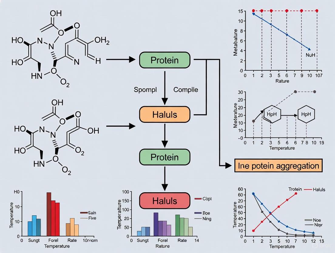
Visualizing the Workflow and Mechanisms
Predictive Stability Workflow
The following diagram illustrates the integrated experimental and computational workflow for applying Arrhenius-based kinetic modeling to predict protein aggregation.
Mechanisms of Protein Aggregation
Understanding the molecular mechanisms leading to aggregation is essential for developing effective mitigation strategies. The diagram below outlines the primary pathways.
Integrating Arrhenius-based kinetic modeling into the biopharmaceutical development pipeline provides a scientifically rigorous and efficient strategy to manage the critical challenge of protein aggregation. The protocols and frameworks outlined in this document enable researchers to quantitatively forecast aggregation, de-risk shelf-life assignments, and ultimately accelerate the delivery of stable, safe, and effective biologic drugs to patients. By moving from empirical observations to predictive, model-based stability assessments, developers can make more informed decisions throughout the drug product lifecycle.
For researchers and drug development professionals working with biotherapeutics, predicting long-term protein stability has represented a significant scientific challenge. Stability studies are vital in biologics development, guiding formulation, packaging, and shelf-life determination [7]. Traditionally, predicting long-term stability based on short-term data has been fundamentally challenging due to the complex behavior of biologics [7]. This application note examines the historical basis for these challenges and outlines how modern Arrhenius-based kinetic modeling has transformed stability prediction from an empirical art to a predictive science.
Historical Challenges in Stability Prediction
The Complexity of Protein Degradation Pathways
The fundamental challenge in predicting protein stability stemmed from the intricate nature of degradation pathways in biological systems. Unlike small molecule drugs, proteins exhibit:
- Multiple simultaneous degradation mechanisms: Proteins can undergo aggregation, fragmentation, deamidation, and oxidation simultaneously through different pathways [7]
- Concentration-dependent behavior: Attributes like aggregation demonstrated concentration-dependent modifications that appeared impossible to model practically [7]
- Non-Arrhenius behavior: Some systems exhibited anomalous temperature dependence that complicated extrapolation [9]
Limitations of Traditional Approaches
Traditional stability assessment relied heavily on:
- Linear extrapolation methods: Using straight-line regression from limited data points [7]
- Trial-and-error formulation: Empirical testing of standard buffers and excipients without predictive capability [10]
- Real-time stability studies: Time-consuming approaches requiring 10.5 years average development time from Phase I to approval [10]
Table 1: Historical Limitations in Protein Stability Prediction
| Challenge Area | Specific Limitation | Impact on Development |
|---|---|---|
| Modeling Complexity | Belief that concentration-dependent modifications couldn't be modeled [7] | Inability to predict aggregation kinetics accurately |
| Experimental Design | Activation of multiple degradation mechanisms at different temperatures [7] | Difficulty identifying dominant relevant pathways |
| Technical Capability | Lack of computational power for complex models [10] | Reliance on oversimplified linear models |
| Knowledge Gaps | Limited understanding of protein energy landscapes [9] | Inaccurate temperature dependence assumptions |
The Shift to Predictive Kinetic Modeling
Theoretical Foundation: Arrhenius Equation
The transformation began with proper application of the fundamental Arrhenius equation:
[k = A\exp\left(-\frac{E_a}{RT}\right)]
where (k) represents the rate constant, (A) is the pre-exponential factor, (E_a) is the activation energy, (R) is the gas constant, and (T) is the absolute temperature [11] [12].
The linearized form of the equation enables practical application:
[\ln(k) = \ln(A) - \frac{E_a}{R}\left(\frac{1}{T}\right)]
This relationship allows researchers to construct Arrhenius plots of (\ln(k)) versus (1/T), where the slope yields (-E_a/R) and the intercept provides (\ln(A)) [11] [12].
Key Modeling Breakthroughs
Recent advances demonstrated that long-term stability predictions for monoclonal antibodies in solution could be achieved using simple first-order kinetics combined with the Arrhenius equation [7]. This approach became possible when stability studies were designed to ensure only one degradation pathway relevant at storage conditions was present across all temperature conditions [7].
Modern Experimental Protocol for Aggregation Prediction
Materials and Equipment
Table 2: Essential Research Reagent Solutions and Materials
| Item | Specification | Function/Purpose |
|---|---|---|
| Protein Samples | IgG1, IgG2, Bispecific IgG, Fc fusion, scFv, Nanobodies, DARPins [7] | Representative biologics for stability assessment |
| Formulation Buffers | Pharmaceutical grade excipients [7] | Maintain protein stability and mimic actual formulations |
| SEC Column | Acquity UHPLC protein BEH SEC column 450 Å [7] | Separation of monomers from aggregates |
| HPLC System | Agilent 1290 HPLC with UV detection at 210 nm [7] | Quantitative analysis of protein species |
| Mobile Phase | 50 mM sodium phosphate, 400 mM sodium perchlorate, pH 6.0 [7] | SEC separation while minimizing secondary interactions |
| Stability Chambers | Temperature-controlled (±0.5°C) [7] | Precise maintenance of accelerated conditions |
Step-by-Step Protocol
Study Design and Temperature Selection
- Select relevant temperature conditions: Choose 3-5 storage temperatures based on the protein's stability profile (e.g., 5°C, 25°C, 30°C, 40°C) [7]
- Critical consideration: Ensure the temperature range activates only the degradation pathway dominant at recommended storage conditions [7]
- Include recommended storage condition: Always include the actual storage temperature (typically 2-8°C) as a reference point [7]
Sample Preparation and Storage
- Filter protein solutions through 0.22 µm PES membrane filter to remove pre-existing particulates [7]
- Aseptically fill glass vials with specified protein concentration [7]
- Determine protein concentration via absorbance at 280 nm using UV-Vis spectrometry [7]
- Incubate samples upright at designated temperatures for predetermined durations (e.g., 12-36 months) [7]
Analytical Monitoring via Size Exclusion Chromatography
- Dilute protein samples to 1 mg/mL for SEC analysis [7]
- Inject 1.5 µL of diluted protein solution [7]
- Perform 12-minute run at 40°C with flow rate of 0.4 mL/min [7]
- Maintain column temperature at 40°C for improved separation of fragments from monomers [7]
- Quantify species by calculating percentage of total area for monomers, fragments, and aggregates [7]
Data Analysis and Kinetic Modeling
- Model aggregation using first-order kinetics:
[\frac{d\alpha}{dt} = k(1-\alpha)^n]
where (\alpha) represents the fraction of aggregates formed, (k) is the rate constant, and (n) is the reaction order [7].
- Apply Arrhenius equation to determine temperature dependence:
[k = A\exp\left(-\frac{E_a}{RT}\right)]
- Extrapolate to storage temperature using the determined activation energy ((E_a)) and pre-exponential factor ((A)) [7].
Case Study Applications and Validation
Successful Applications Across Protein Modalities
Research has demonstrated effective modeling of aggregate predictions for diverse protein formats using first-order kinetic models [7]:
Table 3: Validation Across Protein Modalities
| Protein Format | Concentration | Temperatures Studied | Prediction Accuracy |
|---|---|---|---|
| IgG1 (P1) | 50 mg/mL | 5°C, 25°C, 30°C | Accurate long-term prediction achieved |
| IgG2 (P3) | 150 mg/mL | 5°C, 25°C, 30°C | Reliable aggregation modeling |
| Bispecific IgG (P4) | 150 mg/mL | 5°C, 25°C, 40°C | Successful stability projection |
| Fc-Fusion (P5) | 50 mg/mL | 5°C, 25°C, 35°C, 40°C, 45°C, 50°C | Validated across wide temperature range |
| scFv (P6) | 120 mg/mL | 5°C, 25°C, 30°C | Effective despite smaller size |
| Nanobody (P7) | 150 mg/mL | 5°C, 25°C, 30°C, 35°C | Consistent with larger proteins |
| DARPin (P8) | 110 mg/mL | 5°C, 15°C, 25°C, 30°C | Reliable prediction confirmed |
Comparative Advantage Over Traditional Methods
Compared to linear extrapolation, the kinetic model provided more precise and accurate stability estimates, even with limited data points [7]. The first-order kinetic model enhances reliability by reducing the number of parameters and samples required, preventing overfitting while ensuring better generalizability [7].
The historical challenge of predicting long-term protein stability has been largely addressed through the application of properly designed Arrhenius-based kinetic modeling. The key paradigm shift involved recognizing that through careful temperature selection and simplified kinetic models, accurate predictions become feasible [7]. Emerging approaches integrating artificial intelligence with molecular dynamics simulations show promise for further enhancing prediction accuracy, with recent studies demonstrating correlation coefficients up to 0.91 for aggregation prediction [13]. For researchers, the critical success factors include appropriate temperature selection to isolate dominant degradation pathways and adherence to the described experimental protocols for robust data generation.
The Arrhenius equation, proposed by Svante Arrhenius in the late 19th century, is a fundamental principle in chemical kinetics that describes the temperature dependence of reaction rates. Originally developed based on collision theory for reactions in the gaseous state, it provides a mathematical relationship between the rate of a chemical reaction and the absolute temperature at which it occurs [14].
The equation is expressed as:
( k = A e^{(-E_a / RT)} )
where k is the reaction rate coefficient, A is the pre-exponential factor (related to collision frequency and steric effects), Ea is the activation energy, R is the universal gas constant, and T is the absolute temperature [14].
The logarithmic form of the equation reveals a linear relationship between ln k and the inverse of absolute temperature (1/T):
( ln k = ln A - (E_a / RT) )
This linear relationship allows researchers to determine the activation energy and pre-exponential factor experimentally by measuring reaction rates at different temperatures [14].
Theoretical Framework and Relevance to Protein Systems
Connection to Transition State Theory
With the development of transition state theory, the Eyring equation offered a more theoretically grounded relationship for temperature dependence that maintains mathematical similarity to the Arrhenius equation [14]. The Eyring equation is expressed as:
( k = (k_BT/h) e^{(-ΔG*/RT)} )
where kB is Boltzmann's constant, h is Planck's constant, and ΔG* is the Gibbs free energy of activation [14].
For the relatively small temperature ranges relevant to pharmaceutical product stability, the derivative of ln k with respect to 1/T yields a form effectively identical to the Arrhenius equation, with the activation energy (Ea) replaced by the activation enthalpy (ΔH‡) [14].
Significance in Pharmaceutical Development
The Arrhenius equation provides critical utility for pharmaceutical companies by enabling shelf-life predictions of drug products based on short-term, accelerated stability studies at elevated temperatures. This predictive capability can significantly shorten development timelines, allowing products to reach the market faster [14]. The equation has been widely used—either implicitly or explicitly—for rapid assessment of stability for certain pharmaceutical dosage forms through accelerated aging studies [14].
For protein-based biotherapeutics, the temperature dependence of various degradation pathways follows Arrhenius behavior reasonably well. Chemical reactions involving covalent bond changes in proteins, including oxidation of methionine residues in recombinant human interleukin-1 receptor antagonist (between 5-45°C) and recombinant human granulocyte colony-stimulating factor (between 4-45°C), as well as deamidation in recombinant human interleukin-15 (between 6-40°C), have demonstrated Arrhenius behavior [14].
Application to Protein Aggregation Kinetics
Protein Aggregation as a Critical Quality Attribute
Protein aggregation presents one of the most significant challenges in developing protein biotherapeutics, affecting both product quality and potentially patient safety due to links with cytotoxicity and immunogenicity [14]. Investigations of protein aggregation mechanisms and kinetics remain a major focus for both pharmaceutical companies and academic institutions [14].
The aggregation process typically follows a multi-stage pathway beginning with protein unfolding to reveal aggregation-prone regions, followed by association of these unfolded monomers. The initial stages may involve reversible steps before nucleation of effectively irreversible species, with subsequent growth occurring through various mechanisms including monomer addition and aggregate association [15].
Table 1: Common Protein Aggregation Pathways and Characteristics
| Pathway Type | Key Features | Growth Mechanism | Typical Aggregate Size |
|---|---|---|---|
| Nucleation-Dominated (ND) | Forms irreversible dimers with minimal further growth | Limited to initial association | Small oligomers (dimers, trimers) |
| Chain Polymerization (CP) | Significant monomer consumption via sequential addition | Monomer-addition | Small to medium soluble aggregates |
| Association Polymerization (AP) | Rapid association of existing aggregates | Aggregate-aggregate association | Very large soluble species |
| Phase Separation (PS) | Association leading to physical separation | Aggregation and precipitation | Insoluble particles |
Non-Arrhenius Behavior in Protein Aggregation
Despite the utility of the Arrhenius equation for many chemical degradation pathways, temperature-induced protein aggregation often displays non-Arrhenius behavior even across relatively small temperature ranges relevant to product development [14]. This non-ideal behavior creates significant challenges for extrapolating aggregation rates from accelerated stability studies at high temperatures to recommended storage conditions [14].
Two primary categories of non-linear Arrhenius behavior have been identified [14]:
- Concave-up curves: Aggregation rates at low temperatures are higher than predicted by linear extrapolation from high-temperature data
- Concave-down curves: Aggregation rates at low temperatures are lower than predicted by linear extrapolation from high-temperature data
An extreme form of non-Arrhenius behavior manifests as anti-Arrhenius kinetics, where the observed rate coefficient increases with decreasing temperature (apparent negative activation energy) [16]. This behavior has been observed in the folding rates of proteins like chymotrypsin inhibitor 2, which increases from 25°C to 50°C but decreases above 50°C [14].
The underlying causes of non-Arrhenius behavior include [14]:
- Temperature-dependent changes in reaction mechanisms
- Shifts in the rate-determining step
- Significant heat capacity differences (
Δcp) between ground and transition states - Changes in protein conformational stability with temperature
Experimental Protocols for Aggregation Kinetics
Parallel Temperature Initial Rates (PTIR) Method
The PTIR method provides a sample-efficient approach for quantifying initial aggregation rates across multiple temperatures simultaneously [15].
Materials and Reagents:
- Protein solution of interest (purified)
- Appropriate formulation buffers
- Size exclusion chromatography (SEC) columns (e.g., Acquity UHPLC protein BEH SEC)
- HPLC/UHPLC system with UV detection
- Temperature-controlled incubation system capable of maintaining multiple precise temperatures
Procedure:
- Prepare monomeric protein solution using preparative SEC or filtration to remove pre-existing aggregates
- Aliquot identical protein samples into separate vials for each temperature condition
- Incubate samples simultaneously across a temperature gradient (e.g., -25°C to 60°C) for a predetermined time
t[17] [15] - Terminate aggregation by cooling samples and/or adding stabilization excipients
- Quantify remaining monomer concentration for each temperature using analytical SEC
- Calculate the observed aggregation rate coefficient using:
( k{obs}(T) = \frac{1 - cm(T)/c_{m,0}}{t} )
where
cm(T)is monomer concentration after incubation at temperatureT, andcm,0is initial monomer concentration [15]
Data Interpretation:
In the initial-rate regime with small extents of reaction, many aggregation mechanisms reduce to zero-order kinetics, making kobs a valid reduced initial-aggregation-rate coefficient [15].
Simultaneous Multiple Sample Light Scattering (SMSLS)
SMSLS complements PTIR by providing real-time monitoring of aggregate growth through changes in Rayleigh scattering [15].
Materials and Reagents:
- Protein solution (typically 0.1-10 mg/mL depending on protein)
- Light scattering instrument capable of monitoring multiple samples simultaneously
- Temperature-controlled sample chambers
- Clarified buffers (filtered through 0.02-0.1 µm filters)
Procedure:
- Clarify all protein solutions and buffers by filtration or centrifugation to remove particulate contaminants
- Load identical protein samples into multiple scattering cells
- Simultaneously initiate temperature incubation across all samples
- Continuously monitor the absolute Rayleigh scattering ratio
IR(t)for each sample over time - Determine aggregation rates from the time-dependent increase in scattering intensity
Data Interpretation:
In the limit of low protein concentration and negligible non-idealities, light scattering provides the weight-averaged molecular weight (Mw), offering a different "extent of reaction" measure compared to the number-averaged molecular weight from monomer loss [15].
Arrhenius-Based Kinetic Modeling for Shelf-Life Prediction
Recent advances have demonstrated that long-term stability predictions for complex biotherapeutics can be achieved using simplified first-order kinetic models combined with the Arrhenius equation [7] [18].
Materials and Reagents:
- Therapeutic protein in final formulation
- Stability chambers maintaining precise temperatures (e.g., 5°C, 15°C, 25°C, 30°C, 40°C, 45°C, 50°C)
- Analytical methods for quantifying aggregates (typically SEC-HPLC)
- Appropriate statistical software for kinetic modeling
Procedure:
- Incubate protein samples at multiple temperatures (typically 3-5 different temperatures) for extended periods (up to 36 months) [7]
- At predetermined timepoints, withdraw samples and quantify aggregate levels using validated SEC methods
- Fit aggregation time courses at each temperature to a first-order kinetic model: ( Aggregates(t) = Aggregates0 + (Aggregates\infty - Aggregates_0)(1 - e^{-kt}) )
- Extract rate constants (
k) at each temperature from the fits - Construct Arrhenius plot (
ln kvs.1/T) and fit to Arrhenius equation - Extrapolate rate constant at recommended storage temperature (typically 5°C)
- Predict long-term aggregation profile at storage condition using extrapolated
k
Table 2: Example Temperature Conditions for Stability Studies of Various Protein Modalities
| Protein Modality | Typical Storage Temp (°C) | Accelerated Study Temps (°C) | Stress Study Temps (°C) |
|---|---|---|---|
| IgG1/IgG2 | 5 | 25, 30 | 40 |
| Bispecific IgG | 5 | 25 | 40 |
| Fc-Fusion Protein | 5 | 25, 35, 40 | 45, 50 |
| scFv | 5 | 25, 30 | - |
| Bivalent Nanobody | 5 | 25, 30, 35 | - |
| DARPin | 5 | 15, 25, 30 | - |
Advanced Modeling Approaches
Modified Arrhenius Equations
For complex degradation pathways like drug nitrosation in solid dosage forms, modified Arrhenius equations incorporating additional factors can improve prediction accuracy [19]. A generalized form includes terms for relative humidity and excipient content:
( ln k = 41.38 - 13026 \times (1/T) + 0.038 \times (\%RH) - 0.44 \times (\% w/w(AE)) )
where %RH is relative humidity and % w/w(AE) is alkaline excipient content [19].
Multi-Parameter Kinetic Models
For systems with competing degradation pathways, more comprehensive kinetic models may be necessary. A competitive kinetic model with two parallel reactions can be described as [7]:
( \frac{dα}{dt} = v \times A1 \times \exp\left(-\frac{Ea1}{RT}\right) \times (1-α1)^{n1} \times α1^{m1} \times C^{p1} + (1-v) \times A2 \times \exp\left(-\frac{Ea2}{RT}\right) \times (1-α2)^{n2} \times α2^{m2} \times C^{p2} )
where α represents the sum fraction of degradation products, v is the ratio between competing reactions, n and m are reaction orders, and C is concentration [7].
Research Reagent Solutions
Table 3: Essential Materials for Protein Aggregation Kinetics Studies
| Reagent/Equipment | Function | Example Specifications |
|---|---|---|
| SEC-HPLC System | Quantification of monomer loss and aggregate formation | Acquity UHPLC with protein BEH SEC column, 450 Å, UV detection at 210-280 nm |
| Simultaneous Multiple Sample Light Scattering (SMSLS) | Real-time monitoring of aggregate growth | Multi-cell array, temperature control, Rayleigh scattering detection |
| Stability Chambers | Precise temperature control for accelerated studies | Temperature range: -25°C to 60°C, ±0.5°C stability |
| Citrate Buffer Systems | pH control for aggregation studies | 5-50 mM concentration, pH range 4-6 |
| Isochoric Cooling Systems | Prevention of freezing for sub-zero studies | Enables studies down to -25°C without ice formation |
| Polysorbate Excipients | Suppression of interfacial aggregation | Typically 0.01-0.1% w/v polysorbate 80 or 20 |
Workflow and Pathway Diagrams
Protein Aggregation Pathway and Analysis
Diagram 1: Protein aggregation pathway showing reversible and irreversible stages.
Experimental Workflow for Aggregation Kinetics
Diagram 2: Experimental workflow combining PTIR and SMSLS methodologies for comprehensive aggregation kinetics analysis.
Stability studies are fundamental to biologics development, guiding critical decisions from formulation to shelf-life determination. Traditionally, predicting the long-term stability of complex biotherapeutics based on short-term data was considered exceptionally challenging. However, a significant paradigm shift is underway, moving from overly complex models to the robust application of practical first-order kinetics combined with the Arrhenius equation. This approach now enables accurate long-term stability predictions for various critical quality attributes, including the concentration-dependent phenomenon of protein aggregation, across a wide range of protein therapeutic modalities [7]. This Application Note details the experimental protocols and data analysis frameworks that underpin this modern, simplified kinetic modeling strategy.
Application Notes: The Efficacy of Simplified Kinetic Modeling
Rationale for the Paradigm Shift
The development of biotherapeutics has evolved beyond traditional monoclonal antibodies to include more sophisticated formats like bispecific IgGs, Fc-fusion proteins, and nanobodies. This increase in complexity initially suggested a need for equally complex, multi-parameter kinetic models to describe stability. These models, however, often proved impractical for routine development use, carrying a high risk of overfitting and requiring extensive datasets [7]. The shift towards simplified modeling is grounded in the understanding that by carefully designing stability studies—particularly through strategic temperature selection—a single, dominant degradation pathway relevant to storage conditions can be identified and accurately described using a first-order kinetic model [7].
Key Advantages of the First-Order Kinetic Approach
- Enhanced Reliability and Reduced Overfitting: Simpler models with fewer parameters enhance the robustness and reliability of predictions. They are less sensitive to minor variations in input data, preventing overfitting and ensuring better generalizability to new data [7].
- Resource Efficiency: This approach reduces the number of samples required for analysis and simplifies the experimental and computational workload.
- Regulatory Alignment: The principles of Arrhenius-based Advanced Kinetic Modelling (AKM) are now being incorporated into revised ICH guidelines under the Accelerated Predictive Stability (APS) framework, facilitating their use in regulatory submissions for shelf-life justification [7] [20].
The following table summarizes the successful application of first-order kinetic modeling to predict aggregation in various protein modalities, as demonstrated in a recent comprehensive study [7].
Table 1: Aggregation Kinetics of Various Protein Modalities Modeled with First-Order Kinetics
| Protein Modality | Example Code | Concentration (mg/mL) | Key Stability Temperatures Studied | Model Applicability |
|---|---|---|---|---|
| IgG1 | P1, P2 | 50, 80 | 5°C, 25°C, 30°C, 33°C, 40°C | Confirmed |
| IgG2 | P3 | 150 | 5°C, 25°C, 30°C | Confirmed |
| Bispecific IgG | P4 | 150 | 5°C, 25°C, 40°C | Confirmed |
| Fc-Fusion Protein | P5 | 50 | 5°C, 25°C, 35°C, 40°C, 45°C, 50°C | Confirmed |
| scFv | P6 | 120 | 5°C, 25°C, 30°C | Confirmed |
| Bivalent Nanobody | P7 | 150 | 5°C, 25°C, 30°C, 35°C | Confirmed |
| DARPin (ensovibep) | P8 | 110 | 5°C, 15°C, 25°C, 30°C | Confirmed |
Experimental Protocols
Protocol 1: Quiescent Storage Stability Study
Purpose: To generate the high-quality, time-dependent data necessary for kinetic modeling of protein aggregation under controlled temperature stress.
Materials:
- The Scientist's Toolkit: Key Research Reagent Solutions
- Protein Drug Substance: Fully formulated, sterile-filtered.
- Sterilizing Filter: 0.22 µm PES membrane filter.
- Aseptic Filling Vials: Sterile glass vials for sample containment.
- Stability Chambers: Temperature-controlled chambers capable of maintaining set points from 2°C to 50°C with minimal variation.
- UV-Vis Spectrometer: For precise determination of protein concentration (e.g., via A280 measurement).
Procedure:
- Sample Preparation: Aseptically filter the fully formulated drug substance through a 0.22 µm PES membrane filter.
- Aseptic Filling: Fill the filtered solution into sterile glass vials. Seal the vials according to standard operating procedures.
- Concentration Verification: Confirm the protein concentration in the filled vials using UV absorbance at 280 nm.
- Incubation: Incubate the filled vials upright in stability chambers at pre-defined temperatures. A typical study includes the recommended storage temperature (e.g., 5°C), at least one accelerated temperature (e.g., 25°C), and one or more stress temperatures (e.g., 30°C, 40°C) [7].
- Sampling ("Pull Points"): Remove samples from each temperature condition at pre-determined time intervals (e.g., 0, 1, 3, 6, 12, 18, 36 months). The frequency should be designed to capture the initial, middle, and late stages of the degradation process.
- Analysis: Analyze the pulled samples for the quality attribute of interest (e.g., soluble aggregates via Size Exclusion Chromatography) as described in Protocol 2.
Protocol 2: Analysis of Protein Aggregates by Size Exclusion Chromatography (SEC)
Purpose: To quantitatively monitor the formation of high-molecular-weight species (HMWs or aggregates) over time in stability samples.
Materials:
- UHPLC System: Agilent 1290 HPLC or equivalent, equipped with a quaternary pump, autosampler, column thermostat, and UV detector.
- SEC Column: Acquity UHPLC protein BEH SEC column, 450 Å.
- Mobile Phase: 50 mM sodium phosphate, 400 mM sodium perchlorate, pH 6.0. The use of sodium perchlorate helps minimize secondary interactions between the protein and the column matrix.
- Molecular Weight Markers: For system suitability testing (e.g., BSA, thyroglobulin).
Procedure:
- Sample Preparation: Dilute the protein from stability samples to a standard concentration (e.g., 1 mg/mL) using an appropriate diluent.
- System Preparation and Calibration: Condition the SEC column according to the manufacturer's instructions. Establish system suitability by injecting molecular weight markers and evaluating peak resolution.
- Chromatographic Run:
- Injection Volume: 1.5 µL of diluted sample.
- Column Temperature: 40°C (to improve separation of fragments from the monomer).
- Flow Rate: 0.4 mL/min.
- Run Time: 12 minutes.
- Detection: UV at 210 nm.
- Data Analysis: Integrate the chromatogram peaks. The purity of the main peak (monomer) and the percentage of high-molecular species (aggregates) are determined as a percentage of the total peak area.
Protocol 3: Data Analysis and Kinetic Modeling
Purpose: To fit the experimental aggregation data to a first-order kinetic model and extrapolate the rate to the desired storage temperature using the Arrhenius equation.
Procedure:
- Model Selection: For many degradation processes, including aggregation, the data can be fitted to a first-order kinetic model:
α = 1 - exp(-k * t)whereαis the fraction of aggregate formed at timet, andkis the apparent first-order rate constant. - Determine Rate Constants: At each experimental temperature (
T), fit the time-course aggregation data to the model to extract the rate constant (k). - Apply the Arrhenius Equation: The temperature dependence of the rate constant
kis described by the Arrhenius equation:k = A * exp(-Ea / (R * T))whereAis the pre-exponential factor,Eais the apparent activation energy (in kJ/mol),Ris the universal gas constant (8.314 J/mol·K), andTis the absolute temperature in Kelvin. - Plot and Extrapolate: Plot
ln(k)against1/T(an Arrhenius plot). The data points should ideally form a straight line. The activation energy (Ea) is determined from the slope of this line (-Ea/R). - Long-Term Prediction: Use the fitted Arrhenius parameters to calculate the rate constant (
k) at the recommended storage temperature (e.g., 5°C). Use thiskin the first-order model to predict the level of aggregation over the proposed shelf-life (e.g., 24 or 36 months).
Visualizing the Workflow and Kinetic Relationship
The following diagrams illustrate the core experimental workflow and the fundamental kinetic relationship that enables long-term predictions from short-term data.
Figure 1: Experimental and Modeling Workflow for Predicting Protein Aggregation.
Figure 2: The Kinetic Bridge from Short-Term Data to Long-Term Stability.
Understanding and controlling protein degradation pathways is a fundamental challenge in developing stable biotherapeutics. Among the various degradation mechanisms, chemical modifications and unfolding-driven aggregation represent two critical pathways that can compromise therapeutic efficacy and safety [21]. These pathways are of particular concern during long-term storage and shipment of fragile biomolecules. Arrhenius-based kinetic modeling has emerged as a powerful tool to quantitatively describe these complex degradation processes, enabling researchers to predict long-term stability from short-term accelerated stability studies [7] [21]. This Application Note provides a structured comparison of these pathways, detailed experimental protocols for their study, and practical guidance for integrating this knowledge into stability prediction workflows essential for drug development professionals.
Theoretical Framework and Kinetic Modeling
The integration of degradation pathway analysis with kinetic modeling provides a powerful framework for predicting protein behavior under various conditions.
Fundamental Kinetic Pathways
Protein degradation often proceeds through competing pathways that can be quantitatively described using kinetic models. The unfolding-driven aggregation pathway typically involves a triggering event where a protein unfolds or misfolds, exposing hydrophobic regions and aggregation-prone sequences that subsequently assemble into higher-order structures [8] [22]. This pathway exhibits distinct kinetic coupling where the irreversible unfolding of a protein is often a unimolecular step with a high activation energy barrier, while the subsequent aggregation is frequently a bimolecular reaction characterized by a lower activation energy [8] [22]. For instance, studies on a human antibody light chain (hLC) revealed an unfolding barrier of 260 kJ/mol compared to an aggregation barrier of 40 kJ/mol [8] [22].
In contrast, chemical modification pathways involve covalent changes to the protein structure, such as glycation, oxidation, or deamidation, which can alter protein function and stability. These modifications can sometimes precede and even accelerate physical aggregation processes [23].
Arrhenius-Based Advanced Kinetic Modeling (AKM)
Advanced Kinetic Modeling leverages the Arrhenius equation to describe complex degradation kinetics from accelerated stability data. The reaction rate (( \frac{d\alpha}{dt} )) for competitive degradation pathways can be described by:
Where (A) is the pre-exponential factor, (Ea) is the activation energy, (R) is the universal gas constant, (T) is temperature in Kelvin, (n) and (m) are reaction orders, (v) is the ratio between reactions, and (C^p) accounts for concentration dependence where applicable [7] [21]. This sophisticated modeling approach can describe everything from simple first-order degradation to complex multi-step pathways involving both chemical modifications and physical aggregation.
Quantitative Comparison of Degradation Pathways
The table below summarizes key characteristics and kinetic parameters of the primary degradation pathways.
Table 1: Comparative Analysis of Key Protein Degradation Pathways
| Parameter | Unfolding-Driven Aggregation | Chemical Modification-Driven Aggregation |
|---|---|---|
| Primary Drivers | Thermal stress, mechanical perturbation, surface interactions [8] [24] | Reactive species (e.g., sugars, oxidative compounds), pH extremes [23] [25] |
| Molecularity of Rate-Limiting Step | Often bimolecular for aggregation step [8] [22] | Often unimolecular |
| Typical Activation Energy Range | Unfolding: ~260 kJ/mol; Aggregation: ~40 kJ/mol (hLC example) [8] [22] | Varies widely by modification type |
| Key Structural Changes | Unfolding/misfolding exposing aggregation-prone regions [8] [23] | Covalent modification of amino acid side chains [23] |
| Primary Forces Stabilizing Aggregates | Hydrophobic interactions, hydrogen bonds, van der Waals forces [25] | Covalent bonds (disulfide, advanced glycation end-products) [23] [25] |
| Key Analytical Techniques | SEC, intrinsic/extrinsic fluorescence, turbidity, CD [8] [7] | SEC, MS, CE, specific chemical assays [7] [23] |
| Influence of Protein Concentration | Often strong concentration dependence [8] | Variable concentration dependence |
Table 2: Kinetic Parameters for Unfolding and Aggregation of Model Proteins
| Protein System | Process | Activation Energy (kJ/mol) | Molecularity | Critical Temperature |
|---|---|---|---|---|
| Human antibody light chain (hLC) | Irreversible unfolding | 260 [8] [22] | Unimolecular | - |
| Human antibody light chain (hLC) | Aggregation | 40 [8] [22] | Bimolecular | - |
| Myofibrillar protein (MP) | Head region unfolding | - | - | 40°C [23] |
| Myofibrillar protein (MP) | Tail uncoiling & large aggregate formation | - | - | 47.5°C [23] |
Experimental Protocols
Protocol 1: Quantifying Unfolding-Aggregation Kinetics
This protocol characterizes the kinetic coupling between protein unfolding and aggregation, adapted from studies on antibody light chains and myofibrillar proteins [8] [23] [22].
Materials and Reagents
- Purified protein of interest
- Appropriate buffer system (e.g., PBS for antibodies [8])
- Thioflavin T (ThT) for amyloid detection [8]
- 1-anilino-8-naphthalene sulfonate (ANS) for hydrophobic exposure [8]
- Size-exclusion chromatography (SEC) columns [7]
Procedure
- Sample Preparation: Prepare protein solutions at multiple concentrations (e.g., 5-25 μM) in appropriate buffer [8].
- Temperature Gradient Design: Subject samples to controlled temperature gradients (e.g., 30-60°C for thermosensitive proteins) [23].
- Multi-technique Monitoring:
- Turbidity Measurements: Monitor at 350 nm or similar wavelength to track aggregate formation [23].
- Spectroscopic Probes: Use intrinsic (tryptophan) and extrinsic (ANS, ThT) fluorescence to monitor unfolding and aggregate morphology [8].
- Circular Dichroism (CD): Far-UV CD to track secondary structural changes [8].
- SEC Analysis: Withdraw aliquots at timed intervals, quench on ice, and analyze by SEC to quantify soluble monomer loss and aggregate formation [7].
- Data Analysis: Determine activation energies for unfolding and aggregation steps from temperature-dependent rates using Arrhenius plots [8] [22].
Protocol 2: Evaluating Chemical Modification-Induced Aggregation
This protocol focuses on glycation-induced aggregation, adapted from myofibrillar protein studies [23].
Materials and Reagents
- Target protein (e.g., myofibrillar protein)
- Reducing sugar (e.g., glucose)
- Buffer components for desired pH control
- SEC columns compatible with aggregates [7]
Procedure
- Glycation Reaction Setup: Incubate protein with sugar (e.g., MP with glucose) at controlled temperatures and pH [23].
- Cyclic Continuous Glycation (CCG):
- Implement temperature cycling between moderate (e.g., 37°C) and higher (e.g., 55°C) temperatures [23].
- At moderate temperatures, proteins unfold sufficiently to expose reactive sites without immediate aggregation.
- At higher temperatures, glycation occurs vigorously with fully expanded structures.
- Return to moderate temperature to inhibit excessive aggregation [23].
- Monitoring: Track glycation degree (spectrophotometric methods), aggregation (turbidity, SEC), and structural changes (fluorescence, CD) [23].
- Kinetic Analysis: Model the competition between glycation and aggregation pathways using first-order or more complex kinetics as needed [7].
Visualization of Pathways and Workflows
The following diagrams illustrate the key degradation pathways and experimental workflows.
Diagram 1: Competitive Protein Degradation Pathways. Two main pathways lead to irreversible aggregation: unfolding-driven (blue) and chemical modification-driven (red). Ea denotes activation energy.
Diagram 2: Experimental Workflow for Aggregation Kinetics. Integrated approach combining multiple analytical techniques with kinetic modeling for stability prediction.
The Scientist's Toolkit: Essential Research Reagents and Materials
Table 3: Key Research Reagent Solutions for Aggregation Studies
| Reagent/Material | Function/Application | Example Use Cases |
|---|---|---|
| Thioflavin T (ThT) | Fluorescent dye for amyloid detection | Staining and visualization of amyloid fibrils in hLC aggregates [8] |
| ANS (1-anilino-8-naphthalene sulfonate) | Extrinsic fluorophore detecting hydrophobic surface exposure | Monitoring unfolding transitions in hLC studies [8] |
| Size-Exclusion Chromatography (SEC) Columns | Separation and quantification of soluble monomer and aggregates | Quantifying monomer loss and HMW species formation in stability studies [7] |
| Surfactants (Ionic/Nonionic) | Modifying protein-protein interactions and unfolding behavior | Studying surfactant-driven modifications in protein structure [24] |
| Reducing Sugars (e.g., Glucose) | Inducing glycation-mediated chemical modifications | Glycation studies on myofibrillar proteins [23] |
The strategic differentiation between unfolding-driven aggregation and chemical modification pathways enables more precise stability interventions in biotherapeutic development. Through the application of Advanced Kinetic Modeling and the experimental protocols outlined herein, researchers can quantitatively describe these competing pathways, predict long-term stability, and design more stable biologic formulations. The integrated approach of combining multi-technique experimental data with Arrhenius-based modeling provides a powerful framework for addressing one of the most significant challenges in biopharmaceutical development—ensuring protein stability from manufacturing to patient administration.
Implementing Kinetic Models: A Practical Guide for Stability Prediction
The long-term stability of biotherapeutics, particularly their propensity to aggregate, is a critical determinant of product shelf life, safety, and efficacy. Predicting stability at recommended storage conditions (typically 2-8°C) based on short-term studies represents a significant challenge in pharmaceutical development. Temperature selection in stability studies is not merely a methodological detail but a fundamental strategic consideration that directly determines the validity and predictive power of stability models [7]. When appropriately designed, stability studies leveraging Arrhenius-based kinetic modeling can accurately forecast aggregation behavior, thereby accelerating development timelines and reducing costs [7] [26].
The core challenge stems from the complex nature of protein aggregation, which often proceeds through multiple pathways with distinct temperature dependencies [14] [26]. This application note examines the critical role of temperature selection within the broader context of Arrhenius-based kinetic modeling for protein aggregation research, providing researchers and drug development professionals with structured frameworks and protocols to enhance study design and predictive accuracy.
Strategic Importance of Temperature Selection
Theoretical Foundation: Arrhenius Kinetics and Its Limitations
The Arrhenius equation describes the temperature dependence of reaction rates, forming the cornerstone of accelerated stability studies:
[ k = A \times \exp\left(-\frac{E_a}{RT}\right) ]
Where (k) is the rate constant, (A) is the pre-exponential factor, (E_a) is the activation energy, (R) is the gas constant, and (T) is the absolute temperature [14]. In pharmaceutical stability testing, this relationship theoretically enables the extrapolation of high-temperature degradation data to predict stability at lower storage temperatures.
However, protein aggregation frequently demonstrates non-Arrhenius behavior, manifesting as nonlinearity in Arrhenius plots ((\ln k) versus (1/T)) [14]. This deviation from ideal behavior often arises because aggregation is not a simple elementary reaction but a complex multi-step process whose rate-limiting step can change with temperature [14] [27]. The Lumry-Eyring model describes this scenario, wherein native proteins unfold and the unfolded states subsequently aggregate [27]. As temperature changes, the equilibrium between native and unfolded states shifts, potentially altering the dominant aggregation mechanism.
Mechanism-Driven Temperature Selection
Emerging research reveals that proteins often aggregate through distinct pathways at different temperature regimes [26]. Studies on therapeutic monoclonal antibodies have identified separate low-temperature (LT) and high-temperature (HT) aggregation pathways with different molecular characteristics:
- LT Pathway: Typically dominated by chemical degradation processes such as deamidation and isomerization, exhibiting lower activation energies (10-25 kcal/mol) [26].
- HT Pathway: Primarily driven by conformational unfolding and physical aggregation, displaying higher activation energies (50-150 kcal/mol) [26].
This mechanistic understanding underscores why temperature selection must be guided by the specific degradation processes relevant to intended storage conditions. Studies conducted exclusively at high temperatures may activate unfolding-dominated pathways that poorly represent degradation mechanisms at refrigerated conditions [27] [26].
Table 1: Temperature Selection Strategy Based on Study Objectives
| Study Objective | Recommended Temperature Points | Scientific Rationale | Applicable Protein Modalities |
|---|---|---|---|
| Predicting long-term storage stability | 5°C, 15°C, 25°C | Captures LT aggregation pathway relevant to refrigerated storage | IgG1, IgG2, Bispecific IgG, Fc fusion [7] |
| Rapid formulation screening | 40°C, 45°C, 50°C | Accelerates chemical degradation processes | scFv, DARPins, Nanobodies [7] [26] |
| Comprehensive mechanism mapping | 5°C, 25°C, 40°C, 50°C+ | Identifies both LT and HT aggregation pathways | Therapeutic mAbs [26] |
| Cold denaturation studies | Sub-zero temperatures (isochoric cooling) | Investigates cold unfolding phenomena | Hemoglobin, unstable protein domains [27] |
Experimental Design and Protocol
Temperature Selection Protocol
Objective: Identify temperature conditions that accelerate degradation without altering the fundamental aggregation mechanism relevant to storage conditions.
Materials:
- Purified protein drug substance (>95% purity)
- Formulation buffer (pharmaceutical grade)
- Sterile filtration unit (0.22 µm PES membrane)
- Glass vials with stoppers and seals
- Temperature-controlled stability chambers (±1°C accuracy)
- Size exclusion chromatography (SEC) system with UV detection
Procedure:
- Formulation and Filling:
- Dialyze or dilute protein into target formulation buffer.
- Sterile-filter using 0.22 µm PES membrane.
- Aseptically fill into glass vials (1-2 mL fill volume).
- Seal vials under inert atmosphere if oxidation-sensitive.
Temperature Matrix Design:
- Include at least four temperature points spanning the range from storage temperature to accelerated conditions.
- Recommended progression: 5°C, 25°C, 40°C, and one higher temperature (e.g., 50°C) for mechanism probing [7] [26].
- For proteins prone to cold denaturation, include sub-zero temperatures using appropriate cryoprotectants [27].
Time Point Selection:
- Sample at a minimum of five time points per temperature condition.
- Recommended intervals: 1, 3, 6, 9, and 12 months for refrigerated conditions.
- Accelerated conditions (≥40°C): 2, 4, 8, 12, and 16 weeks.
Stability-Indicating Assays:
- Size Exclusion Chromatography: Quantify soluble aggregates [7].
- Column: Acquity UHPLC protein BEH SEC column 450 Å
- Mobile phase: 50 mM sodium phosphate, 400 mM sodium perchlorate, pH 6.0
- Detection: UV at 210 nm
- Circular Dichroism: Monitor secondary structural changes [28].
- Turbidity Measurements: Assess visible aggregation [28].
- Size Exclusion Chromatography: Quantify soluble aggregates [7].
The following workflow diagram illustrates the strategic approach to temperature selection in stability studies:
Data Generation for Kinetic Modeling
Objective: Generate high-quality data suitable for Arrhenius-based kinetic modeling of aggregation.
Experimental Parameters:
- Protein Concentration: Test multiple concentrations (e.g., 50, 100, 150 mg/mL) to identify concentration-dependent effects [26].
- Time Points: Ensure sufficient data points to establish kinetic curves (minimum 5 points per temperature).
- Replicates: Include triplicate samples for statistical power.
Analytical Measurements:
- SEC Analysis: Quantify monomer loss and aggregate formation at each time point.
- Kinetic Parameter Calculation: Determine apparent rate constants (k_obs) for aggregation at each temperature.
- Arrhenius Plotting: Graph ln(k_obs) versus 1/T to assess linearity and identify mechanism changes.
Table 2: Key Reagents and Research Solutions for Stability Studies
| Reagent/Solution | Function in Study | Application Example | Critical Considerations |
|---|---|---|---|
| Pharmaceutical Grade Buffers | Maintain formulation pH and ionic strength | 50 mM sodium phosphate, pH 6.0 [7] | Buffer capacity must withstand degradation products |
| Stabilizing Excipients | Minimize non-specific aggregation | Sucrose, trehalose, amino acids | Concentration optimization required for each protein |
| Aggregation Suppressors | Reduce surface-induced aggregation | Polysorbate 20/80 [4] | Quality and purity critical for regulatory approval |
| SEC Columns with Enhanced Resolution | Separate monomer from aggregates | Acquity UHPLC protein BEH SEC 450 Å [7] | Regular calibration with molecular weight standards |
| Chemical Stabilizers | Inhibit specific degradation pathways | Methionine (antioxidant) [4] | May interfere with analytical methods |
| Cryoprotectants | Enable sub-zero studies without freezing | Glycerol, DMSO [27] | Can alter protein thermodynamics at high concentrations |
Data Analysis and Modeling Approaches
Kinetic Model Development
For a first-order kinetic model describing monomer loss due to aggregation:
[ \frac{d[M]}{dt} = -k_{obs}[M] ]
Where ([M]) is monomer concentration and (k{obs}) is the apparent rate constant. The temperature dependence of (k{obs}) follows the Arrhenius equation:
[ k{obs} = A \times \exp\left(-\frac{Ea}{RT}\right) ]
For more complex systems involving parallel pathways, a branched mechanism may be required [26]:
[ \frac{d\alpha}{dt} = v \times A1 \times \exp\left(-\frac{E{a1}}{RT}\right) \times (1-\alpha1)^{n1} + (1-v) \times A2 \times \exp\left(-\frac{E{a2}}{RT}\right) \times (1-\alpha2)^{n2} ]
Where (α) is the fraction of degraded product, (v) is the partitioning factor between pathways, (A) is pre-exponential factor, (E_a) is activation energy, and (n) is reaction order [7].
Model Validation Protocol
Objective: Validate the predictive capability of the kinetic model against long-term stability data.
Procedure:
- Reserve a subset of stability samples (e.g., 12-month time points) for validation.
- Develop the kinetic model using data from accelerated conditions only (e.g., 25°C, 40°C, 50°C).
- Predict aggregation levels at storage temperature (5°C) for the validation time points.
- Compare predictions with experimental data using statistical measures (e.g., RMSE, R²).
- Refine the model if predictions deviate by >20% from experimental values.
Success Criteria: The model should predict long-term aggregation within ±15% of measured values to be considered validated [7] [26].
Case Studies and Applications
Successful Implementation Across Protein Modalities
Recent research demonstrates the successful application of temperature-optimized stability studies across diverse biotherapeutic formats:
- Monoclonal Antibodies: For IgG1 and IgG2 antibodies, studies at 5°C, 25°C, and 40°C enabled accurate prediction of aggregation over 3 years at 5°C [7] [26].
- Novel Scaffolds: Bispecific IgGs, Fc-fusion proteins, scFvs, and DARPins have been effectively modeled using first-order kinetics with appropriate temperature selection [7].
- Therapeutic Peptides: SAR441255, a peptide triagonist, showed accurate 2-year stability predictions at 5°C based on 3-month accelerated studies [29].
Regulatory Considerations
The International Council for Harmonisation (ICH) guidelines are evolving to incorporate kinetic modeling approaches for stability prediction [7]. The emerging Accelerated Predictive Stability (APS) framework explicitly acknowledges the value of Arrhenius-based Advanced Kinetic Modeling (AKM) for predicting long-term stability with limited real-time data [7]. Proper temperature selection and mechanism-based modeling are fundamental to successful regulatory submission under these modernized guidelines.
Temperature selection represents a critical design parameter in stability studies for protein-based therapeutics. By strategically choosing temperature conditions that activate degradation mechanisms relevant to storage conditions, researchers can develop predictive kinetic models that accurately forecast long-term aggregation behavior. The protocols and frameworks presented in this application note provide a systematic approach to temperature selection, experimental execution, and data modeling that enhances predictive accuracy while reducing development timelines. As the field advances toward more sophisticated predictive stability frameworks, mechanism-informed temperature selection will remain essential for reliable shelf-life determination of biopharmaceutical products.
Within the development of biotherapeutics, the quantitative analysis of protein aggregates is a critical quality attribute due to concerns over product efficacy and immunogenicity [30] [31]. Size-exclusion chromatography (SEC) stands as a predominant, reproducible technique for the routine analysis of soluble protein aggregates, such as dimers and higher-order multimers [30] [31]. When integrated into a stability-indicating methodology, SEC provides the essential primary data on aggregate formation rates required for Arrhenius-based kinetic modeling. This modeling predicts long-term protein stability under recommended storage conditions, such as 2–8 °C, based on short-term, accelerated stability studies [7]. This application note details the core components of data collection via SEC to support the development of robust kinetic models for protein aggregation.
Principles of Size-Exclusion Chromatography
Separation Mechanism
SEC separates molecules based on their hydrodynamic size in solution [32]. The stationary phase consists of a column packed with porous beads. As a sample passes through the column, larger molecules that cannot enter the pores are excluded and elute first. Smaller molecules that can diffuse into and out of the pore network are temporarily retained and elute later [30] [32]. This mechanism is fundamentally different from other chromatographic modes because it is primarily driven by entropy, not enthalpy [30] [33]. Under ideal conditions, there is no adsorption of the analyte to the stationary phase ( \Delta H = 0 ) , and the separation depends solely on the conformational entropy change as molecules access the pore volume [33].
The elution volume ( VR ) of an analyte is described by the equation: [ VR = V0 + KD Vi ] where ( V0 ) is the interstitial volume, ( Vi ) is the intra-particle pore volume, and ( KD ) is the thermodynamic distribution coefficient, which ranges from 0 (for fully excluded molecules) to 1 (for molecules that fully access the pore volume) [30]. For a given SEC column, the separation range is defined by its exclusion limit (the molecular size too large to enter any pores) and its permeation limit (the molecular size small enough to access all pores) [32].
SEC in the Context of Protein Aggregation
For biopharmaceutical proteins, SEC is routinely used to resolve and quantify the monomeric active ingredient from its smaller fragment and larger aggregate species [31]. A typical chromatogram for an antibody sample might show a main peak (monomer), followed by earlier-eluting peaks representing aggregates (dimers, trimers, etc.), and later-eluting peaks representing fragments [31]. The accurate quantification of the high-molecular-weight species is a direct measurement of a key degradation pathway and serves as the primary data input for stability modeling [7]. It is critical that the analytical method itself does not alter the native aggregation state of the sample through shear forces, interactions with the column, or changes in the mobile phase [31].
Integration of SEC Data into Kinetic Models
From Chromatographic Data to Kinetic Rates
The core of the kinetic model relies on measuring the change in the quantity of a quality attribute over time under different stress conditions. SEC provides the precise data for the percentage of aggregates at each time point. In a simplified, first-order kinetic approach, the degradation rate for aggregation can be described as the conversion from native monomer ( N ) to aggregate ( A ). The rate of aggregate formation is often proportional to the concentration of the native species.
The fundamental relationship is: [ \frac{d[A]}{dt} = k \cdot [N] ] where ( [A] ) is the concentration of aggregates, ( [N] ) is the concentration of the native monomer, and ( k ) is the reaction rate constant at a specific temperature [7]. The SEC data collected over time at multiple elevated temperatures allows for the determination of the rate constant ( k ) at each temperature.
The Arrhenius Relationship
The rate constants ( k ) derived from SEC data at various temperatures are then fitted to the Arrhenius equation to extrapolate the rate at lower, storage temperatures [7]. The Arrhenius equation is: [ k = A \cdot \exp\left(-\frac{E_a}{RT}\right) ] where:
- ( k ) is the rate constant.
- ( A ) is the pre-exponential factor.
- ( E_a ) is the activation energy for the aggregation reaction.
- ( R ) is the universal gas constant.
- ( T ) is the absolute temperature in Kelvin.
By plotting ( \ln(k) ) versus ( 1/T ), a straight line is obtained with a slope of ( -E_a/R ), allowing for the calculation of the activation energy and the prediction of ( k ) at any desired temperature [7]. This model has been successfully applied to predict long-term stability for various protein modalities, including IgG1, IgG2, bispecific antibodies, Fc-fusion proteins, and scFvs [7].
Workflow for Stability Prediction
The following diagram illustrates the logical workflow that integrates SEC data collection with kinetic modeling to predict protein aggregation.
Experimental Protocol for SEC-Based Aggregate Analysis
Materials and Reagents
Table 1: Key Research Reagent Solutions for SEC Analysis
| Item | Function/Description | Example |
|---|---|---|
| SEC Column | Porous bead-packed column for size-based separation. | Acquity UHPLC BEH SEC column, 200 Å, 1.7 µm [7] |
| Mobile Phase Buffer | Aqueous buffer to maintain protein stability and minimize secondary interactions. | 50 mM sodium phosphate, 400 mM sodium perchlorate, pH 6.0 [7] |
| Ionic Strength Additive | Salt (e.g., NaCl) added to shield electrostatic interactions between protein and stationary phase. | 100-150 mM Sodium Chloride [32] |
| Protein Standard Mix | Molecules of known molecular weight for system suitability and calibration. | BioRad Gel Filtration Standard [31] |
Detailed Step-by-Step Method
1. Sample Preparation:
- Filter the fully formulated drug substance through a 0.22 µm PES membrane filter [7].
- If necessary, dilute the sample to the target concentration using the mobile phase or formulation buffer. For the analysis cited, proteins were diluted to 1 mg/mL prior to injection [7].
- Minimize sample handling to prevent mechanical stress-induced aggregation.
2. Instrument and Column Setup:
- Utilize a U/HPLC system (e.g., Agilent 1290) with a quaternary pump, autosampler, and column thermostat [7].
- Equilibrate the SEC column with the mobile phase at the recommended operating pressure and flow rate until a stable baseline is achieved.
- Condition the column by saturating it with a protein solution like BSA to minimize nonspecific binding [7].
- Set the column temperature. While temperature has minimal effect on the SEC mechanism, thermostating the system ensures reproducibility. A temperature of 40°C may be used for optimal separation [7].
3. System Suitability Test:
- Inject a protein standard mix (e.g., thyroglobulin, IgG, ovalbumin) to verify column performance, resolution, and peak shape [31].
- Ensure that the resolution between key peaks meets predefined criteria before analyzing experimental samples.
4. Sample Analysis:
- Inject an appropriate sample volume (typically 1–10 µL for analytical SEC) [7] [32].
- Perform an isocratic elution at a constant flow rate. A flow rate of 0.4 mL/min is typical, though slower rates may improve resolution [7] [32].
- Monitor the eluent using a UV detector, typically at 210 nm or 280 nm [7].
5. Data Analysis:
- Integrate the chromatographic peaks corresponding to aggregates, monomer, and fragments.
- Calculate the percentage of high-molecular-weight species (aggregates) as a percentage of the total peak area [7]. [ \% \text{Aggregates} = \left( \frac{\text{Area of aggregate peaks}}{\text{Total area of all peaks}} \right) \times 100 ]
Method Optimization and Critical Parameters
Optimization Strategies for SEC
To ensure accurate and reproducible data for kinetic modeling, the SEC method must be optimized. Key parameters to consider are summarized in the table below.
Table 2: Key Parameters for SEC Method Optimization
| Parameter | Optimization Goal | Impact and Consideration |
|---|---|---|
| Mobile Phase Composition | Minimize secondary interactions (ionic, hydrophobic). | Use buffers (e.g., phosphate) with sufficient ionic strength (e.g., 100-400 mM salt) to shield electrostatic interactions. Additives like arginine can reduce hydrophobic interactions [32]. |
| Column Selection | Match pore size to target protein and aggregates. | A pore size of 150-300 Å is common for mAbs. Smaller pores resolve fragments; larger pores are needed for high-order aggregates [31]. |
| Flow Rate | Balance resolution and analysis time. | Slower flow rates (e.g., 0.2-0.5 mL/min) generally improve resolution but increase run time [32]. |
| Sample Load | Prevent column overloading. | Keep the injection volume and mass within 5-10% of the total column volume to avoid peak broadening and loss of resolution [32]. |
| Temperature | Ensure reproducibility. | While retention is largely independent of temperature, thermostating the column and system improves baseline stability and retention time reproducibility [30] [33]. |
Troubleshooting Common Issues
- Poor Recovery or Peak Tailing: Often caused by nonspecific interactions with the stationary phase. Remedy: Increase ionic strength of the mobile phase or include additives like arginine [32].
- Unexpected Aggregate Levels: The analytical process can create or dissociate aggregates. Remedy: Review sample preparation (e.g., filtration, dilution) and ensure mobile phase is compatible with the protein formulation [31].
- Loss of Resolution: Can be due to column aging, overloading, or inappropriate flow rate. Remedy: Check system suitability with standards, reduce sample load, or adjust flow rate [32].
Size-exclusion chromatography is an indispensable tool for generating the high-quality, quantitative data on protein aggregation required for building predictive kinetic models. By following the detailed protocols and optimization strategies outlined in this document, researchers can establish robust SEC methods. When these methods are applied within a structured stability study design across multiple temperatures, the resulting data empowers the use of Arrhenius-based kinetic modeling. This integrated approach allows for the reliable prediction of protein aggregation during long-term storage, ultimately accelerating the development of biotherapeutics and ensuring their quality, safety, and efficacy.
The long-term stability of therapeutic proteins, particularly monoclonal antibodies (mAbs), is a critical factor in drug development. Arrhenius-based kinetic modeling provides a powerful tool to predict degradation over time by leveraging data from accelerated stability studies. This approach is grounded in the principle that the rate of chemical reactions, including protein degradation, increases with temperature. For the biopharmaceutical industry, this enables scientists to predict shelf-life and make crucial development decisions without waiting for multi-year real-time stability data, thereby accelerating the delivery of novel biologics to patients [34].
This Application Note provides a detailed, step-by-step protocol for constructing a first-order kinetic model with Arrhenius dependence to predict the aggregation behavior and stability of therapeutic proteins.
Experimental Design & Workflow
A successful kinetic modeling study requires careful planning of the stability study design, data collection, and analysis workflow. The overarching process is designed to maximize predictive power from efficiently collected accelerated data.
The diagram below outlines the core workflow for building and validating the kinetic model.
Materials and Equipment
Research Reagent Solutions
The table below lists essential materials and their functions for conducting stability studies and kinetic analysis.
| Item | Function / Application | Example Specifications |
|---|---|---|
| Therapeutic Protein | Primary molecule for stability assessment. | Monoclonal Antibodies (e.g., IgG1, IgG2), Fusion Proteins (e.g., Etanercept) [34]. |
| Formulation Buffers | Provide stable chemical environment; critical for pH control. | Citrate, phosphate, or histidine buffers at relevant pH (e.g., 5.2 - 6.5 for mAbs) [34]. |
| Stabilizers | Protect against aggregation and surface adsorption. | Sucrose, trehalose, sorbitol, amino acids (e.g., lysine) [34]. |
| Surfactants | Mitigate interfacial stress. | Polysorbate 80 (PS80), Polysorbate 20 (PS20) [34]. |
Specialized Equipment
| Item | Function / Application |
|---|---|
| Stability Chambers | Controlled storage at specified temperatures and humidity (e.g., 5°C, 25°C/60% RH, 40°C/75% RH). |
| Size Exclusion Chromatography (SEC) | Quantification of soluble protein aggregates and fragments [34]. |
| Cation Exchange Chromatography (CEX) | Analysis of charge variants resulting from chemical degradation [34]. |
| Capillary Electrophoresis (CE-SDS) | Monitoring of protein fragmentation under denaturing conditions [34]. |
| Kinetic Analysis Software | Data fitting and outlier cleaning (e.g., Kfits, an open-source Python tool) [35]. |
Step-by-Step Protocol
Step 1: Design and Execute the Stability Study
- Formulation: Prepare the drug product in its final formulation buffer. For a typical monoclonal antibody, this could be at a concentration of 50-150 mg/mL in a buffer such as 5 mM sodium citrate at pH 6.3, containing excipients like sucrose and polysorbate 80 [34].
- Storage: Fill the formulated product into appropriate primary packaging (e.g., type I glass vials). Divide samples for storage at a minimum of three different temperatures.
- Intended storage condition: 5°C ± 3°C
- Accelerated condition: 25°C ± 2°C / 60% ± 5% RH
- Stress condition: 40°C ± 2°C / 75% ± 5% RH [34]
- Sampling: Remove samples from each storage condition at predefined time points. For a 6-month study, typical time points could be T=0, 1, 3, and 6 months. Ensure proper documentation to maintain chain of integrity.
Step 2: Monitor Stability-Indicating Attributes
At each time point, analyze samples using validated, stability-indicating methods to track the progression of degradation.
- For Aggregation: Use Size Exclusion Chromatography (SEC) to quantify the percentage of monomeric protein versus soluble aggregates. The loss of monomer is often the primary metric for kinetic modeling [34] [36].
- For Chemical Modifications: Use Cation Exchange Chromatography (CEX) or imaged Capillary Isoelectric Focusing (iCIEF) to monitor changes in charge variants due to deamidation or oxidation [34].
- For Fragmentation: Use Capillary Electrophoresis-Sodium Dodecyl Sulfate (CE-SDS) to detect and quantify protein fragments [34].
Step 3: Determine Degradation Rate Constants (k)
For each temperature condition, fit the time-course data for the key quality attribute (e.g., % monomer) to a first-order kinetic model.
- Model Fitting: Use the equation for a first-order reaction:
( C = C0 \cdot e^{-kt} )
where:
- ( C ) is the concentration (or percentage) of the monomer at time ( t ).
- ( C0 ) is the initial concentration (or percentage) at ( t = 0 ).
- ( k ) is the apparent first-order rate constant (in units of time(^{-1})).
- ( t ) is the storage time.
- Parameter Estimation: Employ non-linear regression analysis (software such as Kfits is designed for this purpose) to obtain the best-fit value of ( k ) at each temperature [35]. This will yield a set of rate constants: ( k{T1} ), ( k{T2} ), ( k_{T3} ), etc.
Step 4: Apply the Arrhenius Equation
The relationship between the degradation rate constant (( k )) and the absolute temperature (( T )) is described by the Arrhenius equation.
- Linearize the Equation: The equation can be written in linear form:
( \ln(k) = \ln(A) - \frac{Ea}{R} \cdot \frac{1}{T} )
where:
- ( k ) is the rate constant.
- ( A ) is the pre-exponential factor.
- ( Ea ) is the apparent activation energy (J/mol).
- ( R ) is the universal gas constant (8.314 J/mol·K).
- ( T ) is the absolute temperature in Kelvin (K).
- Plot and Perform Linear Regression: Create an Arrhenius plot of ( \ln(k) ) versus ( 1/T ) (where T is in Kelvin). Perform a linear regression on the data points. The slope of the best-fit line is ( -E_a/R ), and the y-intercept is ( \ln(A) ).
Step 5: Predict Long-Term Stability
With the parameters from the Arrhenius plot, you can predict the degradation rate at any temperature, most importantly the intended storage temperature.
- Calculate ( k{5°C} ): Substitute the determined values of ( Ea ) and ( A ) into the Arrhenius equation to calculate the rate constant at the intended storage temperature of 5°C (278.15 K).
- Predict Shelf-Life: Insert the calculated ( k_{5°C} ) back into the first-order kinetic model. Determine the time ( t ) at which the monomeric purity ( C ) reaches the pre-defined critical quality limit (e.g., not less than 95% monomer). This time ( t ) is the predicted shelf-life.
Data Analysis and Modeling
Kinetic and Arrhenius Parameters
The following table summarizes the core parameters obtained through the protocol and their significance in model interpretation.
| Parameter | Symbol | Unit | Interpretation in Stability Assessment |
|---|---|---|---|
| Rate Constant | ( k ) | time⁻¹ (e.g., month⁻¹) | Speed of degradation at a specific temperature. A higher ( k ) means faster degradation. |
| Activation Energy | ( E_a ) | kJ/mol | Represents the temperature sensitivity of the degradation reaction. A higher ( E_a ) indicates a process that accelerates more rapidly with increasing temperature. |
| Pre-exponential Factor | ( A ) | time⁻¹ (e.g., month⁻¹) | Related to the frequency of molecular collisions leading to a reaction. |
Model Validation
A critical final step is to validate the predictive power of the model.
- Comparison with Real-Time Data: The robustness of the model is demonstrated by comparing the predicted degradation profile at 5°C with experimentally obtained long-term data (e.g., from 24 or 36 months of storage). A validated model will show that the vast majority of the real-time data points fall within the 95% prediction interval of the model [34].
- Quantifying Success: One study demonstrated that 96% of experimental long-term data not used to build the model fell within the calculated prediction intervals, confirming the model's accuracy [34].
Logical Relationship Diagram
The diagram below illustrates the logical and mathematical relationships between the experimental data, the kinetic model, and the final shelf-life prediction, integrating the key equations used in the protocol.
Arrhenius-based kinetic modeling has become a cornerstone for predicting the long-term stability of biotherapeutics, a critical aspect of drug development and formulation. While extensively applied to monoclonal antibodies (mAbs), the principles of kinetic modeling are equally vital for a new generation of sophisticated protein modalities. The transition from conventional mAbs to more complex and often smaller structures—such as Fc-fusion proteins, single-chain variable fragments (scFvs), Designed Ankyrin Repeat Proteins (DARPins), and Nanobodies—introduces unique stability challenges. These constructs can exhibit different aggregation pathways and degradation kinetics compared to their full-length antibody counterparts. Recent studies demonstrate that a simplified first-order kinetic model, combined with the Arrhenius equation, can effectively predict aggregation in these diverse modalities, enabling robust shelf-life determination and guiding the development of stable therapeutic formulations [7]. This application note details the protocols and methodologies for applying Arrhenius-based modeling to these novel protein therapeutics.
Theoretical Framework: Kinetic Modeling of Aggregation
Protein aggregation is a complex process often described as a series of steps beginning with the unfolding of the native monomer to reveal aggregation-prone regions, followed by nucleation and subsequent growth of aggregates [15] [36]. The net aggregation rate can change by orders of magnitude with a temperature change of only 5–10 °C, primarily driven by the large enthalpy change associated with the unfolding equilibrium [15].
The Lumry-Eyring Nucleated Polymerization (LENP) model is a foundational framework that introduces the concept of nucleation to aggregation kinetics [37]. This model describes the initial reversible unfolding of a native protein (N) into a reactive, aggregation-prone state (R), which then forms an irreversible nucleus (A(_x)). This nucleus can grow through monomer addition (chain polymerization, CP) or aggregate-aggregate association (association polymerization, AP) [15] [37].
For practical stability prediction in biologics development, a simplified first-order kinetic model has proven effective across diverse protein formats. This model characterizes the stability profiles of quality attributes through exponential functions, providing robustness and high precision [7]. The reaction rate for a dominant degradation pathway like aggregation can be expressed as:
[ \frac{d\alpha}{dt} = A \times \exp\left(-\frac{E_a}{RT}\right) \times (1-\alpha)^n ]
Where:
- (\frac{d\alpha}{dt}) is the rate of change of the degradation product
- (A) is the pre-exponential factor
- (E_a) is the activation energy (kJ/mol)
- (R) is the universal gas constant
- (T) is the absolute temperature (K)
- (n) is the apparent reaction order
By carefully selecting temperature conditions to isolate the dominant degradation mechanism, this simple model reduces the number of parameters needing fitting, minimizes the risk of overfitting, and enhances the reliability of long-term predictions [7].
The diagram below illustrates the core workflow for applying this kinetic modeling approach to stability studies.
Comparative Aggregation Kinetics Across Protein Modalities
The applicability of the first-order kinetic model has been validated across a wide spectrum of protein therapeutic modalities. A 2025 study systematically investigated the aggregation behavior of eight different proteins, demonstrating the model's robustness [7].
Table 1: Summary of Protein Modalities for Aggregation Kinetic Studies
| Protein ID | Modality | Formulation Concentration | Key Stability Finding |
|---|---|---|---|
| P1 | IgG1 | 50 mg/mL | Successfully modeled with first-order kinetics |
| P2 | IgG1 | 80 mg/mL | Successfully modeled with first-order kinetics |
| P3 | IgG2 | 150 mg/mL | Successfully modeled with first-order kinetics |
| P4 | Bispecific IgG | 150 mg/mL | Successfully modeled with first-order kinetics |
| P5 | Fc-Fusion Protein | 50 mg/mL | Successfully modeled with first-order kinetics |
| P6 | scFv | 120 mg/mL | Successfully modeled with first-order kinetics |
| P7 | Bivalent Nanobody | 150 mg/mL | Successfully modeled with first-order kinetics |
| P8 | DARPin (ensovibep) | 110 mg/mL | Successfully modeled with first-order kinetics |
The study highlighted that the simplicity of the first-order kinetic model enhances reliability by reducing the number of parameters and samples required. This approach was effective for predicting aggregate formation for all modalities tested, including the non-antibody scaffolds DARPin (P8) and nanobody (P7) [7]. The critical factor for successful modeling is the careful selection of temperature conditions in the stability study to ensure that only one dominant degradation pathway, relevant to storage conditions, is activated across all temperature conditions [7].
Experimental Protocol: Aggregation Kinetics Study
Materials and Equipment
Table 2: Research Reagent Solutions and Essential Materials
| Item | Function/Description | Example/Specification |
|---|---|---|
| Formulated Drug Substance | Protein sample for stability assessment | Various modalities (see Table 1); 0.22 µm filtered [7] |
| Storage Vials | Aseptic container for sample incubation | Glass vials with inert closures [7] |
| Stability Chambers | Controlled temperature incubation | For conditions such as 5°C, 25°C, 30°C, 40°C, etc. [7] |
| Size Exclusion Chromatography (SEC) System | Quantification of soluble aggregates and monomeric protein | UHPLC system (e.g., Agilent 1290) with UV detector [7] |
| SEC Column | Separation of protein species by hydrodynamic size | Acquity UHPLC Protein BEH SEC Column, 450 Å [7] |
| Mobile Phase | Solvent for chromatographic separation | 50 mM sodium phosphate, 400 mM sodium perchlorate, pH 6.0 [7] |
Step-by-Step Procedure
Sample Preparation and Storage
- Filter and Aseptically Fill: Filter the formulated drug substance through a 0.22 µm PES membrane filter and fill aseptically into glass vials [7].
- Determine Protein Concentration: Verify protein concentration via absorbance at 280 nm using a UV-Vis spectrometer [7].
- Incubate Samples: Place vials upright in stability chambers set at predetermined temperatures. A typical study includes storage at 5°C (all proteins), 25°C (all proteins), and higher stress temperatures (e.g., 30°C, 35°C, 40°C, 45°C, 50°C) selected based on the protein's known stability profile [7].
- Define Pull Points: Establish a time-point schedule for sample analysis (e.g., over 12, 18, or 36 months) [7].
Size Exclusion Chromatography (SEC) Analysis
- Sample Preparation: Dilute the incubated protein solution to 1 mg/mL using the mobile phase or a compatible buffer [7].
- System Setup: Equilibrate the SEC column with mobile phase at 40°C [7].
- Chromatographic Run: Inject 1.5 µL of the diluted sample and run for 12 minutes at a flow rate of 0.4 mL/min [7].
- Data Analysis: Integrate the chromatogram peaks. The purity of the main peak (monomer) and the amount of high-molecular-weight species (aggregates) are determined as a percentage of the total area [7].
Data Analysis and Kinetic Modeling
- Calculate Aggregation Rate: For initial rates, the aggregation rate coefficient ((k{obs})) can be calculated as: [ k{obs}(T) = \frac{1 - cm(T)/c{m,0}}{t} ] where (cm(T)) is the monomer concentration at temperature (T) after incubation time (t), and (c{m,0}) is the initial monomer concentration [15].
- Fit Kinetic Model: Fit the time-dependent aggregation data (percent aggregates vs. time) at each temperature to a first-order kinetic model to determine the apparent rate constant at each temperature [7].
- Perform Arrhenius Analysis: Plot the natural logarithm of the apparent rate constants ((\ln k)) against the reciprocal of the absolute temperature ((1/T)). The activation energy ((Ea)) is derived from the slope of the fitted line ((-\frac{Ea}{R})) [7] [8].
- Predict Long-Term Stability: Use the fitted Arrhenius model to extrapolate the rate of aggregation to the intended storage temperature (e.g., 2–8°C) and predict the level of aggregates over the proposed shelf-life [7].
The application of Arrhenius-based kinetic modeling extends robustly beyond mAbs to a wide array of therapeutic protein modalities. The fundamental steps of protein unfolding and subsequent aggregation are shared across diverse scaffolds, enabling the use of a simplified first-order kinetic model for accurate long-term stability predictions [7]. This approach is sample-sparing, efficient, and enhances the robustness of stability estimates, which is crucial for accelerating the development of novel biotherapeutics.
The structural simplicity of smaller modalities like scFvs and nanodies can confer advantages in stability. Nanobodies, derived from camelid heavy-chain-only antibodies, are known for their excellent thermal and chemical stability, high solubility, and resistance to aggregation [38]. These intrinsic properties may result in lower observed aggregation rates and higher activation energies for unfolding compared to more complex molecules, potentially leading to longer predicted shelf-lives and reduced degradation risks during storage and handling.
In conclusion, the methodology outlined herein provides a standardized, reliable framework for assessing the stability of diverse protein therapeutics. By integrating well-designed stability studies with simplified kinetic modeling, researchers can make confident predictions about product shelf-life, de-risking the development pathway for next-generation biotherapeutics like Fc-fusions, scFvs, DARPins, and Nanobodies.
The Accelerated Predictive Stability (APS) Framework and ICH Guidelines
The Accelerated Predictive Stability (APS) framework represents a modern, science-based approach to predicting the long-term stability of pharmaceutical products, a critical aspect of drug development and regulatory submission. APS moves beyond traditional real-time stability testing by using computational modeling and accelerated stability studies to forecast degradation profiles and shelf-life in a fraction of the time. This methodology is particularly valuable in the context of protein aggregation research, where understanding and predicting stability behavior is essential for ensuring the safety and efficacy of biotherapeutic products [39] [5].
The foundation of APS lies in the principle that chemical degradation processes, including protein aggregation, follow mathematically predictable kinetics. By subjecting drug substances and products to elevated stress conditions and applying kinetic models, scientists can extrapolate long-term stability behavior under recommended storage conditions. This approach is especially crucial for biologics, where the complex degradation pathways and concentration-dependent modifications like aggregation present unique predictive challenges [7]. The APS framework continues to gain regulatory acceptance and is increasingly being incorporated into modern regulatory guidelines, including the ongoing revisions to ICH guidelines, which now introduce Arrhenius-based Advanced Kinetic Modeling (AKM) as a core component of enhanced stability modeling [7].
Theoretical Foundation: Arrhenius-Based Kinetic Modeling
Fundamental Principles
At the core of the APS framework is the modified Arrhenius equation, which describes the relationship between degradation rate and environmental conditions. For protein aggregation studies, this relationship is expressed as:
[k = A \times \exp\left(-\frac{E_a}{RT}\right) \times \exp(B \times RH)]
Where:
- (k) is the reaction rate constant
- (A) is the pre-exponential factor (collision frequency)
- (E_a) is the activation energy (kJ/mol)
- (R) is the universal gas constant
- (T) is the absolute temperature (Kelvin)
- (B) is the humidity sensitivity factor
- (RH) is the relative humidity [39]
This equation forms the mathematical foundation for predicting degradation rates across a range of storage conditions based on data collected from accelerated stress conditions. For protein aggregation, which often follows complex kinetics, the principle of isoconversion time—the time required to reach the specification limit for a given degradant—simplifies modeling by eliminating the need to consider non-linear degradation kinetics [39].
Kinetic Models for Protein Aggregation
Protein aggregation presents unique modeling challenges due to its often concentration-dependent nature and potential for multiple parallel degradation pathways. Research has demonstrated that even complex aggregation behavior can be effectively modeled using simplified kinetic approaches. A competitive kinetic model with two parallel reactions can be described by:
[ \begin{aligned} \frac{d\alpha}{{dt}} = & v \times A{1} \times \exp \left( { -\frac{Ea1}{{RT}} } \right) \times ( {1 - \alpha{1} } )^{n1} \times \alpha{1}^{m1} \times C^{p1} + ( {1 - v} ) \times A{2} \ & \quad \times \exp \left( { -\frac{Ea2}{{RT}} } \right) \times ( {1 - \alpha{2} } )^{n2} \times \alpha{2}^{m2} \times C^{p2} \end{aligned} ]
Where:
- (\alpha) is the sum of the fraction of degradation products 1 and 2
- (n) is the reaction order
- (m) is the autocatalytic-type contribution
- (v) is the ratio between first and second reactions
- (C) is the concentration [7]
Notably, recent research has shown that a first-order kinetic model often provides sufficient accuracy for predicting aggregation of various protein modalities while reducing model complexity and the risk of overfitting [7] [40]. This simplified approach enhances reliability by minimizing the number of parameters that need to be fitted and reduces the number of samples required for accurate predictions [7].
Experimental Design and Methodology
APS Workflow for Protein Aggregation Studies
The implementation of APS for protein aggregation research follows a systematic workflow that integrates experimental design, data collection, and modeling. The diagram below illustrates this comprehensive process:
Figure 1: APS Workflow for Protein Aggregation Studies
Critical Experimental Design Considerations
Pre-Study Requirements
Before initiating an APS study, certain fundamental data about the protein therapeutic must be established:
- Physicochemical Characterization: This includes determining melting point, deliquescence, potential for crystallization, hydration/dehydration behavior, and other relevant properties [39].
- Specification Limits: Establishing acceptable levels for aggregates and other degradation products based on safety and efficacy considerations [39].
- Ideal Storage Conditions: Defining the target storage conditions for which predictions will be made [39].
Stress Condition Selection
The appropriate selection of stress conditions is crucial for generating meaningful predictive models:
- Temperature Range: Studies should include a minimum of five different temperature conditions strategically chosen to accelerate degradation without activating secondary pathways not relevant to storage conditions [39] [7].
- Humidity Control: For solid dosage forms or lyophilized products, relative humidity conditions must be carefully controlled and monitored [39].
- Time Points: Multiple time points must be selected to capture the degradation kinetics adequately, with sufficient repetitions for statistical reliability [39].
Recent research has emphasized that temperature selection is particularly critical for protein aggregation studies, as inappropriate temperatures may activate degradation mechanisms not relevant to actual storage conditions [7].
Analytical Methods for Protein Aggregate Characterization
The accurate quantification and characterization of protein aggregates is fundamental to APS. The table below summarizes key analytical techniques used in APS studies for protein aggregation:
Table 1: Analytical Techniques for Protein Aggregate Characterization
| Method | Principle | Size Range | Applications in APS | Advantages | Limitations |
|---|---|---|---|---|---|
| Size Exclusion Chromatography (SEC) | Separation by hydrodynamic volume | 1-100 nm | Primary method for quantifying soluble aggregates | High resolution, quantitative, ICH compliant | Limited to soluble aggregates, potential interactions with stationary phase |
| Dynamic Light Scattering (DLS) | Fluctuations in scattered light due to Brownian motion | 1 nm - 6 μm | Hydrodynamic size measurement, early aggregation detection | Minimal sample preparation, small sample volume | Limited resolution in polydisperse samples |
| Light Obscuration | Blockage of light by particles | 2-100 μm | Subvisible particle counting | Rapid analysis, size classification | May miss translucent particles, sensitive to contamination |
| Flow Imaging | Microscopic imaging of particles in flow | 1-400 μm | Concentration, size and morphology of particles | Morphological information, differentiates protein from non-protein particles | Limited characterization, high data volume |
| Analytical Ultracentrifugation (AUC) | Sedimentation under centrifugal force | 0.1 nm - 10 μm | Absolute size distribution, no stationary phase | First-principle method, no matrix interactions | Low throughput, specialized equipment |
For APS studies, SEC is typically employed as the primary quantitative method for monitoring aggregation kinetics due to its robustness, precision, and regulatory acceptance [7]. However, orthogonal methods are recommended during method development to fully characterize the aggregation profile [41].
Essential Materials and Reagents
The successful implementation of APS for protein aggregation studies requires specific materials and analytical capabilities. The table below details key research reagent solutions and their functions:
Table 2: Essential Research Reagents and Materials for APS Protein Aggregation Studies
| Category | Specific Items | Function/Application | Technical Considerations |
|---|---|---|---|
| Protein Samples | IgG1, IgG2, Bispecific IgG, Fc fusion, scFv, Nanobodies, DARPins | Representative biologics for aggregation studies | Diverse modalities with different aggregation propensities; formulations represent intellectual property [7] |
| Chromatography Materials | Acquity UHPLC protein BEH SEC column 450 Å, 50 mM sodium phosphate, 400 mM sodium perchlorate (pH 6.0) | SEC analysis for aggregate quantification | Mobile phase additives reduce secondary interactions; column temperature (40°C) improves separation [7] |
| Stability Study Materials | Glass vials, 0.22 μm PES membrane filters, Stability chambers | Sample preparation and controlled stress conditions | Aseptic filtration and filling; precise temperature and humidity control [7] |
| Reference Materials | Molecular-weight markers, Bovine serum albumin, Thyroglobulin | System suitability testing | Ensure analytical method performance and column conditioning [7] |
| Characterization Reagents | Various buffers, electrolytes, mobile phase additives | Sample preparation and analytical method optimization | Maintain protein stability and ensure accurate quantification [41] |
Detailed Experimental Protocols
Protocol 1: APS Study Setup for Protein Aggregation Assessment
Sample Preparation
- Formulation Preparation: Prepare protein formulations at target concentrations (typically 50-150 mg/mL for monoclonal antibodies) using appropriate pharmaceutical buffers [7].
- Filtration: Filter protein solutions through 0.22 μm PES membrane filters to remove pre-existing particles and ensure sterility [7].
- Aseptic Filling: Aseptically fill filtered protein solutions into glass vials under controlled conditions to minimize initial particle introduction [7].
- Initial Characterization: Analyze initial samples using SEC and other orthogonal methods to establish baseline aggregation levels [7] [41].
Stress Study Design
- Condition Selection: Implement a minimum of five different temperature conditions, typically including 5°C (recommended storage), 25°C, and higher temperatures based on protein stability (e.g., 30°C, 35°C, 40°C, 45°C, 50°C) [7].
- Time Point Selection: Define appropriate pull points based on preliminary stability data. For typical studies, time points may range from 2 weeks to 36 months, depending on the temperature condition [7].
- Replication: Include multiple replicates (typically n≥3) for each condition and time point to account for variability and ensure statistical significance [39].
Protocol 2: Size Exclusion Chromatography for Aggregate Quantification
Sample Analysis
- Sample Dilution: Dilute protein samples to 1 mg/mL using mobile phase or an appropriate buffer [7].
- Injection: Inject 1.5 μL of diluted protein solution onto the SEC column [7].
- Chromatographic Conditions:
- Column: Acquity UHPLC protein BEH SEC column 450 Å
- Mobile Phase: 50 mM sodium phosphate, 400 mM sodium perchlorate, pH 6.0
- Flow Rate: 0.4 mL/min
- Column Temperature: 40°C (to improve separation of fragments from monomer)
- Run Time: 12 minutes
- Detection: UV at 210 nm [7]
- System Suitability: Before each measurement series, condition the column by saturation with bovine serum albumin/thyroglobulin/NaCl solution and inject a blank. Establish system suitability by evaluating molecular-weight markers and limit of quantification [7].
Data Analysis
- Peak Integration: Integrate the main monomer peak as well as high-molecular weight species (aggregates) and low-molecular weight species (fragments) [7].
- Quantification: Determine the percentage of aggregates and other species as a percentage of the total area obtained for each sample [7].
- Kinetic Parameter Calculation: Use the degradation data to compute the k, A, Ea, and B rate constants for the modified Arrhenius equation [39].
Protocol 3: Data Analysis and Kinetic Modeling
Data Processing
- Data Compilation: Compile aggregate percentage data from all time points and stress conditions into a unified dataset.
- Model Selection: Based on the degradation profile, select an appropriate kinetic model. For many protein aggregation processes, a first-order kinetic model provides sufficient accuracy [7]:
[ \frac{d\alpha}{dt} = k \times (1 - \alpha)^n ]
Where α is the fraction of aggregate formed, k is the rate constant, and n is the reaction order.
- Parameter Estimation: Use nonlinear regression to estimate the kinetic parameters (k, n) for each temperature condition.
Arrhenius Modeling
- Rate Constant Analysis: Plot ln(k) against 1/T for the different temperature conditions.
- Activation Energy Calculation: Determine the activation energy (Ea) from the slope of the Arrhenius plot:
[ \ln(k) = \ln(A) - \frac{E_a}{R} \times \frac{1}{T} ]
- Model Validation: Validate the model by comparing predictions with actual long-term stability data when available [7].
Data Analysis, Interpretation, and Regulatory Considerations
Kinetic Parameter Extraction
The successful application of APS relies on accurate determination of kinetic parameters from experimental data. The table below summarizes key parameters and their significance in protein aggregation modeling:
Table 3: Key Kinetic Parameters for Protein Aggregation Modeling
| Parameter | Symbol | Units | Typical Range for Proteins | Significance in APS |
|---|---|---|---|---|
| Activation Energy | Ea | kJ/mol | 50-150 | Temperature sensitivity of aggregation; higher values indicate greater temperature dependence |
| Pre-exponential Factor | A | Variable | Reaction-specific | Molecular collision frequency; related to probability of productive interactions |
| Reaction Order | n | Dimensionless | 1-2 | Mechanism indicator; first-order often adequate for protein aggregation [7] |
| Humidity Sensitivity | B | %RH⁻¹ | Formulation-dependent | Critical for solid dosage forms; indicates moisture sensitivity |
| Rate Constant at 5°C | k₅°C | time⁻¹ | Product-specific | Directly used for shelf-life predictions at recommended storage |
Model Optimization and Validation
Recent research has emphasized the importance of model simplification to enhance reliability and regulatory acceptance. Complex models with multiple parameters raise concerns about overfitting, particularly with limited data points [7]. The approach of carefully selecting stress conditions to isolate a single dominant degradation mechanism allows for the use of simpler models that are more robust and predictive [7].
Model validation should include:
- Internal Validation: Using a subset of experimental data not included in model development.
- External Validation: Comparison with actual long-term stability data as it becomes available.
- Accuracy Assessment: Evaluating the difference between predicted and observed aggregation levels at recommended storage conditions [7].
Regulatory Framework and ICH Guidelines
The regulatory landscape for APS is evolving, with ICH guidelines currently under revision to incorporate modern predictive stability approaches [7] [5]. The upcoming revisions introduce:
- Arrhenius-based Advanced Kinetic Modeling (AKM) as a formal approach for predicting long-term stability [7].
- Failure Mode and Effects Analysis (FMEA) for assessing risks related to critical quality attributes that cannot be adequately modeled [7].
- Expanded acceptance of APS data in regulatory submissions for both clinical and commercial stages [7] [5].
The diagram below illustrates the integration of APS within the broader regulatory and development context:
Figure 2: APS in Regulatory and Development Context
The Accelerated Predictive Stability framework represents a paradigm shift in how the pharmaceutical industry approaches stability assessment, particularly for complex biologics susceptible to protein aggregation. By integrating Arrhenius-based kinetic modeling with carefully designed stress studies, APS enables evidence-based stability predictions that can significantly accelerate drug development while maintaining scientific rigor.
The successful implementation of APS for protein aggregation studies requires:
- Strategic experimental design with appropriate stress conditions that activate relevant degradation pathways
- Robust analytical methods, particularly SEC, for accurate aggregate quantification
- Simplified kinetic models that balance predictive accuracy with regulatory acceptability
- Integration with regulatory frameworks including evolving ICH guidelines
As the regulatory landscape continues to evolve with planned ICH guideline revisions, APS methodologies are poised to become increasingly central to stability assessment strategies for biotherapeutics. The approach offers the potential to overcome stability-related bottlenecks in drug development, ultimately accelerating patient access to novel therapies while enhancing scientific understanding of product stability and degradation behavior [5].
Beyond Simple Models: Tackling Non-Arrhenius Behavior and Pathway Switching
Identifying and Understanding Non-Arrhenius Temperature Dependence
Protein aggregation presents a critical challenge in the development of biotherapeutics, impacting both product quality and patient safety. A significant complication in predicting aggregation rates is its frequent deviation from classical Arrhenius behavior, where the logarithm of the rate constant (ln k) does not scale linearly with the inverse of absolute temperature (1/T). This application note examines the non-Arrhenius temperature dependence of protein aggregation, explores its underlying molecular origins, and provides detailed protocols for its accurate characterization within the broader context of Arrhenius-based kinetic modeling for shelf-life prediction.
The Arrhenius equation, k = A exp(-E~a~/RT), has been a cornerstone for predicting the temperature dependence of reaction rates, where k is the rate constant, A is the pre-exponential factor, E~a~ is the activation energy, R is the universal gas constant, and T is the absolute temperature [14]. A plot of ln k versus 1/T (an Arrhenius plot) yields a straight line for simple, elementary reactions. This relationship allows the extrapolation of high-temperature, accelerated stability data to predict shelf-life at lower storage temperatures.
However, complex processes like protein aggregation often deviate from this linearity, exhibiting non-Arrhenius behavior. This poses a substantial hurdle in pharmaceutical development, as straightforward extrapolation can lead to significant under- or over-estimation of actual degradation rates at storage conditions [14] [42]. Understanding and identifying this behavior is therefore essential for accurate stability assessment.
Manifestations and Categories of Non-Arrhenius Protein Aggregation
Non-Arrhenius behavior in protein aggregation typically manifests as a continuous, curved relationship in the Arrhenius plot. These deviations can be systematically categorized, which aids in both diagnosis and modeling.
Table 1: Categories of Non-Arrhenius Behavior in Protein Aggregation
| Category | Shape in Arrhenius Plot | Implication for Low-T Extrapolation | Reported Examples |
|---|---|---|---|
| Concave-Up | Curved upward | Underestimation of aggregation rate at low temperature | General protein aggregation, de-excitation of Trp in LADH and α-crystallin [14] |
| Concave-Down | Curved downward | Overestimation of aggregation rate at low temperature | Refolding of lysozyme, refolding of trypsin inhibitor 2 [14] |
| Anti-thermal (Negative E~a~) | Positive slope | Rate increases with decreasing temperature | Grain boundary migration, antibody cold denaturation [43] [17] |
A prominent feature of protein aggregation is its tendency toward concave-up behavior, meaning the observed aggregation rate at low temperatures is faster than predicted from high-temperature data alone [14] [44]. Furthermore, an extreme form of non-Arrhenius behavior is an apparent negative activation energy, where the rate constant increases as temperature decreases. This "anti-thermal" phenomenon has been observed in material science and recently in antibody systems approaching cold denaturation conditions [43] [17].
Molecular Origins and Mechanisms
The deviation from simple Arrhenius kinetics arises from the complexity of the aggregation pathway, which is often a multi-step process convoluting conformational and colloidal stability.
Key Mechanisms
- Temperature-Dependent Activation Energies: For a single elementary step, if the heat capacity difference (Δc~p~) between the ground state and the transition state is significantly non-zero, the activation enthalpy (ΔH‡) becomes temperature-dependent. This leads to a natural curvature in the Arrhenius plot, as described by the Eyring equation and transition state theory [14] [45].
- Change in Rate-Limiting Step: The overall aggregation rate may be governed by unfolding at high temperatures but by nucleation or cluster formation at lower temperatures. This shift in the rate-determining step results in a biphasic or curved Arrhenius plot [14].
- Competing Forward and Reverse Rates: A classical model that considers both forward and backward reaction rates can inherently produce non-Arrhenius and even anti-thermal behavior. The velocity or rate v of a process can be described by: v = N b ν exp(-Q / k~B~T) 2 sinh(p / 2k~B~T) where Q is the intrinsic barrier height, and p is the driving force. When the driving force p is large relative to the thermal energy k~B~T, the rate can become less sensitive to temperature or even decrease with increasing temperature, explaining phenomena like cryogenic grain boundary migration and analogous cold-induced aggregation [43].
- Cold Denaturation: As temperatures decrease significantly below the thermal unfolding transition, proteins can undergo cold denaturation, a process that can similarly populate unfolded states prone to aggregation. This leads to increased aggregation rates at both high and low-temperature extremes, creating a concave-up profile with a minimum rate at an intermediate temperature [17].
The following diagram illustrates the complex interplay of pathways leading to non-Arrhenius aggregation behavior.
Diagram 1: Pathways leading to non-Arrhenius aggregation. The rate-limiting step shifts from unfolding at high temperatures to nucleation or association at low temperatures, and cold denaturation introduces an additional low-temperature pathway.
Experimental Protocols for Characterizing Non-Arrhenius Aggregation
This protocol outlines a methodology for measuring protein aggregation rates over a wide temperature range to identify and model non-Arrhenius kinetics, with a specific focus on capturing cold-induced aggregation.
Protocol: Multi-Temperature Aggregation Kinetics Using Isochoric Cooling
Principle: To prevent freezing of aqueous formulations below 0°C, an isochoric cooling method is employed. This technique utilizes a closed, rigid container where the pressure increases upon cooling, suppressing ice formation and allowing the study of aggregation in sub-zero liquid states [17].
Materials & Reagents: Table 2: Research Reagent Solutions and Essential Materials
| Item | Function / Specification |
|---|---|
| Protein Formulation | The therapeutic protein of interest in its final formulation buffer. |
| Isochoric Vessels | Sealed, pressure-tolerant containers (e.g., HPLC vials with sealed closures or specialized high-pressure cells). |
| Stability Chambers | Programmable ovens and refrigerators for temperatures from -25°C to 60°C. |
| Size-Exclusion Chromatography (SEC-HPLC) | For quantifying monomer loss and soluble aggregate formation. |
| Dynamic Light Scattering (DLS) | For monitoring hydrodynamic size and detecting subvisible particles. |
Procedure:
- Sample Preparation: Aseptically prepare the protein solution and fill a predetermined volume into the isochoric vessel, ensuring no headspace to maintain constant density. Seal the vessel securely.
- Temperature Incubation: Place the isochoric vessels at designated temperatures covering a broad range (e.g., -25°C, 5°C, 25°C, 40°C, 50°C, 60°C). Include at least five temperatures to adequately define curvature.
- Sampling and Analysis: At predetermined time intervals, remove samples from each temperature condition.
- For SEC-HPLC: Centrifuge samples if necessary to remove large insoluble particles. Inject the supernatant to quantify the percent monomer remaining and the appearance of soluble aggregate peaks.
- For DLS: Analyze samples directly to measure the Z-average diameter and polydispersity index, tracking the growth of aggregates over time.
- Data Collection: Collect data for a sufficient duration to establish a kinetic profile at each temperature (e.g., until <90% monomer remains).
Data Analysis:
- Determine Rate Constants: For each temperature, fit the monomer loss over time to an appropriate kinetic model (e.g., first-order or second-order decay) to extract an apparent rate constant, k~app~.
- Construct Arrhenius Plot: Plot ln(k~app~) versus 1/T for all temperatures studied.
- Identify Behavior: Visually inspect the plot for curvature. Concave-up curvature indicates that low-temperature stability is worse than a simple Arrhenius extrapolation from high temperatures would predict.
Advanced Modeling and Data Interpretation
When non-Arrhenius behavior is confirmed, more sophisticated models beyond the simple Arrhenius equation are required for accurate prediction.
Incorporating the Gibbs-Helmholtz Equation
A powerful approach deconvolutes the contributions of protein unfolding from the association kinetics. The apparent aggregation rate constant (k~app~) can be modeled as proportional to the concentration of the unfolded state, which itself has a strong, non-linear temperature dependence described by the Gibbs-Helmholtz equation [17]:
K~unfold~(T) = exp[-ΔH~m~(1/T - 1/T~m~)/R + (ΔC~p~/R)((T~m~/T) - 1 + ln(T/T~m~*))]]
Where K~unfold~ is the unfolding equilibrium constant, T~m~ is the midpoint melting temperature, ΔH~m~ is the enthalpy of unfolding at T~m~, and ΔC~p~ is the change in heat capacity upon unfolding. By fitting the temperature-dependent aggregation rates to a model that incorporates this unfolding equilibrium, the individual contributions can be separated, enabling more reliable low-temperature predictions.
Two-Activation Energy Model
For systems exhibiting a clear biphasic Arrhenius plot, the data can be modeled using two distinct activation energies, corresponding to different dominant mechanisms in high- and low-temperature regimes [14] [46].
Table 3: Model Comparison for Extrapolating Aggregation Rates
| Model | Equation | Application | Limitations |
|---|---|---|---|
| Simple Arrhenius | ln k = ln A - E~a~/RT | Simple chemical degradations; narrow temp. ranges. | Fails for complex, multi-step processes like aggregation. |
| Two-Regime Arrhenius | ln k = { ln A~1~ - E~a1~/RT (T > T~c~) \n ln A~2~ - E~a2~/RT (T < T~c~) } | Systems with a clear change in rate-limiting step. | Requires extensive data to identify transition temp (T~c~). |
| Gibbs-Helmholtz Coupled | k~app~ ∝ K~unfold~(T) × k~assoc~ | Aggregation linked to unfolding equilibrium. | Requires independent measurement of unfolding thermodynamics. |
The following diagram summarizes the recommended workflow for data analysis and model selection.
Diagram 2: Decision workflow for analyzing temperature-dependent aggregation data and selecting an appropriate predictive model.
Non-Arrhenius temperature dependence is a common and critical characteristic of protein aggregation that invalidates simple extrapolations from accelerated stability studies. Researchers must proactively identify this behavior by conducting studies over a wide temperature range, including sub-ambient conditions where cold denaturation can occur. By employing isochoric methods to prevent freezing and applying more sophisticated physical models that account for the temperature dependence of protein unfolding, the accuracy of shelf-life predictions for biotherapeutics can be significantly enhanced, ensuring product quality and patient safety.
Accurate prediction of protein aggregation is a critical challenge in the development of biotherapeutics, as aggregates can compromise drug efficacy and safety. The Arrhenius equation has traditionally been used to model aggregation kinetics by extrapolating high-temperature accelerated stability data to predict long-term stability at recommended storage conditions (e.g., 2-8 °C). However, emerging evidence reveals that this approach can be fundamentally limited because the dominant aggregation pathways often shift between high and low temperature regimes [17]. Understanding these competing pathways—where high temperatures typically drive unfolding-limited aggregation while low temperatures can promote non-native interactions or cold denaturation—is essential for developing more accurate kinetic models and ensuring product stability.
This Application Note delineates the distinct aggregation mechanisms that prevail at different temperatures and provides detailed protocols for their experimental characterization within an Arrhenius-based kinetic modeling framework. The content is structured to equip researchers with practical methodologies to deconvolute these competing pathways, supported by quantitative data comparisons and standardized experimental workflows.
Mechanistic Insights into Temperature-Dependent Aggregation
Protein aggregation kinetics are highly temperature-dependent, but this relationship is often non-linear and cannot be described by a single Arrhenius model across broad temperature ranges. Recent studies have demonstrated that aggregation rates can actually increase at both extreme high and low temperatures, forming a U-shaped curve when plotted against temperature [17]. This behavior arises because different molecular mechanisms dominate under different thermal conditions.
High-Temperature Regime (Unfolding-Limited Aggregation): At elevated temperatures, aggregation is primarily driven by the thermal unfolding of native protein domains. This process is often well-described by Arrhenius kinetics, as the rate-limiting step is the partial unfolding of the protein structure, which exposes aggregation-prone regions. For instance, in human Fc1, aggregation at temperatures near or above the midpoint-unfolding temperature (Tm) of the CH2 domain follows an Arrhenius relationship [47]. The aggregation rate in this regime is highly sensitive to temperature and is strongly influenced by the protein's conformational stability.
Low-Temperature Regime (Non-Arrhenius Behavior): At lower temperatures, a non-Arrhenius regime emerges where aggregation rates deviate from the linear model [47] [17]. This behavior is attributed to factors other than global unfolding, such as:
- Cold Denaturation: Although less common, some proteins undergo partial unfolding at low temperatures.
- Enhanced Protein-Protein Interactions: Weakened hydrophobic interactions and reduced conformational flexibility at low temperatures can alter the favorability of specific colloidal interactions, leading to alternative aggregation nuclei [17].
- Domain-Specific Effects: The non-Arrhenius behavior in Fc1 was suggested to result from the significant temperature dependence of the unfolding enthalpy of the CH2 domain at lower temperatures [47].
Table 1: Characteristics of Aggregation Mechanisms at Different Temperatures
| Feature | High-Temperature Mechanism | Low-Temperature Mechanism |
|---|---|---|
| Driving Force | Partial unfolding of native structure, exposing hydrophobic residues [47] | Altered protein-protein interactions, potential cold denaturation, and colloidal instability [17] |
| Kinetics | Often follows Arrhenius behavior; unfolding-limited [47] | Non-Arrhenius; often reaction-limited or interface-controlled [47] [17] |
| Primary Cause | Decreased conformational stability of specific domains (e.g., CH2 in Fc1) [47] | Changes in solvation, entropy, and weak interactions [17] |
| Aggregate Morphology | Often large, insoluble particles | Can involve soluble oligomers and dimers [47] |
The following diagram illustrates the competing pathways and the key techniques used to characterize them.
Diagram: Competing Aggregation Pathways and Characterization Methods. The high-temperature pathway (red) is driven by domain unfolding, while the low-temperature pathway (blue) is driven by altered colloidal interactions. DSC = Differential Scanning Calorimetry; SEC-MALS = Size-Exclusion Chromatography with Multi-Angle Light Scattering; DLS = Dynamic Light Scattering.
Quantitative Data Comparison
The following tables consolidate key quantitative findings from research on temperature-dependent aggregation, providing a reference for expected behaviors and values.
Table 2: Experimental Aggregation Rates Across Temperature and pH for Human Fc1 (a model system) [47]
| pH | Temperature Regime | Observed Aggregation Kinetics |
|---|---|---|
| 4.0 - 6.0 | High-Temp (Unfolding-Limited, Arrhenius) | Aggregation rates span ~5 orders of magnitude; dominated by CH2 domain unfolding. |
| 4.0 - 6.0 | Low-Temp (Non-Arrhenius) | Significant deviation from Arrhenius model; rates influenced by temperature dependence of CH2 unfolding enthalpy. |
| 5.0 | Overall | Weakest protein-protein repulsions observed, posing challenges for long-term stability. |
Table 3: Impact of Cold Temperature on Proteostasis (as observed in C. elegans and human cell models) [48]
| Parameter | Effect at Cold Temperature (15°C for C. elegans, 36°C for human cells) |
|---|---|
| Trypsin-like Proteasome Activity | Dramatically increased (via PA28γ/PSME3 activation). |
| Caspase-like Proteasome Activity | Remained similar. |
| Chymotrypsin-like Proteasome Activity | Remained similar or decreased. |
| Aggregation of Disease-Related Proteins | Reduced in models of Huntington's disease and ALS. |
| Lifespan | Extended. |
Experimental Protocols
This section provides detailed methodologies for key experiments used to characterize temperature-dependent aggregation pathways.
Protocol: Determining Aggregation Kinetics Across a Broad Temperature Range
This protocol is adapted from studies that successfully identified non-Arrhenius behavior at low temperatures [17].
I. Materials and Equipment
- Purified protein sample in desired formulation buffer.
- Isochoric Cooling Device: Prevents freezing of samples below 0°C, enabling aggregation studies at sub-zero temperatures [17].
- Stability Chambers or Incubators: For precise temperature control across a wide range (e.g., -25°C to 60°C).
- Size-Exclusion Chromatography with Multi-Angle Light Scattering (SEC-MALS): For quantifying aggregate levels and molecular weight.
- Dynamic Light Scattering (DLS): For monitoring hydrodynamic size and early aggregation.
II. Procedure
- Sample Preparation: Aseptically prepare and fill protein solutions into appropriate vials or the isochoric cooling device. Ensure consistent sample volume and headspace.
- Temperature Incubation: Incubate samples at a minimum of 4-5 different temperatures, strategically chosen to cover both high- and low-temperature regimes. A suggested range is -25°C, 5°C, 25°C, 40°C, and 60°C [17].
- Sampling (Pull Points): Remove samples at pre-defined time intervals. For long-term studies, pull points may extend over months (e.g., 12, 18, or 36 months) [7].
- Analysis: Analyze samples at each pull point using:
- SEC-HPLC or SEC-MALS: To quantify the percentage of high-molecular-weight species (HMWs) and monomers.
- DLS: To monitor changes in particle size distribution and polydispersity.
- Data Modeling: Plot aggregation rate constants (e.g., for HMW formation) against the inverse of absolute temperature (1/T). Fit the data with both Arrhenius and more complex models (e.g., incorporating Gibbs-Helmholtz equations) to identify deviations and regime shifts [17].
Protocol: Arrhenius-Based Kinetic Modeling for Stability Prediction
This protocol outlines the use of a simplified first-order kinetic model for predicting long-term stability [7].
I. Materials and Software
- Data on the degradation of a quality attribute (e.g., % aggregates) over time at multiple accelerated temperatures.
- Software for non-linear regression analysis (e.g., R, Python with SciPy, or dedicated kinetic modeling software).
II. Procedure
- Study Design: Conduct stability studies at a minimum of three elevated temperatures (e.g., 25°C, 30°C, 40°C) in addition to the recommended storage temperature (e.g., 5°C). Careful temperature selection is critical to ensure a single, dominant degradation mechanism is active across all conditions [7].
- Model Fitting: For a quality attribute like aggregates, fit the data at each temperature to a first-order kinetic model: ( \frac{d\alpha}{dt} = k \times (1 - \alpha) ) where ( \alpha ) is the fraction of product degraded (e.g., aggregates formed), and ( k ) is the rate constant.
- Arrhenius Analysis: Plot the natural logarithm of the rate constants (ln(k)) obtained from Step 2 against 1/T (where T is temperature in Kelvin).
- Parameter Determination: Fit the Arrhenius equation to the data: ( k = A \times \exp\left(-\frac{Ea}{RT}\right) ) where ( A ) is the pre-exponential factor, ( Ea ) is the activation energy (kcal/mol), R is the gas constant, and T is the absolute temperature. This fit yields the values for ( E_a ) and ( A ) [7].
- Prediction: Use the fitted Arrhenius parameters to extrapolate the rate constant (( k )) at the desired storage temperature (e.g., 5°C). Then, use this ( k ) in the first-order kinetic model to predict the level of degradation over the proposed shelf-life.
The following diagram visualizes this integrated experimental and modeling workflow.
Diagram: Integrated Workflow for Stability Prediction. The experimental data feeds into a four-step kinetic modeling workflow to predict long-term stability.
The Scientist's Toolkit: Essential Research Reagents and Materials
Table 4: Key Reagents and Materials for Studying Temperature-Dependent Aggregation
| Item | Function/Application | Examples / Notes |
|---|---|---|
| Stability Chambers | Provides precise temperature control for long-term quiescent storage studies [7]. | Capable of maintaining temperatures from -25°C to 60°C or higher. |
| Isochoric Cooling Device | Enables aggregation studies at sub-zero temperatures by preventing freezing [17]. | Critical for investigating cold denaturation and low-temperature non-Arrhenius behavior. |
| SEC-MALS System | Quantifies aggregate levels, molecular weight, and oligomeric state of proteins in solution [47]. | Agilent HPLC systems with UV detection; Acquity UHPLC BEH SEC columns. |
| Differential Scanning Calorimetry (DSC) | Measures thermal unfolding transitions and conformational stability of protein domains [47]. | Identifies Tm of domains like CH2 and CH3 in Fc-containing proteins. |
| Dynamic Light Scattering (DLS) | Monitors changes in hydrodynamic size and detects early oligomer formation. | Useful for high-throughput screening of formulation conditions. |
| Thioflavin T (ThT) | Fluorescent dye that binds to amyloid fibrils; used to monitor amyloidogenic aggregation kinetics [49]. | Commonly used for neurodegenerative disease-related proteins (e.g., Aβ, α-synuclein). |
| Analytical Columns | Separation of monomers from aggregates for quantification. | Acquity UHPLC protein BEH SEC column [7]. |
| First-Order Kinetic & Arrhenius Models | Mathematical framework for fitting kinetic data and predicting stability [7]. | Implemented in software like R, Python, or MATLAB. |
In the development of biopharmaceuticals, particularly monoclonal antibody (mAb) therapeutics, protein aggregation represents a critical challenge that can compromise product efficacy, safety, and stability. Protein aggregation is a non-native process wherein constituent monomers adopt significantly altered secondary structures compared to their native, folded states, ultimately forming soluble or insoluble aggregates [50]. These aggregates can trigger immune responses in patients and are associated with numerous neurodegenerative diseases, creating significant hurdles throughout therapeutic development pipelines [51] [50]. The Extended Lumry-Eyring (ELE) model has emerged as a powerful framework for quantifying and understanding the complex kinetics underlying protein aggregation, particularly for IgG1-based monoclonal antibodies subjected to various environmental stresses during upstream and downstream bioprocessing [51].
The ELE model represents a significant advancement beyond the classical Lumry-Eyring paradigm, which conceptualized aggregation as a two-step process involving rate-limiting reversible conformational transitions of the native protein followed by irreversible congregation into aggregates [51]. This original model provided foundational insights but lacked the granularity to distinguish between different types of aggregated species based on their monomer composition. The ELE model addresses this limitation by offering a more detailed description of intrinsic aggregation kinetics, specifically distinguishing between different kinds of aggregated molecules based on the number of monomer chains constituting them [51]. When further augmented to account for nucleated polymerization phenomena, the framework expands into the Lumry-Eyring Nucleated-Polymerization (LENP) model, which explicitly incorporates kinetic contributions from aggregate-aggregate condensation polymerization [50].
These advanced kinetic models enable researchers to deconvolute the multiple, often overlapping stages of the aggregation process, including partial monomer unfolding, reversible self-association or pre-nucleation, nucleation of the smallest irreversible aggregates, and subsequent aggregate growth via chain polymerization and/or aggregate self-association [50]. For drug development professionals, such mechanistic understanding is indispensable for designing precise experiments, predicting aggregation rates under various conditions, and implementing effective strategies to inhibit aggregation throughout manufacturing and storage.
Theoretical Framework and Key Kinetic Models
The Extended Lumry-Eyring (ELE) Model
The Extended Lumry-Eyring model provides a sophisticated kinetic framework for analyzing protein aggregation, particularly relevant to monoclonal antibody therapeutics. This model describes a two-step, non-native aggregation mechanism wherein native monomers first undergo a reversible conformational change to an altered state, followed by an irreversible step where these altered monomers conglomerate into aggregates [51]. Mathematically, the ELE model offers more detailed intrinsic kinetics compared to the classical Lumry-Eyring approach by specifically differentiating aggregated species based on their monomeric composition [51].
In practical applications for mAb therapeutics, the ELE model has demonstrated remarkable utility in quantifying how external factors—including pH, temperature, buffer species, and salt concentration—affect aggregation rates. These factors significantly influence the kinetic rate constants within the model, allowing researchers to identify critical thresholds and conditions that promote or inhibit aggregation [51]. For instance, studies on IgG1 antibodies have utilized the ELE model to establish safe hold times during bioprocessing by quantifying aggregation rates under different environmental conditions relevant to downstream processing units such as Protein A chromatography, cation exchange chromatography, and anion exchange chromatography [51].
The Lumry-Eyring Nucleated-Polymerization (LENP) Model
The LENP model extends the ELE framework by explicitly incorporating nucleated polymerization mechanisms, providing a more comprehensive description of aggregate formation and growth. This expanded model accounts for six distinct stages in the overall aggregation process [50]:
- Conformational transitions of monomers between folded (F) and unfolded (U) states, with potential stable folding intermediates (I)
- Association of reactive monomers to form reversible pre-nuclei or oligomers (R_i) composed of i molecules
- Nucleation of the smallest aggregate species that is effectively irreversible (A_x) through conformational rearrangement
- Growth of soluble aggregates via chain polymerization
- Soluble aggregate growth through aggregate-aggregate condensation polymerization
- Removal of aggregates via phase separation to form macroscopic particles or precipitates
The LENP model introduces several key parameters that define the characteristic timescales of different aggregation processes, including τn for nucleation, τg for chain polymerization, and τc for condensation polymerization [50]. The ratios of these timescales (βgn = τn/τg and βcg = τg/τ_c) determine which stages dominate the overall aggregation kinetics under specific conditions. A crucial insight from LENP analysis is that condensation reactions may be neglected when considering only early-time data (first few percent loss of monomer), simplifying the model for practical applications in pharmaceutical product stability studies where only small extents of reaction are typically observed [50].
Finke-Watzky (F-W) Model
While the ELE and LENP models provide detailed mechanistic insights, the Finke-Watzky (F-W) model offers an alternative approach that has been successfully applied to various aggregating proteins, including amyloid β and prions [51]. This model conceptualizes aggregation as a two-step process involving continuous nucleation and autocatalytic growth, described by the kinetic scheme: A → B (nucleation) and A + B → 2B (growth) [51]. In comparative studies of mAb aggregation kinetics, the F-W model has demonstrated utility alongside ELE and LENP approaches, with each model offering distinct advantages depending on the specific aggregation behavior and experimental data available [51].
Table 1: Key Parameters in Advanced Aggregation Kinetic Models
| Model | Key Parameters | Physical Significance | Experimental Accessibility |
|---|---|---|---|
| Extended Lumry-Eyring (ELE) | Conformational transition rate constants, Irreversible congregation rate constants | Quantifies the two-step mechanism of initial structural alteration followed by aggregation | Monitor monomer loss via SEC; structural changes via FTIR, DLS |
| LENP | τn (nucleation timescale), τg (chain polymerization timescale), τ_c (condensation timescale) | Characterizes timescales of different aggregation stages and their relative contributions | Requires combination of monomer loss (SEC) and aggregate size distribution (DLS, MALS) |
| Finke-Watzky (F-W) | Nucleation rate constant (k₁), Autocatalytic growth rate constant (k₂) | Describes continuous nucleation and autocatalytic growth mechanism | Fit to monomer decay curves from SEC |
Experimental Protocols for Kinetic Analysis
Sample Preparation and Stress Conditions
To effectively apply kinetic models such as ELE and LENP, researchers must employ rigorous experimental protocols that generate high-quality data under controlled stress conditions. For monoclonal antibody therapeutics, a systematic approach to sample preparation involves several critical steps [51]:
Buffer Exchange: Utilize gel filtration chromatography with Sephadex G-25 resin packed in a Tricon column (100 × 10 mm) to achieve specific buffer compositions relevant to downstream processing conditions. Common buffers include acetate, glycine, and citrate at pH 3.0 for Protein A chromatography; phosphate, citrate, and acetate at pH 6.0–7.5 for cation exchange chromatography; and tris and phosphate at pH 7.2–8.0 for anion exchange chromatography [51].
Sample Concentration Adjustment: After buffer exchange, measure protein concentration by UV-VIS spectroscopy at 280 nm using an extinction coefficient of 1.41 and adjust the final concentration to 10 mg/mL with the respective buffer [51].
Stress Condition Application: Aliquot samples and expose them to different temperature conditions (e.g., 4°C, 15°C, 30°C) for extended time periods (0–120 hours) to monitor aggregation kinetics under thermal stress [51].
Analytical Techniques for Aggregation Monitoring
Size Exclusion Chromatography (SEC) serves as the primary analytical technique for characterizing aggregation kinetics and quantifying monomer loss over time [51]. The standard protocol includes:
- Utilize a Superdex 200, 10 mm × 300 mm high-resolution column mounted on an HPLC system with a variable wavelength detector.
- Perform isocratic elution for 45 minutes at a flow rate of 0.5 mL/min with 50 mM phosphate buffer, 300 mM NaCl, and 0.05% NaN₃ at pH 7.0.
- Monitor UV absorbance at 280 nm and use Chromeleon software to compute the percentage area under the monomer peak in non-normalized SEC chromatograms to estimate residual monomer concentration [51].
Dynamic Light Scattering (DLS) complements SEC data by providing information about the hydrodynamic size of aggregates [51]:
- Use a Zetasizer Nano ZS 90 particle size analyzer with temperature control fitted with a 633-nm He-Ne laser.
- Measure the diffusion coefficient (D) and convert to average hydrodynamic size (RH) using the Stokes-Einstein equation: RH = kBT/(6πηsD), where kB is Boltzmann's constant, T is absolute temperature (25°C), and ηs is solvent viscosity (~0.8 mPa·s) [51].
- Record scattered intensities at a fixed scattering angle of 90° to minimize dust effects, with extensive sample preparation including consecutive filtration through 0.4 μm membrane filters.
Additional characterization techniques may include Fourier-Transform Infrared (FTIR) spectroscopy to monitor changes in β-sheet content under amyloidogenic conditions, Thioflavin T (ThT) fluorescence to detect amyloid formation, and Transmission Electron Microscopy (TEM) to visualize amyloid/amorphous species [52].
Data Analysis and Model Fitting
The process of analyzing aggregation kinetics data and fitting it to advanced models involves several methodical steps:
- Data Collection: Collect time-series data for monomer concentration (via SEC) and aggregate size distribution (via DLS) under different stress conditions.
- Model Selection: Compare the performance of ELE, LENP, and F-W models in describing the experimental data, assessing goodness-of-fit metrics for each.
- Parameter Estimation: Employ numerical regression techniques to obtain separate timescales or inverse rate coefficients for nucleation (τn), chain polymerization (τg), and condensation polymerization (τ_c) from the LENP model [50].
- Mechanistic Interpretation: Use the fitted parameters to identify the rate-limiting steps in the aggregation process under different conditions, enabling informed decisions about process optimization and stabilization strategies.
Table 2: Essential Research Reagents and Equipment for Aggregation Kinetics Studies
| Category | Specific Items | Function in Aggregation Studies |
|---|---|---|
| Chromatography Materials | Sephadex G-25 resin, Superdex 200 column | Buffer exchange and size-based separation of aggregates |
| Buffer Components | Sodium phosphate, acetate, citrate, tris, glycine, NaCl | Create specific pH and ionic strength conditions |
| Analytical Reagents | Thioflavin T (ThT), 1-Anilinonaphthalene-8-sulfonate (ANS) | Fluorescent detection of amyloid aggregates and exposed hydrophobic surfaces |
| Specialized Equipment | HPLC system with VWD, Zetasizer Nano ZS 90, FTIR spectrometer | Quantify monomer loss, measure hydrodynamic size, analyze secondary structure |
| Computational Tools | GROMACS, CHARMM36 force field, Predict-SNP service | Molecular dynamics simulations and stability prediction |
Application Notes and Case Studies
Case Study: IgG1 mAb Aggregation Under Various Buffer Conditions
A comprehensive kinetic study of IgG1 monoclonal antibody aggregation demonstrated the practical application of ELE, LENP, and F-W models under conditions relevant to industrial bioprocessing [51]. Researchers subjected the mAb to different buffer systems mimicking downstream processing conditions—low pH (3.0) for Protein A elution, neutral pH for cation exchange, and slightly basic pH for anion exchange—at temperatures ranging from 4°C to 30°C. Key findings included:
- Buffer-Dependent Kinetics: Aggregation rates showed significant dependence on buffer composition, with certain conditions (particularly low pH) dramatically accelerating aggregation.
- Temperature Acceleration: Elevated temperatures (30°C) substantially increased aggregation rates across all buffer conditions, enabling accelerated stability studies.
- Model Performance: The ELE and LENP models provided superior fits to experimental data compared to simpler models, allowing researchers to identify specific conditions that minimized aggregation during processing hold steps [51].
This case study highlights how kinetic modeling can directly inform process design in therapeutic antibody development, enabling scientists to establish safe hold times and optimal buffer conditions throughout upstream and downstream operations.
Case Study: G41D SOD1 Mutation in Amyotrophic Lateral Sclerosis
Research on the G41D mutation in superoxide dismutase 1 (SOD1) associated with amyotrophic lateral sclerosis (ALS) provides insights into how charge variations influence aggregation kinetics [52]. This study combined computational and experimental approaches to characterize the biophysical consequences of this mutation:
- Computational Analysis: Molecular dynamics simulations revealed that the G41D mutation resulted in loss of conformational stability, increased flexibility, and greater compactness—all factors supporting aggregation propensity.
- Experimental Validation: FTIR spectroscopy confirmed distinct changes in β-sheet content for the G41D mutant compared to wild-type SOD1 under amyloidogenic conditions, while ThT/ANS fluorescence and TEM analysis identified the formation of amyloid/amorphous species.
- Charge-Based Mechanism: The introduction of a negative charge at residue 41 significantly altered the protein's net charge, disrupting local salt-bridge and hydrogen interaction networks and shortening the lag phase in SOD1 aggregation [52].
This case study illustrates how kinetic aggregation models informed by structural insights can elucidate disease mechanisms in neurodegenerative disorders, potentially identifying new therapeutic targets for conditions like ALS.
Diagram: LENP Model Mechanism
The Extended Lumry-Eyring model and its extension to the Lumry-Eyring Nucleated-Polymerization framework represent sophisticated tools for deconvoluting the complex kinetics of protein aggregation, particularly in the context of therapeutic protein development. These models enable researchers to move beyond empirical observations to gain mechanistic insights into the individual stages of aggregation—from initial conformational changes and nucleation to growth via chain polymerization and condensation. The experimental protocols and case studies presented herein provide a roadmap for applying these kinetic models to real-world challenges in biopharmaceutical development, from optimizing downstream processing conditions to understanding disease-related aggregation mechanisms.
For drug development professionals, the ability to quantitatively predict aggregation rates under various stress conditions is invaluable for designing stable formulations, establishing safe processing parameters, and ensuring product quality throughout the product lifecycle. As the field advances, integrating these kinetic models with high-throughput experimental approaches and computational predictions will further enhance our ability to control and mitigate protein aggregation in therapeutic products.
Within the context of Arrhenius-based kinetic modeling for predicting protein aggregation, the selection of experimental temperatures is not merely a logistical consideration; it is a fundamental strategic variable that dictates the success and accuracy of long-term stability predictions. The primary challenge in traditional stability studies has been the activation of multiple degradation pathways at elevated stress temperatures, pathways that are not relevant to real-world storage conditions (e.g., 2-8 °C). This phenomenon introduces significant errors in extrapolations and complicates kinetic models. This Application Note details a targeted strategy for temperature selection designed to suppress these irrelevant pathways, thereby isolating the dominant degradation mechanism operative at intended storage conditions. By framing this within the principles of Accelerated Predictive Stability (APS) and Advanced Kinetic Modeling (AKM), this protocol provides researchers and drug development professionals with a methodology to generate more reliable, simplified models for forecasting the shelf-life of complex biotherapeutics [7].
Scientific Rationale: Linking Temperature to Degradation Pathways
Protein aggregation is a concentration-dependent process often initiated from a partially unfolded state (U). The equilibrium between the native (N) and this aggregation-prone state shifts with temperature. The fundamental relationship between the fraction of unfolded protein ((f{un})) and the observed aggregation rate ((k{obs})) is described by the extended Lumry-Eyring model: (k{obs}(T) = λk{1,1}f{un}^2), where (k{1,1}) is the intrinsic dimerization rate constant [53]. The fraction unfolded ((f{un})) is itself a function of the Gibbs free energy of unfolding, which exhibits a complex, non-linear dependence on temperature due to a large heat capacity change ((\Delta Cp)) [53].
This relationship results in a stability curve for a protein, where its stability is greatest at a specific temperature ((T_H)) and decreases at both higher and lower temperatures, leading to so-called "hot" and "cold" denaturation [54] [53]. Consequently, accelerating aggregation by increasing temperature can inadvertently populate unfolded states with characteristics different from those sampled during long-term, low-temperature storage. These distinct unfolded states can engage in different intermolecular interactions, leading to alternative aggregation pathways—such as the formation of soluble oligomers via exposed hydrophobic patches at high temperatures versus nucleation-limited processes at lower temperatures—that are not representative of the degradation seen during refrigerated storage [55] [56]. The goal of intelligent temperature selection is to conduct accelerated studies within a temperature window that populates the same unfolded state as the intended storage condition, thereby ensuring the studied aggregation mechanism is relevant.
Experimental Protocol for Temperature Selection
The following protocol outlines a systematic procedure for identifying the optimal temperature range for stability studies aimed at predicting aggregation under refrigerated conditions.
Materials and Equipment
- Protein Samples: Purified drug substance at the target formulation and concentration.
- Stability Chambers: Programmable chambers capable of maintaining temperatures from 4 °C to 50 °C with high accuracy (±1 °C).
- Analytical Instrumentation:
- Size Exclusion Chromatography (SEC-HPLC): For quantifying soluble aggregates and monomer loss [7].
- Differential Scanning Calorimetry (DSC): For determining the protein's thermal unfolding transition temperature ((T_m)) [56].
- Circular Dichroism (CD) Spectrophotometer: For monitoring secondary structural changes [54].
- Data Analysis Software: Suitable for non-linear regression and kinetic fitting.
Step-by-Step Procedure
Step 1: Initial Thermodynamic Profiling
- Objective: To identify the upper temperature boundary for relevant stress conditions.
- Procedure:
- Using DSC, perform a thermal scan of the protein (e.g., from 10 °C to 100 °C at 1 °C/min) to determine its melting temperature ((Tm)) [56].
- Using a CD spectrophotometer, obtain a thermal denaturation curve by monitoring the signal at 220 nm from a low temperature (e.g., 2 °C) to a high temperature (e.g., 70 °C). Fit the data to a two-state unfolding model to generate the protein's stability curve and identify the temperature of maximum stability, (TH) [54] [53].
Step 2: Design of Isothermal Stability Studies
- Objective: To select a temperature series that avoids high-temperature specific unfolding.
- Procedure:
- Set up isothermal stability studies at a minimum of four temperatures. The highest temperature should be 10-15 °C below the (Tm) determined by DSC. For a typical mAb with a (Tm) of 65 °C, this would be approximately 50-55 °C [7] [56].
- Include intermediate temperatures (e.g., 25 °C, 40 °C) and the target storage temperature (5 °C) as a control.
- Aseptically fill glass vials with the protein solution and incubate them upright in stability chambers at the designated temperatures [7].
Step 3: Time-Point Sampling and Analysis
- Objective: To monitor the evolution of aggregates over time.
- Procedure:
- At pre-defined time points (e.g., 1, 3, 6, 9, 12 months), remove samples from each temperature condition.
- Quantify the percentage of high molecular weight species (HMWs) using SEC-HPLC. Ensure sample handling minimizes additional stress [7].
Step 4: Data Analysis and Mechanism Verification
- Objective: To confirm a single, dominant aggregation mechanism across the chosen temperatures.
- Procedure:
- Plot the natural logarithm of the aggregation rate constant (ln(k)) against the inverse absolute temperature (1/T) for the data from your isothermal studies.
- Assessment: A straight-line fit indicates that a single aggregation mechanism with a constant activation energy ((E_a)) is dominant across the temperature range. Significant non-linearity or "kinks" in the Arrhenius plot suggest a shift in the dominant degradation pathway and necessitate a re-evaluation of the selected temperature stress conditions [7] [40].
Workflow Visualization
The following diagram illustrates the logical workflow and decision points in the temperature optimization strategy.
Key Research Reagent Solutions
The table below lists essential materials and their critical functions in implementing the described temperature optimization strategy.
Table 1: Essential Research Reagents and Materials for Temperature Selection Studies
| Item | Function/Relevance in Protocol | Key Considerations |
|---|---|---|
| Stability Chambers | Provide precise temperature control for long-term isothermal studies. | Accuracy (±1°C) and uniformity are critical for reliable kinetic data [7]. |
| SEC-HPLC System | Primary analytical tool for quantifying soluble aggregates (% HMWs) over time. | Method must be stability-indicating and minimize artifacts (e.g., column interactions) [7]. |
| DSC Instrument | Determines the protein's thermal unfolding midpoint ((T_m)), defining the upper temperature limit for stress studies. | Helps avoid temperatures that induce massive, non-native unfolding [56]. |
| CD Spectrophotometer | Elucidates the protein's stability curve, identifying (T_H) and potential for cold denaturation. | Sensitive to secondary structure changes, providing a full view of thermal stability [54]. |
| Glass Vials | Inert containers for protein solution storage during stability studies. | Minimize surface-induced aggregation and leachables that could confound results [7]. |
Case Study and Data Presentation
A recent study demonstrated this principle across diverse protein modalities, including IgG1, IgG2, bispecific IgG, Fc-fusion, and nanobodies. By carefully selecting stress temperatures, researchers were able to apply a simple first-order kinetic model to predict long-term aggregation. For example, a first-order model was successfully used to predict the aggregation of an IgG1 (P1) and an Fc-fusion protein (P5) based on data from temperatures at and below 40°C, which were selected based on prior thermodynamic analysis [7].
The quantitative data from this and other studies underscore the value of the optimized strategy. The table below compares the outcomes of using a traditional multi-temperature approach versus the optimized, pathway-specific approach.
Table 2: Comparison of Traditional vs. Optimized Temperature Selection Strategies
| Aspect | Traditional Multi-Temp Approach | Optimized, Pathway-Specific Approach |
|---|---|---|
| Typical Temperature Range | Broad, often including very high temperatures (e.g., >50°C) close to (T_m). | Narrowed, typically 10-15°C below (T_m), based on DSC data [56]. |
| Degradation Pathways | Multiple, competing pathways are often activated. | A single, dominant pathway relevant to storage conditions is isolated [7]. |
| Kinetic Model Complexity | Complex, requiring multi-parameter and competitive models (e.g., Eq. 1 in [7]). | Simplified, often describable with a first-order kinetic model [7] [40]. |
| Prediction Accuracy at 5°C | Lower, due to model overfitting and incorrect mechanistic assumptions. | Higher, as the model accurately reflects the relevant degradation physics [7]. |
| Risk of Overfitting | High, due to the large number of parameters needed to fit complex data. | Low, due to a reduced number of fitted parameters, enhancing robustness [7]. |
Concluding Remarks
The strategic selection of stress temperatures is not a process of maximizing acceleration but of optimizing for mechanistic relevance. By employing thermodynamic profiling to define a rational temperature window, researchers can suppress irrelevant aggregation pathways that would otherwise invalidate long-term stability predictions based on Arrhenius kinetics. This methodology, central to modern APS and AKM frameworks, enhances the reliability of shelf-life estimations, de-risks biologics development, and provides a more scientifically sound basis for regulatory submissions. Adopting this disciplined approach to temperature selection is therefore critical for efficiently advancing stable biotherapeutic products from candidate selection to commercial market.
Leveraging Biophysical Characterization (LC-MS, ciEF) to Decipher Mechanisms
The development of stable biotherapeutic products requires a deep understanding of their degradation pathways. Protein aggregation is a critical degradation route that can impact drug efficacy and safety. This application note details how biophysical characterization techniques, specifically Liquid Chromatography-Mass Spectrometry (LC-MS) and capillary isoelectric focusing (ciEF), are employed to decipher the underlying mechanisms of protein aggregation. When integrated with Arrhenius-based kinetic modeling, these methods provide a powerful framework for predicting long-term stability from short-term accelerated studies, enabling robust shelf-life determination and efficient biopharmaceutical development [7] [57].
Protein aggregation is a complex, multi-stage process that begins with the conformational destabilization of native monomers, leading to the formation of misfolded intermediates, soluble oligomers, and ultimately, insoluble aggregates or amyloid fibrils [57]. For monoclonal antibodies (mAbs) and other biotherapeutics, this poses a significant challenge, as aggregates can affect product quality, efficacy, and immunogenicity.
Stability studies are vital in biologics development, guiding formulation, packaging, and shelf-life determination. Predicting long-term stability based on short-term data has traditionally been challenging due to the complex behavior of biologics. However, the application of simplified kinetic models combined with the Arrhenius equation has demonstrated considerable success in achieving accurate long-term stability predictions for various quality attributes, including protein aggregates [7] [40]. This approach, often termed Accelerated Predictive Stability (APS) or Advanced Kinetic Modelling (AKM), allows for the prediction of stability even with limited real-time data at recommended storage conditions [7]. The effectiveness of first-order kinetic modeling has been validated across diverse protein modalities, including IgG1, IgG2, Bispecific IgG, Fc fusion proteins, scFv, and nanobodies [7].
Table 1: Common Protein Degradation Pathways and Impact
| Degradation Pathway | Description | Impact on Critical Quality Attributes (CQAs) |
|---|---|---|
| Aggregation | Formation of higher-order structures (HMW species) | Purity, potency, potential immunogenicity |
| Fragmentation | Cleavage of peptide bonds (LMW species), often at the hinge region | Purity, potency, biological activity |
| Charge Variants | Post-translational modifications (e.g., deamidation, oxidation) altering surface charge | Potency, stability, biological activity |
| Glycosylation Changes | Alterations in glycan patterns | Effector function, clearance rate, stability |
The Role of LC-MS in Characterizing Aggregation Mechanisms
Liquid Chromatography-Mass Spectrometry (LC-MS) is a versatile and powerful platform for characterizing therapeutic proteins across multiple structural levels, from intact mass analysis to the identification of localized post-translational modifications (PTMs) [58] [59].
Key LC-MS Approaches
Intact and Native MS: Intact mass analysis confirms molecular weight and identifies major proteoforms. When performed under non-denaturing conditions (native MS), it can preserve non-covalent interactions, providing insights into oligomeric states and the stoichiometry of protein complexes [59] [57]. This is particularly useful for characterizing aggregates and antibody-drug conjugates (ADCs).
Middle-up/MS and Peptide Mapping (Bottom-up): Middle-up analysis, involving limited enzymatic digestion (e.g., IdeS), allows for domain-level characterization. Peptide mapping is the gold standard for pinpointing specific chemical modifications. By digesting the protein into peptides and analyzing them with high-resolution MS, researchers can localize and quantify PTMs—such as oxidation, deamidation, and isomerization—that can serve as precursors to aggregation [58] [59]. The emergence of Multi-Attribute Methods (MAM) leverages peptide mapping to monitor multiple CQAs simultaneously [59].
Ion Mobility-MS (IM-MS): IM-MS separates ions based on their size and shape in the gas phase, providing a measurement of the rotationally averaged Collision Cross Section (CCS). This technique is invaluable for resolving different conformational states of a protein and detecting small populations of misfolded monomers or soluble oligomers that are critical in the early stages of aggregation [57].
Hydrogen/Deuterium Exchange-MS (HDX-MS): HDX-MS measures the rate at which backbone amide hydrogens exchange with deuterium in the solvent, providing insights into protein dynamics and higher-order structure. Regions that show altered deuterium uptake upon stress (e.g., temperature) can identify aggregation-prone "hot spots" and localize conformational changes that precede aggregation [58] [57].
Table 2: LC-MS Techniques for Aggregation Mechanism Analysis
| Technique | Analytical Information | Application in Aggregation Studies |
|---|---|---|
| Intact & Native MS | Molecular weight, oligomeric state | Detect and quantify low levels of soluble aggregates; determine ADC DAR |
| Ion Mobility (IM-MS) | Collision Cross Section (CCS), conformational stability | Identify and separate compact, extended, or misfolded conformers |
| Peptide Mapping (MAM) | Sequence confirmation, PTM identification and localization | Pinpoint oxidation, deamidation, or cleavage sites that increase aggregation propensity |
| Hydrogen/Deuterium Exchange (HDX-MS) | Protein dynamics, solvent accessibility, higher-order structure | Map conformational changes and identify aggregation-prone regions exposed under stress |
Experimental Protocol: Online 2D-LC-MS for Variant Characterization
This protocol outlines an automated method for characterizing charge and size variants, significantly reducing turnaround time compared to manual off-line fractionation [59].
Workflow Overview:
- First Dimension (Separation): Separate protein variants using IEX or SEC with MS-incompatible (non-volatile) salts.
- Online Trapping/Desalting: Heart-cut the variant peak of interest onto a trapping column.
- Second Dimension (Analysis): Desalt and elute the trapped variant to the MS for intact mass analysis, or directly to a digestion column for peptide mapping.
- Data Analysis: Identify PTMs and correlate variants with specific modifications.
Materials:
- LC System: 2D-LC system capable of heart-cutting and column switching.
- Columns:
- 1D Column: IEX (e.g., ProPac WCX-10) or SEC column.
- Trapping Column: C4 or C8 desalting column.
- 2D Digestion Column (Optional): Immobilized protease column (e.g., trypsin).
- Analytical Column: C18 column for peptide separation.
- Mass Spectrometer: High-resolution mass spectrometer (e.g., Q-TOF, Orbitrap).
- Reagents: MS-grade water, acetonitrile, formic acid; non-volatile salts for 1D (e.g., sodium chloride); volatile alternatives for MS-compatible methods (e.g., ammonium acetate).
Procedure:
- Sample Preparation: Dilute the protein sample to a concentration of 1-5 mg/mL in the 1D mobile phase. Centrifuge to remove particulates.
- First-Dimension Separation:
- IEX Method: Use a linear salt gradient (e.g., 0-100% B over 30 minutes) in a volatile buffer like 20 mM ammonium acetate, pH 5.5, or a non-volatile buffer with online desalting.
- Flow Rate: 0.2 mL/min.
- Detection: Monitor at UV 280 nm.
- Heart-Cutting and Desalting:
- At the apex of the variant peak, trigger the valve to divert the eluent to the trapping column for 30-60 seconds.
- Wash the trapping column with 0.1% formic acid for 2-3 minutes to remove non-volatile salts.
- Second-Dimension MS Analysis:
- For Intact Mass Analysis: Switch the trapping column in-line with the MS. Elute the protein using a steep gradient of acetonitrile (e.g., 5-95% in 5 minutes) with 0.1% formic acid into the MS.
- MS Settings: Use capillary voltage 4,000 V, fragmentor voltage 150-400 V, drying gas temperature 325°C. Deconvolute spectra to obtain intact mass.
- For Peptide Mapping: Elute the protein from the trapping column onto the immobilized enzyme column for rapid digestion. Then, elute the peptides onto the C18 column for separation and MS/MS analysis.
- Data Processing: Use software for deconvolution (intact analysis) and database searching (peptide mapping) to identify modifications.
Figure 1: Online 2D-LC-MS Workflow for Automated Variant Characterization.
The Role of ciEF in Monitoring Charge Heterogeneity
Capillary isoelectric focusing (ciEF) is a high-resolution technique that separates charge variants based on their isoelectric point (pI). It is orthogonal to IEX chromatography and is critical for monitoring charge-based heterogeneity, which can be indicative of degradation pathways that influence protein stability and aggregation.
Relevance to Aggregation: Many PTMs that trigger aggregation also alter a protein's surface charge. For example:
- Deamidation of asparagine to aspartic acid increases negative charge (lower pI).
- C-terminal Lysine clipping reduces positive charge (lower pI).
- Oxidation of methionine can slightly increase hydrophobicity and may influence pI.
By quantifying the shifts in charge variant profiles under accelerated stress conditions, ciEF provides data that can be correlated with aggregation rates. This data is vital for building kinetic models that predict how a formulation will behave over its shelf life.
Experimental Protocol: ciEF for Stability Stress Studies
Materials:
- ciEF Instrument: e.g., iCE3 or PA 800 Plus system.
- Capillary: Fused silica capillary with fluorocarbon coating.
- Pharmalyte: Broad range (e.g., 3-10) or narrow range carrier ampholytes.
- Methylcellulose: (0.35%) as a sieving agent.
- pI Markers: Broad and narrow range standards.
- Solutions: 1M phosphoric acid (anolyte), 0.1M sodium hydroxide (catholyte).
Procedure:
- Sample Preparation:
- Prepare the master mix containing 4% carrier ampholytes, 1% methylcellulose, and 0.5% pI markers.
- Mix the protein sample with the master mix to a final concentration of 0.1-0.5 mg/mL.
- Centrifuge the sample mixture to remove bubbles.
- Instrument Setup:
- Pre-rinse the capillary with deionized water.
- Install anolyte and catholyte in their respective vials.
- Sample Loading and Focusing:
- Load the sample mixture into the capillary by pressure or vacuum.
- Focus the sample using a step-voltage protocol: 1.5 kV for 1 minute, followed by 3.0 kV for 8-10 minutes.
- Monitor the current to ensure it drops to a stable minimum, indicating completion of focusing.
- Mobilization and Detection:
- Mobilize the focused zones past the UV detector (280 nm) by applying pressure or using chemical mobilization (e.g., adding salt to the catholyte).
- Data Analysis:
- Identify charge variants (acidic, main, basic peaks) based on their migration time relative to pI markers.
- Integrate peak areas to quantify the percentage of each variant.
Integrating Biophysical Data with Arrhenius-Based Kinetic Modeling
The true power of biophysical characterization is realized when its quantitative output is used to parametrize predictive stability models. A simplified first-order kinetic model combined with the Arrhenius equation has proven effective for various protein modalities [7].
The Kinetic Model:
For a dominant degradation pathway (e.g., aggregation), the rate of monomer loss can be described as:
dα/dt = k_obs × (1 - α)
where α is the fraction of degraded product (e.g., aggregates), and k_obs is the observed rate constant.
The Arrhenius Equation:
The temperature dependence of the rate constant is given by:
k_obs = A × exp(-Ea/RT)
where:
Ais the pre-exponential factorEais the activation energy (kcal/mol)Ris the universal gas constantTis the temperature in Kelvin
Integration Workflow:
- Stability Study Design: Incubate the drug product at multiple accelerated temperatures (e.g., 5°C, 25°C, 40°C) and pull samples at predefined time points [7].
- Biophysical Analysis: At each time point, analyze samples using SE-HPLC to quantify monomer loss (% aggregates), ciEF to track charge variants, and LC-MS to identify specific chemical modifications.
- Rate Constant Determination: At each temperature, fit the time-dependent monomer loss data to the first-order kinetic model to determine the observed rate constant (
k_obs). - Arrhenius Plot: Plot
ln(k_obs)against1/T. The slope of the linear fit yields the activation energy (-Ea/R). - Long-Term Prediction: Use the fitted Arrhenius parameters to extrapolate the rate constant (
k_obs) at the recommended storage temperature (e.g., 5°C) and predict the level of degradation over the intended shelf life.
Figure 2: Data Integration Workflow for Kinetic Modeling.
Table 3: Key Parameters for Arrhenius-Based Stability Predictions
| Parameter | Description | How it is Determined | Role in Prediction |
|---|---|---|---|
| Activation Energy (Ea) | Energy barrier for the degradation reaction | Slope of the Arrhenius plot (ln(k) vs. 1/T) | Defines the temperature sensitivity of the degradation rate |
| Pre-exponential Factor (A) | Frequency factor representing collision frequency | Y-intercept of the Arrhenius plot | Scaling factor for the absolute rate constant |
| Observed Rate Constant (k_obs) | Experimentally derived rate of degradation at a given temperature | Fitting time-course data (e.g., % monomer loss) to a kinetic model | The fundamental measured output used for extrapolation |
The Scientist's Toolkit: Essential Research Reagents and Materials
Table 4: Key Research Reagent Solutions for Biophysical Characterization
| Reagent / Material | Function / Application | Example Use-Case |
|---|---|---|
| Size Exclusion Chromatography (SEC) Column | Quantifies soluble aggregates (HMW) and fragments (LMW) by hydrodynamic size. | Monitoring monomer purity and aggregation levels during stability studies [7]. |
| Ion Exchange Chromatography (IEX) Column | Separates charge variants (acidic and basic species). | Resolving deamidated or oxidized species from the main peak [59]. |
| ciEF Capillary & Ampholytes | High-resolution separation of charge variants based on pI. | Tracking charge heterogeneity resulting from degradation in formulation screening [59]. |
| LC-MS Compatible Volatile Buffers | Enable direct hyphenation of LC separation to MS detection. | Ammonium acetate/formate buffers for intact mass analysis and peptide mapping [59]. |
| Immobilized Enzyme Cartridge | Enables rapid, online enzymatic digestion for peptide mapping. | Automated middle-up or bottom-up analysis in multi-dimensional LC-MS setups [59]. |
| Hydrogen/Deuterium Exchange Reagents | Probes protein higher-order structure and dynamics. | Identifying aggregation-prone regions by comparing deuterium uptake under stress vs. native conditions [57]. |
| Stability Chambers | Provide controlled temperature and humidity for ICH-compliant stability studies. | Generating the stress condition data required for kinetic modeling [7]. |
The integration of advanced biophysical tools like LC-MS and ciEF with Arrhenius-based kinetic modeling creates a powerful paradigm for deconstructing protein aggregation mechanisms. This combined approach moves stability assessment from a purely empirical, observational exercise to a predictive science. By providing molecular-level insights into degradation pathways and enabling accurate extrapolation of long-term stability, it significantly de-risks biopharmaceutical development. This allows for more efficient formulation screening, optimized storage conditions, and scientifically justified shelf-life assignments, ultimately ensuring the delivery of safe and effective biologic drugs to patients.
Proving the Model: Validation, Comparative Accuracy, and Regulatory Fit
Experimental Validation and Quantitative Accuracy
Advanced kinetic modeling, particularly Arrhenius-based approaches, has been extensively validated for predicting long-term stability of biotherapeutics. Cross-company case studies demonstrate that data from accelerated stability studies of 3-6 months can accurately predict stability profiles up to 36 months at recommended storage conditions (2-8°C) [21]. The table below summarizes key validation results from published studies:
Table 1: Experimental Validation of Long-Term Stability Predictions
| Biotherapeutic Format | Accelerated Study Duration | Prediction Period | Key Stability Attribute | Prediction Accuracy | Citation |
|---|---|---|---|---|---|
| IgG1, IgG2 mAbs | 6 months | 36 months | Purity, aggregates, charge variants | 96% of experimental data within prediction intervals | [60] |
| Bispecific IgG, Fc fusion | 3-6 months | 36 months | High molecular weight species | Consistent with experimental data | [7] [21] |
| scFv, DARPin, nanobodies | 3-6 months | 36 months | Protein aggregation | Accurate prediction of aggregate fractions | [7] |
| Therapeutic peptide SAR441255 | 3 months | 24 months + 28 days in-use | Purity, HMWP formation | High prediction accuracy confirmed | [29] |
| Multiple vaccine antigens | Short-term accelerated | Shelf-life period | Potency | Comprehensive assessment achieved | [61] |
The robustness of these predictions is evidenced by the finding that 96% of experimental stability data points not used for model building fell within the calculated 95% prediction intervals [60]. Compared to classical linear extrapolation, kinetic modeling provided more precise and accurate stability estimates, even with limited data points [7].
Mechanistic Foundations and Kinetic Pathways
Protein aggregation, a critical quality attribute for biotherapeutic shelf-life determination, proceeds through distinct molecular pathways that exhibit temperature dependence. Research has revealed that antibodies aggregate via competing low-temperature (LT) and high-temperature (HT) pathways with different molecular mechanisms [26].
Table 2: Characteristics of Competing Aggregation Pathways
| Parameter | Low-Temperature (LT) Pathway | High-Temperature (HT) Pathway |
|---|---|---|
| Activation Energy | 10-25 kcal/mol | 50-150 kcal/mol |
| Molecular Trigger | Chemical modifications (deamidation, oxidation) | Partial protein unfolding coupled with chemical modifications |
| Characteristic Modifications | Oxidation of Met-254 in Fc region | Additional oxidation of Met-430, deamidation of Asn-84 and Asn-386 |
| Temperature Range | Dominant at storage conditions (2-8°C) | Dominant at stress conditions (>40°C) |
| pI Shift of Dimers | 0.16 pH units lower than monomer | 0.22 pH units lower than monomer |
The branched kinetic mechanism explains the curvature often observed in Arrhenius plots for protein aggregation, resolving previous limitations in long-term stability prediction [26]. This understanding enables the design of stability studies that focus on the degradation pathway relevant to actual storage conditions.
The following diagram illustrates the relationship between these competing pathways and the experimental workflow for model development:
Detailed Experimental Protocol
Accelerated Stability Study Design
Materials and Formulations:
- Protein modalities: IgG1, IgG2, bispecific IgG, Fc fusion, scFv, DARPin, or nanobodies
- Formulation buffers: Varied pH (4.0-7.4), excipients, and protein concentrations (5-150 mg/mL)
- Primary packaging: Type I glass vials with appropriate closures
- Control: Reference standard in optimized formulation [7] [60]
Temperature Conditions and Timepoints:
- Recommended storage: 5°C for all proteins (36 months)
- Accelerated conditions: 25°C for all proteins (3-6 months)
- Stress conditions: 30°C, 33°C, 35°C, 40°C, 45°C, or 50°C depending on protein (3-6 months)
- Sampling intervals: 0, 1, 2, 3, 6 months for accelerated conditions; additional timepoints for recommended storage [7]
Analytical Methods for Stability-Indicating Attributes
Size Exclusion Chromatography (SEC) for Aggregation Analysis:
- Instrumentation: UHPLC system with UV detection (210 nm)
- Column: Acquity UHPLC protein BEH SEC column 450 Å
- Mobile phase: 50 mM sodium phosphate, 400 mM sodium perchlorate, pH 6.0
- Flow rate: 0.4 mL/min for 12 minutes
- Temperature: 40°C for improved separation
- Sample preparation: Dilute to 1 mg/mL, inject 1.5 μL
- System suitability: BSA/thyroglobulin conditioning, molecular weight markers [7]
Additional Analytical Techniques:
- Peptide mapping (PepMap) for chemical modifications
- Imaged capillary isoelectric focusing (iCIEF) for charge variants
- Capillary zone electrophoresis (CZE) and cation exchange chromatography (CEX)
- SDS capillary electrophoresis (CE-SDS) for fragmentation
- Bioactivity assays (cell-based or surface plasmon resonance) [60]
Implementation Framework and Tools
Kinetic Modeling Methodology
The core kinetic model for aggregation prediction employs a competitive two-step mechanism described by the equation:
Where α represents the fraction of degradation products, A is the pre-exponential factor, Ea is activation energy, n and m are reaction orders, v is the ratio between reactions, R is the gas constant, T is temperature in Kelvin, and C is protein concentration [7] [21].
Model Selection and Validation:
- Screen multiple kinetic models (zero-order, first-order, complex multi-step)
- Use statistical criteria (AIC, BIC) for model selection
- Apply bootstrap methods to determine prediction intervals
- Validate with partial data sets (e.g., use 5-25°C data to predict 5°C stability) [21]
Research Reagent Solutions
Table 3: Essential Materials for Predictive Stability Studies
| Reagent/Category | Specific Examples | Function/Application |
|---|---|---|
| Protein Modalities | IgG1, IgG2, Bispecific IgG, Fc fusion, scFv, DARPin, nanobodies | Representative biotherapeutics for model validation |
| Formulation Buffers | Phosphate, citrate, histidine buffers across pH 4.0-7.4 | Assessing pH-dependent degradation |
| Stabilizing Excipients | Sucrose, trehalose, sorbitol, methionine, polysorbates | Screening optimal formulation conditions |
| Chromatography Columns | Acquity UHPLC protein BEH SEC column 450 Å | Separation of monomers, fragments, and aggregates |
| Mobile Phase Additives | Sodium perchlorate in phosphate buffer | Reduction of secondary interactions in SEC |
| Primary Packaging | Type I glass vials with appropriate closures | Assessment of packaging compatibility |
Regulatory and Practical Applications
The predictive stability approach aligns with the ongoing revision of ICH Q1 guidelines, which introduces Accelerated Predictive Stability (APS) principles [7]. This methodology supports critical development activities including formulation screening, primary packaging selection, shelf-life determination, and assessment of manufacturing process changes [60] [29].
The accuracy of these predictions has been demonstrated across multiple biotherapeutic modalities, with predictions showing excellent agreement with real-time stability data up to 36 months [21]. This approach enables significant time savings in biopharmaceutical development while enhancing scientific understanding of degradation pathways.
The accurate prediction of protein aggregation is critical for developing stable biologic therapeutics. Traditional stability assessments often rely on linear extrapolation of real-time data, a method accepted by regulatory guidelines but limited in predictive power for complex degradation pathways. In contrast, Arrhenius-based kinetic modeling offers a scientifically robust framework for long-term stability prediction from short-term accelerated studies. This Application Note provides a direct comparison of these methodologies, demonstrating through quantitative data that kinetic modeling delivers superior precision and accuracy in predicting aggregation across diverse protein modalities. Detailed protocols are provided for implementing accelerated predictive stability (APS) studies, enabling researchers to make reliable shelf-life determinations for monoclonal antibodies, fusion proteins, and other complex biologics.
Protein aggregation presents a fundamental challenge in biopharmaceutical development, potentially impacting drug efficacy, safety, and shelf life [15] [62]. Stability studies guide critical decisions in formulation development, primary packaging selection, and shelf-life determination [7] [63]. For decades, the biopharmaceutical industry has largely depended on linear regression models for stability extrapolation, as described in ICH Q1 guidelines [7]. This approach assumes that changes in critical quality attributes—including aggregates, fragments, and charge variants—follow a straight-line progression at recommended storage conditions (2-8°C) [7] [63].
The Arrhenius equation provides the fundamental relationship between temperature and reaction rate constants, forming the basis for predicting degradation kinetics at storage temperatures from accelerated conditions. Advanced Kinetic Modelling (AKM) within an APS framework leverages this relationship to predict long-term stability of non-frozen drug substances and products based on short-term accelerated studies [7] [63]. This approach is currently under consideration for inclusion in revised ICH guidelines, representing a paradigm shift in stability assessment for biologics [7].
Quantitative Comparison of Predictive Accuracy
Performance Across Protein Modalities
Recent research demonstrates that first-order kinetic models combined with the Arrhenius equation successfully predict long-term aggregation for diverse protein therapeutics, even for complex aggregation pathways that are concentration-dependent [7] [63]. The table below summarizes quantitative evidence comparing the predictive accuracy of kinetic modeling versus traditional approaches across various protein formats.
Table 1: Predictive Performance of Kinetic Modeling for Protein Aggregation Across Modalities
| Protein Format | Conc. (mg/mL) | Highest Fitted Temp (°C) | Validation Period (Months) | Prediction Correct | Activation Energy, Ea (kcal/mol) |
|---|---|---|---|---|---|
| IgG1 (P1) | 50 | 30 | 36 | Yes | 18.6 |
| IgG1 (P2) | 80 | 40 | 12 | No | 76.8 |
| IgG2 (P3) | 150 | 35 | 36 | Yes | 13.3-14.5 |
| Bispecific IgG (P4) | 150 | 40 | 18 | Yes | 19.9 |
| Fc Fusion (P5) | 50 | 40 | 36 | Yes | 22.3 |
| scFv (P6) | 120 | 30 | 18 | Yes | 62.3-63.1 |
| Bivalent Nanobody (P7) | 150 | 35 | 36 | Yes | 37.5 |
| DARPin (P8) | 110 | 30 | 36 | Yes | 15.0-17.4 |
Data adapted from Scientific Reports 15, Article number: 22355 (2025) [7] [63].
The data demonstrate successful prediction across diverse protein modalities, from simple IgG1 to complex formats like DARPins and nanobodies. The single failure case (P2) highlights the importance of appropriate temperature selection to avoid activating degradation pathways not relevant to storage conditions [7] [63].
Direct Method Comparison
Table 2: Direct Comparison of Linear Extrapolation vs. Kinetic Modeling
| Parameter | Linear Extrapolation | Kinetic Modeling |
|---|---|---|
| Theoretical Basis | Empirical fitting | First principles (Arrhenius equation) |
| Model Complexity | Simple linear regression | First-order or competitive kinetic models |
| Data Requirements | Long-term real-time data | Short-term accelerated data |
| Temperature Dependence | Not explicitly accounted for | Explicitly modeled via activation energy |
| Handling of Complex Mechanisms | Limited | Identifies dominant degradation pathway |
| Prediction Range | Limited to observed data range | Enables extrapolation beyond observed conditions |
| Regulatory Acceptance | ICH Q1 guidelines | Under revision for ICH Q1 (APS framework) |
Kinetic modeling provides superior precision and accuracy even with limited data points, as it captures the fundamental temperature dependence of degradation processes rather than merely extrapolating observed trends [7]. The simplicity of first-order kinetic models enhances reliability by reducing parameters and minimizing overfitting risks [7] [63].
Fundamental Mechanisms of Protein Aggregation
Understanding aggregation pathways is essential for selecting appropriate kinetic models. Protein aggregation typically occurs through a series of steps beginning with unfolding or activation of native monomers, followed by nucleation and subsequent growth phases [15].
Figure 1: Protein Aggregation Pathways. Aggregation proceeds through reversible unfolding, followed by reversible association, irreversible nucleation, and growth through elongation and secondary processes. [15] [64]
Aggregation Mechanisms and Kinetics
The aggregation process can follow different dominant mechanisms depending on solution conditions and protein properties:
- Nucleation-Dominated (ND) Aggregation: Formation of irreversible dimers or small oligomers with minimal subsequent growth [15]
- Chain Polymerization (CP): Nucleation followed by significant monomer addition growth [15]
- Association Polymerization (AP): Rapid association of aggregates forming large soluble species [15]
- Phase Separation (PS): Essentially phase separation of aggregates [15]
These mechanisms can be distinguished by their characteristic kinetic profiles and concentration dependencies [15] [65]. For instance, simple nucleation-dominated aggregation may follow straightforward first-order kinetics, while systems with significant secondary nucleation exhibit characteristic sigmoidal kinetics with clear lag, growth, and plateau phases [15] [64].
Experimental Protocols
Accelerated Predictive Stability (APS) Workflow
The APS approach combines Advanced Kinetic Modelling (AKM) with Failure Mode and Effects Analysis (FMEA) to holistically support shelf-life assignment for biologics [7] [63]. The core experimental workflow for aggregation kinetics is outlined below.
Figure 2: APS Workflow. Key steps include formulation, stress conditioning under multiple temperatures, analytical quantification, kinetic modeling, and model validation. [7] [63]
Protocol: Quiescent Storage Stability Studies
Materials: Fully formulated drug substance, 0.22 µm PES membrane filter, glass vials, stability chambers, Size Exclusion Chromatography (SEC) system [7] [63]
Procedure:
- Filter formulated drug substance through 0.22 µm PES membrane filter
- Aseptically fill into glass vials
- Determine protein concentration via UV absorbance at 280 nm
- Incubate vials at multiple temperatures (e.g., 5°C, 25°C, 30°C, 35°C, 40°C)
- Collect samples at predetermined intervals (e.g., 0, 1, 3, 6 months)
- Analyze aggregate content via SEC
Critical Considerations:
- Temperature selection should activate only degradation pathways relevant to storage conditions
- Include sufficient time points to capture kinetic progression
- Ensure consistent sample handling across all conditions
- Monitor mass balance to confirm analytical accuracy [7] [15] [63]
Protocol: Size Exclusion Chromatography for Aggregate Quantification
Materials: Agilent 1290 HPLC, Acquity UHPLC protein BEH SEC column 450 Å, mobile phase (50 mM sodium phosphate, 400 mM sodium perchlorate, pH 6.0) [7] [63]
Chromatographic Conditions:
- Column temperature: 40°C
- Flow rate: 0.4 mL/min
- Run time: 12 minutes
- Detection: UV at 210 nm
- Injection volume: 1.5 µL of 1 mg/mL protein solution
System Suitability:
- Condition column with BSA/thyroglobulin/NaCl solution
- Inject blank before sample series
- Evaluate molecular-weight markers for peak resolution
- Establish limit of quantification [7] [63]
Data Analysis and Kinetic Modeling
For first-order kinetic modeling of aggregation, the rate of monomer loss can be described as:
[ \frac{d\alpha}{dt} = A \times \exp\left(-\frac{E_a}{RT}\right) \times (1-\alpha)^n ]
Where:
- (\alpha) = fraction of degradation products
- (A) = pre-exponential factor
- (E_a) = activation energy (kcal/mol)
- (R) = gas constant
- (T) = temperature (K)
- (n) = reaction order [7]
For more complex systems with parallel degradation pathways, competitive kinetic models may be employed:
[ \frac{d\alpha}{dt} = v \times A1 \times \exp\left(-\frac{Ea1}{RT}\right) \times (1-\alpha1)^{n1} + (1-v) \times A2 \times \exp\left(-\frac{Ea2}{RT}\right) \times (1-\alpha2)^{n2} ]
Where (v) represents the ratio between competing reactions [7].
The Scientist's Toolkit: Essential Research Reagents and Materials
Table 3: Essential Materials for Protein Aggregation Kinetics Studies
| Category | Specific Items | Function/Application |
|---|---|---|
| Protein Formats | IgG1, IgG2, Bispecific IgG, Fc fusion, scFv, Nanobodies, DARPins | Representative modalities for method validation [7] [63] |
| Analytical Instruments | Agilent 1290 HPLC with SEC column, Simultaneous Multiple Sample Light Scattering (SMSLS) | Aggregate quantification and size distribution analysis [7] [15] |
| Buffer Components | Sodium phosphate, Sodium perchlorate, Citrate buffer | Mobile phase preparation, secondary interaction suppression [7] [15] |
| Stability Equipment | Temperature-controlled stability chambers, Glass vials, 0.22 µm PES filters | Controlled stress conditioning and sample integrity [7] [63] |
| Software Tools | AmyloFit, Custom kinetic modeling scripts | Data analysis and mechanism determination [66] [62] |
Discussion and Outlook
The transition from empirical linear extrapolation to mechanism-based kinetic modeling represents a significant advancement in protein stability assessment. The demonstrated success of first-order kinetic models across diverse protein formats suggests that despite the complexity of biologics, their degradation often follows predictable pathways governed by the Arrhenius equation [7] [63].
The simplicity of first-order models provides distinct advantages in development settings, reducing parameter numbers, minimizing samples required, and enhancing robustness by avoiding overfitting [7]. This approach aligns with the regulatory shift toward Accelerated Predictive Stability (APS) frameworks, which combine Advanced Kinetic Modelling with comprehensive risk assessment through FMEA analysis [7].
Future directions in protein aggregation kinetics include:
- Development of integrated rate laws accounting for continuous protein production and clearance, more accurately modeling in vivo conditions [67]
- Implementation of catalytic aggregation models that account for saturation effects in nucleation and elongation processes [64]
- Application of global fitting approaches to discriminate between competing mechanistic hypotheses [66] [65]
- Integration of computational prediction methods with experimental kinetics for proactive aggregation mitigation [62]
As the field advances, the combination of robust experimental protocols, appropriate temperature selection, and simplified kinetic modeling presented in this Application Note will enable more efficient and accurate stability assessment, ultimately accelerating the development of stable biotherapeutic products.
Within biopharmaceutical development, the stability of a protein therapeutic is not defined by a single formulation or batch but must be demonstrated across a diverse landscape of molecular modalities, formulation compositions, and manufacturing batches. A critical challenge is predicting long-term stability, particularly the aggregation of proteins, which is a key degradation pathway affecting both product efficacy and safety [7] [68]. The Arrhenius equation provides a fundamental bridge between short-term, high-temperature stability data and long-term stability at recommended storage conditions [7] [69]. This application note details a robust framework for assessing the aggregation propensity of biotherapeutics, leveraging Arrhenius-based kinetic modeling to ensure product robustness across molecular, formulation, and batch variations.
Theoretical Foundation: Arrhenius-Based Kinetic Modeling
The core principle of accelerated stability testing is that the rate of a chemical degradation reaction, such as protein aggregation, increases with temperature. The relationship between the reaction rate constant ((k)) and the absolute temperature ((T)) is described by the Arrhenius equation [7] [70] [69]:
$$k = A \cdot \exp\left(-\frac{E_a}{RT}\right)$$
Where:
- (k) is the reaction rate constant
- (A) is the pre-exponential factor
- (E_a) is the activation energy (eV), a critical parameter specific to the degradation pathway
- (R) is the universal gas constant (8.617 x 10⁻⁵ eV/K)
- (T) is the absolute temperature in Kelvin (K)
For a first-order kinetic process, the degradation of a quality attribute, such as the growth of aggregates, can be modeled as:
$$\frac{d\alpha}{dt} = k \cdot (1 - \alpha)$$
Where (\alpha) is the fraction of degradation product. By integrating this rate law and applying the Arrhenius relationship, the time ((t)) to reach a specific level of degradation at a given temperature can be predicted. This allows for the extrapolation of stability from accelerated conditions to recommended storage temperatures [7] [70]. The acceleration factor (AF) between a higher temperature (T2) and a lower storage temperature (T1) is given by:
$$AF = \exp\left[\frac{Ea}{k} \left( \frac{1}{T1} - \frac{1}{T_2} \right) \right]$$
This relationship enables quantitative predictions of shelf-life [69].
The following workflow outlines the critical stages for implementing this modeling approach to assess robustness.
Experimental Protocol for Robustness Assessment
This protocol is designed to systematically evaluate the robustness of protein stability against aggregation.
Materials and Reagents
- Protein Samples: A panel of protein modalities (e.g., IgG1, IgG2, Bispecific IgG, Fc-fusion, scFv, DARPins, Bivalent Nanobodies) at various development stages [7].
- Formulation Buffers: A range of formulations representing the design space, including variations in pH, buffer species, and excipient concentrations.
- Multiple Batches: At least three independent batches for each molecule-formulation combination to quantify batch-to-batch variation [71].
- Size Exclusion Chromatography (SEC) Column: e.g., Acquity UHPLC protein BEH SEC column, 450 Å.
- HPLC System: Agilent 1290 HPLC or equivalent, equipped with a UV detector, quaternary pump, autosampler, and column thermostat.
- Mobile Phase: 50 mM sodium phosphate, 400 mM sodium perchlorate, pH 6.0 (or as appropriate to minimize secondary interactions) [7].
- Stability Chambers: Programmable chambers capable of maintaining temperatures typically between 5°C and 50°C.
Step-by-Step Procedure
Sample Preparation:
- Prepare the drug substance by filtering through a 0.22 µm PES membrane filter.
- Aseptically fill glass vials with the formulated protein solution.
- Determine the exact protein concentration via absorbance at 280 nm.
Stability Study Setup:
- Incubate filled vials upright at a minimum of three elevated temperatures (e.g., 25°C, 30°C, 40°C) in addition to the recommended storage temperature (e.g., 5°C).
- Ensure temperature selection is designed to activate the dominant degradation pathway relevant to storage conditions, avoiding secondary pathways [7].
- For each molecule-formulation-batch combination, include a minimum of n=3 vials per time point to account for analytical variability.
Sample Pull Points and Analysis:
- Withdraw samples at pre-defined intervals (e.g., 0, 1, 3, 6 months).
- Analyze samples using SEC-HPLC to quantify the percentage of high molecular weight (HMW) species (aggregates).
- SEC Method:
- Dilute samples to 1 mg/mL.
- Injection volume: 1.5 µL.
- Column temperature: 40°C.
- Flow rate: 0.4 mL/min.
- Run time: 12 minutes.
- Detect aggregates and monomer by UV absorption at 210 nm.
- Report aggregates as a percentage of the total peak area.
Data Collection and Management:
- Record the % aggregates for each sample at each time point and temperature.
- Organize data by molecule, formulation, and batch identifier for subsequent analysis.
The Scientist's Toolkit: Research Reagent Solutions
Table 1: Essential research reagents and materials for robustness assessment.
| Item | Function/Application in Robustness Assessment |
|---|---|
| UHPLC-grade SEC Column (e.g., Acquity BEH SEC 450 Å) | High-resolution separation of monomeric protein from soluble aggregates and fragments [7]. |
| Stability Chambers | Provide controlled temperature and humidity environments for long-term and accelerated stability studies. |
| Pharmaceutical Grade Excipients | Used to create a range of formulation compositions for testing, ensuring clinical relevance and lot-to-lot consistency. |
| Degenerate Codon Peptide Libraries (e.g., NNK libraries) | For massive parallel quantification of sequence-aggregation relationships, enabling fundamental understanding of aggregation propensity [72]. |
| Structure-Based Models (SBMs) | Coarse-grained molecular dynamics models used in silico to assess protein folding robustness to packing perturbations, identifying aggregation-prone scaffolds [73]. |
Data Analysis and Kinetic Modeling
Model Fitting:
- For the aggregation data at each elevated temperature, fit a first-order kinetic model to the increase in % aggregates over time.
- The simplicity of the first-order model reduces the number of fitted parameters, enhancing robustness and preventing overfitting [7].
- From the fits, obtain the observed rate constant ((k_{obs})) for aggregation at each temperature.
Arrhenius Plot and Activation Energy:
- Construct an Arrhenius plot by graphing the natural logarithm of the observed rate constants (ln(k)) against the reciprocal of the absolute temperature (1/T).
- The slope of the linear fit is equal to (-\frac{E_a}{R}).
- Calculate the activation energy ((E_a)) for the aggregation reaction for each molecule-formulation-batch combination.
Long-Term Prediction:
- Using the determined (E_a) and the rate constant from one accelerated condition, extrapolate the level of aggregation at the recommended storage temperature over the proposed shelf-life (e.g., 24 months).
- The prediction is performed using the integrated form of the first-order rate law and the Arrhenius equation [7] [70].
Visualizing Robustness: Data from Multiple Molecules
The following table summarizes hypothetical long-term aggregation predictions for a diverse set of protein modalities, demonstrating how robustness can be quantified and compared.
Table 2: Exemplified robustness data: Predicted aggregation for various protein modalities after 24 months at 5°C.
| Protein Modality | Formulation | Batch ID | Activation Energy, Ea (eV) | Predicted % Aggregates at 24 months (5°C) | Meets Spec (<2.0%)? |
|---|---|---|---|---|---|
| IgG1 (P1) | Histidine, Sucrose, PS80 | B001 | 0.95 | 0.8 | Yes |
| IgG1 (P1) | Histidine, Sucrose, PS80 | B002 | 0.92 | 0.9 | Yes |
| IgG1 (P1) | Histidine, Sucrose, PS80 | B003 | 0.98 | 0.7 | Yes |
| Bispecific IgG (P4) | Succinate, Mannitol | B001 | 0.81 | 1.5 | Yes |
| Bispecific IgG (P4) | Succinate, Mannitol | B002 | 0.79 | 1.7 | Yes |
| scFv (P6) | Citrate, NaCl | B001 | 0.65 | 3.2 | No |
| DARPin (P8) | Phosphate, Arginine | B001 | 1.10 | 0.5 | Yes |
The relationship between a molecule's inherent stability (represented by (E_a)) and the final predicted quality attribute is central to assessing robustness, as shown in the following conceptual diagram.
Interpreting Results and Assessing Robustness
The analysis provides a multi-faceted view of robustness.
Robust Molecule-Formulation System: A system is considered robust if all tested batches of a molecule in a specific formulation consistently show predicted aggregation levels well below the specification limit (e.g., <2.0%) at the end of shelf-life. A high, consistent (E_a) across batches further confirms robustness, indicating a strong temperature dependence and lower reactivity at colder storage temperatures. This is exemplified by the IgG1 (P1) and DARPin (P8) data in Table 2 [7].
Sensitive Molecule or Formulation: A molecule or formulation is flagged as sensitive if predictions approach or exceed the specification limit. A lower average (E_a) suggests an inherently higher aggregation propensity at storage temperatures. The scFv (P6) data demonstrates this, requiring formulation optimization or process control improvements [7] [72].
Batch-to-Batch Variation: Robustness is confirmed when different production batches of the same molecule-formulation show minimal variation in predicted stability. Significant outliers in (E_a) or predicted aggregation between batches (not seen in the example table) would indicate a process-related robustness issue that must be addressed through improved manufacturing control [71]. The application of a probability-box (p-box) robust process design can be useful here to model and account for such imprecise batch-to-batch uncertainties [71].
A systematic approach combining accelerated stability studies with Arrhenius-based kinetic modeling provides a powerful and reliable framework for assessing the robustness of protein therapeutics. By testing across diverse molecules, formulations, and batches, developers can make data-driven decisions to ensure product quality, identify critical vulnerabilities, and define a robust control strategy, ultimately accelerating the path to patients with a safe and effective medicine.
In the field of protein aggregation research, predicting long-term stability of biologics is critical for formulation development, packaging selection, and shelf-life determination [63]. Overfitting occurs when a model learns the training data so well that it captures noise and outliers instead of generalizable patterns, leading to excellent performance on training data but poor predictive accuracy on new, unseen data [74]. This phenomenon presents a significant risk in computational models used for predicting protein aggregation behavior.
The Arrhenius-based kinetic modeling approach has emerged as a powerful framework for predicting long-term stability of protein therapeutics based on short-term stability data [63]. Traditional approaches to modeling complex biological phenomena like protein aggregation often relied on highly complex models with numerous parameters, making them susceptible to overfitting, particularly when limited experimental data is available [63]. In contrast, simplified kinetic modeling employing reduced parameters represents a paradigm shift toward parsimonious model design that enhances generalizability while maintaining predictive accuracy.
Protein aggregation itself is a complex process driven by environmental factors, amino acid sequence features, and partial unfolding events [3]. The multi-stage aggregation cascade involves partial unfolding, reversible monomer association, nucleation, growth by monomer addition, and eventual aggregate association [3]. Modeling this intricate process with high fidelity while avoiding overfitting requires careful balance between model complexity and generalizability. Research demonstrates that by using simple kinetics and the Arrhenius equation, it is possible to achieve accurate long-term stability predictions for various quality attributes, including protein aggregates across diverse protein modalities [63]. The simplicity of first-order kinetic models enhances reliability by reducing the number of parameters and samples required, directly mitigating overfitting risks while maintaining practical utility in biopharmaceutical development.
Theoretical Foundations: Parameter Reduction and Model Generalization
The Statistical Mechanics of Overfitting
In machine learning, the fundamental relationship between model complexity and generalization error follows a characteristic pattern: as complexity increases, training error typically decreases, but beyond a certain point, validation error begins to increase [75]. This divergence creates the hallmark signature of overfitting, where models memorize training data specifics rather than learning underlying patterns that generalize to new data [75]. The mathematical manifestation of this phenomenon appears in the bias-variance tradeoff, where complex models typically exhibit low bias but high variance, making them sensitive to small fluctuations in training data.
Regularization techniques address overfitting by adding constraint terms to the model's objective function [76]. The general form with regularization is:
J(θ) = Loss(θ) + λR(θ)
Where Loss(θ) measures how well the model fits the training data, R(θ) is the regularization term, and λ is the regularization parameter controlling the trade-off between fitting the data and model simplicity [76]. This mathematical framework formally encodes the principle of parsimony, penalizing unnecessary complexity that does not contribute to genuine predictive power.
Arrhenius Kinetics as a Naturally Regularized Framework
The Arrhenius equation provides a physically-grounded foundation for modeling temperature-dependent degradation processes, including protein aggregation:
k = A exp(-Ea/RT)
Where k is the rate constant, A is the pre-exponential factor, Ea is the activation energy, R is the gas constant, and T is absolute temperature [63] [77]. This established relationship intrinsically constrains the parameter space, reducing the risk of overfitting compared to purely empirical models. The integration of this fundamental physical chemistry principle with first-order kinetic models creates a powerful, yet constrained framework that aligns with the underlying mechanisms of protein aggregation while maintaining mathematical simplicity [63].
Table 1: Comparative Analysis of Modeling Approaches for Protein Aggregation
| Modeling Approach | Parameter Count | Overfitting Risk | Interpretability | Experimental Data Requirements |
|---|---|---|---|---|
| Complex Neural Networks | High (Thousands-Millions) | Very High | Low | Massive datasets |
| Multi-parameter Empirical Models | Medium-High (10-100) | High | Medium | Extensive characterization |
| First-order Kinetic + Arrhenius | Low (2-4 per pathway) | Low | High | Limited stability data |
| Linear Extrapolation | Very Low (1-2) | Very Low | Very High | Limited, but less predictive |
Practical Applications: Simplified Kinetic Modeling in Protein Aggregation Research
Success Across Protein Modalities
Recent research has demonstrated that first-order kinetic models combined with Arrhenius principles can effectively predict aggregation for diverse protein modalities, including IgG1, IgG2, bispecific IgG, Fc fusion proteins, scFv, bivalent nanobodies, and DARPins [63]. The remarkable success of these simplified approaches across such structural diversity highlights the power of parameter reduction. For instance, in one comprehensive study, aggregate predictions remained accurate across formats of varying complexity, with correct predictions validated for 12-36 months using models built from only 3-9 months of stability data [63].
The effectiveness of these simplified models stems from careful experimental design that identifies dominant degradation processes relevant to storage conditions. By selecting appropriate temperature conditions, researchers can design studies focused on a single degradation mechanism, enabling simple kinetic models to accurately describe the process without activation of irrelevant pathways [63]. This strategic simplification aligns with the fundamental principle that at storage conditions (typically 2-8°C), changes in protein quality attributes often follow relatively straightforward kinetics that can be captured with minimal parameters.
Comparative Performance Metrics
Table 2: Performance Metrics of Simplified Kinetic Models for Various Protein Formats
| Protein Format | Complexity Classification | Number of Formulations Tested | Data Points per Model | Prediction Validation Timepoint | Aggregation Predictions Correct |
|---|---|---|---|---|---|
| IgG1 | Simple | 1 | 10 | 36 months | Yes |
| IgG2 | Simple | 2 | 10 | 36 months | Yes |
| Bispecific IgG | Moderate | 1 | 7 | 18 months | Yes |
| Fc fusion | Moderate | 1 | 13 | 36 months | Yes |
| scFv | Moderate | 2 | 6 | 18 months | Yes |
| Bivalent nanobody | Complex | 1 | 9 | 36 months | Yes |
| DARPin | Complex | 4 | 10 | 36 months | Yes |
Experimental Protocols: Implementing Reduced-Parameter Models
Protocol 1: Quiescent Storage Stability Studies for Aggregation Prediction
Purpose: To generate experimental data for building simplified kinetic models of protein aggregation under controlled conditions.
Materials:
- Fully formulated drug substance
- 0.22 µm PES membrane filter (Millex GP - Merck)
- Glass vials for aseptic filling
- UV-Vis spectrometer (NanoDrop One - Thermo Fisher)
- Temperature-controlled stability chambers (±0.5°C accuracy)
Procedure:
- Filter the fully formulated drug substance through a 0.22 µm PES membrane filter to remove pre-existing aggregates and particulates.
- Aseptically fill filtered protein solution into sterile glass vials, ensuring consistent fill volume across all samples.
- Determine protein concentration via absorbance at 280 nm using a UV-Vis spectrometer, applying the appropriate extinction coefficient for the specific protein.
- Incubate filled vials upright at multiple temperature conditions: 5°C (all proteins), 25°C (all proteins), and elevated temperatures based on protein stability (30°C, 33°C, 35°C, 40°C, 45°C, or 50°C).
- Establish pre-defined pull points for stability sampling (e.g., 0, 1, 3, 6, 9, 12, 18, 24, 36 months) based on the protein's stability profile and study objectives.
- At each pull point, remove samples from stability chambers and immediately analyze using Size Exclusion Chromatography (SEC) to quantify high molecular weight species.
- Maintain detailed documentation of storage conditions, handling procedures, and any deviations from protocol.
Data Analysis:
- Determine the percentage of high molecular weight species (aggregates) from SEC chromatograms as a percentage of the total area.
- Plot aggregate formation over time for each temperature condition.
- Apply first-order kinetic modeling to describe aggregation rates at each temperature.
- Utilize Arrhenius relationship to extrapolate kinetics to desired storage temperature (typically 2-8°C).
Protocol 2: Size Exclusion Chromatography for Aggregate Quantification
Purpose: To accurately quantify protein aggregation levels in stability samples.
Materials:
- Agilent 1290 HPLC system or equivalent
- Acquity UHPLC protein BEH SEC column 450 Å (Waters)
- Mobile phase: 50 mM sodium phosphate, 400 mM sodium perchlorate, pH 6.0
- Bovine serum albumin/thyroglobulin/NaCl solution for column conditioning
- Molecular-weight markers for system suitability
Procedure:
- Condition the SEC column by saturation with bovine serum albumin/thyroglobulin/NaCl solution to minimize secondary interactions.
- Establish system suitability by evaluating molecular-weight markers, ensuring proper peak resolution and retention time reproducibility.
- Dilute protein samples to 1 mg/mL using formulation buffer.
- Inject 1.5 µL of diluted protein solution onto the equilibrated SEC column.
- Perform a 12-minute isocratic run at 40°C with a flow rate of 0.4 mL/min.
- Monitor elution at 210 nm UV detection.
- Between measurement series, inject blank runs to ensure no carryover between samples.
- Re-establish system suitability after each column conditioning or maintenance procedure.
Data Processing:
- Integrate chromatograms to identify monomer peak and high molecular weight species.
- Calculate aggregate percentage as (area of high molecular weight peaks / total area) × 100.
- Normalize data against time zero measurements to account for initial aggregate levels.
- Perform statistical analysis on replicate measurements to determine experimental error.
Simplified Kinetic Modeling Workflow
The Scientist's Toolkit: Essential Research Reagents and Materials
Table 3: Essential Research Reagents for Protein Aggregation Studies
| Research Reagent | Function | Application Notes |
|---|---|---|
| Size Exclusion Chromatography Column (Acquity UHPLC protein BEH SEC 450 Å) | Separation and quantification of monomeric protein from aggregates based on hydrodynamic radius. | Maintain at consistent temperature (40°C); precondition with BSA/thyroglobulin to minimize secondary interactions. |
| Stability Chambers | Precise temperature control for long-term stability studies. | Require ±0.5°C accuracy; multiple chambers needed for Arrhenius modeling (typically 4-5 temperatures). |
| Mobile Phase Additives (Sodium perchlorate) | Modifier in SEC mobile phase to reduce secondary interactions between protein and column matrix. | Use at 400 mM concentration in 50 mM sodium phosphate buffer, pH 6.0. |
| Molecular Weight Markers | System suitability verification for SEC method. | Ensure proper resolution and retention time reproducibility before sample analysis. |
| Formulation Excipients | Stabilize protein structure and minimize aggregation during storage. | Include surfactants (e.g., polysorbate 20/80) to reduce interfacial stress, sugars/polyols as stabilizers. |
| 0.22 µm PES Membrane Filters | Remove pre-existing aggregates and particulates from protein solutions before stability studies. | Ensure sterile filtration while minimizing protein adsorption and shear stress. |
Advanced Framework: ADAM-SINDy for Parameter Optimization
The ADAM-SINDy framework represents an advanced approach that synthesizes the advantages of sparse identification with adaptive parameter optimization [78]. This methodology employs the ADAM optimization algorithm to simultaneously optimize nonlinear parameters and coefficients associated with nonlinear candidate functions [78]. For protein aggregation modeling, this enables efficient and precise parameter estimation without requiring prior knowledge of nonlinear characteristics, addressing a key limitation of classical kinetic modeling approaches.
Parameter Optimization with ADAM-SINDy
The framework integrates concurrent hyperparameter optimization during the identification process, mitigating the need for extensive manual tuning [78]. Compared to the classical SINDy approach, which is sensitive to the sparsity knob value and requires delicate balance between accuracy and model complexity, ADAM-SINDy introduces a candidate-wise sparsity knob that selectively penalizes incorrect terms while retaining relevant ones throughout optimization [78]. This approach is particularly valuable for protein aggregation modeling where multiple degradation pathways may compete, and identifying the dominant mechanism with minimal parameters is essential for robust prediction.
The integration of reduced-parameter models within Arrhenius-based frameworks represents a powerful approach for predicting protein aggregation while minimizing overfitting risks. The success of first-order kinetic models across diverse protein modalities demonstrates that strategic simplification enhances rather than diminishes predictive capability [63]. By focusing on dominant degradation pathways and leveraging physically-grounded relationships like the Arrhenius equation, researchers can develop models that balance accuracy with robustness.
The practical implementation of these principles requires careful experimental design, appropriate analytical methods, and disciplined modeling approaches. The protocols and methodologies outlined provide a roadmap for researchers to implement these strategies in their own protein aggregation studies. As the field advances, frameworks like ADAM-SINDy that automate parameter optimization while maintaining model parsimony will further enhance our ability to predict protein behavior without succumbing to overfitting [78]. In an era of increasingly complex biotherapeutics, the advantages of simplicity remain more relevant than ever for developing robust, reliable predictive models.
Stability studies are vital in biologics development, guiding formulation, packaging, and shelf-life determination [7]. Traditionally, predicting long-term stability based on short-term data has been challenging due to the complex behavior of biologics [7]. Arrhenius-based kinetic modeling has emerged as a powerful regulatory-compliant framework for achieving accurate long-term stability predictions for critical quality attributes like protein aggregates [7]. This approach enables scientists to build a robust regulatory case for shelf-life extrapolation, supporting both clinical development and commercial applications.
The Accelerated Predictive Stability (APS) framework, currently under consideration in revised ICH guidelines, utilizes Arrhenius-based Advanced Kinetic Modelling (AKM) to predict the long-term stability of non-frozen drug substances and products based on short-term accelerated studies [7]. This methodology represents a paradigm shift from traditional linear regression approaches, offering greater precision and reliability for complex biotherapeutics.
Kinetic Modeling Foundations
Theoretical Framework
At the core of shelf-life extrapolation lies the application of the Arrhenius equation to model the temperature dependence of degradation reactions. For protein therapeutics, degradation processes such as aggregation often follow predictable kinetic patterns that can be mathematically modeled. The fundamental relationship between reaction rate and temperature is described by:
[ k = A \times \exp\left(-\frac{E_a}{RT}\right) ]
Where (k) is the reaction rate constant, (A) is the pre-exponential factor, (E_a) is the activation energy (kcal/mol), (R) is the gas constant, and (T) is the absolute temperature.
For protein aggregation, a first-order kinetic model has demonstrated remarkable effectiveness across diverse protein modalities [7]. The reaction rate for aggregation can be calculated using a competitive kinetic model with two parallel reactions [7]:
[ \begin{aligned} \frac{d\alpha}{{dt}} = & v \times A{1} \times \exp \left( { -\frac{Ea1}{{RT}} \right) \times \left( {1 - \alpha{1} } \right)^{n1} \times \alpha{1}^{m1} \times C^{p1} + \left( {1 - v} \right) \times A{2} \ & \quad \times \exp \left( { -\frac{Ea2}{{RT}} \right) \times \left( {1 - \alpha{2} } \right)^{n2} \times \alpha{2}^{m2} \times C^{p2} \end{aligned} ]
Where:
- (α) is the sum of the fraction of degradation products 1 and 2 ((α1+α2))
- (A) is the pre-exponential factor
- (Ea) is the activation energy ((\frac{kcal}{mol}))
- (n) is the reaction order
- (m) is the autocatalytic-type contribution
- (v) is the ratio between first and second reactions [7]
Experimental Design Considerations
Temperature selection represents a critical factor in stability study design. By carefully choosing appropriate temperature conditions, scientists can identify the dominant degradation process and accurately describe it using a simple first-order kinetic model [7]. This approach prevents the activation of additional degradation mechanisms not relevant to storage conditions, enabling study designs focused on a single mechanism.
The simplicity of the kinetic model reduces the number of parameters that need fitting and minimizes the samples requiring measurement [7]. This enhances the robustness and reliability of predictions while preventing overfitting, ensuring better generalizability to new data.
Quantitative Stability Data Analysis
Experimental Aggregation Data Across Protein Modalities
Recent investigations have demonstrated the broad applicability of kinetic modeling across diverse protein therapeutic formats. The table below summarizes aggregation data obtained from stability studies conducted across multiple biologic modalities:
Table 1: Protein Aggregation Stability Data Across Biologic Modalities
| Protein Modality | Formulation Concentration (mg/mL) | Storage Temperatures (°C) | Study Duration (Months) | Key Findings |
|---|---|---|---|---|
| IgG1 (P1) | 50 | 5, 25, 30 | 36 | First-order kinetics accurately predicted long-term aggregation at 5°C [7] |
| IgG1 (P2) | 80 | 5, 33, 40 | 12 | Consistent aggregation profile across temperatures; suitable for Arrhenius modeling [7] |
| IgG2 (P3) | 150 | 5, 25, 30 | 36 | Demonstrated predictable aggregation behavior despite high concentration [7] |
| Bispecific IgG (P4) | 150 | 5, 25, 40 | 18 | Complex format exhibited simple kinetic behavior appropriate for extrapolation [7] |
| Fc-Fusion (P5) | 50 | 5, 25, 35, 40, 45, 50 | 36 | Multi-temperature data enabled robust Arrhenius fitting [7] |
| scFv (P6) | 120 | 5, 25, 30 | 18 | Smaller protein domains followed predictable aggregation kinetics [7] |
| Bivalent Nanobody (P7) | 150 | 5, 25, 30, 35 | 36 | Non-antibody scaffold successfully modeled using first-order approach [7] |
| DARPin (P8) | 110 | 5, 15, 25, 30 | 36 | Novel protein architecture compatible with kinetic modeling framework [7] |
Model Performance Comparison
The predictive performance of kinetic modeling was quantitatively compared against traditional linear extrapolation methods across multiple quality attributes:
Table 2: Kinetic Modeling vs. Linear Extrapolation Performance
| Performance Metric | First-Order Kinetic Model | Linear Extrapolation |
|---|---|---|
| Prediction Accuracy | High (validated against real-time data) [7] | Moderate (often underestimated degradation) [7] |
| Data Requirements | Reduced number of parameters and samples [7] | Extensive real-time data points needed |
| Applicability | Broad (various protein formats) [7] | Limited to simple degradation profiles |
| Regulatory Acceptance | Supported under APS framework [7] | Standard approach per ICH Q1 |
| Risk of Overfitting | Low with simplified models [7] | Variable depending on dataset |
| Early Development Utility | High (enables candidate selection) [7] | Limited (requires substantial stability data) |
Experimental Protocols
Quiescent Storage Stability Studies
Objective: To generate stability data under controlled conditions for kinetic modeling and shelf-life prediction.
Materials:
- Fully formulated drug substance
- 0.22 µm PES membrane filter (Millex GP - Merck)
- Glass vials
- Stability chambers with temperature control
- UV-Vis spectrometer (NanoDrop One - Thermo Fisher)
Procedure:
- Filter the drug substance through a 0.22 µm PES membrane filter
- Aseptically fill into glass vials
- Determine protein concentration via absorbance at 280 nm
- Incubate vials upright at predetermined temperatures (e.g., 5°C, 15°C, 25°C, 30°C, 33°C, 35°C, 40°C, 45°C, 50°C)
- Maintain samples for extended durations (12-36 months) based on study design
- At predefined intervals, remove samples for analysis of critical quality attributes [7]
Key Considerations:
- Temperature selection should enable identification of dominant degradation pathway
- Study duration should capture meaningful degradation at each condition
- Sufficient timepoints should be included to establish kinetic profiles
Size Exclusion Chromatography for Aggregate Quantification
Objective: To quantify soluble protein aggregates during stability studies.
Materials:
- Agilent 1290 HPLC system or equivalent
- Acquity UHPLC protein BEH SEC column 450 Å (Waters)
- Mobile phase: 50 mM sodium phosphate, 400 mM sodium perchlorate, pH 6.0
- Bovine serum albumin/thyroglobulin/NaCl solution for column conditioning
Procedure:
- Condition column with BSA/thyroglobulin/NaCl solution
- Inject blank to establish baseline
- Verify system suitability using molecular-weight markers
- Dilute protein samples to 1 mg/mL
- Inject 1.5 µL of diluted protein solution
- Perform 12-minute run at 40°C with 0.4 mL/min flow rate
- Integrate peaks corresponding to monomer and high-molecular-weight species
- Calculate aggregate percentage based on total peak area [7]
Quality Controls:
- Establish system suitability criteria before sample analysis
- Monitor peak resolution and limit of quantification
- Include control samples to verify method performance
Visualization of Workflows and Relationships
Experimental Workflow for Stability Assessment
The following diagram illustrates the comprehensive workflow for conducting stability studies and building a regulatory case for shelf-life extrapolation:
Kinetic Modeling Decision Pathway
This diagram outlines the logical decision process for selecting appropriate kinetic models based on degradation behavior:
The Scientist's Toolkit: Essential Research Reagents and Materials
Successful implementation of stability studies and kinetic modeling requires specific reagents and instrumentation. The following table details key solutions and their applications:
Table 3: Essential Research Reagents and Materials for Stability Assessment
| Category | Specific Items | Function & Application |
|---|---|---|
| Chromatography | Acquity UHPLC protein BEH SEC column 450 Å | Separation of monomeric protein from aggregates [7] |
| Mobile Phase | 50 mM sodium phosphate, 400 mM sodium perchlorate, pH 6.0 | SEC mobile phase with reduced secondary interactions [7] |
| Sample Preparation | 0.22 µm PES membrane filters (Millex GP - Merck) | Sterile filtration before vial filling [7] |
| Storage | Glass vials | Chemically inert container for stability samples [7] |
| Concentration Measurement | UV-Vis spectrometer (NanoDrop One) | Protein concentration determination via A280 [7] |
| Temperature Control | Stability chambers | Precise temperature maintenance for degradation studies [7] |
| Advanced Characterization | Dynamic Light Scattering (DLS) instruments | Size distribution analysis of protein aggregates [79] |
| Particle Analysis | Nanoparticle Tracking Analysis (NTA) | Quantification of subvisible particles [79] |
Regulatory Strategy and Implementation
Building a compelling regulatory case requires careful planning and strategic implementation of kinetic modeling approaches. The APS framework utilizes Arrhenius-based Advanced Kinetic Modeling (AKM) supplemented with Failure Mode and Effects Analysis (FMEA) to comprehensively assess risks associated with critical quality attributes [7]. This holistic approach supports proposals for assigning retest periods or shelf life for various biologics in both clinical and commercial phases.
Regulatory guidelines are evolving to accommodate these advanced modeling approaches. The revision of ICH Q1 guidelines incorporates APS principles, acknowledging that Arrhenius-based modeling can effectively predict long-term stability when limited real-time data exists at recommended storage conditions [7]. This represents a significant advancement from traditional linear regression approaches previously described in ICH guidelines.
When preparing regulatory submissions, scientists should:
- Provide comprehensive dataset from multiple temperature conditions
- Demonstrate model robustness through statistical analysis
- Validate predictions with available real-time data
- Implement risk mitigation strategies for quality attributes not amenable to modeling
- Clearly document the scientific rationale for selected models and experimental conditions
The successful application of this approach across diverse protein formats—including monoclonal antibodies, fusion proteins, bispecific mAbs, and novel scaffolds like DARPins—demonstrates its broad utility in modern biologics development [7].
Conclusion
Arrhenius-based kinetic modeling represents a significant advancement in the development of biologic therapeutics, transforming the prediction of protein aggregation from an intractable challenge into a manageable, data-driven process. The synthesis of insights presented confirms that simple first-order kinetics, when applied with carefully designed stability studies, can yield accurate and reliable long-term stability predictions across a wide range of complex protein modalities. This approach not only outperforms traditional linear extrapolation but also accelerates formulation development, informs smarter packaging decisions, and provides a robust scientific basis for shelf-life determination. Future directions will involve further integration of these models into regulatory frameworks like ICH guidelines, expansion to even more complex therapeutic modalities, and the continued elucidation of aggregation mechanisms to refine predictive accuracy. The adoption of these methodologies promises to enhance the efficiency of biopharmaceutical development and the quality of biologic products available to patients.
References
- 1. Protein Aggregation and Immunogenicity of Biotherapeutics [https://pmc.ncbi.nlm.nih.gov/articles/PMC7362938/]
- 2. Exploring Protein Aggregation in Biological Products [https://link.springer.com/article/10.1208/s12249-025-03189-2]
- 3. Protein aggregation: Challenges approaches for mitigation [https://pipebio.com/blog/protein-aggregation]
- 4. Stabilization challenges and aggregation in protein-based ... [https://pubs.rsc.org/en/content/articlehtml/2023/ra/d3ra06476j]
- 5. Predictive stability in biopharmaceuticals and vaccines [https://www.sciencedirect.com/science/article/pii/S0022354925003259]
- 6. Extend Biologic Drug Shelf Life: A Strategic Advantage [https://www.leukocare.com/blog/how-to-extend-biologic-drug-shelf-life]
- 7. Simplified kinetic modeling for predicting the stability of ... [https://www.nature.com/articles/s41598-025-07037-y]
- 8. A kinetic coupling between protein unfolding and ... [https://pmc.ncbi.nlm.nih.gov/articles/PMC7679964/]
- 9. Protein Folding Kinetics Exhibit an Arrhenius Temperature ... [https://pubmed.ncbi.nlm.nih.gov/9380687/]
- 10. Data-Driven Stability Prediction for Proteins [https://www.leukocare.com/blog/data-driven-stability-prediction-for-proteins]
- 11. 6.2.3.4: The Arrhenius Law - Arrhenius Plots [https://chem.libretexts.org/Bookshelves/Physical_and_Theoretical_Chemistry_Textbook_Maps/Supplemental_Modules_(Physical_and_Theoretical_Chemistry)/Kinetics/06%3A_Modeling_Reaction_Kinetics/6.02%3A_Temperature_Dependence_of_Reaction_Rates/6.2.03%3A_The_Arrhenius_Law/6.2.3.04%3A_The_Arrhenius_Law_-_Arrhenius_Plots]
- 12. Arrhenius plot [https://en.wikipedia.org/wiki/Arrhenius_plot]
- 13. Prediction of aggregation in monoclonal antibodies from ... [https://www.nature.com/articles/s41598-025-13527-w]
- 14. Non-Arrhenius Protein Aggregation - PMC [https://pmc.ncbi.nlm.nih.gov/articles/PMC3691426/]
- 15. Identifying protein aggregation mechanisms and ... [https://pmc.ncbi.nlm.nih.gov/articles/PMC5004974/]
- 16. Simulating replica exchange simulations of protein folding ... [https://pmc.ncbi.nlm.nih.gov/articles/PMC2000486/]
- 17. Native and Non-Native aggregation pathways of antibodies ... [https://www.sciencedirect.com/science/article/pii/S0939641123002692]
- 18. Long-term stability predictions of therapeutic monoclonal ... [https://www.nature.com/articles/s41598-021-99875-9]
- 19. Construction of a modified Arrhenius equation for ... [https://www.pharmaexcipients.com/news/predicting-drug-nitrosation/]
- 20. Aggregation Time Machine: A Platform for the Prediction and ... [https://www.semanticscholar.org/paper/Aggregation-Time-Machine%3A-A-Platform-for-the-and-of-Bunc-Had%C5%BEi/33054d67221e9203547e4dfb40ae8b730f914e54]
- 21. A universal tool for stability predictions of biotherapeutics ... [https://www.nature.com/articles/s41598-023-35870-6]
- 22. A kinetic coupling between protein unfolding and ... [https://pubmed.ncbi.nlm.nih.gov/33030218/]
- 23. An unfolding/aggregation kinetic instructed rational design ... [https://www.sciencedirect.com/science/article/abs/pii/S0308814624005259]
- 24. Surfactant-driven modifications in protein structure [https://pubs.rsc.org/en/content/articlehtml/2025/sm/d5sm00207a]
- 25. Plant-derived insoluble protein aggregates: Formation ... [https://pmc.ncbi.nlm.nih.gov/articles/PMC12483645/]
- 26. Aggregation Time Machine: A Platform for the Prediction ... [https://pmc.ncbi.nlm.nih.gov/articles/PMC8842250/]
- 27. Connecting high-temperature and low-temperature protein ... [https://journals.plos.org/plosone/article?id=10.1371/journal.pone.0176748]
- 28. Protein thermal stability in the undergraduate biochemistry ... [https://pmc.ncbi.nlm.nih.gov/articles/PMC11910175/]
- 29. Long-Term Stability Prediction for Developability ... [https://www.mdpi.com/1999-4923/14/2/375]
- 30. Size-Exclusion Chromatography for the Analysis of Protein ... [https://pmc.ncbi.nlm.nih.gov/articles/PMC3556795/]
- 31. Size-Exclusion Chromatography of Protein Aggregation in ... [https://www.chromatographyonline.com/view/size-exclusion-chromatography-protein-aggregation-biopharmaceutical-development-and-production]
- 32. Size exclusion chromatography: Fundamentals and ... [https://www.abcam.com/en-us/knowledge-center/proteins-and-protein-analysis/size-exclusion-chromatography-fundamentals-and-applications]
- 33. Fundamentals and Properties of Size-Exclusion ... [https://www.chromatographyonline.com/view/fundamentals-and-properties-size-exclusion-chromatography-packings-and-columns]
- 34. Long-term stability predictions of therapeutic monoclonal antibodies in... [https://www.nature.com/articles/s41598-021-99875-9?error=cookies_not_supported&code=46b23bbf-608d-442e-949b-7289ea0e97f6]
- 35. Kfits: a software framework for fitting and cleaning outliers ... [https://pubmed.ncbi.nlm.nih.gov/28968645/]
- 36. Uncovering Protein Mechanisms Aggregation [https://www.news-medical.net/whitepaper/20241015/Uncovering-protein-aggregation-mechanisms.aspx]
- 37. Ensuring mAb Stability Through Modeling [https://www.pharmtech.com/view/ensuring-mab-stability-through-modeling]
- 38. Multifunctional Nanobody Fusion Proteins in Immunoassays [https://pmc.ncbi.nlm.nih.gov/articles/PMC12377495/]
- 39. Making Accelerated Predictive Stability Studies Even ... [https://www.acdlabs.com/blog/making-accelerated-predictive-stability-studies-even-faster-with-software/]
- 40. Simplified kinetic modeling for predicting the stability of ... [https://pubmed.ncbi.nlm.nih.gov/40594607/]
- 41. Strategies for the Assessment of Protein Aggregates in ... - PMC [https://pmc.ncbi.nlm.nih.gov/articles/PMC3063870/]
- 42. Current Perspectives on the Aggregation of Protein Drugs [https://pmc.ncbi.nlm.nih.gov/articles/PMC4012046/]
- 43. A classical equation that accounts for observations of non- ... [https://www.nature.com/articles/s41524-022-00835-2]
- 44. Non-Arrhenius Protein Aggregation [https://www.semanticscholar.org/paper/Non-Arrhenius-Protein-Aggregation-Wang-Roberts/0f3c8d492174c9f89ec7ad997f14a415686c6046]
- 45. Transition state theory [https://en.wikipedia.org/wiki/Transition_state_theory]
- 46. Uniform description of non-Arrhenius temperature ... [https://www.sciencedirect.com/science/article/pii/S0009261413013316]
- 47. and temperature-dependent aggregation of human Fc1 [https://pmc.ncbi.nlm.nih.gov/articles/PMC4966490/]
- 48. Cold temperature extends longevity and prevents disease ... [https://www.nature.com/articles/s43587-023-00383-4]
- 49. General integrated rate law for complex self-assembly ... [https://pubs.rsc.org/en/content/articlehtml/2025/cp/d5cp01288k]
- 50. A Lumry-Eyring Nucleated-Polymerization (LENP) Model of ... [https://pmc.ncbi.nlm.nih.gov/articles/PMC2683240/]
- 51. Aggregation Kinetics for IgG1-Based Monoclonal Antibody ... [https://link.springer.com/article/10.1208/s12248-016-9887-0]
- 52. Uncovering the protein aggregation process through effect ... [https://www.nature.com/articles/s41598-025-16910-9]
- 53. Connecting high-temperature and low-temperature protein ... [https://pmc.ncbi.nlm.nih.gov/articles/PMC5417562/]
- 54. An optimized strategy to measure protein stability ... [https://pmc.ncbi.nlm.nih.gov/articles/PMC5454340/]
- 55. High concentration formulation developability approaches and ... [https://pmc.ncbi.nlm.nih.gov/articles/PMC10190182/]
- 56. Temperature stability of proteins: Analysis of irreversible ... [https://pmc.ncbi.nlm.nih.gov/articles/PMC5706114/]
- 57. Mass spectrometric insights into protein aggregation - PMC [https://pmc.ncbi.nlm.nih.gov/articles/PMC10070474/]
- 58. LC-MS characterization of antibody-based therapeutics [https://www.sciencedirect.com/science/article/pii/B9780081030196000011]
- 59. Multi-dimensional LC-MS - RSC Publishing [https://pubs.rsc.org/en/content/articlehtml/2021/an/d0an01963a]
- 60. Long-term stability predictions of therapeutic monoclonal ... [https://pmc.ncbi.nlm.nih.gov/articles/PMC8519954/]
- 61. Bayesian hierarchical model predicts biopharmaceutical ... [https://www.nature.com/articles/s41598-025-99458-y]
- 62. Computational methods to predict protein aggregation [https://www.sciencedirect.com/science/article/pii/S0959440X22000161]
- 63. Simplified kinetic modeling for predicting the stability of ... [https://pmc.ncbi.nlm.nih.gov/articles/PMC12214494/]
- 64. The catalytic nature of protein aggregation - PMC [https://pmc.ncbi.nlm.nih.gov/articles/PMC7377910/]
- 65. Model Discrimination and Mechanistic Interpretation of ... [https://www.sciencedirect.com/science/article/pii/S0006349509003245]
- 66. Molecular mechanisms of protein aggregation from global ... [https://www.nature.com/articles/nprot.2016.010]
- 67. Kinetic models reveal the interplay of protein production and ... [https://pmc.ncbi.nlm.nih.gov/articles/PMC11151821/]
- 68. Probing protein aggregation through spectroscopic insights ... [https://www.sciencedirect.com/science/article/pii/S240584402403980X]
- 69. 8.1.5.1. Arrhenius - Information Technology Laboratory [https://www.itl.nist.gov/div898/handbook/apr/section1/apr151.htm]
- 70. 5.2. Arrhenius Model [https://bio-protocol.org/exchange/minidetail?id=8545430&type=30]
- 71. Robust Process Design in Pharmaceutical Manufacturing ... [https://www.mdpi.com/2227-9717/7/8/509]
- 72. Massive experimental quantification allows interpretable ... [https://pmc.ncbi.nlm.nih.gov/articles/PMC12042874/]
- 73. A Method for Assessing the Robustness of Protein ... [https://pmc.ncbi.nlm.nih.gov/articles/PMC9271847/]
- 74. Understanding Overfitting: Strategies and Solutions [https://www.lyzr.ai/glossaries/overfitting/]
- 75. Overfitting in Machine Vision Explained [https://www.unitxlabs.com/overfitting-machine-vision-explained/]
- 76. 7 Ways Regularization Parameters Prevent Overfitting in ML Models [https://www.numberanalytics.com/blog/7-ways-regularization-prevents-overfitting-ml-models]
- 77. Sensitivity and uncertainty analysis of Arrhenius ... [https://www.sciencedirect.com/science/article/abs/pii/S037971121630039X]
- 78. ADAM-SINDy: An Efficient Optimization Framework for ... [https://arxiv.org/html/2410.16528v3]
- 79. Analyzing Protein in Biopharmaceuticals Aggregation [https://www.pharmtech.com/view/analyzing-protein-aggregation-biopharmaceuticals]