Beyond Cutting: The CRISPR-Cas9 Toolkit for Precision Metabolic Pathway Engineering
This article provides a comprehensive overview of the expanding CRISPR-Cas9 toolkit for metabolic pathway engineering, moving beyond simple gene knockouts to include transcriptional control, epigenetic editing, and base editing.
Beyond Cutting: The CRISPR-Cas9 Toolkit for Precision Metabolic Pathway Engineering
Abstract
This article provides a comprehensive overview of the expanding CRISPR-Cas9 toolkit for metabolic pathway engineering, moving beyond simple gene knockouts to include transcriptional control, epigenetic editing, and base editing. Tailored for researchers and drug development professionals, it explores foundational principles, delivery and methodological strategies for application, critical troubleshooting for optimization, and robust validation frameworks. By synthesizing current advances and clinical progress, this review serves as a strategic guide for leveraging CRISPR technologies to rewire cellular metabolism for therapeutic and bioproduction goals.
From Molecular Scissors to a Synthetic Biology Swiss Army Knife
The Clustered Regularly Interspaced Short Palindromic Repeats (CRISPR) and CRISPR-associated protein 9 (Cas9) system has revolutionized genetic engineering since its development as a programmable gene-editing tool in 2012 [1]. Derived from a natural adaptive immune system in bacteria and archaea, CRISPR-Cas9 functions as a precise RNA-guided DNA targeting platform [2]. For metabolic pathway engineering, this technology enables unprecedented control over cellular biosynthetic capabilities, allowing researchers to reprogram microorganisms for efficient production of high-value biochemicals, biofuels, and pharmaceutical precursors [2] [3]. The simplicity of retargeting the Cas9 nuclease to new genomic loci by designing complementary guide RNA sequences has dramatically accelerated the engineering of industrial microbial strains, overcoming limitations of previous technologies such as zinc finger nucleases (ZFNs) and transcription activator-like effector nucleases (TALENs) that required protein redesign for each new target [3] [1].
The core CRISPR-Cas9 system consists of two fundamental components: the Cas9 endonuclease, which creates double-strand breaks in DNA, and a single-guide RNA (sgRNA) that directs Cas9 to specific genomic sequences through complementary base pairing [2]. This system has been extensively repurposed for metabolic engineering applications across diverse bacterial hosts including Escherichia coli, Bacillus subtilis, Corynebacterium glutamicum, and Clostridium species, enabling precise gene deletions, insertions, and replacements [2]. Beyond simple gene editing, CRISPR interference (CRISPRi) and CRISPR activation (CRISPRa) systems utilizing catalytically deactivated Cas9 (dCas9) provide powerful tools for fine-tuning metabolic flux without altering chromosomal DNA [2] [3]. The evolution of these technologies has created an expanding toolbox that allows metabolic engineers to balance pathway expression, minimize metabolic burden, and optimize microbial cell factories for industrial biotechnology.
The Expanding CRISPR Toolbox
The fundamental CRISPR-Cas9 system has diversified into numerous specialized tools that greatly expand its applications in metabolic pathway engineering. These developments address key limitations of the original platform, including off-target effects, limited editing efficiency, and the inability to perform precise chemical conversions without double-strand breaks.
Core Editing Nucleases
The CRISPR editing landscape now includes multiple Cas protein variants with distinct properties. While Cas9 from Streptococcus pyogenes remains the most widely used enzyme, Cas12a (Cpf1) offers advantages including T-rich protospacer adjacent motifs (PAMs), staggered DNA cuts, and the ability to process its own crRNA arrays [2]. These features make Cas12a particularly valuable for multiplexed genome editing. Thermostable Cas9 variants such as ThermoCas9, active at 55°C, have enabled genome editing in thermophilic bacteria like B. smithii, expanding the range of industrially relevant hosts accessible to CRISPR engineering [2].
Precision Editing Systems
The development of base editing and prime editing technologies represents a significant advance toward precision genome engineering. Base editors enable direct, irreversible conversion of one DNA base into another without double-strand breaks [1]. Cytosine base editors (CBEs) facilitate C•G to T•A conversions, while adenine base editors (ABEs) enable A•T to G•C transitions [1]. These systems are particularly valuable for installing precise point mutations to optimize enzyme function in metabolic pathways.
Prime editing, described as "search-and-replace" editing, offers even greater versatility by enabling all 12 possible base-to-base conversions, as well as small insertions and deletions, without requiring double-strand breaks [1]. The system utilizes a catalytically impaired Cas9 fused to a reverse transcriptase enzyme, programmed with a prime editing guide RNA (pegRNA) that specifies both the target site and the desired edit [1]. This technology demonstrated promising applications in clinical trials, with the FDA approving the first prime editing trial for chronic granulomatous disease (CGD) in May 2024 [1].
Regulatory Systems
CRISPR interference (CRISPRi) and CRISPR activation (CRISPRa) systems utilizing catalytically dead Cas9 (dCas9) provide powerful tools for fine-tuning gene expression in metabolic pathways [2]. CRISPRi functions as a programmable transcriptional repressor by sterically blocking RNA polymerase binding or elongation, while CRISPRa systems recruit transcriptional activators to enhance gene expression [3]. These approaches enable precise control of metabolic flux without permanently altering the genome, allowing dynamic optimization of pathway performance.
Table 1: Evolution of CRISPR-Based Technologies for Metabolic Engineering
| Technology | Key Features | Applications in Metabolic Engineering | Year Developed |
|---|---|---|---|
| CRISPR-Cas9 | RNA-guided DNA cleavage; requires NGG PAM | Gene knockouts, insertions, and deletions in diverse hosts | 2012 [1] |
| CRISPRi | dCas9 for transcriptional repression | Fine-tuning metabolic pathway expression; essential gene knockdowns | 2013 [2] |
| CRISPRa | dCas9 fused to transcriptional activators | Upregulation of rate-limiting enzymes in biosynthetic pathways | 2013 [2] |
| Base Editing | Direct base conversion without DSBs; various PAM requirements | Point mutations to optimize enzyme activity or regulation | 2016 [1] |
| Prime Editing | Reverse transcriptase fusion; pegRNA-guided editing | Precision editing for diverse mutation types without DSBs | 2019 [1] |
| CRISPR-Cas12a | T-rich PAM; staggered DNA cuts; processes crRNAs | Multiplex genome editing in AT-rich genomes | 2015 [2] |
Diagram 1: The evolution of CRISPR technologies from core editing systems to specialized precision and regulatory tools.
Applications in Metabolic Pathway Engineering
Pathway Optimization and Balancing
CRISPR-based tools have become indispensable for optimizing metabolic pathways in industrial biotechnology. A primary challenge in metabolic engineering is balancing the expression of multiple pathway genes to maximize flux toward desired products while minimizing metabolic burden and intermediate accumulation [3]. CRISPRi has been successfully applied to downregulate competing pathways and fine-tune metabolic flux. In E. coli, CRISPRi-mediated repression of the pck gene increased succinate production by 54%, demonstrating how targeted repression of competing reactions can enhance product yields [3]. Similarly, in Corynebacterium glutamicum, multiplexed CRISPRi was used to simultaneously repress multiple genes (pyc, gltA, idsA, glgC), redirecting carbon flux toward desired biochemical products [2].
Combinatorial CRISPR approaches enable high-throughput optimization of biosynthetic pathways. By creating libraries of guide RNAs targeting different genes with varying repression strengths, researchers can rapidly identify optimal genetic configurations for maximal product synthesis [3]. This approach was successfully applied to improve production of carotenoids and fine chemicals in E. coli, where CRISPRi libraries were used to systematically modulate expression levels of multiple pathway enzymes [3].
Genome-Scale Engineering
The multiplexing capability of CRISPR systems enables genome-scale engineering for complex phenotypic improvements. CRISPR-Cas9 has been used to introduce multiple mutations simultaneously, creating diverse mutant libraries for strain improvement [3]. This approach is particularly valuable for introducing complex traits that require coordinated changes across multiple genomic loci, such as thermotolerance, substrate utilization expansion, or resistance to inhibitory compounds found in lignocellulosic hydrolysates.
In Saccharomyces cerevisiae, CRISPR-based genome editing enabled the construction of strains with multiple integrated gene copies for improved production of isoprenoids [3]. The ability to efficiently integrate large DNA constructs at specific genomic locations allows stable installation of entire biosynthetic pathways without relying on plasmid-based expression, which often suffers from genetic instability and high metabolic burden [3].
In Vivo Metabolic Pathway Reprogramming
A groundbreaking application of CRISPR-Cas9 in metabolic engineering is the concept of metabolic pathway reprogramming, which involves genetically modifying components of metabolic networks to create beneficial phenotypes. A seminal demonstration of this approach was the treatment of hereditary tyrosinaemia type I (HT-I) in mice by converting the disease phenotype into the benign tyrosinaemia type III through Hpd gene deletion [4].
Rather than correcting the disease-causing Fah gene mutation, researchers used CRISPR-Cas9 to delete the Hpd gene in hepatocytes, effectively rerouting tyrosine catabolism through an alternative non-toxic pathway [4]. The edited hepatocytes (Fah⁻⁺/Hpd⁻⁺) displayed a significant growth advantage over non-edited cells and repopulated the liver within 8 weeks, resulting in healthy, asymptomatic mice without dietary restrictions [4]. Metabolic analyses revealed that this genetic approach was superior to pharmacological treatment with nitisinone, showing significantly lower levels of the toxic metabolite succinylacetone [4].
Table 2: Key Applications of CRISPR-Cas9 in Metabolic Pathway Engineering
| Application Area | CRISPR Tool | Host Organism | Outcome | Reference |
|---|---|---|---|---|
| Succinate production | CRISPRi | E. coli | 54% increase in succinate yield by repressing pck gene | [3] |
| Butanol production | CRISPR-Cas9 | Clostridium saccharoperbutylacetonicum | Enhanced butanol production by pta gene deletion | [2] |
| Tyrosinaemia treatment | CRISPR-Cas9 | Mouse model | Metabolic reprogramming via Hpd deletion | [4] |
| Isoprenoid production | CRISPR-Cas9 | E. coli, S. cerevisiae | Multi-gene integration for pathway installation | [3] |
| GABA production | CRISPR-Cas9 | Corynebacterium glutamicum | Genome deletion for gamma-aminobutyric acid production | [2] |
| Carotenoid optimization | CRISPRi library | E. coli | Combinatorial tuning of pathway expression | [3] |
Diagram 2: Metabolic pathway reprogramming for hereditary tyrosinaemia treatment using CRISPR-Cas9 to delete HPD and redirect metabolic flux.
Experimental Protocols
Protocol: CRISPR-Cas9-Mediated Gene Deletion in Bacterial Systems
This protocol describes a standardized approach for targeted gene deletion in industrial bacterial strains using the CRISPR-Cas9 system, adapted from successful applications in E. coli, Bacillus, and Corynebacterium species [2].
Materials and Reagents
- Bacterial strain of interest (e.g., E. coli, B. subtilis, C. glutamicum)
- CRISPR-Cas9 plasmid: pCRISPR or similar containing Cas9 gene and sgRNA scaffold
- Guide RNA oligonucleotides: Designed to target flanking regions of gene to be deleted
- Homology-directed repair (HDR) template: DNA fragment containing homologous arms (500-1000 bp) flanking a selection marker or with desired deletion
- Electrocompetent cells: Prepared using standard methods
- Selection antibiotics: Appropriate for CRISPR plasmid and HDR template
- LB broth and agar plates: With appropriate antibiotics
- PCR reagents for verification: Taq polymerase, dNTPs, primers
- Gel electrophoresis equipment for DNA analysis
Procedure
sgRNA Design and Cloning:
- Design two sgRNAs targeting sequences flanking the gene to be deleted using online tools (e.g., http://crispr.mit.edu) [4].
- Consider PAM requirements (NGG for SpCas9) and minimize potential off-target effects using prediction software.
- Clone annealed oligonucleotides into the CRISPR-Cas9 plasmid using appropriate restriction sites.
- Verify constructs by Sanger sequencing.
HDR Template Preparation:
- Amplify 500-1000 bp homologous regions upstream and downstream of the target gene from the host genome.
- For markerless deletion, design the HDR template to join the homologous regions directly, creating an in-frame deletion.
- For selection-based approaches, include an antibiotic resistance marker between homologous arms.
Transformation:
- Prepare electrocompetent cells of the target bacterial strain.
- Co-transform with the CRISPR-Cas9 plasmid (100-200 ng) and HDR template (500-1000 ng) by electroporation.
- Recover cells in SOC medium at optimal growth temperature for 2-3 hours.
Selection and Screening:
- Plate transformed cells on selective media containing appropriate antibiotics.
- Incubate until colonies appear (typically 24-48 hours).
- Screen colonies by colony PCR using verification primers that flank the deletion site.
- Confirm positive clones by Sanger sequencing of the modified genomic region.
Plasmid Curing:
- For strains with temperature-sensitive replicons, grow confirmed mutants at non-permissive temperature to lose the CRISPR plasmid.
- For other systems, perform serial passage without antibiotic selection.
- Verify plasmid loss by patching colonies on selective and non-selective media.
Protocol: Metabolic Pathway Reprogramming Using CRISPR-Cas9
This protocol outlines the methodology for metabolic pathway reprogramming, based on the successful treatment of hereditary tyrosinaemia in mouse models [4]. The approach can be adapted for other metabolic disorders or engineering applications.
Materials and Reagents
- Animal model or cell line with metabolic pathway of interest
- CRISPR-Cas9 components: Plasmid DNA or ribonucleoprotein complexes
- Guide RNA pairs: Designed to excise critical exons of target metabolic enzyme
- Delivery system: Hydrodynamic injection apparatus for liver delivery or appropriate method for target tissue
- Analytical standards for metabolic profiling
- Antibodies for protein detection (e.g., Western blot, immunohistochemistry)
- Next-generation sequencing reagents for off-target analysis
Procedure
Target Identification and Validation:
- Identify a non-essential enzyme in the metabolic pathway whose inhibition or deletion would create a beneficial phenotype.
- Validate target using pharmacological inhibitors or RNAi if available to confirm expected metabolic effects.
gRNA Design for Exon Excision:
- Design paired gRNAs targeting intronic regions flanking critical exons of the target gene.
- Select target sites >100 bp from exon boundaries to ensure complete exon excision.
- Evaluate potential off-target effects using prediction tools like COSMID [4].
- Validate editing efficiency of gRNA combinations in vitro before in vivo use.
In Vivo Delivery:
- For liver-directed editing, use hydrodynamic tail vein injection for efficient hepatocyte transfection [4].
- Prepare CRISPR-Cas9 construct containing expression cassettes for Cas9 and paired gRNAs.
- For mouse models, inject 10-20 μg of plasmid DNA in a volume equivalent to 8-10% of body weight administered rapidly (5-7 seconds).
- Include controls receiving Cas9 alone or irrelevant gRNAs.
Monitoring and Validation:
- Monitor animals for phenotypic changes and collect tissue samples at appropriate time points.
- Assess editing efficiency by PCR band shift analysis of the target locus.
- Confirm protein knockdown by Western blotting and immunohistochemistry.
- Quantify editing rates by deep sequencing of the target region.
- Analyze potential off-target effects at predicted sites.
Metabolic Analysis:
- Collect plasma and urine samples at regular intervals.
- Quantify relevant metabolites using LC-MS/MS or specialized assays.
- Compare metabolic profiles to control animals and established standards.
- For tyrosinaemia model, measure tyrosine, phenylalanine, and succinylacetone levels [4].
Research Reagent Solutions
Table 3: Essential Research Reagents for CRISPR-Based Metabolic Engineering
| Reagent Category | Specific Examples | Function | Application Notes |
|---|---|---|---|
| Cas Variants | SpCas9, SaCas9, Cas12a (Cpf1) | DNA recognition and cleavage | SpCas9 most common; SaCas9 for smaller delivery packages; Cas12a for different PAM requirements [2] |
| Guide RNA Design Tools | CRISPR.mit.edu, CHOPCHOP, COSMID | Target selection and off-target prediction | COSMID provides enhanced off-target prediction; design >100 bp from exons for excision strategies [4] |
| Delivery Systems | Electroporation, Hydrodynamic injection, Viral vectors | Introduction of CRISPR components | Hydrodynamic injection effective for hepatocytes (up to 30% efficiency) [4] |
| HDR Templates | dsDNA with homologous arms, ssODN | Template for precise edits | 500-1000 bp arms for high efficiency; can include selection markers [2] |
| Validation Reagents | PCR primers, Sequencing kits, Antibodies | Confirmation of edits | Design verification primers flanking target site; use antibodies for protein detection [4] |
| Metabolic Assays | LC-MS/MS kits, ELISA kits | Metabolic profiling | Essential for evaluating pathway engineering outcomes [4] |
The CRISPR-Cas9 toolbox continues to evolve at a rapid pace, with significant implications for metabolic pathway engineering. Several emerging trends are likely to shape future applications in this field. The integration of artificial intelligence with CRISPR technology promises to accelerate the discovery of novel Cas variants with improved properties, including altered PAM specificities, reduced off-target effects, and enhanced editing efficiency [1]. Machine learning approaches are being applied to predict guide RNA efficiency and specificity, optimizing experimental design [5].
The clinical translation of CRISPR-based therapies reached a landmark achievement with the 2023 FDA approval of Casgevy, the first CRISPR-based therapy for sickle cell disease and beta-thalassemia [1]. This approval demonstrates the therapeutic potential of CRISPR technologies and paves the way for applications in metabolic disorders. However, challenges remain in delivery efficiency, editing precision in non-dividing cells, and long-term safety [6]. Ongoing clinical trials, including those for transthyretin amyloidosis (NTLA-2001) and familial hypercholesterolemia (VERVE-101), continue to expand the therapeutic landscape [1].
In industrial biotechnology, CRISPR technologies are driving innovation in sustainable bioproduction of biofuels, biochemicals, and pharmaceutical precursors. The ability to rapidly engineer microbial cell factories with optimized metabolic pathways promises more efficient and environmentally friendly manufacturing processes [2] [3]. As CRISPR tools continue to diversify and improve, they will undoubtedly play an increasingly central role in both therapeutic applications and industrial biotechnology, enabling unprecedented control over biological systems for human benefit.
The expansion of the CRISPR toolkit beyond the standard Cas9 nuclease has ushered in a new era of precision in metabolic engineering. Technologies such as base editing, CRISPR-mediated transcriptional regulation, and epigenetic modulation provide a suite of tools for fine-tuning metabolic pathways without introducing double-strand DNA breaks. This document details the core components of this advanced toolkit, including specific Cas variants, their applications, and detailed protocols for their use in reprogramming microbial, plant, and mammalian cell factories for enhanced production of valuable biochemicals.
The initial adoption of CRISPR-Cas9 in metabolic engineering focused primarily on gene knockouts via targeted double-strand breaks (DSBs). However, the field has rapidly evolved to embrace a wider array of CRISPR-based systems that enable more nuanced control over cellular metabolism [7] [8]. The inherent limitations of DSB-based editing—including reliance on error-prone repair pathways and potential cellular toxicity—have driven the development of more sophisticated tools. These include catalytically impaired Cas variants for transcriptional control, base editors for single-nucleotide conversions without DSBs, and epigenetic editors for stable manipulation of gene expression states [7] [8]. This shift from "cutting" to "editing" and "modulating" allows metabolic engineers to perform large-scale, multiplexed fine-tuning of metabolic pathways, balancing flux and minimizing metabolic burden to achieve optimal yields of target compounds [9] [10]. The subsequent sections will dissect the core components of this next-generation toolkit, providing application notes and detailed protocols for their implementation.
Core Component Specifications and Applications
Cas Protein Variants and Their Metabolic Engineering Roles
The choice of Cas protein is fundamental to any CRISPR-based metabolic engineering strategy. While Cas9 was the pioneering enzyme, the discovery and engineering of alternative variants have significantly expanded the targetable genomic space and application scope.
Table 1: Key Cas Protein Variants for Metabolic Engineering
| Cas Protein | Type & PAM | Key Features | Primary Metabolic Engineering Applications |
|---|---|---|---|
| SpCas9 | Type II5'-NGG-3' | • Pioneer nuclease; well-characterated• Large size (~4100 aa) can hinder delivery | • Gene knockouts to eliminate competing pathways• Knock-in of heterologous pathways [11] [8] |
| Cas12a (Cpf1) | Type V5'-TTTV-3' (T-rich) | • Simplifies multiplexing with single crRNA array• Creates staggered ends• Smaller than Cas9 | • Simultaneous regulation of multiple genes in a pathway• Editing in genomes with high AT content [7] [12] [8] |
| CasMINI | Engineered Type VCompact | • Ultra-compact size (~1.5 kb)• Eases delivery into challenging systems | • Genetic manipulation of microalgae and other cells with rigid walls or small size [7] |
| dCas9 (nuclease-dead) | Type IIBinds but does not cut | • Serves as a programmable DNA-binding scaffold• Can be fused to various effector domains | • CRISPRi (knockdown) and CRISPRa (activation) for flux control• Base editing and epigenetic modulation [10] [8] |
Base Editors and Epigenetic Modulators
For metabolic engineering, precise fine-tuning is often more valuable than complete gene disruption. Base editors and epigenetic modulators provide this precision without relying on DSBs.
Table 2: DSB-Free Editors for Fine-Tuning Metabolism
| Editor Type | Core Components | Editing Outcome | Application in Metabolic Pathway Optimization |
|---|---|---|---|
| Cytosine Base Editor (CBE) | • nCas9 or dCas9• Cytidine deaminase | • Converts C•G to T•A | • Diversifying ribosome binding sites (RBS) and promoters to create expression libraries [9] |
| Adenine Base Editor (ABE) | • nCas9 or dCas9• Adenine deaminase | • Converts A•T to G•C | • Fine-tuning enzyme active sites• Installing regulatory mutations [9] |
| CRISPR Activator (CRISPRa) | • dCas9 fused to transcriptional activators (e.g., VP64, p65) | • Upregulates gene transcription | • Overdriving rate-limiting enzymes in a biosynthetic pathway [7] [8] |
| CRISPR Interference (CRISPRi) | • dCas9 alone or fused to repressors (e.g., KRAB, Mxi1) | • Downregulates gene transcription | • Silencing competing metabolic pathways to redirect flux [10] [8] |
| Epigenetic Editors | • dCas9 fused to chromatin/modifying enzymes (e.g., p300, DNMT3A) | • Alters DNA methylation or histone marks | • Creating stable, long-term changes in gene expression without altering DNA sequence [7] |
A key application of base editors is the BETTER (Base Editor-Targeted and Template-free Expression Regulation) system. This method repurposes CRISPR-guided base editors to create complex libraries of genetic variants in regulatory elements like ribosome binding sites (RBS) in situ. For example, applying BETTER to simultaneously regulate the expression of ten genes in Corynebacterium glutamicum successfully generated variants with improved xylose catabolism, glycerol catabolism, and lycopene biosynthesis [9].
Detailed Experimental Protocols
Protocol 1: Multiplexed Gene Regulation Using Cas12a crRNA Arrays
Purpose: To simultaneously regulate (activate or repress) multiple genes in a metabolic pathway using a single, multiplexed Cas12a crRNA array. Background: Cas12a processes its own crRNA from a single transcript, simplifying the delivery of multiple guides compared to Cas9 systems [10]. This is ideal for manipulating polycistronic operons in bacteria or several genes in a eukaryotic pathway.
Materials:
- Plasmid Backbone: Cas12a expression vector (e.g., pFABrick for Cpf1).
- Assembly Kit: Golden Gate Assembly mix (e.g., BsaI-HF v2, T4 DNA Ligase).
- Host Strain: Chemically competent E. coli (DH5α or similar) and your target production strain.
- Culture Media: LB with appropriate antibiotics.
- Verification: PCR reagents, Sanger sequencing primers.
Procedure:
- crRNA Array Design: Design oligonucleotides for each crRNA target, ensuring each spacer is flanked by the direct repeat (DR) sequence native to your Cas12a variant. The final array structure is:
DR-spacer1-DR-spacer2-DR-spacer3... - Golden Gate Assembly:
- Combine the linearized Cas12a vector and the annealed crRNA array oligonucleotides in a Golden Gate reaction mixture.
- Thermocycle as follows: 10 cycles of (37°C for 5 minutes + 16°C for 5 minutes), followed by 50°C for 5 minutes and 80°C for 5 minutes.
- Transformation and Verification:
- Transform the assembled product into competent E. coli, plate on selective media, and incubate overnight.
- Pick colonies, isolate plasmid DNA, and verify correct assembly by colony PCR and Sanger sequencing across the cloned array.
- Delivery into Production Host:
- Transform the verified plasmid into your production host (e.g., E. coli BL21, C. glutamicum, or S. cerevisiae).
- Phenotypic Screening:
- Screen transformants for the desired metabolic phenotype (e.g., pigment production, substrate utilization).
- Quantitatively validate gene expression changes using qRT-PCR and measure final product titers using HPLC or GC-MS.
Troubleshooting:
- Low Assembly Efficiency: Re-anneal oligonucleotides and ensure the Golden Gate enzyme recognition sites are absent from the crRNA spacers.
- Inefficient Regulation: Verify Cas12a and crRNA expression. Check for potential DNA or chromatin accessibility issues at the target loci.
Protocol 2: Fine-Tuning Pathway Expression using BETTER (Base Editing)
Purpose: To generate and screen a library of genetic variants by creating diverse ribosome binding sites (RBS) via targeted base editing, without the need for donor DNA or library construction [9]. Background: The BETTER system uses a cytosine base editor (CBE) to introduce C-to-T transitions in a tailored, G-rich RBS sequence, generating a large library of RBS variants in the chromosome in situ.
Materials:
- Strains: Target strain with a chromosomal RBS sequence (e.g.,
GGGGGGGG) upstream of your gene of interest. - Plasmids: Target-AID or similar CBE plasmid (expressing nCas9-cytidine deaminase fusion).
- gRNA Plasmid: Vector expressing gRNA(s) targeting the RBS region.
- Media: Selective media for plasmid maintenance and media for serial cultivation/screening.
- Sequencing: Primers for targeted next-generation sequencing (NGS) of the edited RBS region.
Procedure:
- Strain Engineering: Integrate the gene(s) to be tuned into the host chromosome, each preceded by the tailored
GGGGGGGGRBS. - gRNA Design: Design one or two interlaced gRNAs to cover the entire 8-base RBS region, ensuring PAM sites are adjacent.
- Transformation: Co-transform the CBE tool plasmid and the gRNA plasmid into your target strain.
- Induce Base Editing: Add inducer (e.g., anhydrotetracycline) to initiate base editor expression. Culture for a defined period (e.g., 6-8 hours).
- Cure Tool Plasmids: Remove the editing plasmids to stabilize the genotype.
- Variant Enrichment: Use serial cultivation in selective conditions (e.g., minimal media with a target carbon source) to enrich for fast-growing or high-producing variants.
- Library Analysis:
- Extract genomic DNA from the cell population before and after enrichment.
- Amplify the target RBS region by PCR and subject to NGS.
- Analyze sequencing data to identify enriched RBS variants associated with improved growth or production.
- Validation: Clone the enriched RBS sequences into a clean genetic background to confirm the phenotype.
Troubleshooting:
- Biased Editing: If editing is biased towards certain nucleotides, use a weaker RBS to control the translation of the base editor, leading to more moderate and even editing [9].
- Low Diversity: Express a second, interlaced gRNA to expand the editing window and generate a more uniform distribution of RBS variants [9].
Visual Workflows and Schematics
Workflow for a Multiplexed Metabolic Engineering Campaign
The following diagram illustrates a logical workflow for applying advanced CRISPR tools in a metabolic engineering project, moving from design to strain validation.
Mechanism of the BETTER Base Editing System
This schematic details the mechanism of the BETTER system for creating combinatorial RBS libraries, as described in Protocol 2.
The Scientist's Toolkit: Research Reagent Solutions
Table 3: Essential Research Reagents for Advanced CRISPR Metabolic Engineering
| Reagent / Solution | Function | Example Use-Case |
|---|---|---|
| YaliCraft Toolkit | A modular DNA assembly toolkit for CRISPR/Cas9 engineering of Yarrowia lipolytica. | Marker-free gene integration; promoter library characterization; assembly of complex pathways [13] [14] |
| pCas/pTarget System | A two-plasmid system for CRISPR-Cas9 and λ-Red recombineering in E. coli. | Scarless gene deletions and insertions in E. coli BL21 and K-12 strains [15] [8] |
| CRASH Donor DNA | Asymmetric homology arms prepared by single-step PCR for recombineering. | Simplifies and accelerates the generation of donor DNA for homologous recombination [15] |
| Target-AID Plasmid | Expresses a Cas9 nickase-cytidine deaminase fusion for C-to-T base editing. | Implementing the BETTER system for in-situ RBS library generation [9] |
| Golden Gate Assembly Kit | Modular cloning system (e.g., MoClo, GoldenBraid). | Assembly of multigene constructs and crRNA arrays for multiplexed editing [13] [10] [14] |
| dCas9 Effector Fusions | Plasmids encoding dCas9 fused to activators (CRISPRa) or repressors (CRISPRi). | Fine-tuning gene expression levels in metabolic pathways without altering genomic sequence [10] [8] |
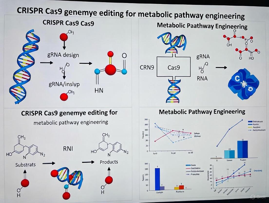
The application of Clustered Regularly Interspaced Short Palindromic Repeats (CRISPR) and CRISPR-associated protein 9 (CRISPR-Cas9) technology has revolutionized the field of metabolic pathway engineering. This powerful gene-editing tool enables researchers to investigate and regulate the biosynthetic pathways of active ingredients in biological systems with unprecedented precision [16]. Metabolic engineering focuses on reprogramming the biochemical networks within cells to enhance the production of valuable compounds or to elucidate complex biological processes. The CRISPR-Cas9 system functions as a bacterial defense mechanism that has been repurposed for precise genome editing, consisting of two main components: the Cas9 endonuclease and a guide RNA (gRNA) that directs the nuclease to specific genomic locations [17]. By precisely regulating the expression of key enzymes and transcription factors, CRISPR technology not only deepens our understanding of secondary metabolic pathways but also opens new avenues for drug development and biotechnology applications [16].
Key Principles of Pathway Targeting
Precision Editing for Metabolic Regulation
The fundamental principle of CRISPR-Cas9 mediated metabolic engineering lies in its ability to create targeted double-strand breaks in DNA, which the cell then repairs through either error-prone non-homologous end joining (NHEJ) or homology-directed repair (HDR) [16] [11]. This precise editing capability allows researchers to strategically manipulate metabolic pathways by knocking out competing pathways, enhancing flux through desired pathways, or introducing new enzymatic functions. The system's efficiency stems from its RNA-guided nature, where a synthetic guide RNA (sgRNA) containing a 20-base variable domain mediates DNA-binding specificity, enabling researchers to quickly retarget the system to different metabolic genes without reengineering protein-DNA interactions [11].
Strategic Multiplexing for Pathway Optimization
Advanced CRISPR-Cas9 applications in metabolic engineering often involve multiplexed approaches, where multiple genes within a metabolic network are targeted simultaneously. This principle recognizes that metabolic pathways are interconnected networks rather than linear sequences, requiring coordinated manipulation of several nodes to effectively redirect metabolic flux. The technology's scalability enables targeting of multiple enzymatic steps in complex metabolic pathways, such as those producing terpenoids, alkaloids, and flavonoids in medicinal plants [16]. This multiplexing capability is particularly valuable for overcoming rate-limiting steps and regulatory feedback mechanisms that often constrain natural metabolic pathways.
Quantitative Framework for Metabolic Engineering
Pharmacokinetic/Pharmacodynamic Modeling of CRISPR Interventions
Quantitative Systems Pharmacology (QSP) platforms provide crucial computational frameworks for predicting the dose-exposure-response relationships of in vivo CRISPR-Cas therapies targeting metabolic pathways. These models characterize the complex journey of CRISPR components from administration to functional metabolic alteration, incorporating mechanisms such as lipid nanoparticle (LNP) binding to opsonins in liver vasculature, phagocytosis into the Mononuclear Phagocyte System (MPS), LNP internalization via endocytosis, and eventual cellular internalization and transgene product release [18]. The following table summarizes key quantitative parameters from established QSP models for CRISPR-based metabolic interventions:
Table 1: Quantitative Parameters for In Vivo CRISPR-Cas Therapy from QSP Models
| Parameter | Species | Value | Biological Significance |
|---|---|---|---|
| Internalization Rate in Interstitial Layer | Non-Human Primate | 0.039 1/h | Determines cellular uptake efficiency in preclinical models |
| Internalization Rate in Interstitial Layer | Human | 0.007 1/h | Predicts slower uptake in human clinical applications |
| Exocytosis Rate | Mouse | 6.84 1/h | Guides preclinical model selection and interpretation |
| Exocytosis Rate | Non-Human Primate | 2690 1/h | Indicates species-specific clearance mechanisms |
| Exocytosis Rate | Human | 775 1/h | Informs clinical dosing frequency and regimen design |
| First-Order Degradation Rate (TTR) | Non-Human Primate | 0.493 1/d | Quantifies target protein reduction in transthyretin amyloidosis model |
| LNP Dose (Total RNA) | Human | 0.75-3 mg/kg | Establishes therapeutic dosing range for metabolic applications |
| Total LNP Dose | Human | 17.2-137.64 mg/kg | Guides formulation development for metabolic pathway targeting |
Metabolic Pathway Editing Efficiency Metrics
Successful metabolic engineering requires not only delivery efficiency but also precise editing outcomes at the target loci. The following quantitative data provides benchmarking metrics for evaluating CRISPR-Cas9 performance in metabolic pathway engineering applications:
Table 2: Editing Efficiency Metrics for Metabolic Pathway Engineering
| Parameter | Efficiency Range | Factors Influencing Efficiency | Optimization Strategies |
|---|---|---|---|
| HDR Efficiency | Generally lower than NHEJ | Cell cycle stage, donor template design, Cas9 version | Synchronize cells in S/G2 phases, use single-stranded DNA templates, employ Cas9 fusions with HDR promoters [19] |
| Base Editing Efficiency | Variable depending on window | PAM positioning, editing window, base editor version | Carefully design gRNA to position target base within optimal editing window (typically positions 4-8 for cytosine base editors) [19] |
| Prime Editing Efficiency | Generally low but highly specific | pegRNA design, Cas9 nickase activity, reverse transcriptase efficiency | Optimize pegRNA scaffold, use dual pegRNA strategies, employ engineered prime editor variants [19] |
| Gene Knockout via NHEJ | High efficiency (often >50%) | gRNA cutting efficiency, chromatin accessibility, Cas9 delivery method | Select exons encoding essential protein domains, use high-fidelity Cas variants, validate multiple gRNAs [19] |
| Multiplexed Editing | Decreasing with target number | gRNA competition, cellular stress, delivery efficiency | Employ tRNA-based polycistronic systems, titrate Cas9-gRNA ratios, use validated gRNA libraries [16] |
Strategic Goals for Metabolic Pathway Engineering
Enhancing Production of Valuable Metabolites
A primary strategic goal in metabolic pathway engineering is the enhanced production of valuable secondary metabolites. CRISPR-Cas9 enables precise manipulation of biosynthetic pathways for compounds with pharmaceutical, agricultural, or industrial significance. In medicinal plants, this technology has been successfully applied to optimize the production of terpenoids (such as tanshinone, artemisinin, and ginsenosides), alkaloids (including morphine, cocaine, and ephedrine), and flavonoids [16]. The strategic approach involves identifying rate-limiting enzymes in these pathways and using CRISPR tools to either enhance their expression or remove metabolic bottlenecks. For example, artemisinin, noted for its antimalarial effects, and andrographolide, known for its antibacterial properties, represent prime targets for pathway optimization [16].
Creating Isogenic Models for Metabolic Studies
The generation of isogenic cell lines with specific metabolic perturbations represents another crucial strategic goal. These engineered models enable researchers to study metabolic pathway functions and identify potential drug targets with minimal genetic background noise. As highlighted in stem cell research, "isogenic models enable more subtle phenotypes to be detected than might be seen when comparing cells derived from two different individuals" [11]. This approach is particularly valuable for modeling metabolic diseases and screening for therapeutic compounds that modulate specific metabolic pathways. The precision of CRISPR-Cas9 allows for the introduction of patient-specific mutations into control cell lines or the correction of disease-causing mutations in patient-derived cells, creating perfectly matched pairs that differ only at the target locus.
Experimental Protocols
Comprehensive Workflow for Metabolic Gene Knockout
The generation of metabolic gene knockouts in human pluripotent stem cells (hPSCs) follows a systematic workflow to ensure high efficiency and specificity [11]:
sgRNA Design and Validation
- Select appropriate online tools for sgRNA design (e.g., CHOPCHOP, CRISPR Design Tool)
- Identify guide sequences with high predicted on-target activity and minimal predicted off-target activity
- Target constitutively expressed regions, 5' exons, or exons coding for essential protein domains
- For metabolic pathway genes, consider targeting catalytic domains of key enzymes
- Clone sgRNA into expression plasmids enabling co-expression with Cas9 and selection markers
- Validate sgRNA efficiency using in vitro cutting assays with Cas9 protein before cellular experiments
CRISPR Delivery and Clone Isolation
- Deliver CRISPR components to hPSCs via electroporation or chemical transfection
- Apply selection (if using selectable markers) 24-48 hours post-transfection
- Isolate single-cell clones by flow cytometry or dilution plating 5-7 days post-transfection
- Expand clones for 10-14 days before genomic DNA extraction and screening
Screening and Validation
- Amplify target region by PCR and analyze by Sanger sequencing or next-generation sequencing
- Identify frameshift mutations that disrupt coding sequences of metabolic enzymes
- Validate functional consequences through metabolic profiling or enzymatic assays
CRISPR Metabolic Knockout Workflow
Homology-Directed Repair for Precise Metabolic Engineering
For introducing specific mutations or inserting reporter genes into metabolic pathways, HDR-based approaches provide precision beyond simple knockouts [11]:
Donor Template Design
- Design single-stranded oligodeoxynucleotides (ssODNs) or double-stranded DNA donors with homology arms
- For point mutations in metabolic enzymes, position the edit within 10 bp of the Cas9 cut site
- Incorporate silent mutations in the PAM site or protospacer to prevent re-cutting
- For reporter knock-ins, ensure proper fusion to metabolic genes and include flexible linkers if needed
CRISPR Component Delivery
- Co-deliver Cas9, sgRNA, and donor template to maximize HDR efficiency
- Time delivery to coincide with S/G2 cell cycle phases when HDR is most active
- Consider using chemical inhibitors of NHEJ pathway to enhance HDR efficiency
- Use Cas9 fused to HDR-promoting factors for improved precise editing
Screening and Validation
- Use droplet digital PCR (ddPCR) or next-generation sequencing to identify precisely edited clones
- Validate proper integration and function through metabolic assays
- Ensure absence of random integration events through appropriate PCR strategies
Research Reagent Solutions
Successful implementation of CRISPR-based metabolic pathway engineering requires carefully selected research reagents and tools. The following table outlines essential components and their applications:
Table 3: Essential Research Reagents for CRISPR-Mediated Metabolic Engineering
| Reagent Category | Specific Examples | Function & Application | Key Considerations |
|---|---|---|---|
| Cas9 Expression Systems | SpCas9 (Addgene #64324) [20], High-fidelity Cas9 variants | Catalyzes DNA cleavage at target sites; base for engineering advanced editors | Consider PAM requirements (NGG for SpCas9); balance activity with specificity [19] |
| gRNA Expression Vectors | pU6-(BbsI)_CBh-Cas9-T2A-mCherry [20] | Express sgRNA with U6 promoter; enable tracking with fluorescent markers | Ensure compatibility with Cas9 system; include selection markers for stable expression |
| Delivery Vehicles | Lipid Nanoparticles (LNPs), Adeno-associated viruses (AAV) | Package and deliver CRISPR components to target cells | Optimize for specific cell types; balance efficiency with cytotoxicity [18] |
| Editing Templates | Single-stranded ODNs, Double-stranded DNA donors with homology arms | Serve as repair templates for HDR; introduce specific mutations or insertions | Design homology arms (typically 800-1000 bp for plasmid donors); include screening markers |
| Validation Tools | Anti-RUNX2 [20], Anti-Collagen antibodies [20], qPCR primers | Confirm successful editing at protein and functional levels | Select validated antibodies for metabolic enzymes; design qPCR assays spanning edit sites |
| Cell Culture Resources | MSOD-B cell line [20], Defined culture media | Provide cellular context for metabolic engineering; maintain pluripotency for differentiation | Use genetically stable lines; employ quality control for consistent experimental conditions |
Metabolic Pathway Manipulation Strategies
Logical Framework for Metabolic Engineering Decisions
Effective metabolic pathway engineering requires strategic decision-making based on the specific engineering goals and pathway characteristics. The following diagram outlines the logical decision process for selecting appropriate CRISPR strategies:
Metabolic Engineering Decision Logic
Advanced Applications in Medicinal Compound Production
The manipulation of plant secondary metabolic pathways represents a particularly promising application of CRISPR-Cas9 technology. Secondary metabolites, including alkaloids, terpenes, flavonoids, and polyphenols, constitute a significant component of human medicines and healthcare products [16]. Research underscores that many active ingredients in modern pharmaceuticals are derived from traditional medicinal plants, with approximately 9% of approved drugs in the United States directly derived from plants, and even higher percentages globally [16]. CRISPR technology enables precise optimization of these valuable compound pathways through several mechanisms:
Terpenoid Pathway Engineering
- Target rate-limiting enzymes such as terpene synthases and cytochrome P450s
- Modify regulatory elements to enhance flux through the mevalonate or methylerythritol phosphate pathways
- Engineer promiscuous enzymes to create novel terpenoid structures with enhanced bioactivity
Alkaloid Biosynthesis Optimization
- Manipulate transcription factors regulating alkaloid biosynthetic gene clusters
- Knock out competing pathways to redirect precursors toward target alkaloids
- Introduce heterologous enzymes to create hybrid alkaloid structures
Flavonoid and Polyphenol Enhancement
- Target phenylpropanoid pathway enzymes to enhance flux toward specific flavonoid classes
- Modify glycosylation patterns to improve bioavailability and stability
- Engineer transcriptional regulators to synchronize pathway expression
Challenges and Future Directions
Current Limitations in Metabolic Pathway Engineering
Despite its transformative potential, CRISPR-Cas9 mediated metabolic engineering faces several significant challenges that must be addressed for broader application. Off-target effects remain a concern, particularly when engineering complex metabolic networks where unintended edits could create undesirable byproducts or disrupt regulatory mechanisms [16] [17]. The efficiency of homology-directed repair, essential for precise metabolic engineering, is generally lower than NHEJ, creating bottlenecks for introducing specific mutations [19]. Delivery efficiency varies considerably across cell types, particularly in industrially relevant organisms and primary cells [16]. Additionally, incomplete understanding of complex metabolic networks and regulatory feedback mechanisms can lead to unexpected outcomes despite precise genetic manipulations [16].
Emerging Solutions and Technological Advances
Several promising approaches are emerging to address these limitations and expand the capabilities of CRISPR-based metabolic engineering. Advanced delivery systems, including improved lipid nanoparticles and viral vectors, are enhancing efficiency particularly for challenging cell types [18]. High-fidelity Cas variants and engineered base editors with reduced off-target activity are improving specificity for metabolic applications [19]. Computational models and QSP platforms are becoming increasingly sophisticated, enabling better prediction of metabolic outcomes following genetic interventions [18]. Machine learning approaches for sgRNA design and metabolic network modeling are further enhancing the precision and predictability of pathway engineering efforts [16]. As these technologies mature, CRISPR-Cas9 is poised to become an increasingly indispensable tool for metabolic engineers seeking to reprogram biological systems for pharmaceutical production, bioenergy applications, and fundamental understanding of metabolic regulation.
Application Note: Clinical Foundations of CRISPR-Cas9 Genome Editing
The transition of CRISPR-Cas9 from a bacterial immune system to a revolutionary clinical technology has established a new paradigm for treating genetic disorders. This foundation is built upon key approved therapies that demonstrate proof-of-concept and provide a framework for future applications in metabolic pathway engineering. The first regulatory approvals in 2023-2024 mark the beginning of clinical CRISPR-based interventions, offering critical insights into effective therapeutic design, safety parameters, and manufacturing protocols.
Foundational Approved Therapies and Mechanisms
The first CRISPR-Cas9 approved therapies target hematological diseases but establish principles applicable to metabolic pathway engineering. These ex vivo approaches modify patient-derived hematopoietic stem cells (HSCs) to achieve therapeutic effect through distinct molecular mechanisms.
Table 1: First Approved CRISPR-Cas9 Genome Editing Therapies
| Therapy Name | Indication | Year Approved | Molecular Target | Editing Outcome | Clinical Efficacy |
|---|---|---|---|---|---|
| Casgevy (exagamglogene autotemcel) | Sickle Cell Disease (SCD); Transfusion-Dependent β-Thalassemia (TDT) | 2023 (UK MHRA, US FDA, EMA) [21] [22] | BCL11A erythroid-specific enhancer region [21] | Disruption of BCL11A, increasing fetal hemoglobin (HbF) production [21] [22] | 93.5% (29/31) of SCD patients free from severe vaso-occlusive crises for ≥12 months; 100% engraftment success [21] [22] |
| Lyfgenia (lovotibeglogene autotemcel) | Sickle Cell Disease (SCD) [22] | 2023 (US FDA) [21] [22] | Addition of HbAT87Q gene via lentiviral vector [21] | Production of gene-therapy derived hemoglobin (HbAT87Q) that resists sickling [21] [22] | 88% (28/32) of patients achieved complete resolution of vaso-occlusive events (6-18 months post-infusion) [22] |
The therapeutic mechanism of Casgevy involves CRISPR-Cas9-mediated disruption of an enhancer region of the BCL11A gene, a repressor of fetal hemoglobin (HbF) production [21]. This reactivation of HbF compensates for the defective adult hemoglobin in sickle cell disease and β-thalassemia. The process involves collecting a patient's CD34+ hematopoietic stem cells, editing them ex vivo using CRISPR-Cas9, and reinfusing them after myeloablative conditioning [21] [22]. The successful engraftment and clinical efficacy across a majority of patients validate this approach for monogenic disorders.
Key Experimental Workflows from Clinical Trials
The foundational protocols for these therapies were established in pivotal clinical trials. The workflow for Casgevy exemplifies a robust ex vivo gene editing protocol applicable to hematopoietic cells.
Protocol 1.1: Ex Vivo HSC Gene Editing (Based on Casgevy Clinical Trial) [21] [22]
- Patient Selection & HSC Collection: Enroll patients meeting specific clinical criteria (e.g., SCD with recurrent vaso-occlusive crises). Collect autologous CD34+ hematopoietic stem and progenitor cells via apheresis.
- CRISPR-Cas9 Delivery & Editing: Electroporate or transduce collected CD34+ cells with CRISPR-Cas9 components:
- RNP Complex: Cas9 protein pre-complexed with sgRNA targeting the BCL11A erythroid-specific enhancer.
- Guide RNA: Designed for high on-target activity and minimal predicted off-target effects.
- Myeloablative Conditioning: Administer busulfan chemotherapy to the patient to create marrow niche space for edited cells.
- Cell Reinfusion & Support: Thaw and administer the edited cell product (Casgevy) as a single-dose infusion. Provide supportive care including monitoring for and management of cytopenias.
- Outcome Assessment: Monitor for successful engraftment (neutrophil and platelet count recovery). Assess primary efficacy endpoint (e.g., freedom from severe vaso-occlusive crises for 12 consecutive months).
Emerging Clinical Platforms: In Vivo and Next-Generation Editing
Building on the success of ex vivo therapies, the clinical CRISPR landscape is rapidly expanding to include in vivo delivery and more precise editing technologies like base and prime editing. These platforms are particularly relevant for engineering metabolic pathways in inaccessible tissues.
Clinical-Stage In Vivo Genome Editing
In vivo editing represents a significant advancement by directly administering CRISPR components to patients, overcoming limitations of ex vivo cell therapy.
Table 2: Select In Vivo CRISPR-Cas Clinical Trials Targeting Metabolic Pathways
| Therapy/Platform | Indication | Phase | Molecular Target | Delivery System | Reported Outcome |
|---|---|---|---|---|---|
| NTLA-2001 (Intellia/Regeneron) | Transthyretin (ATTR) Amyloidosis [23] | III [23] | TTR gene knockout [23] | Lipid Nanoparticle (LNP) [24] | ~90% sustained reduction in serum TTR protein levels [24] |
| VERVE-101 (Verve Therapeutics) | Heterozygous Familial Hypercholesterolemia [23] | Ib (Paused) [23] | PCSK9 gene inactivation [23] | LNP [23] | Proof-of-concept for single-dose in vivo base editing [23] |
| NTLA-2002 (Intellia) | Hereditary Angioedema (HAE) [24] [23] | I/II [23] | KLKB1 gene knockout [24] [23] | LNP [24] [23] | 86% reduction in kallikrein; majority of patients attack-free [24] |
The Intellia Therapeutics trials for NTLA-2001 and NTLA-2002 demonstrate a viable platform for systemic in vivo editing. The use of LNPs that naturally accumulate in the liver enables efficient editing of hepatocytes, making this system ideal for targeting metabolic pathways controlled by the liver [24]. This approach has shown a favorable safety profile, allowing for re-dosing in some cases, which is typically not possible with viral vector delivery due to immune reactions [24].
Workflow for In Vivo Gene Editing
The generalized workflow for LNP-delivered in vivo CRISPR therapies highlights key differences from ex vivo approaches, particularly in delivery and biodistribution.
Protocol 2.1: LNP-Mediated In Vivo Gene Editing (Based on NTLA-2001/2002 Trials) [24]
- Payload Design: Formulate LNPs containing:
- mRNA: Encoding the Cas9 nuclease (e.g., S. pyogenes Cas9).
- sgRNA: Designed for specific knockout of the target gene (e.g., TTR or KLKB1).
- LNP Formulation & Administration: Manufacture LNPs under GMP conditions. Administer a single dose via intravenous infusion.
- Biodistribution & Editing: LNPs traffic to the liver and are taken up by hepatocytes. The CRISPR payload is released into the cytoplasm, enters the nucleus, and performs the intended gene edit.
- Efficacy & Safety Monitoring:
- Biomarker Assessment: Quantify reduction in target protein serum levels (e.g., TTR, kallikrein) as a pharmacodynamic biomarker.
- Clinical Endpoints: Monitor disease-specific clinical outcomes (e.g., attack frequency in HAE).
- Safety Profile: Monitor for infusion-related reactions and potential off-target effects.
The Scientist's Toolkit: Research Reagent Solutions
The translation of CRISPR therapies relies on a standardized set of molecular tools and reagents. The table below details essential components for developing CRISPR-based therapeutics, derived from clinical trial methodologies.
Table 3: Essential Research Reagents for Therapeutic CRISPR Development
| Reagent / Tool | Function in Therapeutic Workflow | Examples & Clinical Context |
|---|---|---|
| Cas9 Nuclease | Creates double-strand breaks (DSBs) in target DNA guided by sgRNA. | S. pyogenes Cas9 (SpCas9) used in Casgevy; engineered variants (SaCas9) with different PAM specificities for broader targeting [25]. |
| Guide RNA (sgRNA) | Directs Cas nuclease to specific genomic locus via 20-nt complementary sequence. | Designed for minimal off-targets; chemically modified for stability in LNP delivery (e.g., NTLA-2001) [24] [25]. |
| Delivery Vehicle | Facilitates intracellular delivery of CRISPR machinery. | Ex vivo: Electroporation of RNP (Casgevy). In vivo: Lipid Nanoparticles (LNP) for liver (NTLA-2001) [24] [22]; Viral vectors (Lentivirus for Lyfgenia) [21]. |
| Hematopoietic Stem Cells (HSCs) | Target cell type for ex vivo editing; capable of self-renewal and reconstituting entire blood system. | Patient-derived CD34+ cells, isolated via apheresis, are the starting material for Casgevy and Lyfgenia [21] [22]. |
| Base Editors (BEs) | Catalyzes direct chemical conversion of one base pair to another without inducing DSBs, reducing genotoxicity. | VERVE-101 uses Adenine Base Editor (ABE) to convert A•T to G•C in PCSK9 gene [25] [23]. |
| Prime Editors (PEs) | Enables all 12 possible base-to-base conversions, plus small insertions and deletions, without DSBs using a reverse transcriptase template. | Prime Medicine's PM359 for CGD uses prime editors for precise correction of NCF1 mutations ex vivo [23]. |
Visualization of Key Signaling Pathways in Approved Therapies
Understanding the molecular pathways targeted by foundational therapies provides a blueprint for engineering novel metabolic interventions. The diagram below illustrates the mechanism of Casgevy.
Pathway 1: BCL11A Enhancer Targeting by Casgevy The CRISPR-Cas9-mediated disruption of the erythroid-specific enhancer of the BCL11A gene reduces the expression of the BCL11A transcription repressor protein. This downregulation de-represses the genes encoding fetal hemoglobin (HbF), specifically the γ-globin genes. The resulting increase in HbF production compensates for the defective β-globin in sickle cell disease, preventing red blood cell sickling and its associated pathologies [21]. This approach demonstrates the power of targeting regulatory elements to orchestrate complex metabolic and developmental pathways.
Strategic Delivery and Application in Metabolic Engineering
The selection of an appropriate delivery vector is a critical step in designing CRISPR-Cas9 experiments for metabolic pathway engineering. The choice between viral and non-viral methods, combined with the decision to perform editing in vivo or ex vivo, directly impacts the efficiency, specificity, and safety of your genomic modifications. For metabolic engineers seeking to reprogram cellular factories for enhanced biochemical production, these decisions must balance editing efficiency with practical constraints such as cargo capacity and scalability. This application note provides a structured comparison of delivery systems and detailed protocols to guide researchers in selecting the optimal strategy for their specific metabolic engineering objectives.
Section 1: Vector System Comparison and Selection Guide
CRISPR-Cas9 components can be delivered in three primary forms: DNA (plasmid), RNA (mRNA with gRNA), or preassembled Ribonucleoprotein (RNP) complexes [26]. The delivery vehicle must be compatible with your chosen cargo format and experimental system.
Table 1: Non-Viral CRISPR Cargo Delivery Methods
| Delivery Method | Compatible Cargo | Key Advantages | Key Disadvantages | Recommended Applications |
|---|---|---|---|---|
| Electroporation [27] | DNA, mRNA, RNP | High efficiency, broad cell type range | Damaging to cells | Ex vivo editing (e.g., Casgevy for sickle cell) [27] |
| Lipid Nanoparticles (LNPs) [27] [28] | DNA, mRNA, RNP | FDA-approved, good stability, low immunogenicity | Low/variable efficiency, endosomal trapping | In vivo therapeutic RNA delivery [27] |
| Microinjection [27] | DNA, mRNA, RNP | Efficient single-cell delivery | Technically demanding, low throughput | Mouse embryo engineering [27] |
Table 2: Viral Vector Delivery Methods
| Delivery Method | Max Cargo Capacity | Integration | Key Advantages | Key Disadvantages | Recommended Applications |
|---|---|---|---|---|---|
| Adeno-associated Virus (AAV) [27] [26] | ~4.7 kb [26] | No [26] | Low immunogenicity, high tissue specificity | Very limited cargo capacity | In vivo delivery (requires small Cas variants like SaCas9) [27] |
| Lentivirus (LV) [27] [26] | >10 kb [26] | Yes (into host genome) | High delivery efficiency, long-term expression | Risk of insertional mutagenesis | In vitro and ex vivo use; CRISPR library screens [27] |
| Adenovirus (AdV) [26] | ~36 kb [26] | No [26] | Very large cargo capacity, infects dividing/non-dividing cells | Can trigger strong immune responses | In vivo delivery requiring large genetic payloads [26] |
The following workflow diagram provides a systematic approach for selecting the appropriate delivery method based on key experimental parameters:
Section 2: In Vivo vs. Ex Vivo Strategy Selection
The choice between in vivo and ex vivo editing is fundamental and influences all subsequent vector decisions.
In Vivo Delivery involves introducing CRISPR components directly into the organism, targeting specific tissues or organs. This approach is less invasive for targeting internal organs but faces challenges including immune recognition, precise targeting, and potential off-target effects [28].
Ex Vivo Delivery involves extracting cells from the organism, performing gene editing in a controlled laboratory environment, and then reintroducing the modified cells back into the host. This method allows for precise control over editing conditions, enables rigorous quality control and screening of edited cells, and minimizes immune responses and off-target risks in the host [29]. The approved drug Casgevy for sickle cell anemia is a prime example, where hematopoietic stem cells are edited ex vivo via RNP electroporation before being reinfused into the patient [27].
Table 3: In Vivo vs. Ex Vivo Editing Comparison
| Parameter | In Vivo Editing | Ex Vivo Editing |
|---|---|---|
| Invasiveness | Less invasive for internal organs | Requires cell extraction & transplantation |
| Control over Editing | Lower; limited by delivery and biodistribution | High; allows for precise control and screening |
| Safety Profile | Higher risk of immune response and off-target effects | Safer; edited cells can be validated pre-infusion |
| Therapeutic Applicability | Suitable for organs that cannot be easily extracted (e.g., liver, brain) | Ideal for blood cells (HSCs), immune cells (T cells) |
| Technical Complexity | Complex delivery challenges (targeting, immune evasion) | Simplified delivery but requires cell culture expertise |
| Clinical Translation | EBT-101 for HIV (Phase 1/2) [27] | Casgevy for Sickle Cell Anemia (FDA-approved) [27] |
Section 3: Detailed Experimental Protocols
Protocol 1: Ex Vivo Metabolic Engineering in E. coli using Plasmid-Based CRISPR-Cas9
This optimized protocol enables iterative genome editing for metabolic engineering in E. coli, facilitating gene knockouts, insertions, and pathway integrations with high efficiency [30].
Research Reagent Solutions:
- pCas9cur Plasmid: Confers chloramphenicol resistance; expresses Cas9 nuclease and λ Red recombinase proteins for recombineering [30].
- gRNA Expression Plasmid: Contains a kanamycin resistance gene and a J23119 promoter driving the expression of the target-specific gRNA [30].
- Donor DNA Template: Double-stranded DNA (dsDNA) fragment containing the desired modification (e.g., gene insertion or deletion) flanked by ~500 bp homology arms for HDR [30].
Procedure:
- Preparation: Transform the pCas9cur plasmid into the target E. coli strain and culture at 30°C. The λ Red system is induced by L-arabinose when needed.
- Co-transformation: Electroporate a mixture of the gRNA expression plasmid and the donor dsDNA fragment into the competent cells containing pCas9cur.
- Selection and Screening: Plate cells on kanamycin plates and incubate at 30°C. Select for colonies that have successfully incorporated the gRNA plasmid, which also indicates successful editing in most cases due to high co-transformation efficiency.
- Curing the gRNA Plasmid: Inoculate a positive colony into liquid medium and culture at 37°C without kanamycin to promote the loss of the temperature-sensitive gRNA plasmid.
- Verification: Screen for kanamycin-sensitive colonies and verify the genetic modification via colony PCR and DNA sequencing.
- Iteration: The pCas9cur plasmid remains in the strain, allowing the process to be repeated from Step 2 for subsequent edits using a new gRNA plasmid.
Protocol 2: In Vivo Metabolic Pathway Reprogramming in Mouse Liver via Hydrodynamic Tail Vein Injection
This protocol demonstrates the conversion of hepatocytes in a mouse model of hereditary tyrosinemia type I (HT-I) to a benign state by deleting the Hpd gene, showcasing the therapeutic potential of in vivo metabolic pathway engineering [4].
Research Reagent Solutions:
- CRISPR Component Plasmids: A plasmid expressing Cas9 nuclease and plasmids expressing two gRNAs (gRNA1 and gRNA3) targeting intronic regions flanking exons 3 and 4 of the murine Hpd gene [4].
- Physiological Saline Solution: Sterile 0.9% NaCl solution for plasmid dilution.
Procedure:
- Solution Preparation: Dilute the plasmid mixture (Cas9 and gRNA plasmids in a 1:1 mass ratio) in a volume of physiological saline equivalent to 8-10% of the mouse's body weight. Filter the solution through a 0.22 µm filter.
- Mouse Restraint: Warm the mouse tail to dilate the veins. Secure the mouse in a tail vein injection restrainer.
- Hydrodynamic Injection: Rapidly inject (within 5-7 seconds) the prepared plasmid solution into the lateral tail vein using a syringe with a 27-gauge needle. The high volume and speed create transient pressure that forces the solution into the hepatocytes.
- Post-Injection Monitoring: Return the mouse to its cage and monitor until it recovers from anesthesia. For the HT-I model, mice are weaned off the protective drug nitisinone post-injection to create a selective advantage for successfully edited (Hpd-/-) hepatocytes.
- Validation: After 4-8 weeks, analyze editing efficiency through PCR of the Hpd locus, immunostaining for HPD protein, and mass spectrometry of plasma tyrosine and succinylacetone levels to confirm metabolic reprogramming [4].
Section 4: The Scientist's Toolkit
Table 4: Essential Research Reagent Solutions for CRISPR Delivery Experiments
| Reagent / Tool | Function / Description | Example Use Case |
|---|---|---|
| CRISPR RNP Complex [27] [26] | Preassembled complex of Cas9 protein and guide RNA. Offers immediate activity, high precision, and reduced off-target effects. | Gold standard for ex vivo therapeutic editing (e.g., Casgevy) [27]. |
| Lipid Nanoparticles (LNPs) [28] | Synthetic, biodegradable particles that encapsulate and protect nucleic acid cargo (mRNA, gRNA). | In vivo delivery of Cas9 mRNA and gRNA for systemic administration [27]. |
| Adeno-associated Virus (AAV) [27] [26] | A non-pathogenic viral vector with high tissue specificity and low immunogenicity. Limited cargo capacity. | In vivo gene editing requiring sustained expression in specific tissues (e.g., liver, muscle). |
| Stimuli-Responsive Nanoparticles [28] | Non-viral vectors designed to release their CRISPR payload in response to specific triggers (e.g., low pH, enzymes). | Precision cancer therapy, ensuring editing occurs primarily in the target tumor microenvironment. |
| Virus-Like Particles (VLPs) [26] | Engineered viral capsids lacking viral genetic material. Combine transduction efficiency of viruses with transient expression of non-viral methods. | Emerging tool for in vivo delivery of base editors or prime editors with improved safety profiles. |
Section 5: Concluding Recommendations
For bacterial metabolic engineering, plasmid-based delivery coupled with recombineering offers a robust, high-efficiency, and iterative platform [30]. For therapeutic ex vivo applications in eukaryotes, such as modifying human stem or immune cells, RNP delivery via electroporation is the preferred method due to its high precision, transient activity, and established clinical success [27]. For direct in vivo therapeutic applications, viral vectors like AAV (for sustained expression) or non-viral vectors like LNPs (for transient expression) are the primary candidates, though both face ongoing challenges in efficiency, safety, and tissue-specific targeting that are the focus of current research [27] [28]. The field is rapidly advancing with the development of novel non-viral nanomaterials and engineered viral capsids, promising ever-more precise and efficient delivery solutions for complex metabolic engineering challenges.
In metabolic pathway engineering research, the precision editing of genomes using CRISPR-Cas9 has emerged as a transformative approach. Central to the success of CRISPR-based methodologies is the efficient delivery of editing components—including Cas nucleases, guide RNAs (gRNAs), and repair templates—into target cells, a process fundamentally reliant on transfection technologies [4] [11]. Transfection, the process of introducing foreign nucleic acids into eukaryotic cells, provides the critical gateway for these components to access the cellular machinery [31] [32]. The choice of transfection method directly impacts key experimental outcomes, including editing efficiency, cell viability, and the fidelity of the resulting metabolic alterations.
The application of transfection in metabolic pathway engineering was powerfully demonstrated in a study treating hereditary tyrosinaemia type I, where researchers used hydrodynamic tail vein injection to deliver CRISPR-Cas9 components targeting the Hpd gene into mouse hepatocytes [4]. This approach successfully reprogrammed tyrosine catabolism by converting hepatocytes from the lethal tyrosinaemia type I into the benign type III, with edited hepatocytes exhibiting a significant growth advantage and repopulating most of the murine liver within weeks [4]. Such successes underscore the critical importance of selecting and optimizing transfection strategies tailored to specific research goals, cell types, and experimental constraints.
Transfection Substrates for Genome Editing
The choice of transfection substrate significantly influences experimental design, timing, and outcome in CRISPR-Cas9 genome editing. Each substrate offers distinct advantages and limitations that must be considered in the context of metabolic engineering applications.
Table 1: Comparison of Transfection Substrates for CRISPR-Cas9 Applications
| Substrate | Key Advantages | Key Limitations | Ideal Applications in Metabolic Engineering |
|---|---|---|---|
| Plasmid DNA | Cost-effective production; stable transfection possible; suitable for a wide range of applications [33] | Must reach nucleus; slower protein expression; risk of genomic integration [33] | Stable cell line generation; long-term pathway modulation |
| mRNA | Rapid protein expression; only needs to reach cytoplasm; higher efficiency in DNA-sensitive cells [33] | Chemically unstable; cannot be used for stable transfection; more expensive production [33] | Rapid, transient protein expression; difficult-to-transfect cells |
| Ribonucleoproteins (RNPs) | Immediate activity; controlled dosage; reduced off-target effects [33] | Protein production can be costly; requires per-protein optimization [33] | CRISPR-Cas9 gene editing; precise, temporary enzyme activity |
| siRNA | Gene silencing via RNA interference; cytoplasmic activity [34] | Temporary effect (typically 4-7 days); requires optimization to minimize off-target effects [34] [35] | Knockdown studies; transient pathway modulation; validation experiments |
For CRISPR-Cas9 mediated metabolic pathway reprogramming, the choice between these substrates depends on the experimental timeline and desired persistence of editing. DNA-based approaches facilitate stable genomic integration but require nuclear access, while RNA and protein-based approaches offer more immediate but transient effects [33]. In the successful tyrosinaemia type I study, researchers employed DNA vectors expressing both Cas9 nuclease and guide RNAs, enabling stable genomic editing of the Hpd gene and permanent metabolic reprogramming [4].
Transfection Methodologies: A Comparative Analysis
Transfection methods can be broadly categorized into viral, chemical, and physical approaches, each with distinct mechanisms, advantages, and optimal applications.
Viral Transfection (Transduction)
Viral-based transfection utilizes modified viral vectors to deliver genetic material into host cells with high efficiency, particularly valuable for difficult-to-transfect cells like primary cells and stem cells [31] [32].
Table 2: Viral Transfection Methods for Genome Editing
| Viral Vector | Transfection Type | Target Cells | Key Advantages | Key Limitations |
|---|---|---|---|---|
| Retrovirus | Stable | Dividing cells | Stable genomic integration; long-term expression [31] | Risk of insertional mutagenesis; limited to dividing cells [31] |
| Lentivirus | Stable | Dividing & non-dividing cells | Broad cellular tropism; stable integration [31] | Complex production; safety concerns for clinical applications [31] |
| Adenovirus | Transient | Dividing & non-dividing cells | High packaging capacity; broad cell type transduction [31] | Immunogenic; transient expression; pre-existing immunity in populations [31] |
| Adeno-associated Virus (AAV) | Primarily transient | Dividing & non-dividing cells | Low immunogenicity; good safety profile [31] | Small packaging capacity (<5 kb); limited insert size [31] |
Non-Viral Transfection Methods
Non-viral approaches encompass both chemical and physical methods, generally offering better safety profiles, easier preparation, and reduced immunogenicity compared to viral methods, though often with lower efficiency in certain cell types [31] [32].
Table 3: Chemical and Physical Transfection Methods
| Method | Mechanism | Best For | Efficiency | Cell Viability | Notes |
|---|---|---|---|---|---|
| Cationic Lipids | Complexes with nucleic acids; fuses with cell membrane [32] | Common cell lines; high efficiency transfer [36] | High | Moderate to low (dose-dependent) [36] | Lipofectamine shows high efficiency but significant cytotoxicity [36] |
| Calcium Phosphate | DNA-calcium phosphate precipitate; endocytosis [32] | Standard cell lines; cost-effective applications | Moderate | Moderate | Classical method; requires optimization of precipitate size |
| Electroporation | Electrical pulses create temporary pores in cell membrane [32] | Primary cells; stem cells; difficult-to-transfect cells [11] | High | Low to moderate (protocol-dependent) | Requires specialized equipment; parameters must be optimized for each cell type |
| Nanoparticles | Nanocarriers encapsulate nucleic acids; endocytosis [36] [37] | In vivo applications; targeted delivery [36] [37] | Moderate | High | Lower cytotoxicity; suitable for in vivo use [36] |
The development of lipid-coated calcium phosphate nanoparticles (LCP nanoparticles) represents an advanced non-viral approach that combines the biocompatibility of calcium phosphate with the delivery efficiency of lipid systems, showing particular promise for in vivo applications including liver-targeted gene therapy [37].
Experimental Protocols for Transfection in Genome Editing
General Guidelines for Transfection Optimization
Successful transfection requires careful optimization of multiple parameters. The following guidelines provide a foundation for developing efficient transfection protocols:
- Cell Health and Density: Cells should be in optimal physiological condition at transfection, typically at 70% confluency for many cell types, though this requires empirical determination for each experimental system [34] [32].
- Nucleic Acid Purity: Ensure high-quality nucleic acid preparation free from contaminants such as endotoxins, salts, or proteins that can significantly reduce transfection efficiency and cell viability [33] [31].
- Reagent:Nucleic Acid Ratio: Systematically optimize the ratio of transfection reagent to nucleic acid for each new cell type and nucleic acid combination [34] [35].
- Serum and Antibiotics: Most chemical transfection reagents require serum-free conditions during complex formation, while antibiotics should typically be omitted during transfection as they can increase cell death [34] [32].
Protocol: siRNA Transfection for Gene Knockdown Studies
Application: Transient knockdown of metabolic enzymes to study pathway flux [34]
Materials:
- Validated siRNA against target gene
- Appropriate transfection reagent (e.g., liposomal agents)
- Opti-MEM or similar serum-free medium
- Cells of interest cultured in appropriate medium
Procedure:
- Day 1: Plate Cells: Seed cells in multiwell plates at optimal density (e.g., 0.4-1.6 × 10⁵ cells/well for 24-well plates) to reach 70% confluency at time of transfection [35].
- Day 2: Prepare Complexes:
- Dilute siRNA in serum-free medium (typical working concentration 5-100 nM; use lowest effective concentration) [34].
- Dilute transfection reagent in separate tube of serum-free medium.
- Combine diluted siRNA and transfection reagent, mix gently, incubate 15-20 minutes at room temperature.
- Transfect Cells: Add complexes dropwise to cells with gentle swirling.
- Incubate: Culture cells for 24-72 hours before analysis. Gene silencing effects can typically be observed as early as 24 hours and may last 4-7 days depending on cell type and protein turnover rate [34].
Controls: Include untransfected cells, mock-transfected cells (reagent only), non-targeting siRNA control, and positive control siRNA if available [34].
Protocol: CRISPR-Cas9 RNP Transfection for Gene Editing
Application: Precise genome editing for metabolic pathway engineering [33] [11]
Materials:
- Purified Cas9 protein
- Synthetic sgRNA targeting gene of interest
- Appropriate transfection reagent (e.g., lipid-based for common cell lines, specialized reagents for stem cells)
- Cells of interest
Procedure:
- Design and Validate sgRNA: Use online tools (e.g., CHOPCHOP, CRISPR Design Tool) to design sgRNAs with high on-target and minimal off-target activity [11].
- Pre-complex RNP: Mix Cas9 protein and sgRNA at optimal molar ratio (typically 1:2 to 1:3), incubate 10-15 minutes at room temperature to form RNP complexes.
- Prepare Transfection Complexes: Combine pre-formed RNP complexes with appropriate transfection reagent according to manufacturer's instructions.
- Transfect Cells: Add complexes to cells at optimal confluency (varies by cell type).
- Analyze Editing Efficiency: Assess editing efficiency 48-72 hours post-transfection using T7E1 assay, tracking of indels by decomposition (TIDE), or sequencing.
For hard-to-transfect cells like human pluripotent stem cells (hPSCs), consider using electroporation-based systems optimized for stem cells [11].
The Scientist's Toolkit: Essential Research Reagents
Table 4: Key Research Reagent Solutions for Transfection-Based Genome Editing
| Reagent/Material | Function | Application Notes |
|---|---|---|
| Liposomal Transfection Reagents | Form complexes with nucleic acids, facilitating cellular uptake [36] | High efficiency but can be cytotoxic; optimize reagent:nucleic acid ratio for each cell type [36] |
| Cationic Polymers | Condense nucleic acids through electrostatic interactions [31] | Includes PEI, DEAE-dextran; can be toxic at high concentrations |
| Calcium Phosphate Nanoparticles | Biocompatible inorganic carriers for nucleic acid delivery [36] [37] | Lower cytotoxicity than liposomal agents; suitable for in vivo applications [36] |
| Electroporation Systems | Apply electrical fields to create temporary pores in cell membranes [32] | Effective for hard-to-transfect cells; parameters must be optimized for each cell type [11] |
| Reporter Genes (eGFP, Luciferase) | Assess transfection efficiency through detectable markers [32] | Essential for optimization and quality control; eGFP enables live-cell imaging [36] |
| Validated Control siRNAs | Control for non-specific effects in RNAi experiments [34] | Include positive, negative, and fluorescent controls for accurate interpretation |
Transfection Workflow and Efficiency Assessment
The following diagram illustrates the decision-making workflow for selecting and optimizing transfection methods in the context of CRISPR-Cas9 genome editing for metabolic engineering:
Transfection Method Selection Workflow
Assessing Transfection Efficiency and Cell Viability
Accurate assessment of transfection success is critical for data interpretation and experimental reproducibility. The following approaches are commonly employed:
- Microscopy and Flow Cytometry: For fluorescent reporters (e.g., eGFP), efficiency can be quantified as the percentage of fluorescent cells relative to total cells using fluorescence microscopy or flow cytometry [36] [38]. Transfection efficiency (%) = (Number of expressing cells / Total number of cells seeded) × 100 [38].
- Viability Assays: Cell health post-transfection can be assessed using trypan blue exclusion, ATP-based assays, or metabolic activity tests like MTT [32]. Live-cell imaging provides temporal assessment of both efficiency and viability [36].
- Functional Assays: For genome editing applications, deep sequencing of target loci provides precise quantification of editing efficiency and specificity [4] [11].
In live-cell imaging studies comparing calcium phosphate nanoparticles to liposomal agents, transfection efficiency and cytotoxicity showed an inverse relationship, with liposomal agents providing higher efficiency but significantly greater cytotoxicity [36].
Advanced Applications in Metabolic Pathway Engineering
The power of transfection-enabled CRISPR-Cas9 genome editing for metabolic engineering is exemplified by innovative approaches such as metabolic pathway reprogramming. In this strategy, rather than directly correcting a disease-causing gene, researchers target alternative genes within disease-associated metabolic pathways to render the phenotype benign [4].
In the seminal study on hereditary tyrosinaemia type I, researchers used hydrodynamic tail vein injection—a specialized physical transfection method—to deliver CRISPR-Cas9 components targeting the Hpd gene into mouse hepatocytes [4]. This approach successfully converted the lethal tyrosinaemia type I hepatocytes into the benign type III by reprogramming tyrosine catabolism, with edited hepatocytes exhibiting a growth advantage and repopulating most of the liver within weeks [4]. The reprogrammed animals showed improved metabolic profiles compared to pharmacologically treated controls, demonstrating the therapeutic potential of this approach [4].
For such advanced applications, careful consideration of transfection parameters is essential. In the tyrosinaemia study, researchers used a combination of two guide RNAs to excise critical exons of the Hpd gene, with editing occurring predominantly in pericentral hepatocytes and reaching up to 99% replacement of diseased hepatocytes within 8 weeks in some animals [4].
Optimizing transfection methodologies is paramount for successful CRISPR-Cas9 genome editing in metabolic pathway engineering research. The selection of appropriate transfection substrates—DNA, RNA, or proteins—coupled with delivery methods tailored to specific cell types and experimental goals, forms the foundation of efficient genetic manipulation. As demonstrated by successful applications in metabolic pathway reprogramming, continued refinement of these techniques, particularly toward enhanced targeted delivery and reduced off-target effects, will further expand the possibilities for precise metabolic engineering and therapeutic intervention.
The liver plays a central role in metabolic homeostasis, making it a prime therapeutic target for a spectrum of genetic and acquired metabolic diseases. The advent of CRISPR-based genome editing, coupled with advances in delivery vehicles, has ushered in a new era of potential one-time, curative treatments. Among delivery systems, lipid nanoparticles (LNPs) have emerged as a particularly promising platform for in vivo liver-directed editing, combining high delivery efficiency with a favorable safety profile and established clinical manufacturability [39] [40]. This Application Note details the principles, protocols, and key applications of LNP-mediated CRISPR delivery for metabolic pathway engineering in the liver, providing a framework for researchers and drug development professionals.
The efficacy of LNPs for liver targeting is not accidental but stems from well-understood physiological mechanisms. Upon intravenous administration, systemically circulating LNPs preferentially accumulate in the liver largely due to their association with apolipoprotein E (ApoE). This ApoE corona facilitates active targeting by binding to low-density lipoprotein (LDL) receptors, which are highly expressed on the surface of hepatocytes [40]. The liver's unique anatomy, including fenestrated endothelial cells, further enhances LNP access to parenchymal cells. Beyond passive accumulation, modern LNP design actively exploits these pathways by incorporating ionizable lipids that optimize ApoE binding and subsequent endosomal escape within hepatocytes, enabling efficient intracellular release of genomic cargo [40].
Table 1: Key Characteristics of LNP Delivery for Liver Editing
| Feature | Description | Therapeutic Implication |
|---|---|---|
| Targeting Mechanism | ApoE protein binding & LDL receptor-mediated uptake on hepatocytes [40] | Natural tropism allows for efficient liver targeting post-intravenous injection. |
| Primary Cell Targets | Hepatocytes (various zones), Kupffer cells [40] | Enables modification of key metabolic cells; Kupffer cell uptake can be a hurdle. |
| Editing Timeline | Transient Cas9 expression (typically days) [39] | Reduces off-target risks compared to persistent viral expression. |
| Packaging Capacity | High (can accommodate large editors like base editors) [39] | More flexible than AAV vectors for delivering sophisticated editing systems. |
CRISPR Tool Selection for Metabolic Engineering
The choice of CRISPR machinery is critical for achieving the desired therapeutic outcome, whether the goal is gene knockout, precise base correction, or gene insertion. While early approaches relied on Cas9 mRNA delivery, recent advances demonstrate the superior efficacy and safety of delivering the pre-assembled ribonucleoprotein (RNP) complex. The RNP format minimizes off-target effects due to its short intracellular half-life and avoids the immune activation sometimes associated with mRNA [41] [42].
A landmark development in this area is the engineering of a thermostable Cas9 from Geobacillus stearothermophilus (GeoCas9). Through directed evolution, researchers created iGeoCas9 variants capable of robust genome editing in mammalian cells and organs. When formulated into LNPs as stable RNP complexes, iGeoCas9 achieved >100-fold higher editing efficiency compared to the wild-type GeoCas9 enzyme. This system has demonstrated impressive results in vivo, with liver editing efficiencies ranging from 16% to 37% in reporter mice following a single intravenous injection [41] [42]. The enhanced stability of iGeoCas9 makes it particularly resilient during the LNP formulation process, which often involves denaturing organic solvents.
For metabolic diseases caused by specific point mutations, base editing offers a powerful alternative. Base editors catalyze precise single-nucleotide changes without creating double-strand breaks, thereby minimizing undesirable indels. The therapeutic potential of this approach is exemplified by CS-121, an in vivo base editing therapy targeting the APOC3 gene for hypertriglyceridemia. CS-121 utilizes a transformer Base Editor (tBE) delivered via LNPs to the liver to mimic beneficial natural loss-of-function variants. In a recent clinical announcement, the first patient treated with a single low dose showed a significant drop in fasting triglyceride levels within three days, underscoring the rapid translational potential of this technology [43].
Diagram 1: Workflow for in vivo liver editing experiment.
Key Applications in Metabolic Disease
LNP-mediated liver editing is being applied to a wide range of metabolic disorders, from common conditions like hypercholesterolemia to rare genetic diseases. The applications can be broadly categorized into gene disruption, gene correction, and gene insertion, each with distinct therapeutic targets.
Disruption of Disease-Causing Genes
A primary application is the knockout of genes whose products have detrimental effects. A prominent target is PCSK9, a regulator of LDL cholesterol levels. Disruption of PCSK9 in hepatocytes leads to increased LDL receptor expression and significant reduction in plasma LDL-C, a well-validated strategy for combating atherosclerotic cardiovascular disease [39]. Similarly, targeting APOC3, a key modulator of triglyceride metabolism, has shown remarkable clinical promise. The base editing therapy CS-121, which knocks down APOC3 function, precipitated a rapid and significant decrease in triglycerides in a patient with chylomicronemia, highlighting its potential for severe hypertriglyceridemia [43].
Insertion or Restoration of Protective Genes
An innovative approach involves reintroducing protective genes that have been lost during evolution. Researchers at Georgia State University used CRISPR-Cas9 to reactivate a long-lost uricase gene in human liver cells. Uricase breaks down uric acid, which in humans—due to the pseudogenization of uricase millions of years ago—can crystallize to cause gout and contribute to fatty liver disease. The reintroduction of uricase in liver cell models not only lowered uric acid levels but also prevented damaging fat accumulation upon fructose exposure, pointing to a potential one-time therapy for gout and related metabolic conditions [44].
Table 2: Key Metabolic Targets for LNP-Mediated Liver Editing
| Therapeutic Target | Metabolic Disease | CRISPR Approach | Reported Editing Efficiency/Outcome |
|---|---|---|---|
| PCSK9 | Hypercholesterolemia | Knockout via NHEJ | ~31% editing in wild-type mice; reduced LDL-C [42] |
| APOC3 | Hypertriglyceridemia | Knockdown via Base Editing (tBE) | Significant TG drop in first patient within 3 days [43] |
| Uricase | Gout, Fatty Liver | Gene Insertion (HITI/HDR) | Reactivation in human liver cells lowered uric acid & fat [44] |
| SFTPC (Lung) | Lung Disease | Knockout via NHEJ (RNP-LNP) | ~19% editing in mouse lung [42] |
Detailed Protocol: iGeoCas9 RNP-LNP Formulation and In Vivo Delivery
This protocol describes the methodology for achieving efficient liver editing using evolved iGeoCas9 RNP complexes encapsulated in tissue-selective LNPs, based on a successful published approach [42].
Reagent Preparation
- iGeoCas9 Protein: Purify the engineered iGeoCas9 protein (e.g., R1W1 mutant) using a standard FPLC system with a heparin column. Confirm purity via SDS-PAGE and concentrate to 5 mg/mL in a storage buffer (e.g., 20 mM HEPES, 300 mM KCl, 10% glycerol, 2 mM DTT, pH 7.4). Aliquot and store at -80°C.
- sgRNA: Synthesize the target-specific sgRNA via in vitro transcription or purchase commercially. Resuspend in nuclease-free water and confirm integrity by denaturing PAGE.
- LNP Lipids: Prepare stock solutions in ethanol for the ionizable lipid (e.g., DLin-MC3-DMA or a biodegradable alternative), phospholipid (e.g., DSPC), cholesterol, and PEG-lipid (e.g., DMG-PEG2000). The molar ratio for a liver-targeting formulation is typically 50:10:38.5:1.5 (ionizable lipid:DSPC:cholesterol:PEG-lipid) [42] [40].
RNP Complex Formation
- Dilute the sgRNA: In a low-binding microcentrifuge tube, dilute the sgRNA in a complexation buffer (e.g., 20 mM HEPES, pH 7.4) to a final concentration of 100 pmol/µL.
- Form the RNP: Add the purified iGeoCas9 protein to the sgRNA at a 1.2:1 molar ratio (Cas9:sgRNA). Mix gently by pipetting.
- Incubate: Allow the RNP complex to form by incubating at room temperature for 10-15 minutes.
LNP Formulation via Microfluidics
- Prepare Aqueous and Organic Phases:
- Aqueous Phase: Dilute the formed RNP complex into a 50 mM sodium acetate buffer, pH 4.0.
- Organic Phase: Combine the lipid stocks in ethanol at the predetermined molar ratio.
- Mixing: Use a microfluidic device (e.g., NanoAssemblr) to mix the aqueous and organic phases at a 3:1 flow rate ratio (aqueous:organic) with a total flow rate of 12 mL/min. This process instantaneously encapsulates the RNP in forming LNPs.
- Buffer Exchange and Purification: Dialyze the freshly formed LNP suspension against a large volume of 1X PBS (pH 7.4) for at least 4 hours at 4°C to remove residual ethanol and adjust the pH. Alternatively, use tangential flow filtration.
- Concentration and Storage: Concentrate the LNPs using centrifugal filters (100 kDa MWCO). Determine the final RNP concentration (e.g., via Ribogreen assay) and adjust to the desired concentration with PBS. Store at 4°C and use within one week.
In Vivo Administration and Analysis
- Animal Model: Use adult C57BL/6 mice (8-12 weeks old). For quantitative editing analysis, Ai9 tdTomato reporter mice are ideal.
- Dosing: Administer a single intravenous injection (e.g., via tail vein) of the iGeoCas9 RNP-LNPs at a dose of 5 mg RNP per kg body weight [42].
- Tissue Collection: After 5-7 days, harvest the liver (and other organs for biodistribution).
Diagram 2: Mechanism of LNP-mediated RNP delivery to hepatocytes.
- Editing Efficiency Analysis:
- Genomic DNA Extraction: Isolate genomic DNA from ~30 mg of liver tissue using a commercial kit.
- Next-Generation Sequencing (NGS): Design amplicons spanning the target site and perform PCR. Prepare NGS libraries and sequence on an Illumina platform.
- Analysis: Use bioinformatics tools (e.g., CRISPResso2) to quantify the percentage of indels or precise base edits from the NGS data.
The Scientist's Toolkit: Research Reagent Solutions
Table 3: Essential Reagents for LNP-Mediated Liver Editing Experiments
| Reagent / Material | Function / Role | Example / Note |
|---|---|---|
| Evolved GeoCas9 (iGeoCas9) | Thermostable, efficient editor for RNP-LNP formulation [42] | iGeoCas9(R1W1) variant; >100x more efficient than wild-type GeoCas9. |
| Ionizable Cationic Lipids | Core LNP component for nucleic acid complexation & endosomal escape [40] | e.g., DLin-MC3-DMA; pKa ~6.5 is optimal for liver delivery. |
| PEG-Lipids | LNP component that stabilizes particles and modulates biodistribution [40] | e.g., DMG-PEG2000; concentration affects circulation time & targeting. |
| ApoE | Serum protein that directs LNPs to hepatocytes via LDL receptor binding [40] | Natural targeting mechanism; LNP composition can be tuned to enhance ApoE binding. |
| Microfluidic Mixer | Device for rapid, reproducible LNP formation [42] | e.g., NanoAssemblr Ignite; enables precise control of LNP size and PDI. |
| tdTomato Reporter Mice (Ai9) | In vivo model for rapid, quantitative assessment of editing efficiency [42] | Successful editing excises a STOP cassette, inducing red fluorescence. |
CRISPR-Cas9 genome editing has revolutionized metabolic pathway engineering, enabling precise rewiring of cellular machinery in diverse organisms. This technology provides researchers with a powerful toolkit to enhance the production of valuable compounds, from sustainable biofuels to high-value pharmaceutical intermediates. Within the context of a broader thesis on CRISPR-Cas9 for metabolic pathway engineering, these application notes detail specific protocols and case studies for two key areas: engineering microalgae for biofuel production and manipulating medicinal plants for enhanced secondary metabolite synthesis. The foundational principle across all applications is the system's ability to create targeted double-strand breaks in DNA, which are then repaired by the cell's own machinery—either through error-prone non-homologous end joining (NHEJ) for gene knockouts or homology-directed repair (HDR) for precise insertions and modifications [45] [46].
Application Note 1: Engineering Microalgae for Enhanced Biofuel Production
Background and Significance
Microalgae represent promising platforms for sustainable biofuel production due to their high photosynthetic efficiency, rapid growth rates, and ability to accumulate substantial lipid droplets, particularly triacylglycerols (TAGs), which can be converted to biodiesel [47]. Compared to terrestrial biofuel crops, microalgae such as Nannochloropsis, Chlorella, and Botryococcus braunii can yield between 20,000 and 80,000 liters of oil per hectare annually, dramatically surpassing the productivity of soybeans (446 L/ha) or oil palm (5,950 L/ha) [47]. However, wild strains typically do not produce sufficient lipids for economic viability, necessitating genetic optimization. CRISPR-Cas9 technology enables targeted manipulation of metabolic pathways to enhance lipid accumulation and improve biomass productivity in microalgae, thereby addressing key bottlenecks in the algal biofuel pipeline [48].
Key Metabolic Engineering Targets and Outcomes
Table 1: Key Metabolic Engineering Targets in Microalgae for Biofuel Production
| Target Pathway/Process | Engineering Strategy | Target Genes | Edited Microalgae Strain | Outcome/Improvement |
|---|---|---|---|---|
| Lipid Biosynthesis & Accumulation | Knockout of lipid catabolism genes; Overexpression of biosynthesis genes | Acetyl-CoA carboxylase (ACC), Diacylglycerol acyltransferase (DGAT), Lipases [47] [48] | Nannochloropsis gaditana, Chlorella vulgaris | Up to 52% increase in lipid content under optimized conditions [47] |
| Carbon Capture & Utilization | Enhancement of CO2 fixation and flux toward precursors | RuBisCO, Carbon concentrating mechanism components [48] | Various freshwater and marine species | Improved growth rate and biomass yield under high CO2 (5%) [47] |
| Photosynthetic Efficiency | Optimization of light-harvesting complexes and electron transport | Chlorophyll a/b binding proteins, Photosystem subunits [48] | Model strains | Increased photosynthetic efficiency and reduced photoinhibition |
| Wastewater Valorization | Engineering tolerance to and utilization of wastewater nutrients | Nitrogen and phosphate assimilation genes [47] | Chlorella spp. | Dual benefit of biofuel feedstock production and wastewater remediation [47] |
Experimental Protocol: CRISPR-Cas9 Mediated Lipid Pathway Engineering inNannochloropsisspp.
Principle: This protocol describes the knockout of a putative lipase gene to reduce lipid turnover and increase net triacylglycerol (TAG) accumulation in the marine microalga Nannochloropsis gaditana.
Materials:
- Strain: Nannochloropsis gaditana CCMP526.
- Growth Medium: F/2 medium prepared in filtered, autoclaved seawater.
- CRISPR Plasmids: A Cas9 expression vector (e.g., with EF1α promoter) and a sgRNA expression vector targeting the lipase gene [48].
- Delivery System: Electroporation system or gene gun for transformation [49].
- Selection Antibiotics: Zeocin or hygromycin, depending on the resistance marker used.
- Analysis: Liquid chromatography-mass spectrometry (LC-MS) for lipid profiling, fluorescence microscopy with lipid stains (e.g., BODIPY), genomic DNA extraction kit, PCR reagents, T7 endonuclease I for mutation detection.
Procedure:
- sgRNA Design and Cloning:
- Identify the target sequence (5'-AAACCTGACGTACCTAGCAT-3') within the lipase gene's first exon, ensuring an adjacent NGG Protospacer Adjacent Motif (PAM) sequence.
- Synthesize and clone the sgRNA oligonucleotide into the Bsal site of the sgRNA expression vector. Verify the construct by Sanger sequencing.
Algal Transformation:
- Grow N. gaditana to mid-log phase (approx. 5-7 days) under continuous light.
- Harvest cells by gentle centrifugation (3,000 x g for 5 min) and concentrate 10-fold in fresh F/2 medium.
- For electroporation, mix 100 µL of cell concentrate with 5 µg of each plasmid (Cas9 + sgRNA). Apply a single pulse (1500 V, 5 ms). Alternatively, use biolistic delivery with a flow guiding barrel (FGB) device, which can improve editing efficiency by 4.5-fold [49].
- Immediately transfer the cells to 10 mL of fresh F/2 medium and incubate under low light for 24 hours for recovery.
Selection and Screening:
- Spread the transformed culture onto F/2 agar plates containing the appropriate antibiotic (e.g., 2.5 µg/mL Zeocin).
- Incubate plates for 2-4 weeks until single colonies appear.
- Pick individual colonies and inoculate into 96-well deep plates containing 1 mL of F/2 medium with antibiotic for growth.
Genotypic Validation:
- Extract genomic DNA from putative transformants.
- PCR-amplify the target region of the lipase gene (~500 bp fragment).
- Use T7 endonuclease I assay or Sanger sequencing of the PCR product to confirm the presence of indel mutations at the target site.
Phenotypic Analysis:
- Grow validated mutant strains and wild-type control under standard and nitrogen-starvation conditions to induce lipid accumulation.
- Quantify total lipid content and TAG levels using gravimetric analysis after solvent extraction or via LC-MS.
- Visualize lipid droplets in vivo using BODIPY 493/503 staining and fluorescence microscopy.
Troubleshooting:
- Low Transformation Efficiency: Optimize electroporation parameters or use the FGB device for biolistic delivery to significantly enhance particle internalization [49].
- No Mutants Obtained: Verify the functionality of the Cas9/sgRNA complex using an in vitro cleavage assay. Re-design the sgRNA if necessary.
- Off-Target Effects: Design multiple sgRNAs targeting the same gene to ensure observed phenotype is due to the intended knockout. Perform whole-genome sequencing on final selected strains if resources allow.
Metabolic Pathway Diagram for Microalgae Lipid Engineering
The following diagram illustrates key metabolic pathways and CRISPR-Cas9 targets for enhancing lipid production in microalgae.
Application Note 2: Engineering Medicinal Plants for Enhanced Secondary Metabolites
Background and Significance
Medicinal plants synthesize a vast array of specialized secondary metabolites—including alkaloids, terpenoids, and phenolics—that serve as vital resources for pharmaceuticals, flavors, and fragrances [46]. Compounds such as the anticancer agent paclitaxel (taxol), the antimalarial artemisinin, and the analgesic morphine are prime examples [45] [50]. However, these compounds are often produced in low quantities in native plants, and their chemical synthesis is complex and economically unviable. CRISPR-Cas9 technology offers a powerful solution by enabling precise manipulation of the biosynthetic pathways of these high-value compounds, thereby increasing yield and enabling the development of novel derivatives with improved pharmacological properties [51] [46] [52].
Key Metabolic Engineering Targets and Outcomes
Table 2: CRISPR-Cas9 Mediated Enhancement of Secondary Metabolites in Medicinal Plants
| Target Compound | Medicinal Plant | Target Gene(s) | Editing Strategy | Outcome/Improvement |
|---|---|---|---|---|
| Tanshinones | Salvia miltiorrhiza (Danshen) | CYP76AK1, CYP76AK3 [52] | Knockout | Clarified gene function and redirected metabolic flux, increasing tanshinone yield [52]. |
| Tropane Alkaloids | Atropa belladonna (Deadly Nightshade) | PYRROLIDINE KETIDE SYNTHASE (PKS) [52] | Knockout | Significant reduction in tropane alkaloid levels, demonstrating successful pathway disruption [52]. |
| Artemisinin | Artemisia annua (Sweet Wormwood) | Genes in competing pathways (e.g., squalene synthesis) | Knockout (CRISPRd) | Increased precursor availability for artemisinin pathway [46]. |
| Sesquiterpene Lactones | Cichorium intybus L. (Chicory) | Germacrene A Synthase (GAS) [52] | Knockout | Complete elimination of sesquiterpene lactone biosynthesis [52]. |
| Withanolides | Withania somnifera (Ashwagandha) | Squalene Synthase (SQS), Cytochromes P450 | Activation (CRISPRa) / Interference (CRISPRi) | Enhanced withanolide production; research ongoing [46]. |
| Ginsenosides | Panax ginseng | Dammarenediol Synthase (DS) | Knockin / Activation | Aims to increase ginsenoside diversity and yield [50]. |
Experimental Protocol: CRISPR-Cas9 Mediated Knockout inSalvia miltiorrhizaHairy Roots
Principle: This protocol describes the generation of knockout mutations in the CYP76AK3 gene, a cytochrome P450 involved in tanshinone biosynthesis, using Agrobacterium rhizogenes-mediated transformation of Salvia miltiorrhiza to produce edited hairy roots.
Materials:
- Plant Material: Sterile leaves of Salvia miltiorrhiza.
- Bacterial Strain: Agrobacterium rhizogenes strain A4 (or C58C1) harboring the CRISPR-Cas9 binary vector.
- CRISPR Vector: A binary vector containing a plant-codon-optimized Cas9 and a sgRNA targeting CYP76AK3.
- Culture Media: Luria-Bertani (LB) medium, Hairy Root Induction Medium (HRIM: MS salts, 3% sucrose, 0.5 mg/L IAA, pH 5.8), Antibiotics (kanamycin, rifampicin, spectinomycin).
- Analysis: Genomic DNA extraction kit, PCR reagents, UPLC-MS for tanshinone quantification.
Procedure:
- sgRNA Design and Vector Construction:
- Design a sgRNA (e.g., 5'-GGCTACGTCAAGATCCTCCA-3') targeting an early exon of the CYP76AK3 gene (GenBank Accession: XXXXXX).
- Clone the sgRNA into the binary vector pBIN19-CRISPR using Golden Gate assembly. Transform the final construct into A. rhizogenes.
Plant Transformation and Hairy Root Induction:
- Grow the A. rhizogenes culture overnight in LB with appropriate antibiotics. Pellet cells and resuspend in liquid HRIM to an OD600 of ~0.6.
- Gently wound sterile S. miltiorrhiza leaves with a scalpel and immerse in the Agrobacterium suspension for 15 minutes.
- Blot-dry the explants and co-cultivate on solid HRIM plates in the dark at 25°C for 2 days.
- Transfer explants to fresh HRIM plates containing antibiotics (e.g., cefotaxime) to kill the Agrobacterium and a selection agent (e.g., kanamycin) to select for transformed cells.
Hairy Root Selection and Propagation:
- Hairy roots typically emerge from infection sites within 2-3 weeks. Excise individual, healthy roots and transfer to fresh HRIM plates with antibiotics for further growth and selection.
- Propagate individual root lines in liquid HRIM on an orbital shaker (100 rpm) in the dark.
Genotypic Analysis:
- Extract genomic DNA from approximately 100 mg of hairy root tissue.
- Amplify the CYP76AK3 target region by PCR and subject the product to Sanger sequencing. Use sequence trace decomposition software (e.g., TIDE) or next-generation sequencing (NGS) of the PCR amplicon to detect and quantify indel mutations.
Metabolite Profiling:
- Lyophilize edited and control hairy root tissues.
- Extract metabolites with methanol and analyze tanshinone I, tanshinone IIA, and cryptotanshinone levels using UPLC-MS.
- Compare metabolite profiles between mutant and wild-type root lines to assess the effect of CYP76AK3 knockout.
Troubleshooting:
- Low Transformation Efficiency: Ensure explants are healthy and optimally wounded. Optimize the Agrobacterium strain and co-cultivation time.
- Chimeric Hairy Roots: Sub-culture the root lines multiple times to obtain homogenous tissue. Perform multiple independent transformations to generate several independent edited lines.
- No Phenotypic Effect: Confirm the gene knockout leads to a frameshift and premature stop codon. Consider multiplexing by targeting multiple genes in the same pathway simultaneously.
Metabolic Pathway Diagram for Medicinal Plant Engineering
The following diagram illustrates a generalized secondary metabolite pathway in medicinal plants and key CRISPR intervention points.
Table 3: Key Research Reagent Solutions for CRISPR-Cas9 Metabolic Engineering
| Reagent / Tool Category | Specific Examples | Function / Application | Notes for Metabolic Engineering |
|---|---|---|---|
| CRISPR Nucleases & Editors | SpCas9, SaCas9, LbCpf1 (Cas12a), dCas9-VPR, dCas9-MXI1, Base Editors (CBE, ABE) [53] | Gene knockout (Cas9), transcriptional activation (dCas9-VPR), interference (dCas9-MXI1), precise base changes (Base Editors). | Combinatorial engineering is possible using orthogonal Cas proteins (e.g., SpCas9 for KO, dSaCas9 for activation, LbCpf1 for interference) [53]. |
| Delivery Tools | Agrobacterium tumefaciens (for plants), A. rhizogenes (for hairy roots), Electroporation, Biolistic Gun (with FGB device) [49] | Introduction of CRISPR constructs into target cells. | The Flow Guiding Barrel (FGB) for biolistic delivery can increase RNP editing efficiency by 4.5-fold in plant tissues [49]. |
| Vector Systems | Modular binary vectors for plants, gRNA multiplexing vectors, All-in-one CRISPR plasmids [45] [54] | Stable expression of Cas9 and gRNA(s). | Vectors with tRNA-based gRNA systems allow efficient multiplexing to target several pathway genes simultaneously [54]. |
| Selection & Screening | Antibiotic resistance markers (Hygromycin, Kanamycin), Visual markers (GFP, mCherry), T7 Endonuclease I assay, NGS [46] [49] | Selection of transformed tissue and identification of edited events. | Fluorescent markers enable early, non-destructive screening of transformants. NGS provides a comprehensive view of editing efficiency and potential off-targets. |
| Analytical Techniques | LC-MS, GC-MS, UPLC-MS, Fluorescence microscopy (BODIPY), Genomic sequencing [47] [46] | Metabolite profiling, lipid visualization, genotypic validation. | Essential for phenotyping and confirming that genetic edits lead to the desired metabolic outcome. |
The case studies and protocols detailed herein demonstrate the profound capacity of CRISPR-Cas9 genome editing to rationally engineer metabolic pathways in both microalgae and medicinal plants. By applying these tools, researchers can directly manipulate the core metabolic networks of these organisms to enhance the production of biofuels and high-value pharmaceuticals, respectively. The continued development of more advanced CRISPR tools—including base editing, CRISPRa/i, and orthogonal systems—promises to further refine our control over cellular metabolism. As transformation protocols improve and our understanding of complex metabolic networks deepens, CRISPR-Cas9 will undoubtedly remain a cornerstone technology in the field of metabolic pathway engineering, accelerating the development of sustainable bioprocesses and novel therapeutic agents.
The engineering of microbial cell factories for the production of valuable chemicals, pharmaceuticals, and biofuels often requires extensive manipulation of metabolic pathways. Traditional genetic engineering methods, which modify genes sequentially, are time-consuming and impractical for complex pathway optimization. The emergence of CRISPR-Cas genome editing has revolutionized metabolic engineering by enabling multiplexed genome editing—the simultaneous modification of multiple genomic loci in a single transformation step [55] [56]. This approach is particularly powerful for metabolic pathway engineering, where balancing the expression of multiple genes is often necessary to maximize product titers and avoid the accumulation of metabolic intermediates [57] [58].
Multiplexed CRISPR editing allows researchers to coordinate multi-gene pathways with unprecedented efficiency, facilitating the rapid construction of microbial strains with enhanced production capabilities for complex metabolic outputs. This protocol focuses specifically on applying multiplexed CRISPR/Cas9 systems in Saccharomyces cerevisiae, a preferred host for metabolic engineering due to its robust growth, well-characterized genetics, and ability to express complex eukaryotic enzymes [58].
Principles of Multiplex CRISPR/Cas9 Editing
The type II CRISPR/Cas9 system from Streptococcus pyogenes has been widely adapted for genome editing in a variety of organisms, including yeast. The system functions through a Cas9 endonuclease that is directed to specific DNA sequences by a guide RNA (gRNA). The gRNA binds to complementary DNA sequences (protospacers) adjacent to a Protospacer Adjacent Motif (PAM), which for Cas9 is 5'-NGG-3'. Successful binding and PAM recognition lead to a double-strand break (DSB) in the DNA [58] [56].
In S. cerevisiae, which has a highly efficient homology-directed repair (HDR) system, these DSBs can be repaired using exogenous donor DNA fragments containing the desired genetic modifications flanked by homology arms. This allows for precise gene insertions, deletions, or substitutions [58].
Multiplexed editing extends this principle by using multiple gRNAs to target several genomic loci simultaneously. This is typically achieved by expressing several gRNAs from a single transcriptional unit, which is then processed into individual functional gRNAs in the cell. The primary strategies for this include:
- CRISPR Array with TracrRNA and RNase III: Native processing of a CRISPR-like array via endogenous RNase III and tracrRNA [10].
- Cas12a-based Processing: The Cas12a nuclease itself can process a CRISPR array due to its inherent RNase activity, eliminating the need for additional processing enzymes [56] [10].
- Ribozyme-flanked gRNAs: Each gRNA in a transcript is flanked by self-cleaving ribozymes (e.g., Hammerhead and HDV ribozymes) that liberate the individual gRNAs [10].
- tRNA-gRNA Arrays: The endogenous tRNA processing machinery (RNases P and Z) processes a transcript where gRNAs are separated by tRNA sequences [58] [10].
- Csy4 Processing: The bacterial endoribonuclease Csy4 is co-expressed to cleave gRNAs from a long transcript at specific recognition sites [10].
Application Note: Multiplex Engineering of the Mevalonate Pathway
Background and Objective
A foundational study demonstrated the power of multiplex CRISPR/Cas9 for metabolic engineering by targeting the mevalonate pathway in S. cerevisiae [57]. The objective was to explore all possible gene disruption combinations to enhance mevalonate production, a key intermediate for isoprenoid biosynthesis, without overexpressing pathway genes.
Experimental Design and Workflow
The experiment involved the systematic disruption of up to five genes in a single transformation step. The target genes were selected to potentially increase carbon flux toward mevalonate by eliminating competing metabolic pathways or regulatory bottlenecks.
Key Results and Quantitative Data
The multiplex editing approach successfully generated a combinatorial library of engineered strains. Genome re-sequencing of the engineered strains revealed no significant off-target effects, demonstrating the high specificity of the CRISPR/Cas9 system in yeast [57].
Table 1: Mevalonate Production in Multiplex-Engineered Yeast Strains
| Gene Disruption Combination | Relative Mevalonate Titer (Fold vs Wild-Type) | Key Finding |
|---|---|---|
| Single disruption | Varied (data not shown) | Identified individual gene effects |
| Double disruption | Increased over single | Demonstrated additive effects |
| Triple disruption | Further increased | Identified synergistic combinations |
| Quadruple disruption | High producers | Uncovered non-obvious optimal combinations |
| Quintuple disruption | Up to >41-fold | Maximum production achieved |
The study identified strains with mevalonate titers greater than 41-fold higher than the wild-type strain, highlighting the success of the multiplexed approach in rapidly optimizing a complex metabolic pathway [57].
Detailed Protocol for Multiplex CRISPR/Cas9 in Yeast
Reagents and Materials
Table 2: Essential Research Reagent Solutions
| Reagent / Material | Function and Specification | Notes |
|---|---|---|
| Cas9 Expression Plasmid | Constitutively expresses S. pyogenes Cas9 codon-optimized for S. cerevisiae. | Typically maintained on a high-copy plasmid with a selectable marker. |
| gRNA Expression Construct | Plasmid or integrated DNA expressing 2-5 gRNAs as an array. | Use tRNA-Gly or ribozyme-based system for processing. Includes a selectable marker. |
| Donor DNA Fragments | Linear double-stranded DNA fragments containing homology arms (40-90 bp) flanking the desired modification. | For gene disruptions, can be a marker gene or a short knockout cassette. |
| Yeast Strain | S. cerevisiae strain with high transformation efficiency (e.g., BY4741). | Ensure compatibility with selection markers. |
| Transformation Kit | High-efficiency yeast transformation kit (e.g., LiAc/SS carrier DNA/PEG method). | |
| Selection Media | Solid and liquid media lacking specific amino acids or containing antibiotics for selection. |
Step-by-Step Methodology
gRNA Design and Donor DNA Preparation
- Design gRNAs: For each gene target, design a 20-nucleotide gRNA sequence that is complementary to the target site and precedes a 5'-NGG-3' PAM. Use computational tools to minimize potential off-target effects.
- Prepare Donor DNA: For gene knockouts, design a donor DNA fragment that contains a selectable marker (e.g., KanMX) flanked by 40-90 bp homology arms that match the sequences immediately upstream and downstream of the target gene's start and stop codons. Synthesize this fragment via PCR.
Assembly of the Multiplex gRNA Expression Construct
- Choose an Array Strategy: This protocol recommends the tRNA-gRNA array system for its efficiency and reliance on endogenous yeast enzymes [58] [10].
- Clone the Array: Synthesize a DNA fragment where each gRNA sequence is separated by a tRNA-Gly sequence. Clone this array into a plasmid under the control of a strong RNA polymerase III promoter (e.g., SNR52 promoter).
Yeast Transformation
- Grow the parent yeast strain to mid-log phase (OD600 ≈ 0.8-1.0).
- Prepare a transformation mix containing:
- 100 µL of competent cells.
- 500 ng of Cas9 expression plasmid.
- 500 ng of the multiplex gRNA expression plasmid.
- 1 µg of each donor DNA fragment.
- Appropriate carrier DNA.
- Perform transformation using the standard LiAc/SS carrier DNA/PEG method.
- After heat shock, resuspend cells and plate onto solid selection media that selects for both the Cas9 and gRNA plasmids.
- Incubate plates at 30°C for 2-3 days until colonies appear.
Screening and Validation
- Colony PCR: Screen individual colonies by PCR using primers that flank the integrated donor DNA to verify correct integration at each target locus.
- Phenotypic Confirmation: For gene knockouts, confirm the phenotype by replica-plating onto media that tests for the loss of gene function (if applicable).
- Sequencing: Sanger sequence the modified genomic regions in the final candidate strains to confirm the intended edits and rule out any unintended mutations.
Troubleshooting and Optimization
| Problem | Potential Cause | Solution |
|---|---|---|
| Low transformation efficiency | Poor quality donor DNA or incorrect homology arm length | Re-prepare donor DNA fragments, ensure homology arms are 40-90 bp. |
| No edits at specific loci | Inefficient gRNA or inaccessible chromatin | Re-design the gRNA for that target; try targeting the non-template DNA strand. |
| Unintended mutations | Off-target activity or incorrect HDR | Re-sequence top candidate strains; design gRNAs with stricter specificity rules. |
Advanced Applications and Future Perspectives
Multiplexed CRISPR editing extends beyond simple gene knockouts. The same principles can be applied for:
- CRISPR Interference (CRISPRi): Using a nuclease-deficient Cas9 (dCas9) fused to repressor domains to simultaneously downregulate multiple genes [56] [10].
- CRISPR Activation (CRISPRa): Using dCas9 fused to activator domains to upregulate endogenous genes [10].
- Combinatorial Metabolic Engineering: Testing multiple pathway configurations by simultaneously integrating heterologous genes while knocking out endogenous genes to create optimal production strains [58].
The continued development of orthogonal Cas proteins (e.g., Cas12a) and inducible CRISPR systems will further enhance the temporal and spatial control of multiplexed gene regulation, opening new frontiers in metabolic engineering for complex biochemical production [56] [10].
Navigating Delivery, Specificity, and Efficiency Challenges
The CRISPR-Cas9 system has emerged as the most robust platform for genome engineering in eukaryotic cells, offering unprecedented precision for modifying metabolic pathways in industrial microorganisms and human cells alike [59] [60]. Despite its transformative potential, the safe and efficient delivery of CRISPR components remains the single greatest challenge to its successful application, particularly in stubborn cell types that resist conventional transfection methods [59] [61]. The delivery bottleneck is especially pronounced in metabolic engineering projects that require multiple genomic modifications or involve hard-to-transfect primary cells [62].
The fundamental challenge lies in transporting the large CRISPR-Cas9 machinery—whether as plasmid DNA, mRNA, or ribonucleoprotein (RNP) complexes—across cellular membranes and into the nucleus, where it can access genomic DNA [61]. This challenge is compounded when working with industrially relevant yeast strains, mammalian stem cells, or primary immune cells, which often exhibit low transformation efficiencies or strong non-homologous end joining (NHEJ) activity that hampers precise genome editing [63] [62]. Overcoming these barriers requires a sophisticated understanding of delivery cargo options, vehicle properties, and cell-specific biological constraints.
This application note provides a structured framework for selecting and implementing delivery strategies for challenging cell types, with a specific focus on applications in metabolic pathway engineering. We present quantitative comparisons of delivery efficiencies, detailed protocols for implementing advanced nanoparticle systems, and visualization tools to guide researchers in navigating the complex delivery landscape.
Understanding Delivery Cargo and Vehicle Options
CRISPR Delivery Cargo Formats
The choice of cargo format significantly influences editing efficiency, specificity, and transient versus persistent expression in target cells. The three primary cargo formats each present distinct advantages and limitations for stubborn cell types [61].
Plasmid DNA (pDNA) offers simplicity and low-cost manipulation but suffers from large size that limits nuclear entry and can cause moderate toxicity in certain cell lines [61]. In metabolic engineering applications, plasmid-based systems enable stable integration but may lead to random integration events that complicate characterization [62].
Cas9 mRNA with gRNA provides rapid, transient expression with low toxicity, making it ideal for sensitive primary cells. This approach decreases off-target editing events and avoids the nuclear entry barrier faced by plasmids [61]. Liu et al. demonstrated high genome editing efficacy and biocompatibility using bioreducible lipid nanoparticles for simultaneous delivery of Cas9 mRNA and gRNA [61].
Ribonucleoprotein (RNP) Complexes, consisting of preassembled Cas9 protein and gRNA, offer the highest gene editing efficiency and specificity while minimizing off-target effects and toxicity [61]. Wei et al. demonstrated that lipid nanoparticles encapsulating RNPs achieve tissue-specific gene editing in mouse lungs and liver [61]. The transient nature of RNP activity makes this format particularly valuable for applications requiring precise editing without persistent Cas9 expression.
Table 1: Comparison of CRISPR-Cas9 Delivery Cargo Formats
| Cargo Format | Editing Efficiency | Specificity | Toxicity | Persistence | Best For |
|---|---|---|---|---|---|
| Plasmid DNA | Moderate | Low-Moderate | Moderate | Long-term | Stable cell line generation |
| mRNA + gRNA | High | High | Low | Short-term | Primary cells, sensitive cells |
| RNP Complexes | Very High | Very High | Very Low | Very Short-term | High-precision editing, clinical applications |
Delivery Vehicle Strategies
Delivery vehicles can be broadly categorized into physical methods, viral vectors, and non-viral nanoparticles, each with distinct mechanisms for overcoming cellular barriers [61].
Physical methods including microinjection, electroporation, and hydrodynamic injection apply physical forces to disrupt cellular membranes and facilitate intracellular uptake of CRISPR components [61]. While electroporation achieves high transfection efficiency in induced pluripotent stem cells (iPSCs), T cells, and zygotes, it can cause significant cellular stress and tissue damage with limited in vivo applicability [61]. Advances in microscale electroporation systems have improved reproducibility and delivery efficiency while reducing cellular damage [61].
Viral vectors such as lentiviruses, adenoviruses (AVs), and adeno-associated viruses (AAVs) offer high transduction efficiency and broad tropism but pose immunogenic risks and have limited packaging capacity [61] [64]. AAVs have become particularly valuable for their non-pathogenic nature and ability to transduce both dividing and non-dividing cells, though their small packaging capacity (~4.7 kb) presents challenges for delivering the standard SpCas9 system [61].
Non-viral nanoparticles represent the safest and most versatile delivery option, with lipid nanoparticles (LNPs) emerging as a leading platform [61] [64]. Traditional LNPs are effective for liver-targeted delivery but often suffer from endosomal entrapment, where particles become trapped in cellular compartments and cannot release their cargo [64]. A recent breakthrough in structural nanomedicine has addressed this limitation through the development of lipid nanoparticle spherical nucleic acids (LNP-SNAs), which feature a dense, protective shell of DNA that enhances cellular uptake and endosomal escape [64].
Table 2: Delivery Vehicle Efficiency Across Cell Types
| Delivery Vehicle | HEK293T | HepG2 | Primary T Cells | iPSCs | S. cerevisiae | Y. lipolytica |
|---|---|---|---|---|---|---|
| Electroporation | 75% | 60% | 45% | 35% | 85% | 25% |
| Adenovirus | 90% | 85% | 30% | 40% | N/A | N/A |
| AAV | 70% | 80% | 20% | 25% | N/A | N/A |
| Standard LNP | 65% | 75% | 15% | 20% | N/A | N/A |
| LNP-SNA | 92% | 95% | 65% | 75% | N/A | N/A |
| Chemical Transformation | N/A | N/A | N/A | N/A | 95% | 70% |
Advanced Delivery Solutions for Stubborn Cell Types
LNP-SNA Platform: A Structural Nanomedicine Approach
The LNP-SNA platform represents a significant advancement in non-viral delivery, combining the cargo protection of lipid nanoparticles with the enhanced cellular uptake properties of spherical nucleic acids [64]. This architecture consists of an LNP core encapsulating CRISPR machinery surrounded by a dense shell of DNA strands that interact with cellular surface receptors to promote active uptake and endosomal escape [64].
In comparative studies across various human and animal cell types, including skin cells, white blood cells, human bone marrow stem cells, and human kidney cells, LNP-SNAs demonstrated remarkable performance improvements [64]. The system achieved cell entry up to three times more effectively than standard LNPs, caused far less toxicity, and boosted gene-editing efficiency threefold while improving the success rate of precise DNA repairs by more than 60% compared to current methods [64].
The DNA shell can be engineered with specific sequences to target particular cell types, making delivery more selective. This modularity enables researchers to adapt the platform for a wide range of systems and therapeutic applications, with Northwestern University spin-out Flashpoint Therapeutics currently commercializing the technology for clinical translation [64].
Protocol: LNP-SNA Preparation and Transfection
Materials Required:
- Cas9 protein or mRNA and target gRNA
- LNP-SNA core components: ionizable lipid, phospholipid, cholesterol, PEG-lipid
- DNA strands for SNA shell (typically 20-30 nt)
- Microfluidic device for nanoparticle formation
- Dialysis membranes for purification
- Target cells for transfection
Procedure:
Day 1: LNP-SNA Assembly
- Prepare CRISPR Cargo: Complex Cas9 protein with sgRNA at a 1:1.5 molar ratio in nuclease-free buffer. Incubate at room temperature for 15 minutes to form RNP complexes.
- Formulate Lipid Mixture: Combine ionizable lipid, phospholipid, cholesterol, and PEG-lipid at optimal molar ratios (typically 50:10:38.5:1.5) in ethanol solution.
- Assemble LNP-SNAs: Use a microfluidic device to mix the aqueous phase (containing CRISPR RNP complexes) with the lipid phase at a 3:1 flow rate ratio. Collect the resulting LNP-SNAs in collection vial.
- Add DNA Shell: Incubate LNPs with thiol-modified DNA strands (100 μM final concentration) for 16 hours at 4°C with gentle agitation to form complete LNP-SNAs.
- Purify and Concentrate: Dialyze against PBS for 4 hours to remove ethanol, then concentrate using centrifugal filters to desired concentration.
Day 2: Transfection and Analysis
- Cell Preparation: Seed target cells at appropriate density (typically 50,000-100,000 cells per well in 24-well plates) in complete growth medium.
- Transfect Cells: Add LNP-SNAs to cells at optimized concentration (typically 10-100 nM final concentration). Include controls of untreated cells and standard LNP-treated cells.
- Assess Efficiency: After 48-72 hours, harvest cells and analyze editing efficiency using T7E1 assay, TIDE analysis, or next-generation sequencing.
- Evaluate Toxicity: Measure cell viability using MTT assay or similar method to confirm low toxicity of the delivery system.
AI-Enhanced gRNA Design and Optimization
Artificial intelligence (AI) and machine learning (ML) have revolutionized gRNA design by predicting on-target efficiency and minimizing off-target effects [65]. Deep learning models like DeepCRISPR and CRISPRon leverage large-scale datasets to identify sequence and epigenetic features that influence editing success [65] [66].
For stubborn cell types with low editing efficiency, AI-based tools can significantly improve outcomes by selecting optimal gRNA sequences based on chromatin accessibility, DNA methylation status, and other epigenetic features [65] [66]. Rule Set 2 and DeepSpCas9 models have demonstrated superior generalization across different cell types compared to earlier prediction algorithms [65].
Implementation Protocol:
- Input Target Sequence: Submit 200-500 bp genomic DNA sequence spanning your target site to multiple prediction tools (DeepCRISPR, CRISPRon, Rule Set 2).
- Integrate Epigenetic Data: Incorporate ATAC-seq or ChIP-seq data when available to account for chromatin accessibility.
- Rank gRNA Candidates: Select top 3-5 gRNAs based on predicted efficiency and specificity scores.
- Validate Empirically: Test selected gRNAs in validation experiments before proceeding to large-scale editing.
Metabolic Engineering Case Study: Multiplexed Genome Editing in Yeast
Application in Pathway Engineering
Metabolic engineering for bioproduction often requires simultaneous modification of multiple genomic loci to optimize precursor supply, eliminate competing pathways, and introduce heterologous enzymes [60] [62]. The CRISPR/Cas9 system has been successfully applied for multiplex metabolic pathway engineering in Saccharomyces cerevisiae and Yarrowia lipolytica, enabling targeted integration of marker-free DNA constructs [63] [60] [62].
A representative study demonstrated quintuple gene disruption in a single transformation step in S. cerevisiae, resulting in mevalonate titers greater than 41-fold compared to wild-type strains [60]. This approach leveraged the high homologous recombination efficiency of S. cerevisiae combined with CRISPR-induced double-strand breaks to eliminate the need for selection markers [60].
For non-conventional yeasts like Y. lipolytica with lower homologous recombination efficiency, specialized toolkits such as YaliCraft have been developed to streamline CRISPR-assisted integration [62]. These systems address key limitations including easy switching between marker-free and marker-based integration, rapid exchange of homology arms to target different genomic loci, and simplified gRNA cloning procedures [62].
Protocol: Multiplexed Integration in S. cerevisiae
Materials:
- Cas9-expressing S. cerevisiae strain (e.g., ST7574)
- Donor DNA fragments with 40 bp homology arms
- gRNA expression plasmids targeting multiple loci
- Lithium acetate transformation reagents
- Selection media appropriate for your system
Procedure:
- Design gRNAs and Donor Templates: Select 20 bp target sequences proximal to PAM sites for each genomic locus. Design donor DNA with 40 bp homology arms flanking the integration cassette.
- Assemble CRISPR Plasmids: Clone gRNA expression cassettes using Golden Gate assembly or similar method into appropriate vectors.
- Prepare Transformation Mix: Combine 1 μg of each gRNA plasmid with 500 ng of each donor DNA fragment.
- Transform Yeast Cells: Use lithium acetate method to introduce DNA mixtures into Cas9-expressing yeast strain.
- Screen and Validate: Plate transformations on appropriate media and screen 10-20 colonies per expected modification by colony PCR and sequencing.
The Scientist's Toolkit: Essential Research Reagents
Table 3: Key Research Reagent Solutions for CRISPR Delivery
| Reagent/Category | Specific Examples | Function | Applications |
|---|---|---|---|
| Cas9 Expression Systems | pX330, pX260 | Express Cas9 and gRNA from single plasmid | Mammalian cells, yeast |
| gRNA Cloning Systems | EasyClone, YaliCraft toolkit | Simplify gRNA vector assembly | Metabolic engineering in yeast |
| Lipid Nanoparticles | LNP-SNA, Standard LNPs | Encapsulate and deliver CRISPR cargo | In vivo delivery, primary cells |
| Electroporation Systems | Neon Transfection System, Amaxa Nucleofector | Physical delivery via electrical pulses | Immune cells, stem cells, zygotes |
| Viral Delivery Systems | AAVs, Lentiviruses, Adenoviruses | High-efficiency transduction | Difficult-to-transfect cells |
| AI Design Tools | DeepCRISPR, CRISPRon, Rule Set 2 | Predict gRNA efficiency and specificity | Optimizing editing in stubborn cells |
| Analytical Tools | T7E1 assay, TIDE, NGS | Quantify editing efficiency and specificity | Validation across all applications |
Overcoming the delivery bottleneck for stubborn cell types requires a multifaceted approach that matches appropriate cargo formats with advanced delivery vehicles. The LNP-SNA platform represents a significant leap forward in non-viral delivery, offering enhanced efficiency and reduced toxicity across diverse cell types [64]. For metabolic engineering applications, integrated systems like the YaliCraft toolkit streamline multiplexed genome editing by addressing key limitations in marker-free integration and gRNA assembly [62].
The convergence of structural nanomedicine, AI-guided design, and synthetic biology toolkits is rapidly dismantling delivery barriers, bringing us closer to the full realization of CRISPR's potential for metabolic pathway engineering and therapeutic development. As these technologies mature, researchers must remain current with the accelerating pace of innovation in delivery strategies to maximize editing efficiency in their specific experimental systems.
The application of CRISPR-Cas9 in metabolic engineering enables precise rewiring of cellular metabolism for high-value compound production. However, CRISPR off-target editing—non-specific activity at unintended genomic sites—poses a significant risk to experimental reliability and therapeutic safety. These effects occur due to the inherent tolerance of wild-type Cas9 nucleases for mismatches between the guide RNA (gRNA) and target DNA, potentially leading to unintended mutations that can confound phenotypic analyses, reduce production titers, and introduce genotoxic risks in therapeutic contexts [67]. For metabolic engineers, off-target effects are particularly problematic when multiplexed editing is required to manipulate entire biosynthetic pathways, as cumulative genotoxic stress can impair cellular fitness and productivity [60]. This application note provides a comprehensive framework for minimizing off-target effects through the selection of high-fidelity Cas9 variants and optimized gRNA design strategies, specifically contextualized for metabolic pathway engineering applications.
High-Fidelity Cas9 Variants: Mechanisms and Performance Metrics
High-fidelity Cas9 variants have been engineered through rational design to reduce off-target cleavage while maintaining robust on-target activity. These variants typically feature point mutations that destabilize non-specific interactions between the Cas9-gRNA complex and DNA substrate [68]. The table below summarizes key high-fidelity SpCas9 variants and their performance characteristics.
Table 1: High-Fidelity Cas9 Variants and Their Specificity Profiles
| Cas9 Variant | Key Mutations | Specificity Mechanism | On-Target Efficiency | Specificity Ratio (On:Off-Target) | Primary Applications |
|---|---|---|---|---|---|
| eSpCas9(1.1) | K848A, K1003A, R1060A | Weakened non-specific DNA interactions [68] | Comparable to WT-SpCas9 [68] | Significantly improved over WT [68] | Multiplexed metabolic pathway engineering [60] |
| SpCas9-HF1 | N497A, R661A, Q695A, Q926A | Reduced protein-DNA interaction energy [68] | Slightly reduced in some contexts [68] | Significantly improved over WT [68] | Precision editing for therapeutic development [67] |
| HypaCas9 | N692A, M694A, H698A | Enhanced proofreading mechanism [68] | High maintenance | High fidelity | Applications requiring ultra-high precision |
| WT-SpCas9 | - | - | Reference standard | Baseline | General purpose editing |
These high-fidelity variants address the promiscuity of wild-type SpCas9, which can tolerate between three and five base pair mismatches, particularly in the PAM-distal region [67]. The strategic mutations in these variants preserve the catalytic efficiency for on-target cleavage while introducing a stricter requirement for perfect complementarity between the gRNA and target DNA.
gRNA Design Optimization for Enhanced Specificity
gRNA design parameters significantly influence both on-target efficiency and off-target potential. Deep learning models that incorporate biological features have demonstrated superior performance in predicting gRNA activity and specificity compared to earlier algorithms [68]. The following design strategies are critical for minimizing off-target effects:
Sequence-Specific Design Rules
- GC Content Optimization: gRNAs with higher GC content (40-80%) in the seed region (PAM-proximal 10-12 nucleotides) stabilize the DNA:RNA heteroduplex, improving on-target efficiency while reducing off-target binding [67].
- gRNA Length Considerations: Truncated gRNAs (17-18 nucleotides) demonstrate reduced off-target activity while maintaining sufficient on-target efficiency for many applications [67].
- Specificity-Promoting Modifications: Incorporation of 2'-O-methyl analogs (2'-O-Me) and 3' phosphorothioate bonds (PS) in synthetic gRNAs reduces off-target editing while potentially enhancing on-target activity [67].
Computational Design Tools
Tools such as DeepHF employ recurrent neural networks (RNN) combined with important biological features to predict gRNA activity for wild-type and high-fidelity Cas9 variants, outperforming earlier design tools [68]. These platforms enable researchers to select gRNAs with optimal on-target to off-target activity ratios specific to their chosen Cas9 variant.
Experimental Protocol: Comprehensive Workflow for Specificity Validation
The following protocol provides a systematic approach for designing, executing, and validating CRISPR experiments with minimal off-target effects, specifically tailored for metabolic engineering applications.
gRNA Selection and Design Phase
- Target Identification: Identify 3-5 potential gRNA target sequences within your gene of interest using design tools (e.g., CRISPOR, CHOPCHOP).
- Specificity Screening: Input candidate sequences into prediction algorithms (e.g., DeepHF) to rank gRNAs based on predicted on-target efficiency and off-target potential [68].
- Genomic Context Evaluation: Verify that selected target sites avoid regions with high sequence similarity to other genomic loci, particularly in coding regions.
- Experimental Validation Planning: Pre-identify top potential off-target sites (≥3 base mismatches) for subsequent analysis.
Delivery and Expression Optimization
- Cas9 Selection: Choose appropriate high-fidelity variant based on required balance between efficiency and specificity (refer to Table 1).
- Delivery Method Selection:
- Transient Expression: Utilize plasmid-based systems with short-term expression to limit Cas9/gRNA exposure.
- Viral Delivery: For in vivo applications, optimize viral titer to minimize copy number and duration of expression.
- Expression Control: Employ inducible promoters where feasible to precisely control the timing and duration of Cas9 expression.
Validation and Analysis Phase
- On-Target Efficiency Assessment:
- PCR-amplify target region 3-5 days post-editing.
- Sequence using next-generation sequencing (NGS) platforms.
- Calculate indel percentage using analysis tools (e.g., ICE, CRISPResso) [67].
- Off-Target Screening:
- Candidate Approach: Sequence the top 5-10 bioinformatically-predicted off-target sites.
- Comprehensive Methods: For therapeutic applications, employ methods like GUIDE-seq or CIRCLE-seq for genome-wide off-target profiling [67].
- Whole Genome Sequencing: For complete characterization, perform WGS to identify chromosomal rearrangements and distant off-target effects [67].
- Phenotypic Validation:
- For metabolic engineering, correlate genotypic changes with product titers (e.g., mevalonate production in yeast) [60].
- Assess cellular fitness and growth parameters to identify potential deleterious off-target effects.
Diagram 1: CRISPR Specificity Validation Workflow. This workflow outlines the comprehensive process for designing and validating highly specific CRISPR editing systems, from initial gRNA selection through final phenotypic confirmation.
Bioinformatics Tools for Off-Target Analysis and Experimental Validation
A suite of bioinformatics tools has been developed to predict potential off-target sites and analyze editing outcomes from high-throughput sequencing data. The table below summarizes key computational resources for CRISPR specificity analysis.
Table 2: Bioinformatics Tools for CRISPR Off-Target Analysis
| Tool Name | Primary Function | Input Data | Analysis Method | Key Features | Compatibility |
|---|---|---|---|---|---|
| CRISPOR | gRNA design & off-target prediction | Target sequence | Alignment-based scoring | Off-target scoring, provides specificity scores | Web-based, stand-alone |
| Cas-OFFinder | Genome-wide off-target site identification | gRNA sequence + reference genome | Pattern matching | Identifies off-targets with bulges/mismatches | Web-based, stand-alone |
| CRISPResso | NGS data analysis | FASTQ files + amplicon sequence | Alignment & statistical analysis | Quantifies editing efficiency, visualizes indels | Web-based, stand-alone |
| MAGeCK | CRISPR screen analysis | NGS read counts | Robust Rank Aggregation (RRA) | Identifies essential genes, pathway analysis [69] | Command line, R package |
| CRISPRMatch | High-throughput analysis | NGS data from protoplasts | BWA alignment + mutation calling | Automated pipeline, batch processing [70] | Stand-alone toolkit |
These tools enable researchers to implement a comprehensive specificity validation pipeline, from initial gRNA selection through final analysis of editing outcomes. For metabolic engineering applications, tools like MAGeCK can further identify essential genes that impact pathway performance and cellular viability [69].
Table 3: Research Reagent Solutions for High-Fidelity CRISPR Editing
| Reagent Category | Specific Examples | Function & Application | Key Considerations |
|---|---|---|---|
| High-Fidelity Nucleases | eSpCas9(1.1), SpCas9-HF1, HypaCas9 | Reduce off-target cleavage while maintaining on-target activity [68] | Balance between on-target efficiency and specificity; variant-specific gRNA design |
| Promoter Systems | mU6, hU6, Inducible promoters | Drive gRNA expression; mU6 expands targeting beyond G-start sites [68] | Promoter choice affects expression level and potential for off-target editing |
| Chemical Modifications | 2'-O-Me, 3' phosphorothioate bonds | Enhance gRNA stability and reduce off-target effects [67] | Particularly important for synthetic gRNAs and therapeutic applications |
| Delivery Vehicles | Lentivirus, AAV, Electroporation | Introduce CRISPR components into cells | Short-term expression reduces off-target risk; optimize MOI to minimize copy number |
| Analysis Tools | ICE, CRISPResso, CRISPRMatch | Quantify editing efficiency and detect off-target events [70] [67] | Validation essential for publication and therapeutic development |
Concluding Recommendations for Metabolic Engineering Applications
For metabolic engineers implementing CRISPR technology, a multi-layered approach to minimizing off-target effects is recommended:
- Variant Selection: Begin with eSpCas9(1.1) or SpCas9-HF1 for optimal balance of efficiency and specificity in pathway engineering applications.
- gRNA Optimization: Utilize deep learning-based tools like DeepHF for variant-specific gRNA design, prioritizing gRNAs with higher GC content in the seed region.
- Delivery Strategy: Implement transient expression systems with optimized delivery parameters to limit Cas9/gRNA exposure duration.
- Comprehensive Validation: Employ a tiered validation approach combining targeted sequencing of predicted off-target sites with phenotypic screening for metabolic output.
This systematic approach to managing off-target effects will enhance the reliability of metabolic engineering outcomes while accelerating the development of high-performance microbial cell factories for compound production. As CRISPR technology continues to evolve, emerging platforms that combine multiple functionalities will further streamline the implementation of high-fidelity genome editing in metabolic engineering workflows [71].
In the field of metabolic pathway engineering, the precision and efficiency of CRISPR-Cas9 genome editing are paramount for successfully rewiring cellular metabolism in industrially relevant microorganisms and human cells. The core thesis of this application note posits that maximizing editing outcomes requires a holistic understanding of three fundamental determinants: the local chromatin state, which governs DNA accessibility; Protospacer Adjacent Motif (PAM) requirements, which constrain target site selection; and endogenous DNA repair pathways, which ultimately define the mutational outcome. This document provides a synthesized framework of optimized protocols and data-driven solutions to navigate these factors, enabling researchers to systematically enhance editing efficiency for applications ranging from bacterial metabolic engineering to therapeutic pathway reprogramming.
The Critical Triad of Editing Efficiency
Chromatin Accessibility and gRNA Design
The compaction of DNA into chromatin can significantly impede the binding of the Cas9 nuclease to its target site. Accessible chromatin regions are more efficiently edited than heterochromatic regions. Consequently, the selection of guide RNAs (gRNAs) must account for this architectural barrier.
Key Considerations and Reagents:
- Predictive Models: Leverage deep learning tools like CRISPRon, which is trained on large-scale gRNA activity datasets to predict on-target efficiency by integrating sequence features and, implicitly, accessibility information [72].
- gRNA Design: Favor target sites with a GC content between 40% and 90% and avoid gRNA sequences with stable secondary structures (Minimum Folding Energy < -7.5 kcal/mol), as these can hinder the gRNA's ability to engage with the target DNA [72].
Table 1: Features Governing gRNA Efficiency
| Feature | Optimal Characteristic | Impact on Efficiency |
|---|---|---|
| GC Content | 40% - 90% | Higher binding stability; outside this range efficiency drops [72]. |
| gRNA Secondary Structure | MFE > -7.5 kcal/mol | Unstable gRNA structures are favorable for binding [72]. |
| gRNA-DNA Binding Energy (ΔGB) | A key predictive feature | Encapsulates hybridization and DNA-opening energy penalties [72]. |
PAM Requirements and Cas Nuclease Selection
The PAM requirement is a primary constraint for CRISPR editing, as it defines the genomic locations available for targeting. The ongoing engineering of Cas nucleases has dramatically expanded the PAM landscape.
Key Considerations and Reagents:
- Wild-Type and Engineered Cas Variants: The classic SpCas9 nuclease requires an NGG PAM immediately downstream of the target site. To overcome this limitation, several engineered variants with altered PAM specificities have been developed [73] [74].
- PAM-Flexible Nucleases: Enzymes like SpCas9-NG and SpG recognize NG PAMs, while SpRY achieves near-PAMless editing by accommodating NRN (preferentially) and NYN PAMs, vastly increasing targetable genomic space [74].
- Cas Orthologs: Alternative Cas proteins, such as Cas12a (Cpf1) from Acidaminococcus sp., recognize TTTV PAMs, which are advantageous for targeting AT-rich genomic regions [73].
Table 2: PAM Specificities of Selected CRISPR-Cas Nucleases
| Cas Nuclease | PAM Sequence (5'—3') | Notes |
|---|---|---|
| SpCas9 (S. pyogenes) | NGG | Standard nuclease; well-characterized [73]. |
| SaCas9 (S. aureus) | NNGRRT | Smaller size for viral delivery [73]. |
| CjCas9 (C. jejuni) | NNNNACAC | Very long PAM; high specificity [73]. |
| AsCas12a (Cpf1) | TTTV | Useful for AT-rich regions [73] [74]. |
| SpCas9-NG | NG | Rationally engineered; expanded targeting [74]. |
| xCas9 | NG | Isolated via phage-assisted evolution [74]. |
| SpRY | NRN > NYN | Near-PAMless; greatly expands flexible targeting [74]. |
DNA Repair Pathway Manipulation
After Cas9 induces a double-strand break (DSB), the cell's repair machinery determines the editing result. In most microbial and eukaryotic systems, including non-dividing cells, the error-prone Non-Homologous End Joining (NHEJ) pathway dominates, leading to insertions or deletions (indels) that can knockout a gene. The Homology-Directed Repair (HDR) pathway, which is active in the S/G2 phases of the cell cycle, can be used to introduce precise point mutations or gene insertions using a donor DNA template [2] [4]. For metabolic engineering in bacteria, the λ Red recombineering system is often coupled with CRISPR-Cas9 to dramatically enhance recombination efficiency with a donor template, enabling seamless gene insertions, deletions, and replacements [30].
Experimental Protocols
Protocol: High-Throughput PAM Determination (HT-PAMDA)
This protocol allows for the scalable characterization of the PAM preferences of any Cas nuclease in a relevant cellular context (e.g., mammalian cells, bacteria) [75].
Workflow Diagram: HT-PAMDA
Detailed Methodology:
- Library Design and Cloning: Synthesize a plasmid library containing a constant protospacer target sequence adjacent to a fully randomized PAM region (e.g., NNNN for a 4-bp PAM).
- Cell Transfection and Selection: Co-transfect the target cells (e.g., HEK293T, E. coli) with:
- The plasmid library from step 1.
- An expression plasmid for the Cas nuclease of interest.
- A gRNA expression plasmid targeting the constant protospacer. Cells in which the Cas nuclease successfully cleaves the target plasmid (due to a permissive PAM) will not retain the plasmid, while those with a non-permissive PAM will survive antibiotic selection.
- Sequencing and Analysis: Isolve plasmids from the surviving cell population and subject the PAM region to high-throughput sequencing. Compare the sequence reads to the initial library to identify PAM sequences that were enriched (i.e., not cleaved). The functional PAMs for the nuclease are the sequences depleted in the final population.
Protocol: CRISPR-Cas9 Mediated Iterative Genome Editing inE. coli
This optimized protocol enables seamless, marker-free genome modifications for metabolic pathway engineering in bacteria with high efficiency [30].
Workflow Diagram: Bacterial Genome Editing
Detailed Methodology:
- Strain Preparation: Transform the target bacterial strain (e.g., E. coli MG1655) with pCas9cur, a plasmid that constitutively expresses Cas9 and inducibly expresses the λ Red recombineering genes (Gam, Bet, Exo).
- Recombineering Preparation: Grow the transformed strain and induce the expression of the λ Red proteins to make the cells competent for recombination.
- Editing Reaction: Co-transform the induced cells with two key components:
- A gRNA expression plasmid targeting the specific genomic locus.
- A double-stranded DNA (dsDNA) donor template containing the desired modification (e.g., gene insertion, point mutation) flanked by homology arms (~500 bp). The Cas9-induced DSB at the target site eliminates non-edited cells, while the λ Red system promotes HDR using the donor template.
- Selection and Verification: Plate the transformation on selective media. The editing efficiency can reach 100% for various modifications. Screen colonies by PCR and sequencing to confirm the correct edit.
- Plasmid Curing: To enable iterative editing, cure the gRNA plasmid from the edited cells. The pCas9cur plasmid can be maintained throughout multiple cycles. One complete editing cycle can be finished in two days.
Protocol: Metabolic Pathway ReprogrammingIn Vivo
This protocol demonstrates a therapeutic application of CRISPR, converting a lethal metabolic disorder (HT-I) into a benign phenotype by reprogramming a metabolic pathway in mouse liver [4].
Detailed Methodology:
- gRNA Design for Exon Excision: Design two gRNAs targeting intronic sequences flanking critical exons (e.g., exons 3 and 4 of the Hpd gene). This strategy excises the entire exon, ensuring complete gene knockout without relying on error-prone NHEJ.
- In Vivo Delivery: Formulate the CRISPR components (Cas9 mRNA and the two gRNAs) and deliver them into the mouse liver via hydrodynamic tail vein injection. This method efficiently transfects up to 30% of hepatocytes.
- Validation and Phenotypic Analysis:
- Editing Efficiency: Quantify exon excision by PCR band shift and deep sequencing. Immunostaining for the target protein (HPD) confirms knockout at the protein level.
- Functional Advantage: Monitor the expansion of edited hepatocytes (Hpd−/−) over non-edited, diseased cells (Fah−/−). Edited cells have a massive proliferative advantage and can repopulate up to 99% of the liver within 8 weeks.
- Metabolic Analysis: Measure plasma and urine levels of relevant metabolites (e.g., tyrosine, succinylacetone) to confirm successful pathway rerouting. This genetic blockade has been shown to be superior to pharmacological inhibition.
The Scientist's Toolkit: Research Reagent Solutions
Table 3: Essential Reagents for Optimized CRISPR Workflows
| Reagent / Tool | Function / Application | Example / Note |
|---|---|---|
| SpCas9 Nucleases | ||
| Alt-R S.p. HiFi Cas9 | High-fidelity variant; dramatically reduces off-target effects [73]. | |
| PAM-Flexible Cas9 | ||
| SpCas9-NG | Engineered to recognize NG PAMs for expanded targeting [74]. | |
| SpRY | Near-PAMless nuclease (NRN/NYN) for maximal target flexibility [74]. | |
| Cas12a Nucleases | ||
| Alt-R Cas12a (Cpf1) Ultra | Recognizes TTTN PAM; higher on-target potency & temp tolerance [73]. | |
| CRISPR Plasmids | ||
| pCas9cur | All-in-one plasmid for bacterial editing (Cas9 + λ Red) [30]. | |
| gRNA Design Tools | ||
| CRISPRon | Deep learning model for highly accurate gRNA efficiency prediction [72]. | |
| Nuclear Localization Signals (NLS) | ||
| Hairpin Internal NLS (hiNLS) | Engineered into Cas9 backbone to enhance nuclear import and editing efficiency in primary human cells [76]. | |
| Delivery Formulation | ||
| Ribonucleoprotein (RNP) Complexes | Cas protein pre-complexed with gRNA; transient activity, high efficiency, low off-targets [76]. |
Optimizing CRISPR-Cas9 editing efficiency is a multi-faceted challenge that demands simultaneous consideration of chromatin architecture, PAM constraints, and cellular repair mechanisms. By adopting the structured protocols and reagent solutions outlined in this document—such as employing PAM-flexible nucleases like SpRY, leveraging predictive models like CRISPRon for gRNA design, and utilizing high-efficiency editing systems in bacteria—researchers can overcome these barriers. This systematic approach enables robust and precise genome editing, paving the way for advanced metabolic engineering in both industrial and therapeutic contexts.
Addressing Financial and Scaling Hurdles in Clinical Translation
The transition of CRISPR-Cas9 technology from a powerful laboratory tool to a clinically viable therapeutic modality is fraught with significant financial and scaling challenges. While CRISPR-based therapies, such as the approved treatment Casgevy for sickle cell disease and beta-thalassemia, demonstrate the profound potential of genome editing, their development and manufacturing present unprecedented hurdles [24]. The high costs associated with clinical trials, complex manufacturing processes, and the need for specialized facilities constitute major financial barriers. Concurrently, scaling these therapies to meet patient demand requires overcoming obstacles in delivery, efficacy, and safety monitoring. This application note details structured protocols and analytical frameworks designed to help researchers and drug development professionals systematically address these challenges within the context of metabolic pathway engineering and therapeutic development.
Quantitative Landscape of Financial and Clinical Progress
Tracking the clinical pipeline and associated costs is crucial for strategic planning. The data below summarizes the current state of CRISPR clinical trials and the financial pressures impacting the field.
Table 1: Clinical Trial Progress and Associated Financial Considerations
| Therapeutic Area | Example Target(s) | Clinical Phase | Key Financial/Scaling Challenges |
|---|---|---|---|
| Rare Genetic Diseases | Hereditary Transthyretin Amyloidosis (hATTR), Hereditary Angioedema (HAE) [24] | Phase I-III [24] | High R&D cost per patient; small target patient populations; complex value-based pricing and reimbursement negotiations [24]. |
| Oncology | CAR-T cell engineering (e.g., allogenic CAR-T) [77] [78] | Multiple ongoing trials (Phase I/II) [78] [25] | Cost of goods (COGs) for autologous therapies; manufacturing complexity for ex vivo editing; managing immunogenicity [79] [77]. |
| In Vivo Therapies | Liver-derived proteins (TTR, Kallikrein) [24] | Early to Mid-Stage Trials [24] | Optimization of lipid nanoparticle (LNP) delivery systems; risk of immune responses to Cas proteins or delivery vectors; potential for re-dosing [79] [24]. |
| Infectious Diseases | Antiviral therapy (e.g., HIV, HBV) [77] | Preclinical & Early Clinical [77] [25] | Demonstrating long-term efficacy; delivery to target cells (e.g., latent viral reservoirs); high clinical trial costs for chronic conditions [77]. |
Table 2: Analysis of Financial and Scaling Pressures in 2025
| Pressure Factor | Impact on CRISPR Therapeutics Landscape |
|---|---|
| Reduced Venture Capital | Biotech companies are narrowing their therapeutic pipelines, focusing on a smaller set of products with the highest probability of rapid market return, thereby reducing investment in broader early-stage trials [24]. |
| High Cost of Clinical Trials | The immense expense of running trials, especially for in vivo therapies, forces companies to prioritize lead candidates and deprioritize other promising targets, slowing the overall pace of innovation [24]. |
| Government Funding Cuts | Proposed cuts to U.S. science funding (e.g., National Institutes of Health, National Science Foundation) threaten to reduce the basic and applied biomedical research that forms the foundation for future therapies and trials [24]. |
Experimental Protocols for Mitigating Key Hurdles
Protocol 1: Assessment of Pre-existing Immunity to CRISPR Effector Proteins
Background: Immunogenicity against bacterial-derived Cas proteins is a major safety and efficacy concern that can derail clinical trials and impact scalability. Pre-existing adaptive immune responses to commonly used Cas9 orthologs like SpCas9 and SaCas9 have been detected in a significant proportion of the healthy population [79]. This protocol outlines a method to screen for and characterize these responses.
Materials:
- Research Reagent Solutions:
- Human Serum/PBMCs: Isolated from healthy donor blood samples.
- Recombinant Cas Protein: The specific Cas ortholog (e.g., SpCas9, SaCas9) intended for therapeutic use.
- ELISA Kit: For quantifying antigen-specific antibodies.
- ELISpot Kit: (e.g., IFN-γ ELISpot) for detecting antigen-specific T-cell responses.
- MHC Tetramers: Loaded with predicted immunodominant Cas epitopes [79].
Methodology:
- Sample Collection: Collect peripheral blood from healthy human donors to obtain serum and peripheral blood mononuclear cells (PBMCs).
- Antibody Detection (Humoral Immunity):
- Coat ELISA plates with recombinant Cas protein.
- Incubate with serial dilutions of donor serum.
- Detect bound antibodies using an enzyme-conjugated secondary antibody and substrate. Quantify titers against a standard curve.
- T-Cell Response Detection (Cellular Immunity):
- Isolate PBMCs from donor blood.
- Perform an IFN-γ ELISpot assay by stimulating PBMCs with pools of overlapping peptides covering the entire Cas protein sequence.
- Count the spots, each representing a single Cas-specific T-cell, to determine the frequency of responsive cells [79].
- Epitope Mapping:
- For donors showing a positive T-cell response, use MHC tetramers loaded with in silico-predicted immunodominant epitopes to confirm specificity and quantify the population of reactive T-cells [79].
Interpretation and Mitigation: A high prevalence of pre-existing immunity may necessitate the selection of alternative, less immunogenic Cas orthologs or the engineering of "immunosilenced" Cas variants with mutated T-cell epitopes [79].
Protocol 2: Optimizing In Vivo Delivery and Assessing Re-dosing Potential with LNPs
Background: Effective in vivo delivery remains a primary bottleneck. Lipid nanoparticles (LNPs) have emerged as a promising vehicle, particularly for liver-targeted therapies. A key advantage of LNPs over viral vectors is the potential for re-dosing, which is critical for achieving therapeutic efficacy in a scalable manner [24].
Materials:
- Research Reagent Solutions:
- CRISPR Payload: Cas9 mRNA and chemically synthesized sgRNA with 5'-hydroxylation to minimize innate immune activation [79].
- LNP Formulation: Commercially available or proprietary lipid mixtures (e.g., ionizable lipid, phospholipid, cholesterol, PEG-lipid).
- Animal Model: Disease-relevant animal model.
- ELISA Kits: For quantifying target protein (e.g., TTR) and anti-Cas antibodies.
Methodology:
- LNP Formulation: Encapsulate Cas9 mRNA and sgRNA using microfluidics or other mixing techniques to form stable LNPs. Characterize particle size, polydispersity, and encapsulation efficiency.
- In Vivo Dosing: Adminivate CRISPR-LNP intravenously to animal models. Include a cohort for re-dosing at a predetermined interval (e.g., 4-8 weeks).
- Efficacy Assessment:
- Molecular Analysis: Harvest target tissues (e.g., liver) and quantify editing efficiency at the target locus using next-generation sequencing (NGS).
- Functional Analysis: Monitor levels of the target protein (e.g., serum TTR for hATTR models) over time using ELISA [24].
- Immunogenicity and Safety Monitoring:
- Collect serial blood samples to measure the induction of anti-Cas antibodies.
- Monitor clinical chemistry and biomarkers for liver damage or immune activation.
Interpretation and Scaling: Successful re-dosing without loss of efficacy or severe immune reactions, as demonstrated in recent clinical trials for hATTR and a personalized therapy for CPS1 deficiency, validates a scalable dosing regimen [24]. This approach allows for titration to a therapeutic effect and potential maintenance dosing, significantly impacting clinical trial design and commercial viability.
The Scientist's Toolkit: Essential Research Reagents
Table 3: Key Reagents for CRISPR Therapeutic Development
| Research Reagent | Function/Application | Considerations for Scaling |
|---|---|---|
| Cas mRNA | Template for in vivo production of the editor protein. | High-quality, modified nucleotides can reduce immunogenicity and enhance stability [79] [24]. Scalable synthesis via in vitro transcription is crucial. |
| Chemically Synthesized sgRNA | Guides the Cas protein to the specific genomic target. | 5'-hydroxylated synthesis avoids triphosphate groups that trigger innate immunity [79]. Cost-effective, GMP-grade production is a key scaling factor. |
| Lipid Nanoparticles (LNPs) | In vivo delivery vehicle, particularly for liver tropism. | Formulation must balance efficiency, stability, and safety. Manufacturing at clinical-grade scale is complex and costly [24]. |
| Adeno-Associated Virus (AAV) | In vivo delivery vehicle for persistent expression. | Targets tissues beyond the liver. Pre-existing immunity is common, and re-dosing is typically not possible [79] [80]. |
| Electroporation System | Ex vivo delivery of CRISPR components to cells (e.g., T-cells, HSCs). | Critical for cell therapy manufacturing. Optimization is needed to maintain high cell viability and editing efficiency at large scale [77] [78]. |
A paradigm shift is underway in the application of CRISPR-Cas9 for metabolic pathway engineering. The ability to administer multiple doses of a genome-editing therapy—a long-elusive goal—is now being realized through advances in lipid nanoparticle (LNP) delivery systems. Redosing is critical for achieving therapeutic levels of gene editing, particularly for in vivo treatments of complex metabolic diseases where a single dose may be insufficient. Unlike viral vectors, which often elicit immune responses that preclude repeated administration, the low immunogenicity of certain LNP formulations enables this repeated dosing, opening the door to dose escalation strategies and the treatment of a wider array of diseases. This document outlines the foundational principles, presents key quantitative evidence, and provides detailed protocols for leveraging LNP-delivered CRISPR therapies in a redosing regimen.
Quantitative Evidence for LNP Redosing Potential
The following tables summarize preclinical and clinical data that validate the feasibility and efficacy of redosing LNP-delivered CRISPR/Cas9 therapies.
Table 1: Clinical Evidence for LNP-CRISPR Redosing
| Therapeutic Program | Dosing Regimen | Key Efficacy Findings | Key Safety/Tolerability Findings | Source |
|---|---|---|---|---|
| NTLA-2001 (for ATTR amyloidosis) | Initial low dose (0.1 mg/kg) followed by a 55 mg follow-on dose at ~2 years. | - 52% median serum TTR reduction after initial dose.- 90% median reduction after follow-on dose.- 95% total reduction from original baseline. | The 55 mg follow-on dose was well-tolerated. One patient experienced a mild infusion-related reaction. Safety was consistent with single-dose profile. | [81] |
| Personalized CRISPR for Urea Cycle Disorder | Three separate LNP-CRISPR doses administered to an infant patient. | Successful editing demonstrated. | No adverse events reported across all three administrations. | [82] |
Table 2: Preclinical Evidence for LNP-CRISPR Redosing and Immunogenicity
| Study Focus / LNP System | Dosing Regimen | Key Findings on Efficacy & Immunogenicity | Citation |
|---|---|---|---|
| LNP for Duchenne Muscular Dystrophy | Repeated intramuscular injections in mouse model. | - Induced stable genomic exon skipping and restored dystrophin.- Demonstrated low immunogenicity, allowing repeated administration without loss of efficacy. | [83] |
| Acuitas LNP Platform | Monthly intravenous administration to non-human primates (NHPs) for three months. | - LNP formulations were highly active with consistent pharmacodynamic effects.- Exhibited consistent and predictable toxicity profiles with repeated administration, supporting redosing feasibility. | [82] |
Experimental Protocols for Redosing Studies
The following protocols provide a framework for designing and validating redosing regimens for LNP-CRISPR therapies in preclinical research.
Protocol 1: Evaluating Redosing Efficacy for a Metabolic Liver Target
This protocol is adapted from studies where LNPs were used to edit metabolic genes in the liver [84] [4] [85].
- LNP Formulation: Encapsulate CRISPR-Cas9 mRNA and target-specific sgRNA (e.g., targeting ANGPTL3 or LPA for cardiovascular metabolic diseases) using a clinically relevant ionizable lipid (e.g., ALC-0315) and helper lipids via an ethanol-based precipitation method. [84]
- Animal Model Administration:
- Group 1 (Single High Dose): Administer a single high dose of LNP-CRISPR (e.g., 3 mg/kg) intravenously to a cohort of mice or NHPs.
- Group 2 (Redosing Regimen): Administer a lower initial dose (e.g., 1 mg/kg) of the same LNP formulation, followed by one or two subsequent doses at 4-week intervals.
- Control Group: Administer LNP containing a non-targeting sgRNA.
- Efficacy Assessment:
- Blood Collection: Collect plasma at regular intervals (e.g., weekly) to quantify reduction in target proteins (e.g., ANGPTL3, Lp(a)).
- Tissue Analysis: At the study endpoint, harvest the liver. Use next-generation sequencing of the target genomic locus to quantify editing efficiency. Confirm editing at the protein level via Western blot or immunohistochemistry.
- Data Interpretation: Compare the total editing efficiency and protein reduction in the redosing group against the single high-dose group. Successful redosing is indicated by an additive or synergistic effect on editing and phenotypic correction.
Protocol 2: Assessing Immunogenicity and Safety of Repeat Dosing
A critical component of redosing is confirming the lack of an anti-drug antibody response. This protocol is based on work highlighting the low immunogenicity of LNP systems [82] [83].
- Study Design: Utilize wild-type mice or other relevant animal models. Divide into two groups: one receiving the LNP-CRISPR formulation and another receiving a vehicle control. Administer at least two doses, 3-4 weeks apart.
- Humoral Immune Response Monitoring:
- Sample Collection: Collect serum samples pre-dose, 14 days after the first dose, and 14 days after the second dose.
- Anti-Cas9 Antibody ELISA: Use a standardized ELISA to detect and quantify IgG and IgM antibodies against the Cas9 protein. The absence of a significant titer increase after the second dose indicates low immunogenicity.
- Cellular Immune Response and Toxicity:
- Cytokine Analysis: Measure pro-inflammatory cytokines (e.g., IL-6, IFN-γ) in plasma shortly after (e.g., 6-24 hours) each administration.
- Clinical Pathology: Perform clinical chemistry and hematology analysis after each dose to monitor for signs of organ toxicity or immune activation.
- Histopathology: Upon termination, conduct a thorough histological examination of the liver, spleen, and other target organs for evidence of immunopathology. [82]
- Data Interpretation: A formulation suitable for redosing will show no or minimal increase in anti-Cas9 antibodies, transient and non-adverse cytokine profiles, and no evidence of cumulative toxicity upon histopathological review.
Visualization of the Redosing Mechanism and Workflow
The diagrams below illustrate the core concept of the LNP redosing revolution and a generalized experimental workflow.
Diagram 1: The Core Redosing Loop. This illustrates how the low immunogenicity of LNPs permits repeated administration, leading to cumulative gene editing and a successful therapeutic outcome.
Diagram 2: Preclinical Redosing Workflow. A generalized protocol for evaluating the efficacy and safety of a two-dose LNP-CRISPR regimen in an animal model.
The Scientist's Toolkit: Essential Research Reagents
Table 3: Key Reagents for LNP-CRISPR Redosing Studies
| Reagent / Material | Function and Role in Redosing Studies | Example / Note |
|---|---|---|
| Ionizable Lipid | The critical LNP component that enables efficient RNA encapsulation and endosomal escape. Its chemical structure influences potency, tropism, and immunogenicity. | ALC-0315, ALC-0307, SM-102, or novel lipids like TCL053. [82] [83] |
| Cas9 mRNA | The template for in vivo production of the Cas9 nuclease. Using mRNA (vs. DNA) results in transient expression, reducing off-target risks and immunogenicity. | Modified nucleotides (e.g., N1-methylpseudouridine) can enhance stability and reduce immunogenicity. [86] |
| sgRNA | Guides the Cas9 protein to the specific genomic target sequence. Chemical modification enhances stability and reduces innate immune activation. | Chemically modified sgRNAs (e.g., 2'-O-methyl, phosphorothioate) are recommended for in vivo use. [83] |
| Animal Disease Models | Preclinical models for evaluating the metabolic correction and safety of the redosing regimen. | Models for metabolic liver diseases (e.g., tyrosinemia [4]), or muscular dystrophies (e.g., mdx mice [83]). |
| Anti-Cas9 Antibody Assay | A critical assay to monitor the humoral immune response against the bacterial Cas9 protein, which is a major potential barrier to redosing. | ELISA-based kits or custom assays to quantify anti-Cas9 IgG/IgM in serum. [83] |
| Next-Generation Sequencing (NGS) | The gold-standard method for quantifying on-target editing efficiency and screening for potential off-target effects in target tissues. | Amplicon sequencing of the edited genomic locus; tools like CIRCLE-seq for off-target prediction. [83] [86] |
Ensuring Fidelity: Robust Methods for Editing Validation
The advent of CRISPR-Cas9 technology has revolutionized metabolic pathway engineering, enabling precise multiplexed genome editing in microbial hosts. While traditional methods like T7 Endonuclease I (T7E1) cleavage have been widely used for mutation detection, they lack the sensitivity and comprehensive analysis capabilities required for sophisticated metabolic engineering. Targeted Next-Generation Sequencing (NGS) has emerged as the superior analytical platform, providing unparalleled depth, accuracy, and quantitative data for characterizing engineered microbial strains. This application note details why targeted NGS has become the gold standard for validation in CRISPR-Cas9 mediated metabolic engineering, complete with structured protocols, comparative analyses, and implementation frameworks for research scientists.
CRISPR-Cas9 genome editing has become an indispensable tool for multiplex metabolic pathway engineering in industrial microorganisms, including Saccharomyces cerevisiae, Escherichia coli, and Corynebacterium glutamicum [57] [2]. These engineering efforts often involve simultaneous modification of multiple genomic loci to optimize the production of valuable biochemicals, such as mevalonate, a key intermediate in isoprenoid biosynthesis [57]. Where CRISPR-Cas9 creates the genetic modifications, validation methods must accurately characterize the outcomes.
Traditional validation methods, particularly T7 Endonuclease I (T7E1) cleavage assays, have significant limitations:
- Qualitative nature: Provides only indirect evidence of editing efficiency without precise sequence information
- Limited sensitivity: Cannot reliably detect low-frequency editing events or rare variants
- Multiplexing incapability: Challenged by complex, multi-gene editing projects common in pathway engineering
- No phasing information: Cannot determine whether mutations occur in cis or trans configurations
Targeted NGS addresses these limitations by providing:
- Base-pair resolution: Exact sequence changes at each edited locus
- Quantitative accuracy: Precise variant allele frequency (VAF) measurement down to 1% with unique molecular identifiers (UMIs) [87]
- Multiplexing capability: Simultaneous analysis of dozens to hundreds of targets in a single run
- Comprehensive variant detection: Identification of all mutation types—SNVs, indels, structural variations
The integration of targeted NGS is particularly crucial for metabolic engineering applications where understanding the complete genetic landscape of engineered strains is essential for optimizing production titers, which have demonstrated 41-fold improvements over wild-type strains through systematic CRISPR-Cas9 multiplex editing [57].
Comparative Analysis: Targeted NGS vs. Traditional Validation Methods
Technical Performance Metrics
Table 1: Comparative analysis of genome editing validation methods
| Feature | T7E1 Assay | Sanger Sequencing | Targeted NGS |
|---|---|---|---|
| Detection Limit | ~5-10% VAF | ~15-20% VAF | 1% VAF (with UMIs) [87] |
| Multiplexing Capacity | Single target | Limited | Virtually unlimited targets per panel [87] |
| Quantitative Output | Semi-quantitative | No | Yes, precise VAF measurements |
| Variant Type Detection | Indels only | All types but limited sensitivity | All variant types with high sensitivity |
| Phasing Ability | No | Limited | Yes, with appropriate library design |
| Data Richness | Indirect inference | Limited to chromatogram | Base-pair resolution across all targets |
| Workflow Throughput | Low | Low | High (96+ samples per run) |
| Cost per Target | Low | Medium | Competitive for multi-target analyses |
Application-Specific Advantages for Metabolic Engineering
For metabolic pathway engineering, targeted NGS provides distinct advantages that align with project requirements:
- Comprehensive pathway analysis: Simultaneously monitors all engineered enzymes in a biosynthetic pathway
- Rare variant detection: Identifies low-frequency subpopulations that may affect production stability
- Structural validation: Confirms precise knock-in of heterologous genes and regulatory elements
- Off-target assessment: Enables focused screening of potential off-target sites when using CRISPR-Cas9 [57]
The deeper coverage provided by targeted NGS (typically 500-1000x) compared to whole genome sequencing (30-50x) enables exceptional sensitivity for detecting heterogeneous editing outcomes common in microbial populations [87].
Targeted NGS Methodologies: Experimental Design and Implementation
Target Enrichment Strategies for Metabolic Engineering Applications
Two primary target enrichment methods are employed in targeted NGS, each with distinct advantages for CRISPR-Cas9 validation:
Hybridization Capture
Principles: Biotinylated oligonucleotide probes complementary to regions of interest hybridize with fragmented genomic DNA, followed by magnetic pull-down of target regions [87].
Best Applications:
- Large target sets (entire metabolic pathways plus regulatory elements)
- Exome-scale verification studies
- Detection of rare variants and low-frequency somatic SNVs/indels [87]
Key Characteristics:
- Input: 1-250 ng for library prep, 500 ng of library into capture
- Virtually unlimited targets per panel
- Higher sensitivity (down to 1% VAF without UMIs)
- More processing steps but greater flexibility [87]
Amplicon Sequencing
Principles: PCR amplification using primers flanking target regions, with integration of sequencing adapters.
Best Applications:
- Focused gene panels (dozens of targets)
- Rapid screening of editing efficiency
- Detection of germline inherited SNPs and indels [87]
Key Characteristics:
- Input: 10-100 ng DNA
- Fewer than 10,000 amplicons practical limit
- Faster workflow with fewer steps
- Generally lower cost per sample [87]
Table 2: Selection guide for target enrichment methods in metabolic engineering
| Consideration | Hybridization Capture | Amplicon Sequencing |
|---|---|---|
| Number of Targets | >50 targets | <50 targets |
| Sample Multiplexing | Before or after capture | After library preparation |
| Input DNA Requirements | Higher (500ng library) | Lower (10-100ng) |
| Variant Sensitivity | Higher (1% VAF) | Lower (5% VAF) |
| Complex Regions | Better for GC-rich/repetitive | Challenging in complex regions |
| Cost Considerations | Higher reagent cost | Lower overall cost |
| Workflow Duration | Longer (2-3 days) | Shorter (1-2 days) |
| CRISPR Validation Fit | Large-scale multiplex editing | Focused gene editing |
Experimental Protocol: Targeted NGS for CRISPR-Cas9 Editing Validation
Protocol 1: Hybridization Capture Workflow for Metabolic Pathway Analysis
Step 1: Sample Preparation and Quality Control
- Extract genomic DNA from CRISPR-Cas9 edited microbial cultures using standard protocols
- Assess DNA quality and quantity via fluorometry (Qubit) and fragment analysis (TapeStation)
- Quality Threshold: DNA integrity number (DIN) >7.0, concentration ≥10 ng/μL
Step 2: Library Preparation
- Fragment genomic DNA to 200-300bp using acoustic shearing
- Repair DNA ends and adenylate 3' ends using library preparation kit
- Ligate Illumina-compatible adapters with unique dual indexes (UDIs) to enable sample multiplexing
- Critical: Use UMIs to enable accurate variant calling and PCR duplicate removal
Step 3: Target Enrichment by Hybridization Capture
- Pool up to 96 libraries in equimolar ratios
- Hybridize with biotinylated probes targeting:
- All edited metabolic pathway genes
- Potential off-target sites predicted by in silico tools
- Control regions (unmodified genomic loci)
- Capture probe-bound fragments using streptavidin magnetic beads
- Wash stringently to remove non-specifically bound DNA
- Amplify captured libraries with limited PCR cycles (≤12)
Step 4: Sequencing
- Quantify final libraries by qPCR
- Pool enriched libraries at appropriate molarity
- Sequence on Illumina platform (MiSeq, NextSeq, or NovaSeq)
- Recommended: 2×150bp reads, 500-1000x mean coverage
Step 5: Data Analysis
- Demultiplex using bcl2fastq or similar tools
- Align to reference genome using BWA-MEM or Bowtie2
- Process UMIs using tools like fgbio to generate consensus reads
- Call variants using GATK or specialized CRISPR editing analysis tools
- Quantify editing efficiency and allele frequencies
Protocol 2: Amplicon Sequencing Workflow for Rapid Editing Assessment
Step 1: Panel Design
- Design primers flanking each CRISPR target site (amplicon size: 180-280bp)
- Include UMIs in primer overhangs for error correction
- Validation: Test primer specificity and amplification efficiency
Step 2: Library Preparation
- Amplify target regions from genomic DNA using pooled primer pairs
- Use high-fidelity DNA polymerase to minimize amplification errors
- Index PCR to add full Illumina adapters and sample barcodes
- Clean up reactions using AMPure XP beads
Step 3: Sequencing and Analysis
- Pool libraries equimolarly
- Sequence on MiSeq or iSeq with 2×150bp or 2×250bp reads
- Process data with amplicon-specific variant callers
Essential Reagents and Computational Tools
Research Reagent Solutions
Table 3: Essential reagents and materials for targeted NGS validation
| Reagent Category | Specific Products | Function in Workflow |
|---|---|---|
| Library Preparation | IDT xGen Library Preparation Kit, Illumina DNA Prep | Fragments DNA, adds adapters and indexes for sequencing |
| Hybridization Capture | IDT xGen Hybridization Panel, Twist Target Panels | Biotinylated probes for specific target enrichment |
| Amplicon Sequencing | Illumina AmpliSeq, QIAseq Targeted DNA Panels | Primer panels for PCR-based target enrichment |
| Target Enrichment | IDT xGen Exome Research Panel v2 | Predesigned panels for exome or custom metabolic pathways |
| Unique Molecular Identifiers | IDT xGen UMI Adapters | Molecular barcodes for error correction and accurate quantification |
| Sequencing Platforms | Illumina MiSeq/NextSeq, Ion Torrent | Instruments for generating sequence data |
| Quality Control | Agilent TapeStation, Qubit Fluorometer | Assess DNA quality, quantity, and library integrity |
Bioinformatics Pipeline for CRISPR Editing Analysis
A specialized bioinformatics approach is required for comprehensive analysis of CRISPR-Cas9 editing outcomes:
Primary Analysis
- Read alignment to reference genome (BWA-MEM, Bowtie2)
- UMI processing and consensus generation (fgbio, Picard)
Variant Calling and Characterization
- CRISPR-specific variant callers (CRISPResso2, Cas-analyzer)
- General variant callers for comprehensive analysis (GATK)
- Allele frequency calculation and visualization
Advanced Analyses
- Haplotype phasing to determine mutation configurations
- Off-target assessment at predicted sites
- Correlation of genotype with phenotypic data (production titers)
Workflow Visualization: Targeted NGS in CRISPR-Cas9 Metabolic Engineering
Experimental Workflow Diagram
Decision Framework for Enrichment Method Selection
Applications in Metabolic Pathway Engineering: Case Examples
Multiplex Engineering for Mevalonate Production in Yeast
A landmark study demonstrated the power of combining CRISPR-Cas9 with targeted NGS for multiplex metabolic engineering in Saccharomyces cerevisiae [57]. Researchers systematically disrupted up to five different genomic loci in a single transformation step to enhance mevalonate production. Targeted NGS provided:
- Comprehensive genotyping: Simultaneous verification of all edited loci
- Quantitative assessment: Precise measurement of editing efficiencies for each target
- Off-target analysis: Genome-wide screening confirmed high specificity of the editing process
- Strain validation: Correlation of specific genotypic combinations with improved mevalonate titers (41-fold increase over wild-type)
The depth and multiplexing capacity of targeted NGS enabled researchers to efficiently screen all possible single, double, triple, quadruple, and quintuple gene disruption combinations, accelerating the identification of optimal genotypes for metabolic production.
Bacterial Metabolic Engineering for Biochemical Production
In bacterial systems including E. coli, C. glutamicum, and various Bacillus species, targeted NGS has become integral to CRISPR-Cas9 mediated metabolic engineering [2]. Specific applications include:
- Pathway optimization: Verification of precise integrations of heterologous genes
- Regulatory element engineering: Assessment of promoter and RBS modifications
- Gene knockdown validation: Confirmation of CRISPRi-mediated repression of competing pathways
- Production strain stability: Monitoring genetic stability over multiple generations
The ability to customize target panels makes targeted NGS particularly valuable for monitoring entire biosynthetic pathways and their regulatory elements in a single assay.
Targeted NGS has unequivocally established itself as the gold standard for validation in CRISPR-Cas9 metabolic engineering, surpassing traditional methods like T7E1 in sensitivity, quantitative accuracy, and comprehensive data output. The technology's ability to provide base-pair resolution across multiple edited loci with exceptional depth makes it indispensable for sophisticated metabolic engineering projects.
The synergy between CRISPR-Cas9 genome editing and targeted NGS validation creates a powerful framework for accelerating metabolic engineering cycles. As sequencing costs continue to decline and bioinformatics tools become more accessible, targeted NGS will likely become even more deeply integrated into the metabolic engineering workflow, enabling real-time monitoring of microbial population dynamics and faster iteration of design-build-test-learn cycles.
For research scientists embarking on CRISPR-Cas9 metabolic pathway engineering, implementing targeted NGS as the primary validation method provides the data richness and accuracy required to understand complex genotype-phenotype relationships and optimize microbial production strains for industrial applications.
In the field of metabolic pathway engineering, the precision of CRISPR-Cas9 genome editing is paramount. Unintended off-target edits can disrupt critical genes, compromise product yields, and confound experimental results in efforts to construct efficient cellular factories [88]. Accurately identifying these off-target sites is a critical step in developing safe and effective therapeutic agents and engineered organisms [89]. The methods for off-target discovery fall into two primary categories: in silico (computational prediction) tools and empirical (experimental detection) methods. This application note provides a comparative analysis of these approaches, offering detailed protocols and a structured framework to guide researchers in selecting and implementing the most appropriate strategies for their metabolic engineering projects.
Comparative Performance of Off-Target Discovery Methods
A head-to-head comparative study analyzed the performance of various off-target nomination tools after ex vivo editing of CD34+ hematopoietic stem and progenitor cells. The study employed 11 different guide RNAs with both wild-type and high-fidelity Cas9 and evaluated the nominated sites via targeted next-generation sequencing [89].
Table 1: Performance Metrics of Off-Target Discovery Methods
| Method Type | Method Name | Key Principle | Sensitivity | Positive Predictive Value (PPV) | Key Findings |
|---|---|---|---|---|---|
| In Silico | COSMID | Computational algorithm | High | High | Identified nearly all OT sites found by other methods |
| CCTop | Computational algorithm | High | Moderate | ||
| Cas-OFFinder | Computational algorithm | High | Moderate | ||
| Empirical | GUIDE-seq | Unbiased in cellula detection | High | High | All OT sites from HiFi Cas9 were identified by all methods except SITE-Seq |
| DISCOVER-Seq | In vivo detection via MRE11 recruitment | High | High | ||
| CIRCLE-Seq | In vitro circularized genome digestion | High | Moderate | ||
| SITE-Seq | In vitro digested genomic DNA | Lower | Moderate |
The study concluded that, in this context, empirical methods did not identify off-target sites that were not also identified by bioinformatic methods. This supports the development of refined bioinformatic algorithms that maintain high sensitivity and PPV for a more efficient screening process [89].
Detailed Experimental Protocols
Below are standardized protocols for two widely used empirical methods and a general workflow for in silico prediction.
Protocol 1: GUIDE-seq (Genome-wide, Unbiased Identification of DSBs Enabled by Sequencing)
GUIDE-seq is a cell-based method that profiles off-target cleavage genome-wide by capturing double-strand breaks (DSBs) [90].
- Transfection: Co-transfect cells with the following reagents:
- CRISPR-Cas9 ribonucleoprotein (RNP) complex.
- A short, double-stranded oligodeoxynucleotide (dsODN) tag.
- Integration: The dsODN tag is integrated into DSB sites created by Cas9 during the cell's DNA repair process.
- Genomic DNA Extraction: Harvest cells 48-72 hours post-transfection and extract genomic DNA.
- Library Preparation & Sequencing: Perform next-generation sequencing library preparation using primers specific to the dsODN tag. This selectively amplifies genomic regions flanking the integrated tag.
- Data Analysis: Map the sequenced reads to the reference genome to identify the locations of DSBs, both on-target and off-target.
Protocol 2: CIRCLE-seq (Circularization for In Vitro Reporting of Cleavage Effects by Sequencing)
CIRCLE-seq is a sensitive, cell-free method that uses circularized genomic DNA for in vitro cleavage [90].
- Genomic DNA Extraction & Shearing: Isolate high-molecular-weight genomic DNA from the target cells or organism and fragment it by sonication.
- Circularization: Ligate the sheared DNA fragments into circular molecules under dilute conditions to favor self-ligation.
- Cas9 Cleavage: Incubate the circularized DNA library with the pre-complexed CRISPR-Cas9 RNP.
- Linearization & Purification: Cleaved circles are linearized, while uncleaved circles remain intact. Use exonucleases to digest the linear DNA fragments, thereby enriching for sequences that were cleaved by Cas9.
- Library Preparation & Sequencing: Prepare a next-generation sequencing library from the enriched, linearized DNA and sequence.
- Data Analysis: Map the sequencing reads to the reference genome to identify off-target sites with high sensitivity.
Protocol 3: In Silico Off-Target Prediction Workflow
Computational prediction provides a rapid, cost-effective first pass for off-target assessment.
- Sequence Input: Input the 20-nucleotide guide RNA (gRNA) spacer sequence and the Protospacer Adjacent Motif (PAM) into the chosen software.
- Genome Scanning: The algorithm scans the reference genome for sequences that are homologous to the gRNA spacer and adjacent to the required PAM.
- Scoring & Ranking: Potential off-target sites are scored and ranked based on factors such as the number, position, and type (e.g., RNA-DNA bulge) of mismatches relative to the on-target site. More advanced tools may incorporate additional features.
- Output & Nomination: The tool generates a list of nominated off-target sites for downstream experimental validation.
Table 2: Key Research Reagent Solutions for CRISPR Off-Target Analysis
| Reagent / Solution | Function | Example Use Case |
|---|---|---|
| CRISPR-Cas9 RNP Complex | The active editing machinery; Cas9 protein complexed with sgRNA. Reduces off-targets compared to plasmid delivery. | Direct use in GUIDE-seq transfection or in vitro CIRCLE-seq cleavage assays [89]. |
| dsODN Tag | Short, double-stranded DNA molecule that integrates into DSBs for genome-wide tagging. | Essential component for the GUIDE-seq protocol to mark cleavage sites [90]. |
| High-Fidelity Cas9 Variants | Engineered Cas9 protein with enhanced specificity, reducing off-target activity while maintaining on-target efficiency. | Critical for metabolic engineering to minimize unintended edits in pathway genes [89]. |
| Next-Generation Sequencing (NGS) Kits | Reagents for preparing sequencing libraries from tagged or cleaved DNA fragments. | Required for all empirical methods (GUIDE-seq, CIRCLE-seq, DISCOVER-Seq) to identify off-target loci [89] [90]. |
| Epigenomic Data (e.g., ATAC-seq, ChIP-seq) | Data on chromatin accessibility (ATAC-seq) and histone modifications (H3K4me3, H3K27ac). | Integration into advanced in silico models like DNABERT-Epi to improve prediction accuracy in specific cell types [91]. |
Recent Advances and Future Outlook
The field of off-target prediction is rapidly evolving with the integration of deep learning and molecular modeling.
- Integration of Deep Learning and Epigenetics: Newer in silico models, such as DNABERT-Epi, are overcoming previous limitations by integrating pre-trained DNA language models with epigenetic features like chromatin accessibility (ATAC-seq) and histone marks (H3K4me3, H3K27ac). This multi-modal approach significantly enhances predictive accuracy by accounting for the cellular context, as open chromatin is more susceptible to Cas9 cleavage [91].
- Leveraging Molecular Dynamics Simulations: The CRISOT tool suite incorporates molecular dynamics (MD) simulations to derive RNA-DNA interaction fingerprints (CRISOT-FP). These fingerprints describe hydrogen bonding, binding free energies, and base-pair geometry at an atomic level, providing a deep mechanistic understanding that improves genome-wide off-target prediction and enables sgRNA optimization for better specificity [92].
For researchers engineering metabolic pathways, a combined and strategic approach to off-target identification is recommended:
- Primary Screening with In Silico Tools: Begin with advanced computational tools like DNABERT-Epi or CRISOT that leverage deep learning and epigenetic data relevant to your host organism or cell line. This provides a rapid, cost-effective initial assessment.
- Experimental Validation with Empirical Methods: For critical gRNAs, especially those intended for therapeutic development or stable integration in production strains, follow up with empirical validation. GUIDE-seq is ideal for the specific cell type of interest, while CIRCLE-seq offers unparalleled sensitivity for in vitro profiling.
- Utilize High-Fidelity Nucleases: Always employ high-fidelity Cas9 variants (e.g., HiFi Cas9) to minimize the off-target burden from the outset, a practice particularly important in metabolic engineering where preserving genomic integrity is essential for high yield [89] [88].
- Leverage Specificity Scores: Use tools like CRISOT-Spec to calculate a specificity score for your sgRNA by aggregating the off-target scores across the genome, providing a single metric to guide the selection of optimal guides for metabolic pathway engineering [92].
This tiered strategy ensures a thorough and efficient identification of off-target sites, accelerating the development of safer and more effective CRISPR-based metabolic engineering applications.
In the field of metabolic engineering, the introduction of genetic edits using CRISPR-Cas9 is only the first step. The crucial subsequent phase is functional validation—a rigorous process that definitively links these genomic modifications to measurable changes in cellular metabolism and ultimately, to the yield of a target product. This protocol details a comprehensive framework for this validation, encompassing experimental design, analytical techniques, and data integration. The principles outlined are applicable across a broad range of host organisms, from prokaryotes like E. coli to eukaryotes like yeasts and plant cells [13] [3] [2].
The cornerstone of this approach is a multi-tiered validation strategy that progresses from confirming the genetic alteration itself to quantifying the resulting metabolic flux and final product titer. This article provides detailed methodologies for key experiments, standardized protocols for consistent execution, and visual workflows to guide researchers through the entire process.
Experimental Design and Workflow
A systematic, multi-stage workflow is essential for robust functional validation. The process begins with genomic verification and proceeds through intermediate phenotypic analysis to final product quantification.
The following diagram illustrates the integrated, iterative pipeline for connecting genetic edits to metabolic outcomes.
Key Analytical Stages and Techniques
- Genotype Verification: Following genomic DNA extraction, confirm the intended edit using PCR amplification of the target locus and Sanger sequencing. Digital PCR (dPCR) provides an advanced, highly sensitive method for quantifying edit efficiency and assessing allelic integration, especially in polyploid strains [93].
- Transcriptomic Analysis: Isolate total RNA and perform quantitative Reverse Transcription PCR (qRT-PCR) to measure expression changes in the edited gene(s) and related pathway components. For discovery-oriented studies, RNA-seq enables unbiased profiling of global transcriptional changes resulting from the edit [3].
- Proteomic Analysis: Confirm the presence and relative abundance of the engineered enzyme(s) via Western Blot. For a systems-level view, Liquid Chromatography-Mass Spectrometry (LC-MS) can identify and quantify proteome-wide changes [88].
- Metabolomic Analysis: Use LC-MS or Gas Chromatography-MS (GC-MS) to profile intracellular and extracellular metabolites. This identifies potential bottlenecks (accumulated intermediates) or off-target effects [13] [3].
- Product Yield Quantification: Employ High-Performance Liquid Chromatography (HPLC) or GC for precise, absolute quantification of the target product and key byproducts in the culture medium over a fermentation time course [13] [94].
Key Methodologies and Protocols
Protocol 1: Genotypic Validation of CRISPR Edits
Objective: To confirm the presence and sequence fidelity of a CRISPR-Cas9-induced genetic modification in the host genome.
Materials:
- Edited microbial strain (e.g., E. coli, Yarrowia lipolytica)
- Control strain (wild-type or empty vector)
- DNA extraction kit
- PCR reagents (polymerase, dNTPs, buffer)
- Primers flanking the edited genomic locus
- Agarose gel electrophoresis equipment
- Sanger sequencing facilities
Procedure:
- Genomic DNA Extraction: Culture strains and extract genomic DNA using a standard kit. Determine DNA concentration via spectrophotometry.
- PCR Amplification: Set up a 25 µL PCR reaction with primers designed to amplify the region encompassing the edit. Use touchdown PCR if high specificity is required.
- Amplicon Analysis: Resolve PCR products on an agarose gel. A size shift relative to the control confirms large insertions/deletions.
- DNA Sequencing: Purify the PCR product and submit for Sanger sequencing using the same flanking primers.
- Sequence Alignment: Align the resulting chromatograms with the reference (control) sequence using software like Geneious or SnapGene to verify the precise edit.
Protocol 2: Metabolite Profiling via LC-MS
Objective: To identify and quantify intermediate metabolites in an engineered pathway, providing insight into metabolic flux.
Materials:
- Quenched cell pellets from mid-log phase cultures
- Cold methanol:water (80:20, v/v) extraction solvent
- LC-MS system (e.g., Q-TOF or Orbitrap mass spectrometer)
- Reversed-phase C18 column
- Authentic chemical standards for metabolites of interest
Procedure:
- Metabolite Extraction: Resuspend cell pellets in 1 mL of cold extraction solvent. Vortex vigorously and incubate at -20°C for 1 hour. Centrifuge and collect the supernatant.
- LC-MS Analysis:
- Column: C18, 1.7 µm, 2.1 x 100 mm.
- Mobile Phase A: Water with 0.1% formic acid.
- Mobile Phase B: Acetonitrile with 0.1% formic acid.
- Gradient: 2% B to 98% B over 15 minutes, hold for 3 minutes.
- Flow Rate: 0.3 mL/min.
- MS Detection: Full scan in negative/positive electrospray ionization mode.
- Data Processing: Use the instrument's software to integrate peak areas for specific ion masses corresponding to target metabolites. Quantify levels using calibration curves from authentic standards.
Metabolic Pathway Engineering Approaches
CRISPR-Cas9 enables diverse strategies for pathway engineering. The choice of strategy depends on the engineering goal, such as knocking out a competing pathway or fine-tuning the expression of multiple genes.
CRISPR-Derived Systems for Metabolic Engineering
- CRISPR Nuclease (Cas9) is used for disruptive edits, such as deleting a non-essential gene like poxB in E. coli to redirect carbon flux, achieving editing efficiencies of up to 100% in some protocols [94] [2].
- CRISPR Interference (CRISPRi) employs a deactivated Cas9 (dCas9) fused to repressor domains to downregulate, but not eliminate, gene expression. This is ideal for balancing flux in essential pathways or reducing the burden of heterologous gene expression without killing the cell [3] [88] [2].
- CRISPR Activation (CRISPRa) uses dCas9 fused to transcriptional activators to overexpress rate-limiting enzymes in a pathway. For example, fusing dCas9 to the RNAP ω subunit has been shown to activate gene expression up to threefold in bacteria [3] [2].
- Base Editing allows for single-nucleotide changes without creating double-strand breaks, enabling precise engineering of enzyme active sites or regulatory regions to improve catalytic efficiency or modify allosteric regulation [95].
Data Presentation and Analysis
Quantitative Data from CRISPR-Mediated Metabolic Engineering
The following table summarizes representative quantitative outcomes from the application of CRISPR tools in metabolic engineering, as reported in the literature.
Table 1: Representative Quantitative Data from CRISPR-Mediated Metabolic Engineering
| Host Organism | CRISPR Tool | Engineering Target | Product / Outcome | Efficiency / Yield | Citation |
|---|---|---|---|---|---|
| Yarrowia lipolytica | Cas9 Nuclease | Library of 137 promoters; pathway integration | Homogentisic Acid | 373.8 mg/L | [13] |
| Escherichia coli | Cas9 Nuclease | Deletion of poxB gene | Gene Deletion Efficiency | ~100% | [94] |
| Escherichia coli | CRISPRi | Multigene repression for isopropanol production | Isopropanol | Increased titer reported | [2] |
| Corynebacterium glutamicum | Cas9 Nuclease / CRISPRi | Multiple gene deletions (pyc, gltA, etc.) | Gamma-aminobutyric acid (GABA) | Increased production | [2] |
| Clostridium spp. | Cas9 Nuclease | Gene deletion & insertion (up to 3.6 kb) | Butanol | Increased production | [2] |
The Scientist's Toolkit: Essential Research Reagents
A successful functional validation pipeline relies on a suite of key reagents and tools.
Table 2: Essential Research Reagents for Functional Validation
| Category | Reagent / Tool | Function in Validation | Example Application |
|---|---|---|---|
| CRISPR Machinery | Cas9 Nuclease Expression Plasmid | Induces double-strand breaks for gene knockout or insertion. | Deleting a competitive pathway gene [94]. |
| dCas9 Repressor / Activator Plasmid | Enables CRISPRi/a for tunable gene regulation without cutting DNA. | Fine-tuning expression of a toxic enzyme [3] [2]. | |
| sgRNA Expression Cassette | Guides Cas9/dCas9 to the specific genomic target. | Targeting a key promoter or open reading frame. | |
| Delivery & Assembly | Temperature-Sensitive Plasmid | Facilitates plasmid curing after editing for sequential modifications. | pRedCas9recA system in E. coli [94]. |
| Golden Gate Assembly Kit | Modular assembly of multiple DNA fragments (e.g., homology arms, gRNAs). | Building multigene integration constructs [13]. | |
| Validation & Analysis | dPCR Assay Kits | Absolute quantification of edit efficiency and allelic copy number. | Detecting homozygous knock-in in polyploid lines [93]. |
| Metabolite Standards (Authentic) | Identification and absolute quantification via LC-MS/GC-MS. | Quantifying pathway intermediates and final product [13]. | |
| Pathway-Specific Antibodies | Detection of protein expression and abundance via Western Blot. | Confirming overexpression of a heterologous enzyme. |
Functional validation is the critical bridge between genetic manipulation and the successful creation of a high-performing microbial cell factory. The integrated framework presented here—combining genotypic confirmation with multi-omics phenotypic analysis—provides a robust roadmap for researchers. By systematically applying these protocols and utilizing the referenced toolkit, scientists can not only confirm the success of their CRISPR edits but also generate the deep, actionable insights needed for iterative strain optimization, ultimately accelerating the development of strains for the sustainable production of biofuels, pharmaceuticals, and biochemicals.
Assessing Long-Term Stability and Genetic Safety of Engineered Cell Lines
The application of CRISPR-Cas9 in metabolic pathway engineering represents a transformative approach for producing valuable biomolecules. However, the long-term stability and genetic safety of engineered cell lines are critical for ensuring consistent production and safe application in therapeutic and industrial contexts. Recent studies have revealed that structural variations and off-target effects pose significant risks that must be systematically addressed through comprehensive assessment protocols [96]. This application note provides detailed methodologies for evaluating these parameters, framed within the broader context of metabolic pathway engineering research using industrially relevant yeast and mammalian systems.
Safety Assessment Challenges in CRISPR-Edited Cell Lines
Undetected Genetic Alterations
Traditional CRISPR editing validation often focuses on small insertions and deletions (indels) at the target site. However, emerging evidence reveals more concerning large-scale structural variations that frequently escape detection by conventional methods like short-read amplicon sequencing [96]. These include:
- Megabase-scale deletions affecting extensive genomic regions
- Chromosomal translocations between different chromosomes
- Chromosomal arm losses and truncations
- Complex rearrangements including chromothripsis
These alterations are particularly problematic in metabolic engineering applications where genomic integrity is essential for stable pathway expression and function. The use of DNA-PKcs inhibitors to enhance homology-directed repair (HDR) efficiency has been shown to exacerbate these structural variations, increasing their frequency by up to a thousand-fold in some cases [96].
Off-Target Editing Landscape
Beyond on-target structural variations, off-target editing remains a significant concern. Current detection methods have varying strengths and limitations that must be considered when designing safety assessment protocols [97]. The chromatin context significantly influences off-target activity, making cell-based detection methods potentially more relevant than cell-free approaches [97].
Experimental Protocols for Comprehensive Safety Assessment
Protocol 1: Genome-Wide Off-Target Analysis Using CIRCLE-Seq
Principle: CIRCLE-seq is a highly sensitive, cell-free method that uses circularized genomic DNA to identify potential off-target cleavage sites across the entire genome [97].
Procedure:
- Genomic DNA Isolation: Extract high-molecular-weight genomic DNA from CRISPR-edited cells using standard phenol-chloroform extraction.
- DNA Shearing and Size Selection: Fragment DNA to 300-500 bp fragments using controlled ultrasonication, then size-select using SPRI beads.
- Adapter Ligation: Ligate sequencing adapters to both ends of fragmented DNA.
- Circularization: Incubate DNA with circligase enzyme to create single-stranded DNA circles.
- Cas9 Cleavage Reaction: Incubate circularized DNA with Cas9-gRNA ribonucleoprotein (RNP) complex in NEBuffer 3.1 at 37°C for 16 hours.
- Library Preparation: Break circles at cleavage sites and prepare sequencing library using Illumina compatible primers.
- Bioinformatic Analysis: Map sequencing reads to reference genome using Cas-OFFinder or similar algorithms to identify off-target sites [97].
Critical Parameters:
- Use 1-5 µg of input genomic DNA for optimal results
- Include negative control without Cas9 enzyme to identify background cleavage
- Sequence to minimum depth of 50x coverage
Protocol 2: Structural Variation Detection Using CAST-Seq
Principle: CAST-Seq specifically detects chromosomal translocations and large deletions resulting from CRISPR-Cas9 editing, providing critical safety data missed by conventional methods [96].
Procedure:
- DNA Extraction and Fragmentation: Isolate genomic DNA and fragment to 2-5 kb fragments.
- Adapter Ligation: Ligate biotinylated adapters to fragment ends.
- Target Enrichment: Perform PCR using primers specific to the on-target site and adapter sequences.
- Library Preparation and Sequencing: Prepare Illumina-compatible library and sequence on MiSeq or HiSeq platform.
- Bioinformatic Analysis: Use CAST-Seq pipeline to identify:
- Translocation breakpoints
- Large deletions (>1 kb)
- Complex rearrangements
Validation: Confirm identified structural variations by Sanger sequencing or digital PCR.
Protocol 3: Long-Term Stability Assessment of Engineered Metabolic Pathways
Principle: This protocol evaluates the functional stability of engineered metabolic pathways over extended cell culture periods, critical for industrial applications.
Procedure:
- Cell Line Expansion: Passage edited cell lines for 50-100 generations under selective and non-selective conditions.
- Periodic Sampling: Collect samples every 10 generations for analysis.
- Metabolic Product Quantification: Measure pathway-specific products (e.g., β-carotene, mevalonate) using HPLC or LC-MS [63].
- Genomic Stability Assessment:
- Perform targeted amplicon sequencing of integrated pathway genes
- Analyze copy number variations by digital PCR
- Assess plasmid retention if episomal systems are used
- Phenotypic Stability: Monitor growth rates, morphology, and pathway-specific markers
Success Criteria: <20% reduction in product titer and >90% retention of integrated pathway elements after 50 generations.
Quantitative Comparison of Detection Methods
Table 1: Comparison of Methods for Detecting CRISPR-Induced Genetic Alterations
| Method | Detection Capability | Sensitivity | Validation Rate | Key Limitations |
|---|---|---|---|---|
| CIRCLE-seq [97] | Genome-wide off-target sites | Very High (cell-free) | Moderate (lacks chromatin context) | False positives from in vitro conditions |
| CAST-Seq [96] | Structural variations, translocations | High for large events | High | Focused on known target regions |
| LAM-HTGTS [96] | Translocations, rearrangements | High | High | Requires a priori knowledge of potential off-targets |
| Amplicon Sequencing | Small indels at target site | High for on-target | High | Misses large structural variations |
| Dot Immunoblot [98] | Protein-level knockout validation | Medium | High (32/44 validated) | Limited to known proteins with good antibodies |
Table 2: Research Reagent Solutions for Safety Assessment
| Reagent/Category | Specific Examples | Function/Application |
|---|---|---|
| Nuclease Variants | HiFi Cas9 [97], SpCas9-NG [99] | Enhanced specificity with reduced off-target activity |
| Detection Kits | ScanLater Western Blot System [100], CIRCLE-seq kit | Validation of editing at protein and DNA levels |
| Bioinformatics Tools | Cas-OFFinder [97], CRISPRon [99] | In silico prediction of off-target sites and editing efficiency |
| Cell Lines | HEK293 [100], Prototrophic S. cerevisiae [63] | Standardized cellular context for editing validation |
| Selection Markers | Puromycin resistance [100], ADE2 knockout [63] | Enrichment for successfully edited cells |
Risk Mitigation Strategies
Improved Nuclease Design
The selection of high-fidelity Cas variants significantly reduces off-target risks:
- HiFi Cas9 demonstrates improved on-to-off-target ratios while maintaining editing efficiency [97]
- Cas12a Ultra variants (M537R, F870L mutations) enhance knockin efficiency in T cells to 60% for single transgenes and 40% for double knockins [101]
- Paired nickase systems using two Cas9 nickases reduce off-target effects but may still introduce structural variations [96]
Guide RNA Optimization
Advanced computational tools now enable more specific gRNA design:
- AI-driven models like CRISPRon integrate sequence features with epigenomic context to predict gRNA efficacy and specificity [99]
- Multitask deep learning models jointly optimize for both on-target activity and off-target minimization [99]
- Chemical modifications (MS, MSP) to guide RNAs improve editing efficiency 2.4-fold in primary human T cells [101]
Safety Assessment Workflow
CRISPR Safety Assessment Workflow
Comprehensive assessment of long-term stability and genetic safety in CRISPR-engineered cell lines requires a multi-faceted approach that addresses both off-target editing and structural variations. The protocols outlined here provide a framework for rigorous safety evaluation, specifically contextualized for metabolic engineering applications. As CRISPR-based therapies advance toward clinical approval, with over 100 ongoing clinical trials and recent regulatory approvals, these assessment strategies become increasingly critical for ensuring both efficacy and safety [97] [96]. Implementation of these protocols will enable researchers to better characterize their engineered cell lines, mitigate risks associated with unintended genomic alterations, and develop more robust metabolic engineering platforms for therapeutic and industrial applications.
Conclusion
CRISPR-Cas9 has fundamentally transformed metabolic pathway engineering from a blunt instrument into a precision toolkit capable of sophisticated genetic rewiring. The integration of base editing, transcriptional control, and multiplexing strategies allows for unprecedented manipulation of metabolic flux. While challenges in delivery efficiency and off-target effects persist, advancements in high-fidelity Cas variants, optimized delivery methods like LNPs, and robust NGS-based validation are steadily overcoming these hurdles. The successful clinical application of CRISPR therapies, coupled with promising early-stage trials for liver-directed and in vivo treatments, underscores the immense therapeutic potential. Future progress will be driven by the continued expansion of the CRISPR toolbox, AI-assisted design of editing systems, and a deeper understanding of cellular repair mechanisms, ultimately enabling the creation of next-generation cell factories and novel therapeutic modalities for metabolic diseases.
References
- 1. - CRISPR : The gene Cas tool changing the world 9 editing [https://www.labiotech.eu/in-depth/crispr-cas9-review-gene-editing-tool/]
- 2. Applications of CRISPR/Cas System to Bacterial Metabolic Engineering - PMC [https://pmc.ncbi.nlm.nih.gov/articles/PMC5979482/]
- 3. CRISPR-based metabolic pathway engineering - ScienceDirect [https://www.sciencedirect.com/science/article/abs/pii/S1096717620301592]
- 4. Reprogramming metabolic pathways in vivo with CRISPR/Cas9 genome editing to treat hereditary tyrosinaemia | Nature Communications [https://www.nature.com/articles/ncomms12642]
- 5. GitHub - Profluent-AI/Protein2PAM: PAM prediction models for CRISPR-Cas nucleases [https://github.com/Profluent-AI/Protein2PAM]
- 6. Frontiers | Mapping the therapeutic landscape of CRISPR - Cas for... 9 [https://www.frontiersin.org/journals/genome-editing/articles/10.3389/fgeed.2025.1558432/full]
- 7. Beyond Cutting: CRISPR-Driven Synthetic Biology Toolkit for Next-Generation Microalgal Metabolic Engineering [https://www.mdpi.com/1422-0067/26/15/7470]
- 8. Application of different types of CRISPR /Cas- based systems in bacteria [https://microbialcellfactories.biomedcentral.com/articles/10.1186/s12934-020-01431-z]
- 9. In-situ generation of large numbers of genetic... | Nature Communications [https://www.nature.com/articles/s41467-021-21003-y?error=cookies_not_supported&code=210e3700-016d-46ac-be86-0572c9d234fb]
- 10. Multiplexed CRISPR technologies for gene editing and transcriptional regulation | Nature Communications [https://www.nature.com/articles/s41467-020-15053-x]
- 11. Comprehensive protocols for CRISPR/Cas9-based gene editing in human pluripotent stem cells - PMC [https://pmc.ncbi.nlm.nih.gov/articles/PMC4988528/]
- 12. CRISPR-based metabolic editing: Next-generation metabolic engineering in plants - ScienceDirect [https://www.sciencedirect.com/science/article/abs/pii/S0378111920306624]
- 13. A DNA assembly toolkit to unlock the CRISPR / Cas potential for... 9 [https://pubmed.ncbi.nlm.nih.gov/37596335/]
- 14. A DNA assembly toolkit to unlock the CRISPR / Cas potential for... 9 [https://www.nature.com/articles/s42003-023-05202-5?error=cookies_not_supported&code=386ce6ce-8751-4caa-a239-f6bcc8af439c]
- 15. Metabolic engineering of Escherichia coli BL21 strain using simplified CRISPR-Cas9 and asymmetric homology arms recombineering | Microbial Cell Factories | Full Text [https://microbialcellfactories.biomedcentral.com/articles/10.1186/s12934-022-01746-z]
- 16. Application and development of CRISPR technology in the secondary... [https://pmc.ncbi.nlm.nih.gov/articles/PMC11753916/]
- 17. / CRISPR in Cas Genetics 9 Quantitative [https://www.numberanalytics.com/blog/crispr-cas9-quantitative-genetics-guide]
- 18. Frontiers | A quantitative systems pharmacology (QSP) platform for... [https://www.frontiersin.org/journals/pharmacology/articles/10.3389/fphar.2024.1454785/full]
- 19. Plan Your CRISPR Experiment [https://www.addgene.org/guides/crispr/plan-your-experiment/]
- 20. Figures and data in Compositional editing of extracellular matrices by... [https://elifesciences.org/articles/96941/figures]
- 21. Editorial: First Regulatory Approvals for CRISPR - Cas ... 9 Therapeutic [https://pmc.ncbi.nlm.nih.gov/articles/PMC10913280/]
- 22. FDA Approves First Gene Therapies to Treat Patients with... | FDA [https://www.fda.gov/news-events/press-announcements/fda-approves-first-gene-therapies-treat-patients-sickle-cell-disease]
- 23. CRISPR Clinical Trials to Follow [https://www.synthego.com/blog/crispr-clinical-trials/]
- 24. Clinical CRISPR : A Trials Update - Innovative Genomics Institute... 2025 [https://innovativegenomics.org/news/crispr-clinical-trials-2025/]
- 25. Current trends of clinical trials involving CRISPR/Cas systems - PMC [https://pmc.ncbi.nlm.nih.gov/articles/PMC10666174/]
- 26. CRISPR Delivery Methods: Cargo, Vehicles, and Challenges [https://www.synthego.com/blog/delivery-crispr-cas9/]
- 27. This Way: Choosing Between CRISPR System | VectorBuilder Delivery [https://en.vectorbuilder.com/resources/vector-academy/lessons/crispr-delivery-systems.html]
- 28. - CRISPR Gene Therapy: Cas - 9 and... Non Viral Delivery [https://pmc.ncbi.nlm.nih.gov/articles/PMC11820414/]
- 29. The CRISPR / Cas system: Their 9 , delivery and in ... vivo ex vivo [https://pubmed.ncbi.nlm.nih.gov/28440027/]
- 30. Metabolic engineering of Escherichia coli using CRISPR–Cas9 meditated genome editing - ScienceDirect [https://www.sciencedirect.com/science/article/abs/pii/S1096717615000750]
- 31. , Transfection and strategies: a technical review - PMC types methods [https://pmc.ncbi.nlm.nih.gov/articles/PMC8067914/]
- 32. : Transfection , Applications and Protocols | Danaher Life... Methods [https://lifesciences.danaher.com/us/en/library/transfection.html]
- 33. Culture Cell | Lonza Transfection Methods [https://bioscience.lonza.com/lonza_bs/CH/en/transfection]
- 34. 10 tips on how to best optimize siRNA transfection | Proteintech Group [https://www.ptglab.com/news/blog/10-tips-on-how-to-best-optimize-sirna-transfection/]
- 35. Guidelines for transfection of siRNA [https://www.qiagen.com/us/knowledge-and-support/knowledge-hub/bench-guide/transfection/guidelines-for-transfection/guidelines-for-transfection-of-sirna]
- 36. Live-cell imaging to compare the transfection and gene silencing efficiency of calcium phosphate nanoparticles and a liposomal transfection agent | Gene Therapy [https://www.nature.com/articles/gt201713]
- 37. Lipid-Coated Calcium Phosphate Nanoparticles for Nonviral Gene Therapy - ScienceDirect [https://www.sciencedirect.com/science/article/abs/pii/B9780128001486000079]
- 38. and transformation Transfection ... - efficiency Conversion Calculators [https://www.calculatorsconversion.com/en/transfection-and-transformation-efficiency-calculations/]
- 39. Treating Cardiovascular Disease with Liver Genome Engineering [https://link.springer.com/article/10.1007/s11883-022-00986-z]
- 40. Why do lipid nanoparticles target the liver? Understanding of biodistribution and liver-specific tropism - PMC [https://pmc.ncbi.nlm.nih.gov/articles/PMC11919328/]
- 41. Lung and liver by editing delivery of a stable... lipid nanoparticle [https://pubmed.ncbi.nlm.nih.gov/38014175/]
- 42. Lung and liver editing by lipid nanoparticle delivery of a stable... [https://www.nature.com/articles/s41587-024-02437-3?error=cookies_not_supported&code=6c7d99a5-2a3d-48d9-9619-ddcd880f4d48]
- 43. The World's First Gene-Editing Therapy Targeting APOC3 for Hyperlipidemia | The AI Journal [https://aijourn.com/the-worlds-first-gene-editing-therapy-targeting-apoc3-for-hyperlipidemia/]
- 44. brings back ancient gene that prevents gout and fatty CRISPR liver [https://www.sciencedaily.com/releases/2025/11/251114041214.htm]
- 45. - CRISPR -based Cas - 9 technologies in genome ... editing engineering [https://pmc.ncbi.nlm.nih.gov/articles/PMC11611024/]
- 46. Frontiers | Enhancement of specialized metabolites using CRISPR / Cas ... [https://www.frontiersin.org/journals/plant-science/articles/10.3389/fpls.2024.1279738/full]
- 47. Frontiers | Hybrid biofactories: integrating microalgae and engineered... [https://www.frontiersin.org/journals/energy-research/articles/10.3389/fenrg.2025.1654079/full]
- 48. Advances in CRISPR / Cas technology: shaping the future of... 9 [https://pubmed.ncbi.nlm.nih.gov/39932844/]
- 49. Enhancing biolistic plant transformation and genome editing with a flow guiding barrel - PMC [https://pmc.ncbi.nlm.nih.gov/articles/PMC12216195/]
- 50. Frontiers | Application and development of CRISPR technology in the... [https://www.frontiersin.org/journals/plant-science/articles/10.3389/fpls.2024.1477894/full]
- 51. Integration of CRISPR / Cas with multi-omics technologies to engineer... 9 [https://pubmed.ncbi.nlm.nih.gov/39496976/]
- 52. / CRISPR : A Novel Genetic Tool to Manipulate Cas ... 9 Plant Secondary [https://link.springer.com/chapter/10.1007/978-981-99-4939-7_3]
- 53. Combinatorial metabolic engineering using an orthogonal tri-functional CRISPR system | Nature Communications [https://www.nature.com/articles/s41467-017-01695-x]
- 54. Advancements in Plant Gene Editing Technology: From Construct Design to Enhanced Transformation Efficiency - PMC [https://pmc.ncbi.nlm.nih.gov/articles/PMC11653234/]
- 55. Multiplexed CRISPR technologies for gene editing and transcriptional regulation - PubMed [https://pubmed.ncbi.nlm.nih.gov/32152313/]
- 56. Multiplex genome editing of microorganisms using CRISPR-Cas - PMC [https://www.ncbi.nlm.nih.gov/pmc/articles/PMC6522427/]
- 57. using Multiplex / metabolic in... pathway engineering CRISPR Cas 9 [https://pubmed.ncbi.nlm.nih.gov/25638686/]
- 58. in Yeast by CRISPR/Cas9 – A Potent and... Multiplex Genome Editing [https://pmc.ncbi.nlm.nih.gov/articles/PMC8374951/]
- 59. of the Delivery - Strategies Gene-Editing System for... CRISPR Cas 9 [https://pmc.ncbi.nlm.nih.gov/articles/PMC5723556/]
- 60. Multiplex metabolic using pathway / engineering in... CRISPR Cas 9 [https://orbit.dtu.dk/en/publications/multiplex-metabolic-pathway-engineering-using-crisprcas9-in-isacc]
- 61. / CRISPR Systems to Enhance Gene... | Preprints.org Cas 9 Delivery [https://www.preprints.org/manuscript/202503.2038]
- 62. A DNA assembly toolkit to unlock the CRISPR / Cas potential for... 9 [https://www.nature.com/articles/s42003-023-05202-5?error=cookies_not_supported&code=749faaf0-e4be-47e5-a116-719b26f2f229]
- 63. A teaching protocol demonstrating the use of EasyClone and... [https://pmc.ncbi.nlm.nih.gov/articles/PMC8260333/]
- 64. CRISPR’s efficiency triples with DNA-wrapped nanoparticles - Northwestern Now [https://news.northwestern.edu/stories/2025/09/crisprs-efficiency-triples-with-dna-wrapped-nanoparticles]
- 65. Revolutionizing CRISPR technology with artificial intelligence | Experimental & Molecular Medicine [https://www.nature.com/articles/s12276-025-01462-9]
- 66. Artificial intelligence-driven epigenetic CRISPR therapeutics: a structured multi-domain meta-analysis of therapeutic efficacy, off-target prediction, and gRNA optimization | Functional & Integrative Genomics [https://link.springer.com/article/10.1007/s10142-025-01725-8]
- 67. - CRISPR Editing: Prediction, Analysis, and More Off Target [https://www.synthego.com/blog/crispr-off-target-editing]
- 68. Optimized CRISPR guide RNA design for two high - fidelity ... [https://www.nature.com/articles/s41467-019-12281-8?error=cookies_not_supported&code=c9920d8b-3032-400c-be41-1b036b8e30de]
- 69. Bioinformatics approaches to analyzing CRISPR screen data: from dropout screens to single-cell CRISPR screens - PMC [https://pmc.ncbi.nlm.nih.gov/articles/PMC10019185/]
- 70. CRISPRMatch: An Automatic Calculation and Visualization Tool for High-throughput CRISPR Genome-editing Data Analysis - PMC [https://pmc.ncbi.nlm.nih.gov/articles/PMC6036748/]
- 71. CRISPR-Cas9 and Its Bioinformatics Tools: A Systematic Review [https://www.mdpi.com/1467-3045/47/5/307]
- 72. Enhancing CRISPR-Cas9 gRNA efficiency prediction by data integration and deep learning | Nature Communications [https://www.nature.com/articles/s41467-021-23576-0]
- 73. Unlock CRISPR Gene Editing Potential: PAM sequences | IDT [https://blast.idtdna.com/pages/education/decoded/article/crispr-cas-nucleases-understanding-pam-requirements-and-cas-diversification-strategies]
- 74. Progress Towards a PAM -Free CRISPR [https://blog.addgene.org/progress-towards-eliminating-the-pam-requirement-in-crispr-editing]
- 75. Scalable characterization of the PAM of requirements -Cas... CRISPR [https://pubmed.ncbi.nlm.nih.gov/33547443/]
- 76. New Strategy Enhances CRISPR Editing Efficiency for Therapeutic Use [https://www.genengnews.com/topics/genome-editing/new-strategy-enhances-crispr-editing-efficiency-for-therapeutic-use/]
- 77. Progress and challenges towards CRISPR/Cas clinical translation - ScienceDirect [https://www.sciencedirect.com/science/article/abs/pii/S0169409X20300806]
- 78. Advancements and Obstacles of CRISPR-Cas9 Technology in Translational Research - PMC [https://pmc.ncbi.nlm.nih.gov/articles/PMC6447755/]
- 79. Immunogenicity of CRISPR therapeutics—Critical considerations for... [https://pmc.ncbi.nlm.nih.gov/articles/PMC9978118/]
- 80. applications of the Clinical / CRISPR - Cas system... 9 genome editing [https://pubmed.ncbi.nlm.nih.gov/37588217/]
- 81. Intellia Announces Positive Clinical Proof-of-Concept Data for Redosing... [https://finance.yahoo.com/news/intellia-announces-positive-clinical-proof-193000323.html]
- 82. Acuitas Therapeutics Showcases Collaboration with Children’s Hospital... [https://www.businesswire.com/news/home/20250516086034/en/Acuitas-Therapeutics-Showcases-Collaboration-with-Childrens-Hospital-of-Philadelphia-for-Personalized-CRISPR-Therapy-and-New-LNP-Research-at-ASGCT-2025]
- 83. Low immunogenicity of LNP allows repeated... | Nature Communications [https://www.nature.com/articles/s41467-021-26714-w?error=cookies_not_supported&code=18ccd2aa-4726-4031-9749-9f14c98aacb4]
- 84. Therapeutics Highlights Strategic Priorities and... - BioSpace CRISPR [https://www.biospace.com/press-releases/crispr-therapeutics-highlights-strategic-priorities-and-anticipated-2025-milestones]
- 85. Lung and liver editing by lipid nanoparticle delivery of a stable CRISPR-Cas9 ribonucleoprotein - PubMed [https://pubmed.ncbi.nlm.nih.gov/39415058/]
- 86. Preclinical Advances in LNP - CRISPR Therapeutics for Solid Tumor... [https://pmc.ncbi.nlm.nih.gov/articles/PMC11011435/]
- 87. ( Targeted ) | IDT next generation sequencing NGS [https://www.idtdna.com/pages/technology/next-generation-sequencing/dna-sequencing/targeted-sequencing]
- 88. CRISPR-based metabolic pathway engineering - PubMed [https://pubmed.ncbi.nlm.nih.gov/33152516/]
- 89. analysis of Comparative - CRISPR following... off target discovery tools [https://pubmed.ncbi.nlm.nih.gov/36793210/]
- 90. Identifying CRISPR editing off-target sites | IDT [https://www.idtdna.com/pages/support/faqs/how-do-i-identify-crispr-editing-off-target-sites]
- 91. Improved CRISPR /Cas9 off - target with... | PLOS One prediction [https://journals.plos.org/plosone/article?id=10.1371/journal.pone.0335863]
- 92. Genome-wide CRISPR off-target prediction and optimization using RNA-DNA interaction fingerprints | Nature Communications [https://www.nature.com/articles/s41467-023-42695-4]
- 93. Rapid generation of homozygous fluorescent... | Nature Protocols [https://www.nature.com/articles/s41596-024-01043-6?error=cookies_not_supported]
- 94. Development of a fast and easy method for Escherichia coli genome... [https://microbialcellfactories.biomedcentral.com/articles/10.1186/s12934-016-0605-5]
- 95. -derived genome CRISPR technologies for editing ... metabolic [https://pubmed.ncbi.nlm.nih.gov/33307189/]
- 96. The hidden risks of CRISPR/Cas: structural variations and genome integrity | Nature Communications [https://www.nature.com/articles/s41467-025-62606-z]
- 97. Frontiers | Beyond the promise: evaluating and mitigating off-target effects in CRISPR gene editing for safer therapeutics [https://www.frontiersin.org/journals/bioengineering-and-biotechnology/articles/10.3389/fbioe.2023.1339189/full]
- 98. Immunoblot screening of CRISPR/Cas9-mediated gene knockouts without selection - PMC [https://www.ncbi.nlm.nih.gov/pmc/articles/PMC4818936/]
- 99. Artificial Intelligence for CRISPR Guide RNA Design: Explainable Models and Off-Target Safety [https://arxiv.org/html/2508.20130v1]
- 100. Validate CRISPR-Edited Cells using Imaging and Western Blot Detection on a Microplate Reader [https://www.moleculardevices.com/en/assets/app-note/br/validate-crispr-edited-cells-using-imaging-and-western-blot-detection-on-a-microplate-reader]
- 101. / CRISPR -based CAR-T Cas : production and application cells [https://biomarkerres.biomedcentral.com/articles/10.1186/s40364-024-00602-z]