Biosensors vs. Traditional Analytics: A Strategic Comparison for Advanced Metabolic Engineering
This article provides a comprehensive comparison between genetically encoded biosensors and traditional analytical methods like chromatography and mass spectrometry in metabolic engineering.
Biosensors vs. Traditional Analytics: A Strategic Comparison for Advanced Metabolic Engineering
Abstract
This article provides a comprehensive comparison between genetically encoded biosensors and traditional analytical methods like chromatography and mass spectrometry in metabolic engineering. Aimed at researchers and scientists, it explores the foundational principles of both approaches, details their methodological applications in dynamic regulation and high-throughput screening, addresses key optimization challenges, and delivers a direct performance comparison. The synthesis offers a strategic framework for selecting the appropriate tool based on project goals, from early pathway discovery to industrial-scale optimization, and discusses the future of intelligent, data-driven strain engineering.
Core Principles: From Chromatography to Genetic Circuits
The Design-Build-Test-Learn (DBTL) Cycle and the Analytics Bottleneck
The Design-Build-Test-Learn (DBTL) cycle serves as the core engineering framework in modern metabolic engineering and synthetic biology, enabling the systematic development of microbial cell factories for producing valuable chemicals, pharmaceuticals, and materials [1] [2]. While revolutionary advances in DNA synthesis, genome editing, and computational design have dramatically accelerated the Design and Build phases, the Test component has consistently lagged, creating a significant analytics bottleneck that impedes overall progress [2]. Traditional analytical methods, particularly chromatography-based approaches like LC-MS/MS, provide highly accurate quantification but are slow, low-throughput, destructive, and require extensive sample preparation [2]. This critical limitation has driven the emergence of biosensor technology as a transformative solution, enabling real-time, high-throughput monitoring of metabolic pathways and fundamentally reshaping the DBTL paradigm [3] [4].
Analytical Approaches in Metabolic Engineering
Traditional Analytics: The Foundation with Limitations
Traditional analytical methods have formed the bedrock of metabolic engineering validation, offering precise, targeted quantification of metabolites and pathway intermediates.
- Chromatography-Mass Spectrometry (LC-MS/GC-MS): These techniques combine physical separation with mass-specific detection, providing confident identification and highly accurate quantification of target molecules and pathway intermediates within complex biological matrices [2]. The methodology involves sample extraction, separation via liquid or gas chromatography, and detection through mass spectrometry, which fragments molecules for highly specific identification [1] [2].
- Spectroscopic Techniques: Methods such as UV-Vis spectroscopy offer higher throughput in microplate formats but generally lack specificity unless coupled with separation techniques or specific chromogenic assays [2].
The primary limitation of these traditional methods is their low throughput and scalability. As noted in research on DBTL pipelines, "the data extraction and processing are based on custom-developed and open-source R scripts," indicating manual, time-intensive processes [1]. This creates a fundamental mismatch with modern Build capabilities that can generate thousands of microbial variants.
Biosensor Technology: A Paradigm Shift in Analytics
Genetic biosensors are engineered biological components that detect specific intracellular or environmental signals and convert them into a measurable output [3] [5]. They fundamentally address the analytics bottleneck by providing real-time, in vivo monitoring of metabolic fluxes.
- Transcription Factor (TF)-Based Biosensors: These protein-based sensors utilize natural transcriptional regulators that bind specific metabolites (ligands), leading to conformational changes that regulate promoter activity and subsequent output gene expression (e.g., fluorescence) [3] [5]. For example, a recently engineered succinate-responsive biosensor based on the PcaR transcription factor demonstrated a 33-fold improvement in dynamic range, enabling precise monitoring of this central metabolic intermediate [6].
- RNA-Based Biosensors: Including riboswitches and toehold switches, these sensors rely on ligand-induced RNA conformational changes that affect translation initiation or termination [3]. They are compact and integrate well into metabolic regulation.
- Enzyme-Based Sensors: These utilize substrate-specific catalytic activity to generate a measurable output, often with high specificity and rapid response times [3].
- Whole-Cell Biosensors: Genetically engineered microbial systems that integrate sensing, signal transduction, and reporting within a living cell, enabling dynamic environmental monitoring [5].
The methodology for employing biosensors typically involves cloning the biosensor genetic circuit (e.g., TF and its cognate promoter fused to a reporter gene) into the production host, culturing the engineered strain, and measuring the output signal (e.g., fluorescence) in response to varying metabolite concentrations [6] [7].
Table 1: Core Performance Metrics for Analytical Methods in Metabolic Engineering
| Performance Metric | Traditional Analytics (LC-MS/MS) | Biosensor Technology |
|---|---|---|
| Temporal Resolution | End-point measurements (hours to days) | Real-time monitoring (seconds to minutes) [3] |
| Throughput | Low (10-100 samples/day) [2] | Very High (>10,000 samples/day via FACS) [2] |
| Spatial Resolution | Bulk population average | Single-cell resolution possible [2] |
| Sample Preparation | Extensive (extraction, purification) | Minimal to none (in vivo sensing) |
| Destructive Sampling | Required | Not required |
| Information Depth | Comprehensive metabolite profiling | Targeted to specific analytes |
Comparative Analysis: Biosensors vs. Traditional Analytics
Performance in the DBTL Cycle
The integration of biosensors directly addresses specific constraints within the Test phase, creating ripple effects throughout the entire DBTL cycle.
- Accelerating the Test Phase: In a notable application for naringenin production, a biosensor-driven screening allowed researchers to rapidly screen a combinatorial library of pathway variants, identifying optimal configurations that ultimately achieved a competitive titer of 286 mg/L in E. coli without precursor supplementation [7]. This high-throughput screening would be prohibitively time-consuming with traditional LC-MS.
- Enabling Dynamic Control: Beyond mere monitoring, biosensors facilitate dynamic regulation of metabolic pathways. By coupling biosensor output to pathway control elements, engineered systems can automatically adjust flux in response to metabolite levels, improving robustness against environmental fluctuations [3]. This closed-loop control is impossible with traditional end-point analytics.
- Enhancing Learning Through Rich Datasets: The sheer volume of data generated by biosensor screening provides superior statistical power for machine learning algorithms. As demonstrated in automated DBTL pipelines, biosensor-generated data enables the identification of complex interactions between genetic parts and pathway performance, informing better designs in subsequent cycles [1] [7].
Table 2: Impact on DBTL Cycle Stages
| DBTL Stage | With Traditional Analytics | With Biosensor Integration |
|---|---|---|
| Design | Relies on limited historical data | Informed by rich datasets from high-throughput biosensor screens [7] |
| Build | Constructs small libraries due to screening limitations | Enables construction of large combinatorial libraries (e.g., 160,000 variants for naringenin) [7] |
| Test | Slow, low-throughput, end-point analysis | Real-time, high-throughput, in-line monitoring [3] [2] |
| Learn | Limited by data scarcity; slow design rule generation | Accelerated by massive datasets; enables machine learning and predictive modeling [1] [7] |
Limitations and Complementary Roles
Despite their transformative advantages, biosensors are not a panacea and present distinct challenges.
- Biosensor-Specific Challenges: Key limitations include the limited diversity of well-characterized biosensors for many metabolites, potential cross-reactivity with non-target molecules, and the need for extensive engineering to optimize performance metrics like dynamic range, sensitivity, and response time [3] [6]. Signal noise and slow response times can also hinder controllability [3].
- The Continued Role of Traditional Analytics: Mass spectrometry remains indispensable for pathway discovery and validation, as it provides untargeted profiling that can reveal unexpected intermediates or byproducts [2]. Furthermore, traditional methods are often required for absolute quantification to calibrate and validate biosensor responses, ensuring accurate correlation between signal output and metabolite concentration [2] [7].
The most effective metabolic engineering strategies therefore employ a hybrid approach: using biosensors for high-throughput strain sorting and dynamic control, while relying on traditional analytics for detailed pathway characterization, model validation, and final strain verification.
Experimental Validation: Case Studies
Case Study 1: Optimizing Naringenin Biosynthesis
An illustrative example of biosensor-driven optimization comes from the orthogonal expression of the naringenin pathway in E. coli [7].
- Experimental Protocol: Researchers assembled a combinatorial library of 160,000 potential pathway variants by varying promoter strength and enzyme isozymes for the four-gene pathway. This library was co-transformed with a naringenin-responsive biosensor plasmid (pSynSens1.100). The biosensor generated a fluorescent signal proportional to intracellular naringenin concentration. They screened 190 colonies via microtiter plate assays and fluorescence measurement, selected top producers, and characterized them further [7].
- Results and Impact: The biosensor-enabled screen identified optimal pathway configurations that were non-obvious. By feeding this high-quality data into statistical models, researchers improved naringenin production titer by 32% compared to a random screen. The final optimized strain produced 286 mg/L naringenin in a bioreactor, a highly competitive titer achieved without precursor feeding [7]. This case demonstrates how biosensors convert the Test phase from a bottleneck into a catalyst for rapid learning.
Case Study 2: Engineering a Central Metabolite Sensor
The development of a succinate-responsive biosensor highlights the engineering efforts behind creating effective biosensors for central metabolism [6].
- Experimental Protocol: The study characterized and engineered the IclR-family transcription factor PcaR from Pseudomonas putida that naturally responds to succinate. Through fine-tuning PcaR expression and employing site-directed mutagenesis and promoter engineering based on structural analysis of the PcaR-succinate complex, they created a library of biosensor variants [6].
- Results and Impact: One variant, P1-AII, exhibited a 33-fold improvement in dynamic range over the wild-type biosensor. This engineered system enables real-time monitoring of succinate, a key node in the TCA cycle, opening possibilities for dynamic control of succinate-derived pathways and high-throughput screening of strain libraries for enhanced succinate production [6].
The Scientist's Toolkit: Essential Research Reagents
The development and application of biosensors in metabolic engineering rely on several key reagents and tools.
Table 3: Key Research Reagents for Biosensor Development and Application
| Research Reagent | Function/Application | Example Use Case |
|---|---|---|
| Transcription Factor Parts | Core sensing component; ligand-binding proteins that regulate transcription [3] [6] | PcaR protein for succinate sensing; TtgR for flavonoid detection [6] [8] |
| Reporter Genes (eGFP, RFP) | Generate measurable output linked to sensor activation [6] [7] | eGFP used in succinate biosensor; fluorescence indicates metabolite concentration [6] |
| Orthogonal Expression Systems | Enable independent control of multiple pathways/modules without crosstalk [7] | Sigma factor (σB) toolbox used in naringenin pathway optimization [7] |
| Quorum Sensing Molecules (AHL) | Serve as shared signals in multicellular biosensor consortia for coordinated activity [9] | 3OC6HSL used as coupling signal in heme/lactate diagnostic consortia [9] |
| Chemical Inducers | Used for controlled expression of biosensor components or as reference analytes [6] | Succinate and other dicarboxylic acids used to characterize dose-response [6] |
The integration of biosensor technology successfully addresses the longstanding analytics bottleneck in the DBTL cycle, transforming the Test phase from a slow, data-poor step into a rapid, data-rich engine for optimization. While traditional analytics retain a crucial role in validation and discovery, the superior throughput and real-time capabilities of biosensors enable unprecedented scalability in strain development, facilitate dynamic control of metabolic pathways, and provide the dense datasets necessary for machine learning [3] [1] [7]. The future of metabolic engineering analytics lies in hybrid approaches that leverage the strengths of both methodologies. Emerging frontiers include the development of biosensor consortia for multi-analyte detection [9], the integration of biosensors with optogenetics and electrogenetics for cyber-physical control [4], and the application of machine learning to guide both biosensor design and pathway optimization [3] [1]. As the synthetic biology toolbox expands, biosensor-driven DBTL cycles will undoubtedly accelerate the development of robust microbial cell factories for sustainable biomanufacturing.
In the field of metabolic engineering, the success of the design–build–test–learn (DBTL) cycle is heavily dependent on robust analytical tools to characterize engineered organisms [10]. Among these, traditional analytics, particularly Gas Chromatography (GC) and Liquid Chromatography-Mass Spectrometry (LC-MS), represent the gold standards for measuring target molecules, intermediates, and broader metabolomic profiles [10] [11]. These methods provide the definitive, quantitative data required for initial pathway validation and the verification of hits from high-throughput screening [10]. While emerging technologies like genetic biosensors offer transformative potential for real-time, in vivo monitoring, their development and evaluation still rely on the analytical authority of GC and LC-MS for calibration and validation [12] [3] [13]. This guide provides an objective comparison of these foundational analytical platforms against the emerging capabilities of biosensors, framing their performance within the context of modern metabolic engineering research.
Performance Comparison: Traditional Analytics vs. Biosensors
The selection of an analytical method involves balancing throughput, sensitivity, flexibility, and informational depth. The table below summarizes the core performance characteristics of traditional chromatography methods and biosensors.
Table 1: Performance Comparison of Analytical Methods in Metabolic Engineering
| Performance Metric | Traditional Chromatography (GC, LC) | Whole-Cell Biosensors |
|---|---|---|
| Sample Throughput (per day) | 10 - 100 [10] | 1,000 - 10,000 [10] |
| Sensitivity (LLOD) | mM [10] | pM - nM [10] |
| Flexibility | ++ (Easily adapted to new targets with standards) [10] | + (Requires extensive engineering for new analytes) [10] |
| Linear Response | +++ (Excellent linearity over a wide range) [10] | + (Can be nonlinear, requiring careful characterization) [10] |
| Dynamic Range | +++ (Broad dynamic range) [10] | + (Limited dynamic range) [10] |
| Key Advantage | Confident identification & precise quantification of multiple analytes [10] [11] | Real-time, in vivo monitoring within a cellular context [3] [13] |
Experimental Protocols and Methodologies
Protocol for GC–MS-Based Metabolomics
GC–MS is prized for its robust separation and reproducible fragmentation patterns, making it a cornerstone for metabolomics [11]. A typical workflow for analyzing microbial metabolomes is detailed below.
Sample Preparation:
- Quenching and Extraction: Rapidly quench microbial metabolism (e.g., using cold methanol). Extract intracellular metabolites from cell pellets using a solvent system like methanol/water/chloroform.
- Derivatization: Dry the extract under a nitrogen stream. Derivatize the sample to increase volatility and thermal stability by using silylating agents (e.g., N-Methyl-N-(trimethylsilyl)trifluoroacetamide, MSTFA) after methoximation [11].
Instrumental Analysis:
- Chromatography: Inject the derivatized sample onto a GC system equipped with a non-polar capillary column (e.g., DB-5MS). Use a temperature gradient (e.g., 60°C to 330°C) to separate compounds based on their volatility and interaction with the stationary phase [11].
- Mass Spectrometry Detection: Utilize an electron ionization (EI) source, typically operated at 70 eV. Scan across a mass range (e.g., m/z 50-600) to capture full spectral data [11].
Data Processing and Annotation:
- Peak Picking and Alignment: Use software (e.g., XCMS) to pick peaks, align features across samples, and filter out noise [11].
- Compound Identification: Annotate metabolites by comparing acquired spectra and retention indices against reference libraries (e.g., NIST Mass Spectral Library) [11]. High-resolution GC-Orbitrap systems can provide accurate mass for sum formula calculation, supporting the identification of unknowns [11].
Protocol for Developing a Biosensor for Metabolite Sensing
Biosensor development is a multi-stage process that culminates in validation against traditional analytics.
1. Biosensor Assembly:
- Sensor Selection: Choose a sensing element (e.g., transcription factor, RNA aptamer) specific to the target metabolite [10] [3].
- Circuit Construction: Genetically fuse the sensor to a reporter gene (e.g., fluorescent protein, enzyme) in a plasmid vector. Transform this construct into a microbial host [10].
2. Characterization and Dose-Response Calibration:
- Cultivation and Induction: Grow the engineered cells and expose them to a range of known concentrations of the target metabolite.
- Output Measurement: Measure the reporter output (e.g., fluorescence intensity) for each concentration.
- Curve Fitting: Plot the dose-response curve to determine key performance parameters: dynamic range (span between min/max output), sensitivity (response slope), and operating range (concentration window of optimal performance) [3].
3. Validation with LC-MS/GC-MS:
- Parallel Analysis: Apply the characterized biosensor to a set of microbial production strains. For the same set of strains, quantify the target metabolite concentration using the gold-standard method (e.g., GC-MS or LC-MS).
- Correlation Analysis: Plot the biosensor output (e.g., fluorescence) against the analytically measured metabolite concentration to establish a correlation and validate the biosensor's accuracy [13].
Workflow Visualization
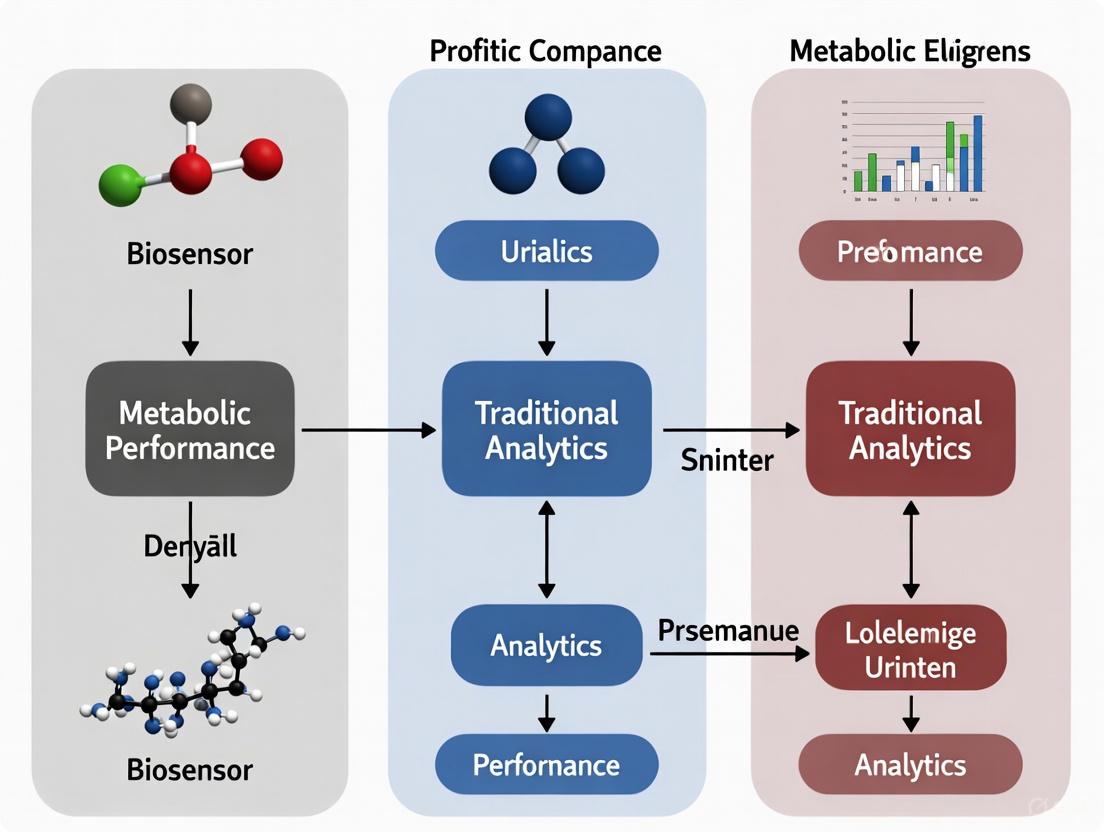
The following diagrams illustrate the logical and experimental workflows for both analytical approaches.
Diagram 1: GC-MS Metabolomics Workflow. This workflow highlights the sample preparation and data processing steps for a GC-MS based metabolomics study, culminating in metabolite identification.
Diagram 2: Biosensor Development and Application. This workflow outlines the process for developing a genetic biosensor, from initial design to its use in screening and subsequent validation by traditional analytics.
Essential Research Reagent Solutions
The following table lists key reagents and materials essential for executing the protocols described in this guide.
Table 2: Key Research Reagents for Analytical Metabolic Engineering
| Reagent / Material | Function / Application | Example Use Case |
|---|---|---|
| MSTFA (N-Methyl-N-(trimethylsilyl)trifluoroacetamide) | Derivatizing agent for GC-MS; increases metabolite volatility [11] | Preparation of polar metabolites (e.g., organic acids, sugars) for GC-MS analysis. |
| NIST Mass Spectral Library | Reference database for metabolite identification [11] | Annotating unknown peaks from GC-EI-MS data by spectral matching. |
| Pyruvate Oxidase (POx) / Glutamate Oxidase (GlOx) | Biorecognition element in enzyme-based biosensors [14] | Detecting alanine aminotransferase (ALT) activity via H₂O₂ production in an amperometric biosensor. |
| Transcription Factor (e.g., TtgR) | Protein-based sensor for small molecules [8] | Engineering whole-cell biosensors to detect specific ligands or antibiotics. |
| PVA-SbQ (Polyvinyl alcohol with steryl pyridinium groups) | Photo-crosslinkable polymer for enzyme immobilization [14] | Entrapping and stabilizing oxidase enzymes on electrode surfaces in biosensor fabrication. |
In metabolic engineering, the Design-Build-Test-Learn (DBTL) cycle is a fundamental framework for developing efficient microbial cell factories [10] [15]. However, the "Test" phase has traditionally been a major bottleneck, relying on slow, low-throughput conventional analytical methods like liquid chromatography (LC) and mass spectrometry (MS) [10] [15]. Genetically encoded biosensors are revolutionizing this process by serving as intracellular tools that detect specific metabolites and link their concentration to a measurable reporter output [16] [3]. This capability enables real-time, high-throughput monitoring of metabolic states, dramatically accelerating strain development [12] [10] [15]. This guide provides a comparative analysis of two primary biosensor classes—transcription factor-based and RNA-based biosensors—contrasting their performance with traditional analytics and detailing their actuation mechanisms.
Biosensor Performance vs. Traditional Analytics
The table below summarizes the critical performance characteristics of major analytical methods used in metabolic engineering.
Table 1: Performance Comparison of Analytical Methods in Metabolic Engineering
| Method | Sample Throughput (per day) | Sensitivity (LLOD) | Flexibility | Key Advantages | Key Limitations |
|---|---|---|---|---|---|
| Chromatography (e.g., GC, LC) | 10 - 100 | mM range | ++ | High confidence in identification; broad applicability [10] | Low throughput; requires sample preparation [15] |
| Direct Mass Spectrometry | 100 - 1,000 | nM range | +++ | Fast analysis; no derivatization needed [10] | Complex data analysis; instrument cost [10] |
| Biosensors (General) | 1,000 - 10,000+ | pM - nM range | + | Highest throughput; real-time, intracellular data [10] [15] | Requires engineering; susceptible to cellular noise [3] |
| Selections | > 10,000,000 | nM range | + | Extremely high throughput; direct coupling to growth [10] | Limited to conditions that confer survival [10] |
As evidenced in Table 1, biosensors offer a transformative advantage in screening throughput, which is crucial for evaluating the vast libraries of strain variants generated by modern DNA synthesis and genome editing tools [10]. Unlike chromatography, which provides a snapshot of extracellular metabolite levels, biosensors operate in vivo, providing high temporal and spatial resolution of intracellular analyte concentrations without the need for sample preparation [12] [15]. The main trade-offs are that biosensors must be engineered for each specific analyte and their performance can be influenced by the host's physiological state [3].
Transcription Factor-Based Biosensors
Mechanism of Action
Transcription factor (TF)-based biosensors are typically composed of a TF that acts as the sensor and a promoter it regulates, which drives the expression of a reporter gene (e.g., GFP) [16]. In the absence of the target small molecule (effector), the TF resides in a state that either represses or activates the promoter. Upon effector binding, a conformational change in the TF alters its DNA-binding affinity, leading to a change in reporter gene expression [16]. This mechanism provides a direct, genetically encoded link between intracellular metabolite concentration and a quantifiable optical signal.
Figure 1: Signaling mechanism of a repressor-type transcription factor biosensor.
Experimental Application & Protocol
TF-based biosensors are widely used for high-throughput screening of enzyme libraries or mutant strains using Fluorescence-Activated Cell Sorting (FACS) [16] [10].
Detailed Protocol: FACS-Based Screening with a TF Biosensor
- Biosensor Integration: Construct a genetic circuit where the promoter regulated by a chosen TF (e.g., LysG for L-lysine in C. glutamicum) drives the expression of a fluorescent protein like eYFP [16].
- Library Transformation: Introduce a plasmid or genomic library of enzyme variants or pathway modifications into the host chassis organism harboring the biosensor.
- Cultivation: Grow the library population under conditions that induce the production of the target metabolite.
- FACS Analysis and Sorting: Use a FACS instrument to analyze and sort the cell population.
- The instrument measures the fluorescence intensity of individual cells, which corresponds to the intracellular concentration of the target metabolite.
- Cells exhibiting fluorescence above a predetermined threshold (indicating high metabolite production) are isolated into a recovery medium.
- Strain Recovery & Validation: Sorted cells are cultured to regenerate populations. The improved production phenotype of these sorted strains is then validated using traditional analytical methods like HPLC [16].
Performance and Example Applications
Table 2: Exemplary Transcription Factor-Based Biosensors
| Transcription Factor | Target Analyte | Host Chassis | Reported Application |
|---|---|---|---|
| LysG | L-lysine, L-arginine | C. glutamicum | HTP FACS screening of mutant libraries for amino acid production [16] |
| FapR | Malonyl-CoA | E. coli | Dynamic control of fatty acid biosynthesis pathways [16] |
| TtgR | Resveratrol, Naringenin | E. coli | Screening enzyme activity and monitoring production of flavonoids [15] |
| BmoR | 1-Butanol | E. coli | Biosensor-based selection for improved 1-butanol production [16] |
RNA-Based Biosensors
Mechanism of Action
RNA-based biosensors, such as riboswitches and toehold switches, are synthetic RNA elements that undergo ligand-induced conformational changes [17] [3]. They are typically located in the 5' untranslated region (5' UTR) of mRNA. In the absence of the ligand, the secondary structure of the RNA may sequester the Ribosome Binding Site (RBS), preventing translation. Upon binding a specific target metabolite or RNA sequence, the RNA structure reconfigures, exposing the RBS and allowing translation of the downstream reporter or actuator gene to proceed [17] [3]. This mechanism offers a rapid response as it occurs at the transcriptional and translational level without the need for protein synthesis.
Figure 2: Signaling mechanism of a translational-activating RNA biosensor.
Experimental Application & Protocol
Riboswitches are particularly useful for implementing dynamic control in metabolic pathways, allowing the cell to self-regulate flux based on metabolite levels [3].
Detailed Protocol: Implementing Dynamic Control with a Riboswitch
- Riboswitch Selection/Design: Identify or engineer a riboswitch that responds to a key pathway intermediate or co-factor (e.g., a riboswitch responsive to intracellular FMN levels) [3].
- Circuit Construction: Place the riboswitch in the 5' UTR of a gene that encodes an enzyme crucial for pathway flux. This gene could be an early pathway enzyme that consumes a central metabolite.
- Strain Cultivation & Evaluation: Cultivate the engineered strain in a production bioreactor.
- As the pathway intermediate accumulates, it binds the riboswitch.
- This binding triggers the conformational change, turning on (or off) the translation of the downstream enzyme.
- This real-time feedback loop dynamically balances metabolic flux to optimize product yield and reduce intermediate accumulation [3].
- Metabolite & Titer Analysis: Periodically sample the culture and use LC-MS to quantify the concentrations of the final product, key intermediates, and by-products to validate the efficacy of the dynamic control strategy.
Performance and Example Applications
Table 3: Exemplary RNA-Based Biosensors and Applications
| RNA Biosensor Type | Target Analyte | Host Chassis | Reported Application |
|---|---|---|---|
| Riboswitch | FMN, Purines, Lysine | Various | Natural and engineered systems for real-time regulation of metabolic fluxes [3] |
| Toehold Switch | Specific RNA Sequences | E. coli | Programmable, logic-gated control of metabolic pathways; diagnostics [3] |
| Self-cleaving Aptazyme | N-acetylneuraminate | E. coli | Screening for optimal enzyme activity in a pathway [15] |
| glmS Ribozyme | N-acetylglucosamine | B. subtilis | Regulating and screening for optimal genetic variants [15] |
The Scientist's Toolkit: Essential Research Reagents
Table 4: Key Research Reagents for Biosensor Development and Application
| Reagent / Material | Function in Biosensor Research |
|---|---|
| Fluorescent Reporters (e.g., GFP, mCherry, eYFP) | Provide a quantifiable optical output for biosensor activation, enabling detection via plate readers or FACS [16] [15]. |
| Selection Markers (e.g., TetA, antibiotic resistance) | Allow for coupling biosensor activation to cell survival, enabling high-throughput selection without specialized equipment [16]. |
| Metagenomic Libraries | Serve as a source of novel transcription factors or regulatory elements for developing biosensors for new analytes [16]. |
| Chiral Dopants (e.g., R5011) & Nematic LCs (e.g., E44) | Components of advanced optical biosensing platforms like cholesteric liquid crystal (CLC) sensors for label-free detection [18]. |
| Vertical Alignment Agents (e.g., DMOAP) | Used to prepare surfaces for CLC-based biosensors, where biomolecule adsorption disrupts alignment, causing a color change [18]. |
In metabolic engineering, the transition from static optimization to dynamic regulation represents a paradigm shift, and biosensors are the linchpin of this transformation [3]. These biological tools allow researchers to move beyond simple endpoint measurements, offering instead a window into real-time cellular metabolic states. When objectively comparing biosensor performance against traditional analytical methods, three metrics stand out as fundamental: dynamic range, sensitivity, and throughput [3] [19]. Dynamic range defines the operational window of a biosensor, quantifying the span between the minimal detectable signal and the maximum quantifiable signal. Sensitivity determines the lowest concentration of a target metabolite that can be reliably detected, directly impacting a sensor's ability to identify subtle metabolic variations. Throughput, crucial for modern strain engineering, measures the capacity to screen vast genetic libraries, a task where traditional analytics often become bottlenecks [3] [20]. This guide provides a structured comparison of these core performance metrics between biosensor-driven and traditional analytical approaches, equipping researchers with the data needed to select optimal tools for metabolic engineering and drug development.
Performance Metric Comparison: Biosensors vs. Traditional Analytics
The table below provides a direct, data-driven comparison of key performance metrics for biosensors and traditional analytical methods, highlighting their respective strengths and limitations in metabolic engineering research.
Table 1: Performance Metric Comparison of Biosensors and Traditional Analytical Methods
| Performance Metric | Biosensors | Traditional Analytics (HPLC, GC-MS, LC-MS) |
|---|---|---|
| Dynamic Range | Typically spans 2-3 orders of magnitude; tunable via engineering (e.g., promoter/RBS modification) [3]. | Very wide, often 4-6 orders of magnitude, but requires sample dilution for accurate quantification across the range. |
| Sensitivity | Varies by type; can detect down to pM-fM levels with nanomaterials and signal amplification [21]. | Extremely high (pM-fM range); considered a gold standard for low-concentration analyte detection [22]. |
| Throughput | Very high (10⁵-10⁸ cells per hour) with FACS; enables real-time, single-cell monitoring in living systems [3] [19]. | Low to medium; time-consuming sample preparation and serial analysis create a significant bottleneck [20] [19]. |
| Measurement Context | In vivo, real-time monitoring within living cells, preserving native metabolic context [3] [5]. | In vitro, endpoint measurements requiring cell lysis, which destroys the cellular context and stops metabolism. |
| Key Advantage | Enables high-throughput screening and dynamic regulation of pathways in their native environment [3]. | Provides highly precise, absolute quantification and is capable of multiplexing many analytes simultaneously. |
| Primary Limitation | Limited analyte scope; potential for cross-talk and false positives in complex cellular environments [3] [6]. | Low throughput and destructive nature prevent real-time monitoring and rapid iterative screening [20]. |
Experimental Protocols for Biosensor Characterization
To ensure the reliability of biosensor data, rigorous and standardized experimental characterization of the above metrics is essential. The following protocols are widely adopted in the field.
Protocol for Characterizing Dynamic Range and Sensitivity
This protocol outlines the steps to generate a dose-response curve, from which dynamic range and sensitivity are derived [3] [6].
Strain Preparation and Cultivation:
- Transform the biosensor plasmid into an appropriate microbial host (e.g., E. coli BW25113 or S. cerevisiae) [6].
- Inoculate a single colony into liquid growth medium with appropriate antibiotics and grow overnight to saturation.
- The following day, dilute the overnight culture into fresh medium and grow until the mid-exponential phase (OD600 ≈ 0.4-0.6).
Dose-Response Assay:
- Aliquot the culture into multiple wells of a microtiter plate.
- Add a series of known concentrations of the target analyte (e.g., succinate, malonyl-CoA) to the wells, creating a concentration gradient. Include replicates and a negative control (no inducer) [6].
- Continue incubation for a defined period to allow the biosensor response to stabilize.
Signal Measurement and Data Analysis:
- Measure the output signal (e.g., fluorescence intensity for GFP/RFP) and the cell density (OD600) for each well using a plate reader [6].
- Normalize the fluorescence signal by the cell density (e.g., Fluorescence/OD600).
- Plot the normalized output signal against the logarithm of the analyte concentration.
- Fit a sigmoidal curve (e.g., Hill function) to the data. The dynamic range is calculated as the ratio between the maximum and minimum output signal across the saturation points. The sensitivity (often reported as the EC50 or KD) is the analyte concentration required to produce a half-maximal response [3].
Protocol for High-Throughput Screening Using FACS
This protocol leverages biosensors with fluorescent outputs to screen large libraries of microbial variants [3].
Library Preparation and Induction:
- Generate a diverse library of microbial strains harboring the biosensor and genetic variations (e.g., pathway enzyme mutants, promoter libraries).
- Grow the library population under conditions that activate the biosensor, such as by adding a precursor or allowing an intermediate to accumulate.
Cell Sorting and Analysis:
- Dilute the culture and load it into a Fluorescence-Activated Cell Sorter (FACS).
- Set sorting gates based on fluorescence intensity. For example, to select high-producing strains, a gate is set to isolate the top 1-5% most fluorescent cells [3].
- Sort the population, collecting the cells from the desired gate into a recovery medium.
Validation and Iteration:
- Plate the sorted cells to form single colonies and characterize them using the dose-response protocol or traditional analytics (e.g., HPLC) to validate the production tier.
- The validated high-performing strains can be used for further rounds of cultivation and sorting to iteratively enrich for the best producers.
Visualizing Biosensor Mechanisms and Workflows
Transcriptional Biosensor Mechanism
The diagram below illustrates the core mechanism of a transcription factor-based biosensor, a common tool in metabolic engineering for sensing intracellular metabolites.
High-Throughput Screening Workflow
This workflow visualizes the iterative "Design-Build-Test-Learn" cycle enabled by biosensors for metabolic engineering, highlighting the high-throughput "Test" phase.
The Scientist's Toolkit: Essential Research Reagent Solutions
The successful implementation and optimization of biosensors rely on a specific set of biological and chemical reagents. The table below details these key components and their functions.
Table 2: Key Research Reagents for Biosensor Development and Application
| Research Reagent | Function & Application |
|---|---|
| Transcription Factors (TFs) | Core sensing element; proteins that bind a target metabolite and regulate transcription (e.g., PcaR for succinate) [3] [6]. |
| Aptamers | Single-stranded DNA/RNA oligonucleotides that bind targets with high specificity; used in electrochemical and optical biosensors as recognition elements [23] [21]. |
| Reporter Genes (GFP, RFP) | Encodes a measurable output (e.g., fluorescent protein); expression is linked to biosensor activation, enabling quantification [6]. |
| Engineered Promoters | DNA sequence controlled by a TF; engineered to fine-tune biosensor performance characteristics like dynamic range and sensitivity [3] [5]. |
| Functional Nanomaterials (AuNPs, Graphene, CNTs) | Enhance electrochemical biosensor performance by improving electron transfer, signal amplification, and biocompatibility [24] [21]. |
| Microfluidic Chips & FACS | Hardware platforms that enable the automation and high-throughput operation of biosensors for rapid library screening [3] [23]. |
The comparative analysis of performance metrics reveals a clear complementarity between biosensors and traditional analytics. Biosensors excel in throughput and real-time, in vivo monitoring, making them indispensable for dynamic regulation and the high-throughput screening phases of metabolic engineering campaigns [3] [20]. Conversely, traditional methods provide unrivaled sensitivity and broad analyte coverage, cementing their role as gold standards for validation and absolute quantification [22]. The choice between these tools is not a matter of superiority but of strategic application. For drug development professionals and researchers, the future lies in integrated workflows: using biosensors to rapidly navigate vast design spaces and identify promising candidates, followed by traditional analytics for rigorous, final validation. This synergistic approach, leveraging the unique strengths of each methodology, will ultimately accelerate the development of robust microbial cell factories for bioproduction and therapeutic applications.
Application in Action: High-Throughput Screening and Dynamic Control
In metabolic engineering, selecting the appropriate analytical method is critical for validating research findings. Traditional analytics, such as chromatography and mass spectrometry, have long been the cornerstone for proof-of-concept demonstration and targeted validation in pathway engineering. While biosensors are revolutionizing high-throughput dynamic monitoring, traditional methods remain indispensable for their definitive quantification and high specificity, particularly in the final stages of strain validation and pathway confirmation [3] [20] [5].
This guide objectively compares the performance of these analytical paradigms, providing experimental data and detailed protocols to help researchers make informed choices for their specific applications in metabolic engineering and drug development.
Performance Comparison: Biosensors vs. Traditional Analytics
The table below summarizes the core performance characteristics of biosensors compared to established traditional analytical techniques.
Table 1: Performance Comparison Between Biosensors and Traditional Analytics
| Performance Characteristic | Biosensors (Whole-Cell & Cell-Free) | Traditional Analytics (e.g., HPLC, GC-MS) |
|---|---|---|
| Temporal Resolution | Real-time to minutes (dynamic monitoring) [3] | Minutes to hours (single time-point measurements) |
| Throughput | Very High (supports screening of large libraries) [3] [20] | Low to Medium (serial analysis is time-consuming) |
| Sensitivity | Variable; can be very high (e.g., LOD of 0.078 mM for lactate aptasensor) [25] | Consistently high (e.g., detection of nanomolar concentrations) |
| Specificity | Programmable, but potential for cross-talk [3] | Very High (excellent separation of analytes) |
| Spatial Resolution (in vivo) | High (can target subcellular compartments) [12] | None (requires cell lysis) |
| Key Application | High-throughput screening, dynamic pathway control [3] [26] | Definitive product quantification, proof-of-concept validation [26] |
| Quantitative Data | Relative or semi-quantitative; requires calibration [3] | Absolute quantification with high accuracy [26] |
| Key Advantage | Enables real-time, dynamic control of metabolism [3] | Provides gold-standard validation for regulatory approval |
Experimental Protocols for Traditional Analytics in Metabolic Engineering
Protocol: Validating Microbial Cadaverine Production using HPLC
This protocol was used to provide the definitive, quantitative validation for a dynamically regulated cadaverine production strain in E. coli [26]. It serves a critical role in confirming the titers reported by biosensor-driven screening.
- Objective: To absolutely quantify cadaverine and precursor lysine concentrations in fermentation broth to validate the success of a biosensor-driven metabolic engineering strategy [26].
- Sample Preparation: Culture samples are centrifuged (e.g., 13,000 rpm for 10 min) to separate microbial cells from the supernatant. The supernatant is then filtered through a 0.22 µm membrane to remove particulate matter. For intracellular metabolite measurement, cell pellets are resuspended and subjected to a quenching and extraction protocol, typically using cold methanol/water solutions [26].
- Instrumentation & Analysis: High-Performance Liquid Chromatography system equipped with a UV/Vis or fluorescence detector. Separation is achieved using a reversed-phase C18 column. The mobile phase is often a mixture of aqueous buffer (e.g., phosphate or acetate) and an organic solvent like methanol or acetonitrile, run under a gradient elution program. Quantification is performed by comparing peak areas of samples to a standard curve generated from pure cadaverine and lysine standards [26].
- Data Interpretation: The concentration of cadaverine in the fermentation broth is calculated from the chromatographic data. In the referenced study, this method confirmed a final titer of 33.19 g/L in the optimized strain, a 48.1% increase over the control, providing the key validation metric for the project's success [26].
Protocol: Quantifying Lignocellulosic Sugars and Inhibitors using GC-MS
This protocol is essential for the initial proof-of-concept in projects utilizing lignocellulosic biomass, as it accurately characterizes the feedstock and identifies potential fermentation inhibitors [5].
- Objective: To identify and quantify the sugar composition (e.g., glucose, xylose) and fermentation inhibitors (e.g., furfural, hydroxymethylfurfural) in lignocellulosic hydrolysates [5].
- Sample Preparation: The liquid hydrolysate is derivatized to increase the volatility of the analytes for gas chromatography. A common derivatization method involves oximation and silylation, where carbonyl groups are stabilized and polar functional groups are replaced with non-polar trimethylsilyl groups [5].
- Instrumentation & Analysis: Gas Chromatography-Mass Spectrometry system. Separation is performed on a non-polar or mid-polar capillary column using a temperature ramp program. The mass spectrometer operates in Selected Ion Monitoring mode for high sensitivity and selectivity. Analytes are identified by their unique retention times and mass fragmentation patterns, and quantified against a calibration curve [5].
- Data Interpretation: The data provides a precise profile of fermentable sugar concentrations, which is critical for designing fermentation media. Simultaneously, it quantifies inhibitory compounds, guiding the need for and optimization of detoxification steps prior to microbial fermentation [5].
Signaling Pathways and Experimental Workflows
The following diagrams illustrate a classic biosensor-regulated pathway and a typical workflow integrating both biosensor-driven and traditional analytical methods.
Diagram 1: The Lysine-Responsive CadC Biosensor Pathway. This genetic circuit dynamically regulates cadaverine production in E. coli. Extracellular lysine is transported by LysP. Under low pH, the transcription factor CadC is activated, binding to the Pcad promoter to drive expression of the cadBA operon. The enzyme CadA then converts lysine into cadaverine. A GFPuv reporter gene can be linked to Pcad for real-time monitoring [26].
Diagram 2: A Hybrid Workflow for Strain Development. This workflow leverages the strengths of both biosensors and traditional analytics. A large library of engineered strains is first rapidly screened using a biosensor (e.g., for fluorescence). The top-performing "hit" strains from this primary screen are then rigorously validated using traditional analytics like HPLC or GC-MS for absolute quantification of the target metabolite. This confirmed strain is scaled up, with traditional methods providing the final, authoritative production titer [3] [20] [26].
The Scientist's Toolkit: Key Research Reagent Solutions
The table below details essential reagents and materials used in the experiments cited in this guide.
Table 2: Key Research Reagents and Their Functions in Metabolic Engineering Analytics
| Reagent / Material | Function / Description | Featured Application |
|---|---|---|
| MOPS Medium | A defined, buffered microbial growth medium that maintains a stable pH during fermentation. | Used in shake-flask fermentation to analyze GFP expression in the lysine biosensor [26]. |
| L-Lactate Aptamer | A synthetic, single-stranded DNA molecule with high binding affinity and specificity for L-lactate. | The core sensing element in the FRET-based lactate aptasensor for sweat analysis [25]. |
| Core-Shell UCNPs | Upconversion nanoparticles with a core-shell structure that convert near-infrared light to visible light, minimizing background noise. | Served as the fluorescent energy donor in the lactate aptasensor, enabling high sensitivity [25]. |
| Fe3O4-MoS2 Nanosheets | A nanocomposite material acting as a fluorescence quencher; its magnetic properties allow for rapid separation from solution. | Used as the energy acceptor and separation matrix in the lactate aptasensor to reduce background interference [25]. |
| CRISPR/Cas9 System | A genome editing system used for precise gene knockouts, knock-ins, and modifications in microbial hosts. | Employed for metabolic engineering of the E. coli host, such as knocking out genes related to metabolic bypasses [26]. |
| C18 Chromatography Column | A reversed-phase chromatography column with C18-functionalized silica, used for separating non-polar to moderately polar molecules. | The stationary phase for HPLC analysis and quantification of compounds like cadaverine and lysine [26]. |
Biosensor-Driven High-Throughput Screening of Strain Libraries
In metabolic engineering, maximizing the productivity of microbial strains is paramount for industrial application. Historically, the optimization of biosynthetic pathways has been hindered by the complex nature of living systems, making rational engineering a time- and labor-intensive process with limited success [27]. Traditional analytical methods, such as mass spectrometry and chromatography, though accurate, are low-throughput and create a significant bottleneck in the discovery of improved strains from vast genetic libraries [27]. The emergence of biosensor-driven screening represents a paradigm shift, offering a powerful alternative to traditional analytics. This guide provides a comparative evaluation of these approaches, focusing on the performance of various biosensor platforms in high-throughput screening (HTS) contexts. By examining throughput, sensitivity, and applicability, we aim to furnish researchers and drug development professionals with the data necessary to select the optimal screening strategy for their metabolic engineering projects.
Biosensor Technology vs. Traditional Analytics: A Core Comparison
At its core, a biosensor is a device that detects a biological analyte and produces a measurable signal. In microbial strain screening, genetically encoded transcription factor (TF)-based biosensors are most common. They detect internal stimuli like metabolite concentration and transduce this input into a quantifiable output, such as fluorescence [27] [28]. This allows for the indirect, real-time monitoring of product formation within living cells, bypassing the need for lengthy sample preparation and analysis.
The table below summarizes the fundamental differences between biosensor-driven HTS and traditional analytical methods.
Table 1: Core Comparison of Screening Methodologies
| Feature | Biosensor-Driven HTS | Traditional Analytics (e.g., LC-MS) |
|---|---|---|
| Throughput | Very High (10^5-10^9 variants) [27] [29] | Low (10^1-10^2 variants) [27] |
| Measurement Speed | Real-time or near-real-time within cells | Slow, requiring sample extraction and processing |
| Key Advantage | Enables screening of vast library sizes; direct linkage of genotype to phenotype | High accuracy and sensitivity for absolute quantification |
| Primary Limitation | Often requires extensive sensor engineering and may have limited dynamic range [28] | Throughput is a major bottleneck for library screening |
| Typical Cost per Sample | Very low once established | High |
Comparative Performance of Biosensor Screening Modalities
Biosensor-based screening is not a monolithic approach. Different modalities offer varying degrees of throughput, instrumentation requirements, and practical constraints. The choice of method depends on the library size and specific experimental goals [27].
Table 2: Comparison of Biosensor-Based High-Throughput Screening Modalities
| Screening Modality | Throughput (Library Size) | Key Principle | Pros & Cons |
|---|---|---|---|
| Fluorescence-Activated Cell Sorting (FACS) | High (10^8-10^9 cells) [27] | Biosensor fluorescence is used to sort single cells from a suspension. | + Highest throughput+ Quantitative selection- Requires specialized equipment- Sensor performance critical |
| Droplet-Based Screening | High (10^7-10^9 variants) [29] | Cells are encapsulated in water-in-oil droplets with assay reagents, acting as picoliter-scale bioreactors. | + Ultra-high throughput+ Compartmentalization prevents cross-talk- Complex microfluidic setup |
| Agar Plate Screening | Medium (10^3-10^6 variants) [27] | Colonies grown on solid media are screened based on fluorescence or colorimetric output. | + Low technical requirement+ Can be highly sensitive (e.g., blue-white) [27]- Lower throughput- Qualitative or semi-quantitative |
| Well Plate/Microtiter Plates | Low-Medium (10^2-10^4 variants) [27] [30] | Cultivation and screening in multi-well plates with online monitoring of fluorescence, OD, pH, etc. | + Quantitative and controlled conditions (e.g., BioLector) [30]+ Easily automated- Lowest throughput of biosensor methods |
Performance Data from Comparative Studies
Independent comparisons of biosensor platforms highlight trade-offs between throughput, data quality, and sensitivity.
Table 3: Experimental Performance Data from Platform Comparison Studies
| Platform/Technology | Application Context | Key Performance Finding | Reference |
|---|---|---|---|
| ECIS vs. xCELLigence vs. cellZscope | Measuring endothelial barrier integrity in response to cytokines (impedance) | ECIS was the most sensitive platform for detecting transient changes in impedance [31]. | |
| Biacore T100 vs. ProteOn XPR36 vs. Octet RED384 vs. IBIS MX96 | Evaluating antibody-antigen binding kinetics | Biacore T100 and ProteOn XPR36 showed excellent data quality and consistency, while Octet RED384 and IBIS MX96 offered higher throughput with compromises in accuracy/reproducibility [32]. | |
| Dual-Gate BioFETs | General biomarker detection (pH sensing) | Signal amplification can surpass the Nernst limit, but noise increases proportionally, resulting in no net improvement to the intrinsic detection limit [33]. | |
| Mach-Zehnder Interferometer (MZI) | Label-free biosensing | The detection limit is highly dependent on the dominant noise source, which can be unrelated to the sensor, affect one arm, or both arms, guiding design optimization [34]. |
Experimental Protocols for Key Biosensor Applications
Protocol: FACS-Based Screening with a TF Biosensor
This protocol is adapted from studies that successfully isolated strains with improved production of metabolites like L-lysine, fatty acids, and acrylic acid [27].
- Strain Library Preparation: Generate a diverse library of microbial strains (e.g., via error-prone PCR, ARTP mutagenesis, or RBS library generation) harboring the biosensor system.
- Biosensor Configuration: Use a TF-based biosensor where the target metabolite binds the transcription factor, leading to the expression of a reporter gene, such as Green Fluorescent Protein (GFP).
- Cultivation and Induction: Grow the library under conditions that induce the production of the target metabolite. This can be in deep-well plates or flasks.
- Cell Preparation for FACS: Harvest cells during the production phase and resuspend them in an appropriate buffer for sorting.
- FACS Sorting: Use a flow cytometer equipped with a cell sorter. Set gates to isolate the top 0.1-1% of cells with the highest GFP fluorescence intensity, which correlate with high metabolite production.
- Recovery and Validation: Sort the selected cells directly into recovery media. After outgrowth, validate improved production of the target metabolite in the enriched population using traditional analytical methods (e.g., HPLC).
Protocol: Dynamic Regulation for Autonomous Pathway Control
This protocol outlines the use of biosensors not just for screening, but for autonomous, real-time pathway optimization within a production strain [28].
- Circuit Design: Design a genetic circuit where a metabolite-responsive biosensor (e.g., responsive to a toxic intermediate or the final product) controls the expression of critical pathway genes.
- Bifunctional Control Implementation:
- Activation: The biosensor can be designed to activate genes in the synthesis pathway upon detection of a key intermediate.
- Repression (CRISPRi/asRNA): The same biosensor can simultaneously guide a repression system (e.g., CRISPRi or antisense RNA) to downregulate competing metabolic pathways or genes related to byproduct generation [28].
- Strain Transformation: Integrate the dynamic regulation circuit into the production host chromosome or a stable plasmid.
- Fermentation and Monitoring: Cultivate the engineered strain in a bioreactor. The biosensor will autonomously rewire metabolic flux in response to changing intracellular metabolite levels, balancing cell growth and production.
- Titer Assessment: Quantify the final product titer at the end of fermentation. Studies have reported significant improvements using this method, such as a 49% increase in muconic acid production and a 40-fold improvement in menaquinone-7 titer [28].
Visualizing Biosensor Mechanisms and Screening Workflows
Diagram: Mechanism of a Transcription Factor-Based Biosensor
Diagram: High-Throughput Screening Workflow Comparison
The Scientist's Toolkit: Essential Research Reagent Solutions
The successful implementation of biosensor-driven HTS relies on a suite of specialized reagents and instruments.
Table 4: Key Reagents and Platforms for Biosensor Screening
| Tool / Reagent | Function | Example Use in Screening |
|---|---|---|
| Transcription Factor (TF) Biosensors | Core detection element; converts metabolite concentration into gene expression. | Engineered to respond to target molecules like L-lysine, fatty acids, or vanillin for FACS or plate screening [27] [28]. |
| Error-Prone PCR Kits | Creates randomized mutagenesis libraries for enzyme evolution. | Generating diverse variant libraries of a rate-limiting pathway enzyme [27]. |
| ARTP Mutagenesis System | A physical mutagenesis method for generating whole-cell random mutant libraries. | Creating genomic diversity in production chassis like E. coli or C. glutamicum [27]. |
| Microfluidic Droplet Generators | Encapsulates single cells and assays in picoliter droplets for ultra-HTS. | Screening cell-free enzymatic reactions or millions of microbial variants with a fluorescent biosensor readout [29]. |
| BioLector / RoboLector Systems | Microbioreactor platforms for online monitoring of growth & fluorescence in microtiter plates. | Provides quantitative, controlled parallel fermentation for screening 10^2-10^3 strains in a batch or fed-batch mode [30]. |
| FACS Instruments | The core platform for the highest-throughput screening of biosensor-equipped libraries. | Isolating the top 0.1% of a library of 10^8 cells based on biosensor GFP intensity in a few hours [27]. |
Implementing Dynamic Regulation for Robust Pathway Performance
Metabolic engineering aims to reprogram microbial cell factories for sustainable chemical production, yet a significant bottleneck persists in effectively evaluating and optimizing pathway performance [2]. Traditional analytical chemistry methods, while highly accurate, are inherently low-throughput and destructive, creating a critical capability gap in the Design-Build-Test-Learn (DBTL) cycle [2]. Biosensors—genetically encoded components that convert metabolite concentrations into measurable outputs—have emerged as a powerful alternative, enabling real-time, dynamic monitoring and control of metabolic pathways [3] [35]. This guide provides an objective comparison between biosensor technology and traditional analytics, offering experimental data and protocols to help researchers select the optimal tool for enhancing pathway robustness and productivity.
Performance Comparison: Biosensors vs. Traditional Analytics
The choice between biosensors and traditional methods involves trade-offs between throughput, information depth, and analytical precision. The following tables summarize their core characteristics and performance metrics.
Table 1: Core Characteristics and Application Fit
| Feature | Biosensors | Traditional Analytics (Chromatography/MS) |
|---|---|---|
| Throughput | Very High (10^4-10^8 variants/day) [35] [2] | Low (10^1-10^3 variants/day) [35] [2] |
| Measurement Context | In vivo, real-time, dynamic [3] | Ex vivo, end-point, static [2] |
| Key Strength | Dynamic control & high-throughput screening [3] [36] | Broad metabolite detection & high accuracy [2] |
| Primary Limitation | Limited analyte scope; requires engineering [3] | Low throughput; destructive sampling [35] [2] |
| Ideal Use Case | Screening large libraries; dynamic pathway regulation [3] [37] | Validating top hits; analyzing unknown pathways [2] |
Table 2: Quantitative Performance Metrics for Specific Analytes
| Analyte | Analytical Method | Limit of Detection | Dynamic/Linear Range | Key Metric |
|---|---|---|---|---|
| 4'-O-Methylnorbelladine | RamR Biosensor (4NB2.1) [37] | ~2.5 µM [37] | 2.5 - 250 µM [37] | Sensitivity |
| 4'-O-Methylnorbelladine | HPLC [37] | ~25 µM [37] | 25 - 1000 µM [37] | Sensitivity |
| Malonyl-CoA | FapR TF-Biosensor + FACS [35] | N/A | N/A | Screened ~10^6 cDNA variants [35] |
| General Metabolites | RapidFire MS [35] | N/A | N/A | ~15 seconds/sample [35] |
| General Metabolites | Traditional LC-MS [35] | N/A | N/A | Minutes to hours/sample [35] |
Experimental Protocols for Implementation
Protocol 1: Developing a Transcription Factor-Based Biosensor
This protocol outlines the creation of a TF-based biosensor for a target metabolite, based on the successful engineering of a sensor for 4'-O-Methylnorbelladine (4NB) [37].
- Sensor Selection and Cloning: Select a promiscuous transcription factor as a starting scaffold (e.g., RamR from Salmonella typhimurium). Clone the TF and its cognate promoter upstream of a reporter gene (e.g., sfGFP) into a plasmid system [37].
- Library Generation: Create site-saturation mutagenesis libraries targeting residues in the ligand-binding pocket. Molecular docking simulations can inform which residues to target for altering specificity [37].
- High-Throughput Screening (SELIS Method):
- Growth-Based Selection: First, apply a growth-based selection to eliminate sensor variants that fail to repress transcription in the absence of the ligand [37].
- Fluorescence Activation Screen: Use fluorescence-activated cell sorting (FACS) to isolate variants that show strong fluorescence activation in the presence of the target metabolite. To enhance specificity, a counter-selection can be applied in the presence of a structurally similar analog [37].
- Biosensor Validation: Characterize top hits by measuring dose-response curves (dynamic range, EC50), specificity against analogs, and response time [3] [37].
Protocol 2: Using a Biosensor for High-Throughput Enzyme Evolution
This protocol details the application of a developed biosensor to engineer enzymes, as demonstrated for norbelladine 4'-O-methyltransferase (Nb4OMT) [37].
- Strain and Pathway Setup: Engineer a microbial host to produce the biosensor's target metabolite by introducing the requisite pathway genes. For Nb4OMT, this involved producing the precursor norbelladine in E. coli [37].
- Library Creation: Generate a variant library of the target enzyme. This can be done via directed evolution or, more efficiently, using a machine learning-guided approach like a structure-based residual neural network (e.g., MutComputeX) to generate activity-enriched designs [37].
- Biosensor Screening: Transform the enzyme variant library into the sensor-equipped production strain. Use FACS or microplate fluorescence screening to rapidly isolate clones exhibiting high biosensor signal, indicating superior production of the target metabolite [37].
- Validation with Traditional Analytics: Cultivate the top-performing isolates from the biosensor screen and quantify the final product titer and byproduct formation using HPLC or LC-MS to confirm biosensor findings [37].
Pathway and Workflow Visualization
The following diagrams illustrate the fundamental mechanism of a transcription factor-based biosensor and the integrated experimental workflow for biosensor-driven enzyme engineering.
Biosensor Mechanism: TF-Based
Workflow: Biosensor-Driven Engineering
Case Study: Biosensor-Driven Engineering of a Plant Methyltransferase
A 2024 study provides a compelling case for the biosensor-driven approach. The goal was to improve the activity of norbelladine 4'-O-methyltransferase (Nb4OMT), a plant enzyme critical for synthesizing Amaryllidaceae alkaloids like galantamine [37].
- Methodology: Researchers first evolved a highly sensitive and specific biosensor for the alkaloid branchpoint intermediate 4'-O-methylnorbelladine (4NB). This biosensor was then used to screen a library of Nb4OMT variants generated by a machine learning model (MutComputeX) [37].
- Results: The biosensor-enabled screen successfully identified engineered enzyme variants with significantly improved performance compared to the wild-type enzyme [37].
- Performance Data:
- Conclusion: This case demonstrates that the integration of bespoke biosensors with ML-guided design creates a powerful technology stack for rapid biocatalyst development, drastically accelerating the DBTL cycle [37].
The Scientist's Toolkit: Essential Research Reagents
The following table lists key materials required for implementing biosensor-driven metabolic engineering protocols.
Table 3: Essential Reagents for Biosensor Development and Application
| Reagent / Material | Function | Example(s) |
|---|---|---|
| Transcription Factor Scaffold | Starting point for biosensor engineering; provides DNA-binding and basic regulatory framework. | RamR from Salmonella typhimurium [37] |
| Reporter Plasmid | Carries the reporter gene under the control of the TF's cognate promoter for signal output. | Plasmid with P_ramR driving sfGFP expression [37] |
| Expression Host | Microbial chassis for hosting the biosensor and/or the metabolic pathway. | Escherichia coli [37] |
| Fluorescence-Activated Cell Sorter (FACS) | Essential equipment for high-throughput screening and isolation of high-performing biosensor variants or production strains. | Used in SELIS protocol [37] |
| Machine Learning Protein Design Tool | Computational tool to generate smart, activity-enriched variant libraries, reducing screening burden. | MutComputeX (3DResNet) [37] |
| Liquid Chromatography-Mass Spectrometry (LC-MS) | Gold-standard analytical instrument for validating metabolite production and purity from top hits. | Used for final validation of 4NB production [37] |
In the pursuit of sustainable biomanufacturing and advanced therapeutic synthesis, metabolic engineering faces a fundamental bottleneck: the inability to monitor and control cellular processes in real-time. Traditional analytical methods, such as high-performance liquid chromatography (HPLC) and mass spectrometry, require sample extraction, lengthy processing, and provide only static snapshots of metabolic states [3]. This limitation becomes particularly acute in complex processes like lignocellulosic biomass conversion, where dynamic metabolic fluxes determine overall efficiency, and in therapeutic compound synthesis, where precision is paramount. Genetically encoded biosensors represent a paradigm shift in this landscape. These molecular devices, typically constructed from transcription factors, RNA switches, or two-component systems, detect specific intracellular metabolites and convert this recognition into a quantifiable signal, enabling real-time monitoring and dynamic control of metabolic pathways [36] [3].
This guide objectively compares biosensor performance against traditional analytics through detailed case studies spanning industrial bioconversion and therapeutic synthesis. We present quantitative data, standardized experimental protocols, and analytical frameworks to evaluate the performance characteristics—including sensitivity, dynamic range, and throughput—that define the operational advantages of biosensor-based approaches. By examining both successful implementations and persistent challenges, this analysis provides researchers and drug development professionals with a comprehensive evidence base for selecting appropriate analytical strategies for their metabolic engineering applications.
Biosensor Performance Fundamentals and Evaluation Metrics
Technical Foundations of Biosensor Operation
Biosensors function as integrated biological circuits within living cells, comprising two core modules: a sensing module that specifically binds to a target analyte (e.g., metabolite, ion, or protein), and an actuator module that generates a measurable output signal [3]. The most common architectures include:
- Transcription Factor (TF)-Based Biosensors: These utilize allosteric transcription factors that undergo conformational changes upon ligand binding, regulating the expression of a reporter gene (e.g., GFP) [36]. The core components include a promoter region containing the TF binding site and a reporter gene encoding a measurable output protein.
- RNA-Based Biosensors: Devices such as riboswitches and toehold switches undergo ligand-induced structural rearrangements that modulate translation initiation or transcriptional termination [3]. Toehold switches, for instance, use sequence complementarity to control ribosomal access to the ribosome binding site.
- Two-Component Systems (TCSs): Common in prokaryotes, these involve a sensor kinase that autophosphorylates upon signal detection and transfers the phosphate group to a response regulator, which then activates target gene expression [3].
The critical performance differentiator lies in biosensors' ability to perform continuous, non-destructive monitoring of living cultures, contrasting sharply with the endpoint measurements characteristic of traditional chromatography-based methods.
Key Performance Metrics for Comparative Analysis
Standardized metrics are essential for objective comparison between biosensor and traditional analytical approaches. Key quantitative evaluation parameters include [3]:
- Dynamic Range: The ratio between the maximum and minimum output signals (e.g., fluorescence) in response to analyte concentration, determining the biosensor's ability to distinguish between different metabolite levels.
- Operating Range: The concentration window of the target analyte where the biosensor functions optimally, critical for matching biosensor selection to expected metabolite concentrations.
- Response Time: The speed at which the biosensor reaches its maximum output signal after analyte exposure, ranging from minutes for some RNA-based sensors to hours for certain TF-based systems.
- Signal-to-Noise Ratio: The ratio between the specific output signal and background variability, directly impacting detection reliability and sensitivity.
- Limit of Detection (LOD): The lowest analyte concentration that can be reliably distinguished from background noise.
- Sensitivity: The change in output signal per unit change in analyte concentration.
Table 1: Fundamental Performance Metrics for Biosensor Evaluation
| Metric | Definition | Impact on Performance | Ideal Range |
|---|---|---|---|
| Dynamic Range | Ratio between max and min output signal | Determines ability to distinguish metabolite levels | >100-fold |
| Operating Range | Analyte concentration window for optimal function | Must match expected metabolite concentrations | μM to mM |
| Response Time | Speed to reach maximum output after analyte exposure | Affects real-time control capability | Minutes to hours |
| Signal-to-Noise Ratio | Ratio between specific signal and background | Impacts detection reliability | >10:1 |
| Limit of Detection (LOD) | Lowest detectable analyte concentration | Determines sensitivity to low metabolite levels | nM to μM |
| Sensitivity | Change in output per unit change in analyte | Affects precision of quantification | Steep dose-response |
Figure 1: Biosensor Architecture and Performance Metrics. This diagram illustrates the core components of genetically encoded biosensors and their relationship to key performance evaluation parameters that determine operational effectiveness in metabolic engineering applications.
Case Study 1: Lignocellulosic Biomass Conversion
Application Context and Implementation Challenge
Lignocellulosic biomass, comprising cellulose, hemicellulose, and lignin, represents a abundant renewable resource for producing biofuels and biochemicals [36]. However, its efficient bioconversion faces significant challenges, including the inherent recalcitrance of the biomass structure, metabolic imbalances in engineered microbes, and the presence of inhibitory compounds generated during pretreatment [36] [38]. Traditional monitoring approaches require repeated sampling and offline analysis of sugar consumption and product formation, creating significant time lags that prevent real-time process optimization and scale-up.
Biosensor-Enabled Strain Engineering for Improved Bioconversion
Biosensors have been successfully implemented to address these limitations through dynamic metabolic regulation and high-throughput screening. A representative application involves engineering transcription factor-based biosensors that respond to key intermediates in lignocellulosic conversion pathways, such as vanillin or ferulic acid from lignin breakdown [36]. These biosensors link intracellular metabolite concentrations to fluorescent output signals, enabling fluorescence-activated cell sorting (FACS) to isolate high-performing microbial variants from combinatorial libraries.
In one implementation, researchers developed a biosensor for real-time monitoring of cellulose-derived sugars during microbial conversion. The biosensor utilized a native transcription factor that responds to cellobiose (a cellulose degradation product) to control GFP expression [36]. This setup allowed continuous tracking of sugar utilization dynamics without culture disruption, revealing metabolic bottlenecks that were undetectable through traditional endpoint sampling.
Table 2: Performance Comparison: Biosensor vs. Traditional Analytics in Lignocellulosic Conversion
| Parameter | Biosensor-Based Approach | Traditional Analytics (HPLC/MS) | Performance Advantage |
|---|---|---|---|
| Measurement Frequency | Continuous real-time monitoring | Discrete time points (hours between samples) | 100-1000x more data points |
| Sample Processing | Non-destructive, in vivo | Destructive, requires extraction | Enables longitudinal studies |
| Analysis Time | Seconds to minutes | 10-30 minutes per sample | >10x faster |
| Throughput | 10^7-10^9 cells per hour (with FACS) | 10-100 samples per day | 1000x higher for screening |
| Information Content | Single-cell resolution | Population average | Reveals population heterogeneity |
| Detection Limit | μM range | nM range | Traditional methods more sensitive |
| Dynamic Range | 10-100 fold | >1000 fold | Traditional methods superior |
Experimental Protocol: Biosensor Implementation for Lignocellulosic Conversion
Objective: Implement a transcription factor-based biosensor for high-throughput screening of microbial strains with enhanced lignocellulosic sugar utilization.
Materials and Reagents:
- Microbial Chassis: E. coli or S. cerevisiae engineered with biosensor circuit
- Biosensor Plasmid: Vector containing transcription factor (e.g., for cellobiose sensing) and GFP reporter
- Lignocellulosic Hydrolysate: Pretreated biomass (e.g., hemp straw, corn stover) enzymatically digested to release sugars
- Fluorescence-Activated Cell Sorter (FACS): For high-throughput screening based on fluorescence intensity
- Control Analytics: HPLC system for validation measurements
Methodology:
- Strain Transformation: Introduce the biosensor plasmid into the microbial production host using appropriate transformation techniques (e.g., electroporation for bacteria, lithium acetate method for yeast).
- Culture and Induction: Grow transformed strains in medium containing lignocellulosic hydrolysate as the sole carbon source. Monitor culture growth and fluorescence development over time.
- FACS Screening: At mid-log phase, harvest cells and sort using FACS based on fluorescence intensity, selecting the top 1-5% brightest cells.
- Validation and Scale-Up: Culture sorted cells in bioreactors with continuous monitoring of both fluorescence and product formation. Validate performance using traditional analytics (HPLC) for correlation.
- Iterative Cycling: Repeat the screening process for multiple rounds to progressively enhance strain performance.
Key Performance Data: In a published case utilizing a g-C3N4/L. reuteri biohybrid system for 1,3-propanediol production from lignocellulosic hydrolysate, biosensor-enabled optimization resulted in a 66% increase in product titer (11.3 g/L vs. 6.8 g/L in dark controls) and significantly improved redox balance, with intracellular NADH/NAD+ ratio increasing by 98.3% and ATP levels rising by 378.5% [39].
Case Study 2: Therapeutic Compound Synthesis
Application Context and Implementation Challenge
The synthesis of complex therapeutic compounds, including flavonoids, cannabinoids, and antibiotics, often relies on engineered microbial factories. Optimizing these production systems requires precise balancing of metabolic fluxes to avoid intermediate accumulation and toxicity while maximizing titers [40] [8]. Traditional approaches involve tedious extraction and quantification of pathway intermediates, creating significant delays in the design-build-test cycle and limiting the exploration of genetic design space.
Biosensor-Enabled Pathway Optimization for Therapeutic Molecules
Biosensors have demonstrated remarkable success in accelerating the engineering of microbial cell factories for therapeutic compounds. A prominent example involves the development of TtgR-based whole-cell biosensors for quantifying bioactive flavonoids and resveratrol [8]. In this system, the TtgR transcriptional repressor from Pseudomonas putida was engineered to regulate a fluorescent reporter gene in response to specific therapeutic compounds.
Implementation typically involves:
- Biosensor Engineering: Modification of the ligand-binding pocket of TtgR through directed evolution to alter specificity toward target therapeutic compounds.
- Circuit Integration: Incorporation of the engineered biosensor into production hosts to enable real-time monitoring of metabolite levels during fermentation.
- Dynamic Regulation: Using biosensor output to control expression of rate-limiting enzymes in the therapeutic compound pathway, enabling automatic flux balancing.
In optimized systems, researchers have achieved quantitative monitoring of resveratrol and quercetin at concentrations as low as 0.01 mM with >90% accuracy, performance comparable to many traditional analytical methods but with continuous monitoring capability [8].
Table 3: Performance Comparison: Biosensor vs. Traditional Analytics in Therapeutic Compound Synthesis
| Parameter | Biosensor-Based Approach | Traditional Analytics (HPLC/LC-MS) | Performance Advantage |
|---|---|---|---|
| Measurement Type | In vivo, real-time | Ex vivo, endpoint | Enables dynamic control |
| Cost per Sample | Low after initial development | High (reagents, columns) | >100x cost reduction at scale |
| Automation Potential | High (integrated with fermenters) | Moderate (auto-samplers) | Better for continuous processes |
| Multiplexing Capacity | Limited by spectral overlap | High (multiple compounds) | Traditional methods superior |
| Accuracy at Low μM | Moderate (>90% at 10 μM) | High (>95% at 1 μM) | Traditional methods more accurate |
| Strain Screening Rate | 10^4-10^6 variants per day | 10^2-10^3 variants per day | 100x faster screening |
Experimental Protocol: TtgR-Based Biosensor for Flavonoid Detection
Objective: Implement an engineered TtgR-based biosensor for quantitative monitoring and high-throughput screening of flavonoid-producing microbial strains.
Materials and Reagents:
- Engineered TtgR Biosensor Strain: E. coli containing TtgR regulator and GFP reporter under control of PttgABC promoter
- TtgR Variants: Wild-type and engineered mutants (e.g., N110F) with altered ligand specificity
- Target Therapeutic Compounds: Flavonoids (e.g., quercetin, resveratrol) for calibration and testing
- Microplate Reader: For fluorescence quantification in high-throughput format
- Analytical Standards: Pure compounds for HPLC validation
Methodology:
- Biosensor Calibration: Grow biosensor strains with varying concentrations of target therapeutic compound (0-100 μM). Measure fluorescence intensity using microplate reader at regular intervals. Generate dose-response curve to determine dynamic range and sensitivity.
- Specificity Profiling: Expose biosensor to structural analogs to quantify cross-reactivity and specificity. Engineered TtgR variants typically show altered selectivity profiles compared to wild-type.
- Production Strain Screening: Transform production strains with biosensor circuit and culture in production medium. Sort high-producing cells based on fluorescence intensity using FACS or microplate-based screening.
- Validation Analytics: Correlate fluorescence intensity with actual product titer using HPLC analysis of extracted samples. Establish regression model for quantitative predictions.
- Process Monitoring: Implement biosensor strains in bioreactor cultures for continuous monitoring of therapeutic compound production during scale-up.
Key Performance Data: Engineered TtgR biosensors have demonstrated the ability to accurately quantify resveratrol and quercetin at 0.01 mM concentration with >90% accuracy, enabling rapid screening of high-producing strains [8]. Computational structural analysis and ligand docking confirmed the mechanism of interaction between TtgR variants and flavonoids, facilitating further rational design of improved biosensors.
Figure 2: Workflow Comparison: Therapeutic Compound Screening. This diagram contrasts the procedural steps and time requirements for strain screening using traditional analytics versus biosensor-enabled approaches, highlighting the significant efficiency gains achieved through biosensor implementation.
Comparative Performance Analysis and Implementation Guidelines
Integrated Performance Assessment Across Applications
When evaluating the overall performance profile of biosensor versus traditional analytical methods, distinct patterns emerge across critical operational parameters:
- Temporal Resolution: Biosensors provide a decisive advantage in measurement frequency, enabling continuous real-time monitoring compared to the discrete time points offered by traditional methods. This capability is particularly valuable for capturing transient metabolic states and dynamic flux changes [3].
- Sensitivity and Dynamic Range: Traditional analytical methods maintain superiority in detection limits and dynamic range, with HPLC and MS capable of detecting compounds at nM concentrations across 3-5 orders of magnitude, while biosensors typically operate in the μM range with 10-100 fold dynamic range [3] [8].
- Throughput and Scalability: Biosensor-enabled screening using FACS achieves unparalleled throughput, processing 10^7-10^9 cells per day compared to 10^2-10^3 samples with traditional methods, making biosensors indispensable for combinatorial library screening [36] [8].
- Information Content: Biosensors provide single-cell resolution data, revealing population heterogeneity that is masked in population-averaged traditional analyses. This capability is crucial for identifying rare high-performing variants in engineered cultures [36].
Implementation Decision Framework
Selecting between biosensor and traditional analytical approaches requires careful consideration of application-specific requirements:
Choose biosensor-based approaches when:
- Screening large combinatorial libraries (>10^4 variants)
- Real-time process monitoring and control is required
- Single-cell resolution and population heterogeneity data are valuable
- Rapid design-build-test cycles are prioritized over absolute quantification accuracy
Prefer traditional analytical methods when:
- Absolute quantification with high accuracy and precision is required
- Detecting multiple compounds simultaneously (multiplexing)
- Analyzing compounds at very low concentrations (nM range)
- Regulatory compliance requires validated analytical methods
Hybrid approaches often deliver optimal outcomes, using biosensors for high-throughput screening followed by traditional analytics for validation and precise quantification of selected top performers.
The Scientist's Toolkit: Essential Research Reagents and Materials
Table 4: Essential Research Reagents for Biosensor Implementation
| Reagent/Material | Function | Application Examples | Key Considerations |
|---|---|---|---|
| Transcription Factor Plasmids | Core sensing element for metabolite detection | TtgR-based sensors for flavonoids [8] | Specificity, dynamic range, host compatibility |
| Reporter Proteins | Generate measurable output signal | GFP, RFP, luciferase | Brightness, stability, spectral properties |
| Engineered Host Strains | Chassis for biosensor implementation | E. coli, S. cerevisiae, P. putida | Transformation efficiency, growth characteristics |
| Fluorescence-Activated Cell Sorter (FACS) | High-throughput screening based on fluorescence | Isolation of high-producing strains [36] | Sorting speed, purity, cell viability maintenance |
| Microplate Readers | Quantitative fluorescence measurement | Dose-response characterization [8] | Sensitivity, detection range, temperature control |
| Ligand/Analyte Standards | Biosensor calibration and validation | Pure flavonoids, sugars, metabolic intermediates | Purity, solubility, stability in assay conditions |
| Molecular Biology Kits | Biosensor engineering and optimization | Site-directed mutagenesis, cloning | Efficiency, fidelity, compatibility with biosensor parts |
Biosensor technology has established itself as a transformative analytical modality that complements and, in specific applications, surpasses traditional analytics for metabolic engineering research. The case studies presented demonstrate that while HPLC and MS maintain advantages in absolute quantification accuracy and sensitivity, biosensors provide unparalleled capabilities in temporal resolution, throughput, and single-cell analysis. The operational data indicate that biosensor-enabled approaches can reduce strain development timelines by >70% and increase screening throughput by 100-fold compared to traditional methods [36] [8].
Future developments are likely to focus on expanding the biosensor toolbox through de novo protein design and multi-omics-guided discovery of novel sensing elements [41], enhancing dynamic range and sensitivity through circuit engineering and signal amplification strategies [3], and integrating biosensors with machine learning algorithms for predictive control of metabolic pathways [3] [38]. As these advancements mature, biosensors will increasingly become the analytical method of choice for dynamic monitoring and high-throughput optimization across both industrial bioconversion and therapeutic synthesis applications, ultimately accelerating the development of more efficient and sustainable biomanufacturing processes.
Overcoming Limitations: Tuning Biosensors and Bridging Capability Gaps
The field of metabolic engineering is undergoing a significant transformation, moving away from reliance on traditional analytical chemistry techniques toward the adoption of dynamic, genetically encoded biosensors. Traditional methods, such as chromatography and mass spectrometry, are the gold standard for sensitive, label-free detection of a wide range of metabolites. However, their destructive nature, low throughput (typically 10-100 samples per day), and requirement for extensive sample preparation create a major bottleneck in the design-build-test-learn (DBTL) cycle for strain development [10]. In contrast, biosensors—genetic components that convert an input signal (e.g., metabolite concentration) into a measurable output (e.g., fluorescence)—offer a revolutionary alternative. They enable real-time, in vivo monitoring with a throughput of 1,000 to 10,000 samples per day, directly addressing the critical need for high-throughput screening in metabolic engineering [42] [10]. Despite this promise, the full potential of biosensors is often hampered by three fundamental challenges: leakiness, noise, and slow response times, which impact their reliability and scalability in industrial and clinical applications [3] [43].
Biosensor Fundamentals and Performance Metrics
Architectures and Signaling Mechanisms
Biosensors are typically composed of a sensor module, which detects a specific intracellular or environmental signal, and an actuator module, which drives a measurable or functional response [3]. They are broadly categorized into protein-based and RNA-based sensors, each with distinct operating principles and characteristics, as detailed in Table 1 [3].
Table 1: Major Classes of Biosensors and Their Characteristics
| Category | Biosensor Type | Sensing Principle | Key Advantages | Common Challenges |
|---|---|---|---|---|
| Protein-Based | Transcription Factors (TFs) | Ligand binding induces DNA interaction to regulate gene expression [3]. | Suitable for high-throughput screening; broad analyte range [3]. | Context-dependent performance; can be slow [3] [42]. |
| Protein-Based | Two-Component Systems (TCSs) | Sensor kinase autophosphorylates and transfers signal to a response regulator [3]. | Modular signaling; adaptable to varied environments [3]. | Engineering complexity; potential for crosstalk. |
| Protein-Based | G-Protein Coupled Receptors (GPCRs) | Ligand binding activates intracellular G-proteins and downstream pathways [3]. | High sensitivity; complex signal amplification [3]. | More common in eukaryotic systems; complex to engineer. |
| RNA-Based | Riboswitches | Ligand-induced RNA conformational change affects translation [3]. | Compact size; reversible; integrates well into metabolic regulation [3]. | Limited number of natural switches; engineering new specificities is difficult. |
| RNA-Based | Toehold Switches | Base-pairing with a trigger RNA activates translation of a downstream gene [3]. | High specificity; programmable; enables logic-gated control [3]. | Can exhibit leaky expression; performance depends on sequence design. |
The following diagram illustrates the general signaling pathways for the major biosensor classes.
Figure 1: Signaling pathways of major biosensor classes. Arrows indicate the flow of signal transduction from input detection to functional output.
Quantitative Performance Metrics
Evaluating biosensors requires a standard set of performance metrics, which also serve to define their common challenges [3] [43].
- Dynamic Range: The ratio between the maximal and minimal output signals, defining the span of detectable analyte concentrations [3].
- Operating Range: The concentration window where the biosensor performs optimally and provides a reliable signal [3].
- Sensitivity: The ability of the biosensor's signal to respond to incremental changes in analyte concentration, crucial for detecting low-abundance targets [43].
- Leakiness: Undesired background output (e.g., gene expression) in the absence of the input signal. This is a failure to maintain a tight "off" state.
- Signal-to-Noise Ratio: The clarity and reliability of the output signal, where "noise" refers to unwanted variability in the output under constant input conditions [3].
- Response Time: The speed at which the biosensor reaches its maximum output signal after exposure to the target analyte, critical for real-time monitoring and dynamic control [3] [43].
Deep Dive into Core Challenges and Engineering Solutions
Leakiness: Unwanted Background Activity
Leakiness, the unintended expression or output of a biosensor in the absence of its inducer, reduces the effective dynamic range and can lead to high false-positive rates during screening. It is a common issue in protein-based transcriptional systems and RNA-based switches.
Experimental Protocol for Quantification:
- Strain Preparation: Transform the biosensor construct, typically a plasmid containing the sensor and a reporter gene (e.g., GFP), into the host chassis (e.g., E. coli).
- Control Cultivation: Grow replicates of the engineered strain in a defined medium without the target analyte.
- Signal Measurement: After reaching the mid-log or stationary phase, measure the fluorescence of the reporter using a plate reader or flow cytometer.
- Data Analysis: Leakiness is quantified as the mean fluorescence intensity (MFI) of the uninduced control samples. The signal-to-leakage ratio is calculated as
MFI_induced / MFI_uninduced.
Engineering Solutions:
- Promoter and RBS Engineering: Fine-tuning the promoter strength and ribosome binding site (RBS) sequences to reduce basal transcription and translation initiation rates [3].
- Operator Region Modification: Increasing the number of operator sites or optimizing their position to enhance the binding and repression efficiency of transcription factors [3].
- Advanced Circuit Design: Incorporating multi-layer repression logic or AND-gate architectures that require the presence of multiple inputs to activate the output, thereby suppressing leaky expression [3].
Noise: Stochastic Signal Fluctuations
Noise represents the cell-to-cell variability in the output signal even when the population is exposed to a constant and uniform input. High noise can obscure subtle differences in metabolite concentrations, complicating the identification of high-performing strains [3].
Experimental Protocol for Quantification:
- Cultivation: Grow the biosensor strain at a specific, saturating analyte concentration.
- Single-Cell Measurement: Analyze the population using flow cytometry to measure the fluorescence output of thousands of individual cells.
- Statistical Analysis: Calculate the coefficient of variation (CV), which is the standard deviation of the fluorescence distribution divided by the mean fluorescence. A lower CV indicates a more precise, less noisy biosensor.
Engineering Solutions:
- Materials Engineering: Utilizing carbon-based nanomaterials like three-dimensional porous graphene (Gii) can provide stable immobilization of bioreceptors and efficient electron transfer, preserving signal stability across repeated measurements and reducing noise [43].
- Non-Covalent Functionalization: Stable attachment of receptor molecules to transducer surfaces without introducing lattice defects helps maintain consistent conductivity and signal fidelity [43].
- Circuit-Level Buffering: Implementing negative feedback loops within the genetic circuit can dampen intrinsic noise and make the output more robust to fluctuations in cellular components.
Slow Response Times: Delayed Feedback
Slow response times hinder the ability of biosensors to provide real-time feedback on metabolic states, which is critical for dynamic pathway control and monitoring fast cellular processes [3] [43].
Experimental Protocol for Quantification:
- Synchronization: Grow the biosensor strain to the desired phase and then transfer a sample to a medium containing a saturating concentration of the analyte. This time point is T=0.
- Kinetic Monitoring: At regular intervals (e.g., every 10-30 minutes), sample the culture and measure the output signal (e.g., fluorescence).
- Kinetic Analysis: Plot the signal intensity over time. The response time is typically defined as the time taken to reach 50% or 90% of the maximum signal amplitude.
Engineering Solutions:
- Hybrid System Design: Combining slower, stable systems (like TFs) with faster-acting components (like riboswitches) can improve overall response kinetics [3].
- Utilizing Protein Turnover: Engineering biosensors based on mechanisms like protein stability and induced degradation can facilitate more rapid measurement of the cellular state, especially in eukaryotic and slower-growing prokaryotic systems [12].
- Porous Nanomaterials: Using three-dimensional porous carbon scaffolds in electrochemical biosensors facilitates rapid diffusion of analytes to the transducer surface, enabling fast biorecognition kinetics and near-instantaneous signal generation [43].
Table 2: Summary of Core Challenges and Corresponding Engineering Solutions
| Challenge | Impact on Performance | Key Engineering Solutions |
|---|---|---|
| Leakiness | Reduces dynamic range; increases false positives in screening [3]. | Promoter/RBS engineering; operator site optimization; multi-layer repression circuits [3]. |
| Noise | Obscures concentration differences; reduces resolution and screening accuracy [3]. | Stable carbon nanomaterial interfaces; non-covalent functionalization; negative feedback circuits [3] [43]. |
| Slow Response Time | Limits real-time monitoring and controllability in dynamic processes [3] [43]. | Hybrid fast/slow systems; protein degradation-based sensors; porous nanomaterial scaffolds [3] [43] [12]. |
Comparative Analysis: Biosensors vs. Traditional Analytics
The choice between biosensors and traditional analytical methods is not a matter of simple replacement but of strategic application based on the stage of the metabolic engineering pipeline. Table 3 provides a direct comparison of their core capabilities.
Table 3: Performance Comparison: Biosensors vs. Traditional Analytical Methods [10]
| Performance Metric | Biosensors | Chromatography/MS | Direct MS |
|---|---|---|---|
| Sample Throughput (per day) | 1,000 - 10,000 [10] | 10 - 100 [10] | 100 - 1,000 [10] |
| Sensitivity (LLOD) | pM [10] | mM [10] | nM [10] |
| Flexibility | Low (requires sensor development per analyte) [10] | High (broadly applicable) [10] | High (broadly applicable) [10] |
| Temporal Resolution | Seconds to Minutes (real-time, in vivo) [3] | Hours (end-point, destructive) [10] | Minutes (destructive) [10] |
| Spatial Resolution | Single-Cell (via microscopy/FACS) [3] | Population Average [10] | Population Average [10] |
The following workflow diagram illustrates how these methods are integrated into a metabolic engineering DBTL cycle.
Figure 2: Integration of biosensors and traditional analytics in the metabolic engineering DBTL cycle. Biosensors enable high-throughput screening of large libraries, while traditional methods provide validation and detailed analysis.
Case Studies and Experimental Data
Case Study 1: Engineered TtgR Biosensor for Flavonoid Detection
A recent study demonstrates the engineering of a transcription factor-based biosensor to address specificity, a challenge related to noise and leakiness.
- Biosensor System: The TtgR repressor protein from Pseudomonas putida and its native promoter (PttgABC) coupled to an EGFP reporter in E. coli [8].
- Challenge: Wild-type TtgR has broad specificity, responding to diverse flavonoids, which can be a source of cross-talk noise [8].
- Solution: Genetic engineering of the TtgR ligand-binding pocket (e.g., creating the N110F mutant) to alter its sensing profile and enhance selectivity for specific ligands like resveratrol and quercetin [8].
- Experimental Data: The engineered biosensors demonstrated high accuracy (>90%) in quantifying resveratrol and quercetin at concentrations as low as 0.01 mM. Computational structural analysis and ligand docking were used to understand the improved interaction mechanisms [8].
Case Study 2: SERS-Based Immunoassay for α-Fetoprotein
This case study highlights the interplay between sensitivity and noise in an optical biosensor platform.
- Biosensor System: A surface-enhanced Raman scattering (SERS) immunoassay using Au-Ag nanostars for the detection of the cancer biomarker α-fetoprotein (AFP) [44].
- Engineering Strategy: The sharp-tipped morphology of the nanostars provides intense plasmonic enhancement, directly boosting the signal-to-noise ratio of the Raman readout. The platform was simplified by using the intrinsic vibrational modes of AFP, eliminating dependence on external Raman reporters [44].
- Experimental Data: The optimized biosensor detected AFP antigens across a range of 500–0 ng/mL, with a calculated limit of detection (LOD) of 16.73 ng/mL, demonstrating high sensitivity suitable for diagnostic applications [44].
The Scientist's Toolkit: Essential Research Reagents and Materials
The development and application of high-performance biosensors rely on a suite of specialized reagents and materials.
Table 4: Key Research Reagent Solutions for Biosensor Development
| Reagent / Material | Function in Biosensor Development | Application Example |
|---|---|---|
| Transcription Factors (e.g., TtgR) | Core sensing element that binds a target analyte and regulates transcription [3] [8]. | Engineered for selective monitoring of bioactive compounds like flavonoids [8]. |
| RNA Aptamers / Riboswitches | RNA-based recognition elements that undergo conformational change upon ligand binding [3] [10]. | Used for real-time regulation of metabolic fluxes and logic-gated control in microbial factories [3]. |
| Carbon Nanomaterials (e.g., Gii) | A high-surface-area, conductive transducer material that enhances sensitivity, signal stability, and response time [43]. | Used in electrochemical biosensors for low-LOD detection in complex biofluids [43]. |
| Plasmid Vectors & Reporter Genes (e.g., EGFP) | Genetic backbone for biosensor construction and a measurable output (actuator) module [3] [8]. | Standard for building genetic circuits and quantifying biosensor response in high-throughput screens [3] [8]. |
| Au/Ag Nanostars | Plasmonic nanoparticles that greatly enhance optical signals in techniques like SERS [44]. | Form the basis of highly sensitive optical immunoassays for biomarker detection (e.g., α-fetoprotein) [44]. |
| Polymer Matrix (e.g., PVA) | A biocompatible material used to form the structure of dissolvable microneedles (DMNs) for transdermal biosensing [8]. | Enables painless penetration of the skin stratum corneum for rapid drug delivery or analyte sampling [8]. |
In metabolic engineering research, the analysis of microbial strain performance and metabolic activity has traditionally relied on analytical chemistry techniques such as high-performance liquid chromatography (HPLC), mass spectrometry, and enzyme assays. These methods provide precise quantification of metabolites but share significant limitations: they are end-point analyses requiring sample destruction, low-throughput, time-consuming, and expensive. Most importantly, they cannot provide real-time data on dynamic metabolic processes within living cells [3].
In contrast, genetically encoded biosensors represent a paradigm shift in analytical capability. These molecular devices convert the presence of a specific biochemical signal into a quantifiable output, typically fluorescence or luminescence, enabling real-time, non-destructive monitoring of cellular metabolism [3]. Biosensors function through a sensing module that detects specific intracellular or environmental signals and an actuator module that drives a measurable response. Key performance metrics include dynamic range (the span between minimal and maximal detectable signals), operating range (the concentration window for optimal performance), response time, and signal-to-noise ratio [3].
The integration of directed evolution and modular part tuning has transformed biosensor development, creating tools with dramatically enhanced performance characteristics that are increasingly competitive with—and in some aspects superior to—traditional analytics for specific research applications in metabolic engineering and synthetic biology.
Directed Evolution of Biosensors
Directed evolution mimics natural selection in the laboratory to optimize biosensor properties through iterative cycles of diversification and screening. This approach is particularly valuable for enhancing biosensor characteristics that are difficult to engineer rationally.
Case Study: Expanding Dynamic Range via Transcription Factor Engineering
Research on the CaiF-based biosensor for l-carnitine detection demonstrates the power of directed evolution. Using computer-aided design and alanine scanning, researchers identified key residues in the CaiF transcription factor. Through a Functional Diversity-Oriented Volume-Conservative Substitution Strategy, they developed a CaiF variant (Y47W/R89A) with dramatically improved properties [45].
Table 1: Performance Comparison of Wild-Type vs. Evolved l-Carnitine Biosensor
| Parameter | Wild-Type CaiF Biosensor | Evolved CaiF-Y47W/R89A Biosensor | Improvement Factor |
|---|---|---|---|
| Detection Range | Not specified in study | 10⁻⁴ mM – 10 mM | Not applicable |
| Dynamic Range | Reference = 1x | 3.3-fold higher signal intensity | 3.3x |
| Response Range | Limited range | 1000-fold wider | 1000x |
The evolved biosensor's 1000-fold wider response range and significantly enhanced signal output make it particularly valuable for monitoring l-carnitine production processes, where it can provide real-time feedback across a broad concentration spectrum [45].
Case Study: Affinity Maturation Through Yeast Surface Display
Directed evolution extends beyond dynamic range optimization to include affinity enhancement. In developing immunotherapeutics, researchers applied yeast surface display-based directed evolution to engineer an ICOS-L variant (Y8) with dramatically improved binding affinity [46].
Table 2: Affinity Enhancement of ICOS-L via Directed Evolution
| Variant | Mutations | Binding Affinity to hICOS | Improvement vs. Wild-Type |
|---|---|---|---|
| Wild-Type ICOS-L | None | ~100s of nM (weak) | Reference |
| Engineered Y8 | Q51P and N57H | ~100-fold higher affinity | ~100x |
The Q51P mutation enhances backbone rigidity through proline-induced conformational constraint, while N57H introduces a salt bridge with Asp86 in hICOS. These synergistic mutations reconfigure the receptor-binding interface for high-affinity engagement [46]. This case illustrates how directed evolution can optimize molecular recognition elements that may be incorporated into future biosensor designs.
Directed Evolution Workflow for Biosensor Optimization
Experimental Protocol: Directed Evolution of Biosensors
A generalized protocol for biosensor directed evolution involves these key steps [45] [46]:
- Library Creation: Generate diversity through error-prone PCR, DNA shuffling, or site-saturation mutagenesis focused on key functional residues.
- Display Platform: Clone the mutant library into an appropriate display system (yeast surface display, phage display) or maintain as a cellular library.
- Screening Selection: Implement high-throughput screening using fluorescence-activated cell sorting (FACS) for biosensors with optical outputs or selective pressure for other outputs.
- Characterization: Isolate individual clones and characterize biosensor performance parameters (dynamic range, sensitivity, specificity).
- Iteration: Perform additional rounds of diversification and screening until desired performance metrics are achieved.
Modular Part Tuning in Biosensor Optimization
Modular part tuning involves the systematic adjustment of individual biosensor components to optimize performance. This approach recognizes biosensors as systems of interchangeable parts that can be independently modified to achieve desired characteristics.
Case Study: Design of Experiments for Terephthalate Biosensor Optimization
A study on TphR-based terephthalate biosensors exemplifies the power of systematic part tuning. Researchers employed a Design of Experiments (DoE) approach to simultaneously engineer the core promoter and operator regions of the responsive promoter [47].
The methodology included:
- Dual Refactoring: Simultaneous modification of multiple genetic components to explore an enhanced biosensor design space.
- Statistical Modeling: Assigning causative performance effects to specific genetic modifications.
- Performance Mapping: Correlating sequence modifications with functional outputs including dynamic range, sensitivity, and response steepness.
This systematic approach enabled the development of tailored biosensors for primary screening of PET hydrolases and enzyme condition screening, demonstrating how modular tuning can adapt biosensors for specific industrial applications [47].
Case Study: Exporter Integration to Shift Detection Range
A particularly innovative approach to modular tuning involves engineering cellular export systems to modulate intracellular ligand concentrations. Researchers developed a strategy using ligand-related exporters to overcome saturation and toxicity limitations in biosensors operating at high ligand concentrations [48].
Table 3: Exporter Systems for Shifting Biosensor Detection Range
| Exporter Type | Example | Biosensor Application | Performance Improvement |
|---|---|---|---|
| Specific Exporter | NisFEG | Nisin A TCS biosensor | Expanded detection 100-fold for high-yield screening |
| Nonspecific Exporter | AcrAB-TolC (regulated by MarA) | Quorum sensing biosensors | Enabled control of autolysis timing and threshold |
The specific NisFEG exporter optimized the nisin A biosensor by actively removing intracellular nisin, shifting the detection range toward higher concentrations and mitigating toxicity effects. Similarly, the nonspecific AcrAB-TolC exporter, regulated by the transcriptional activator MarA, shifted the detection range of quorum sensing biosensors, enabling applications in controlling cell autolysis and enhancing bioconversion efficiency [48].
Exporter-Mediated Detection Range Tuning
Experimental Protocol: Modular Part Tuning via DoE
A framework for implementing Design of Experiments in biosensor optimization includes [47]:
- Factor Identification: Select tunable genetic elements (promoter strength, RBS sequences, operator sites, transcription factor expression levels).
- Experimental Design: Create a structured experimental matrix that efficiently samples the multidimensional design space.
- Parallel Construction: Assemble biosensor variants using standardized genetic assembly techniques (Golden Gate, Gibson Assembly).
- High-Throughput Characterization: Measure biosensor performance parameters across a range of analyte concentrations.
- Response Surface Modeling: Apply statistical analysis to identify optimal combinations of genetic elements and predict performance.
- Validation: Construct and test predicted optimal designs to verify model accuracy.
Comparative Performance: Biosensors vs. Traditional Analytics
The integration of directed evolution and modular part tuning has produced biosensors with capabilities that address specific limitations of traditional analytical methods in metabolic engineering research.
Table 4: Performance Comparison Between Engineered Biosensors and Traditional Analytics
| Performance Metric | Traditional Analytics | Engineered Biosensors | Advantage Factor |
|---|---|---|---|
| Measurement Time | Minutes to hours per sample | Real-time, continuous monitoring | Biosensors: 100-1000x faster |
| Throughput | Low (individual samples) | High (single-cell resolution in populations) | Biosensors: >10,000x higher |
| Cost per Sample | High (reagents, instrumentation) | Low after initial development | Biosensors: >100x lower at scale |
| Destructive Sampling | Required | Not required | Biosensors enable longitudinal studies |
| Dynamic Range | Limited by instrument calibration | Tunable through engineering (up to 1000x) | Comparable with engineering |
| Sensitivity | Excellent (pM for MS) | Improving through evolution (pM range demonstrated) | Traditional: Generally superior |
| Multiplexing Capacity | Limited | Emerging with orthogonal systems | Traditional: Currently superior |
Biosensors particularly excel in applications requiring real-time monitoring, high-throughput screening, and non-destructive analysis. For example, biosensors enabled high-throughput screening of enzyme libraries for PET hydrolase activity [47] and real-time monitoring of metabolite concentrations in fermentations [3] [48]. However, traditional analytics maintain advantages in absolute quantification, sensitivity for low-abundance metabolites, and multiplexed detection of diverse compounds without requiring engineering of specific sensors for each target.
The Scientist's Toolkit: Essential Research Reagents
The implementation of biosensor engineering strategies requires specialized reagents and genetic tools. The following table details key research reagents essential for directed evolution and modular part tuning approaches.
Table 5: Essential Research Reagents for Biosensor Engineering
| Reagent / Tool | Function | Application Example |
|---|---|---|
| Error-Prone PCR Kits | Introduce random mutations throughout biosensor coding sequences | Creating diverse mutant libraries for directed evolution [45] |
| Yeast Surface Display System | Display biosensor variants on yeast surface for screening | Affinity maturation of binding domains through FACS [46] |
| Golden Gate Assembly Kit | Modular assembly of genetic parts | Combinatorial testing of promoter, RBS, and coding sequence variants [47] [48] |
| Cell-Sorting Compatible Vectors | Express biosensors in format compatible with FACS screening | High-throughput isolation of improved biosensor variants [3] |
| Site-Directed Mutagenesis Kits | Introduce specific mutations in key residues | Testing hypotheses from structural models or alanine scanning [45] [46] |
| Broad-Host-Range Expression Vectors | Test biosensor performance in different microbial chassis | Evaluating context-dependence and modularity [3] [48] |
| Fluorogenic/Affinity Tags | Provide measurable outputs for biosensor activity | Quantifying biosensor performance and dynamic range [49] |
Directed evolution and modular part tuning represent complementary, powerful strategies for overcoming the limitations of natural biosensory systems and creating engineered tools with performance characteristics competitive with traditional analytics. Directed evolution excels at optimizing molecular recognition elements and improving fundamental biosensor parameters like dynamic range and affinity without requiring complete mechanistic understanding. Modular part tuning enables systematic optimization of biosensor performance through rational design and combinatorial testing of interchangeable genetic components.
The integration of these approaches has produced biosensors with dramatically expanded detection ranges (up to 1000-fold), significantly improved signal output, and tailored performance characteristics for specific applications in metabolic engineering. While traditional analytical methods maintain advantages in absolute quantification and sensitivity for some applications, engineered biosensors offer unprecedented capabilities for real-time, non-destructive monitoring at single-cell resolution and high-throughput screening of strain libraries.
As these engineering strategies continue to mature and incorporate emerging technologies like machine learning and de novo protein design, biosensors are poised to become increasingly indispensable tools in metabolic engineering research, potentially surpassing traditional analytics for many applications requiring dynamic, high-throughput, or in vivo monitoring of metabolic processes.
The Integration of Machine Learning for Biosensor Design and Data Analysis
Metabolic engineering aims to harness microbes for the renewable production of valuable chemicals and biofuels, yet a significant bottleneck exists in the "Test" phase of the design-build-test-learn (DBTL) cycle [2]. While advancements in DNA synthesis and editing enable the creation of vast strain libraries, traditional analytical methods like chromatography and mass spectrometry are destructive, low-throughput (10^1–10^3 samples per day), and ill-suited for evaluating millions of variants [42] [2]. Genetically encoded biosensors—comprising transcription factors, riboswitches, or aptamers—address this gap by converting intracellular metabolite concentrations into measurable outputs like fluorescence or gene expression, enabling high-throughput, real-time monitoring within living cells [42] [50] [28]. However, the complexity and high dimensionality of data generated by modern biosensor arrays and optical systems present a new interpretive challenge [51] [52]. Machine learning (ML) is now emerging as a transformative tool that enhances biosensor performance by efficiently processing complex data, extracting actionable insights, and reintroducing specificity—effectively acting as a computational bioreceptor [51] [52]. This guide objectively compares the performance of ML-enhanced biosensors against traditional analytical methods, providing researchers with a clear framework for technology selection in metabolic engineering and drug development.
Performance Comparison: ML-Enhanced Biosensors vs. Traditional Analytics
The table below provides a quantitative comparison of performance metrics for ML-enhanced biosensors against traditional analytical methods commonly used in metabolic engineering research.
Table 1: Performance Comparison of Analytical Methods in Metabolic Engineering
| Analytical Method | Throughput (Samples/Day) | Key Strengths | Key Limitations | Reported Performance in Metabolite Detection |
|---|---|---|---|---|
| ML-Enhanced Biosensors | >10^5 (with FACS) [42] [28] | Real-time, in vivo monitoring; high specificity via ML pattern recognition; enables single-cell resolution [51] [52] | Requires extensive training data; model generalizability can be a challenge [51] [52] | LOD for AFP: 16.73 ng/mL (SERS-immunoassay) [44]; Phase Sensitivity: 3.1x10^5 deg/RIU (THz SPR sensor) [44] |
| Chromatography/Mass Spectrometry | 10^1 – 10^3 [42] [2] | Gold standard for sensitivity; label-free; can detect 100+ metabolites simultaneously [42] [2] | Destructive sampling; low throughput; requires specialized equipment and training [42] [2] | High sensitivity (varies by analyte); used for validation of other methods [2] |
| Fluorescence-Based Screening | ~10^4 (microplate) [2] | High throughput; relatively easy to implement | Often requires chemical derivatization of target molecules; limited flexibility [2] | Dependent on fluorophore and assay development; typically used for screening, not absolute quantification [2] |
| Traditional Biosensors (without ML) | >10^5 (with FACS) [28] | High throughput; in vivo operation; genetic encodability | Can lack specificity and sensitivity; dynamic range may require optimization [50] [28] | Effective for high-throughput screening but generally lower accuracy and specificity than ML-enhanced versions [52] |
Experimental Protocols for Key ML-Biosensor Applications
Protocol: Developing a Photonic Crystal Biosensor with ML Classification
This protocol details the process for creating and validating a 2D photonic crystal (PC) biosensor integrated with machine learning for high-sensitivity classification, as demonstrated for skin disease detection [53].
Sensor Design and Simulation:
- Material Selection: Choose a substrate material with a high refractive index, such as Gallium Nitride (GaN), for strong light confinement [53].
- Parameter Definition: Using a tool like Opti FDTD, define the sensor's physical parameters, including lattice spacing and the radius of the dielectric crystals or air holes [53].
- Band Gap Simulation: Simulate the photonic band gap to identify the range of optical wavelengths the structure can control. A well-defined band gap containing one or more resonant modes is crucial for sensitivity [53].
Data Acquisition via Simulation:
- Input Definition: Expose the simulated sensor design to different analytes (e.g., healthy vs. diseased skin components). Each analyte is defined by its unique refractive index [53].
- Output Collection: Simulate the sensor's response for each analyte. The primary output is a transmission spectrum showing resonant wavelength shifts and intensity changes induced by the refractive index change [53].
- Feature Extraction: From each spectrum, extract key optical features for ML analysis. These typically include resonant wavelength peak(s), peak intensity, Full Width at Half Maximum (FWHM), and derived metrics like Quality Factor and Figure of Merit [53].
Machine Learning Model Training and Classification:
- Data Preparation: Structure the extracted features (wavelength, intensity, FWHM, etc.) and their corresponding analyte labels into a dataset for supervised learning [53].
- Algorithm Selection: Train multiple ML classification algorithms on the dataset. Commonly used models in biosensing include:
- Support Vector Machine (SVM): Effective for finding optimal boundaries between classes in high-dimensional spaces [53] [52].
- K-Nearest Neighbor (K-NN): A simple, instance-based learning algorithm [52].
- Random Forest: An ensemble method that combines multiple decision trees for robust performance [53].
- Multi-Layer Perceptron (MLP): A class of artificial neural network capable of learning non-linear relationships [53].
- Performance Evaluation: Evaluate and compare models based on accuracy, sensitivity, and specificity in classifying the test data. The best-performing model is selected for the final sensor system [53].
The following diagram illustrates the core workflow and data flow of this experimental protocol:
Protocol: Metabolic Pathway Optimization with TF-Based Biosensors
This protocol outlines the use of transcription factor (TF)-based biosensors for dynamic regulation and high-throughput screening in metabolic engineering [42] [28].
Biosensor Selection and Engineering:
- Identify Sensor: Select a native or engineered transcription factor (e.g., HucR, CatR, FdeR) that responds to the target metabolite (e.g., vanillin, muconic acid, naringenin) [28].
- Genetic Construction: Place a reporter gene (e.g., Green Fluorescent Protein - GFP) under the control of the promoter regulated by the chosen TF [42] [28].
- Characterization: Measure the biosensor's performance, including its dynamic range (ratio of output in induced vs. uninduced state) and sensitivity (lowest effective metabolite concentration) [50] [28].
Library Creation and Screening:
- Pathway Diversification: Generate a diverse library of strain variants. This can be achieved through methods like promoter engineering, enzyme mutagenesis, or multiplex automated genome engineering (MAGE) [28] [2].
- High-Throughput Sorting: Use Fluorescence-Activated Cell Sorting (FACS) to screen the library. Cells are sorted based on the fluorescence intensity of the biosensor's reporter, which correlates with intracellular metabolite concentration [42] [28].
- Validation: Isolate top-performing clones and validate metabolite production titers using gold-standard methods like LC-MS to confirm the biosensor's sorting accuracy [28] [2].
Implementation of Dynamic Control:
- Circuit Design: For advanced strain engineering, integrate the biosensor into a dynamic regulatory circuit. The biosensor can be designed to:
- Fermentation and Analysis: Cultivate the engineered strain and monitor product titer, yield, and productivity over time to assess the effectiveness of the dynamic control strategy [28].
The logical workflow for this screening and regulation protocol is as follows:
The Scientist's Toolkit: Key Research Reagent Solutions
The following table catalogues essential reagents and tools for implementing ML-enhanced biosensor experiments in a metabolic engineering context.
Table 2: Essential Research Reagents and Tools for ML-Biosensor Development
| Category | Item | Function in Experiment |
|---|---|---|
| Biosensor Components | Transcription Factors (e.g., HucR, CatR, FdeR) [28] | Genetically encoded sensor core; binds specific metabolite to actuate output. |
| Reporter Proteins (e.g., GFP, RFP) [42] [28] | Provides a measurable, often fluorescent, output linked to metabolite concentration. | |
| Riboswitches/Aptamers (e.g., glmS ribozyme) [28] | RNA-based biosensors that undergo structural change upon metabolite binding. | |
| ML & Data Analysis | ML Algorithms (SVM, Random Forest, ANN, CNN) [51] [53] [52] | Classifies sensor data, predicts analyte concentration, and enhances specificity. |
| FDTD Simulation Software (e.g., Opti FDTD) [53] | Designs and simulates the optical characteristics of photonic crystal biosensors. | |
| Strain Engineering | CRISPR/Cas9 Systems [28] [2] | Enables precise genomic integration of biosensor circuits and pathway genes. |
| Promoter Libraries (e.g., synthetic promoters) [28] [2] | Fine-tunes the expression levels of biosensor components and pathway enzymes. | |
| Validation Analytics | Liquid Chromatography-Mass Spectrometry (LC-MS) [42] [2] | Gold-standard method for validating metabolite titers and biosensor accuracy. |
| High-Throughput Screening | Fluorescence-Activated Cell Sorter (FACS) [42] [28] | Enables ultra-high-throughput screening of cellular libraries based on biosensor fluorescence. |
Integrated Workflow: From Biosensor Data to Engineered Strain
The final diagram synthesizes the complete DBTL cycle, highlighting the integrated role of ML-enhanced biosensors and the iterative nature of metabolic engineering.
In metabolic engineering, the journey from designing a microbial cell factory to achieving high product titers is complex and iterative. Biosensors—biological components that convert metabolite concentrations into measurable outputs—and traditional analytical methods are often presented as competing technologies. However, a hybrid methodology that leverages the high-throughput screening capabilities of biosensors for initial strain selection, followed by rigorous analytical validation for definitive quantification, is emerging as a powerful paradigm. This approach integrates the speed and scalability of biosensors with the precision and accuracy of analytics, such as HPLC or MS, to accelerate the engineering cycle [20] [28]. This guide objectively compares the performance of these tools within integrated workflows, providing the data and protocols necessary for researchers to effectively implement this strategy.
Performance Comparison: Biosensors vs. Traditional Analytics
The table below summarizes the core performance characteristics of biosensors and traditional analytical methods, highlighting their complementary roles.
Table 1: Performance Comparison of Biosensors and Traditional Analytical Methods
| Performance Characteristic | Biosensors (for Screening) | Traditional Analytics (for Validation) |
|---|---|---|
| Throughput | Very High (e.g., >10⁶ clones/day via FACS) [28] | Low to Medium (dozens to hundreds of samples/day) |
| Temporal Resolution | High (Real-time, in vivo monitoring) [12] [3] | Low (Endpoint, single time-point measurements) |
| Measurement Context | In vivo, within the native or engineered cellular environment [12] | In vitro, requires cell lysis and sample preparation [54] |
| Key Output | Relative metabolite levels or production capacity | Absolute quantification of metabolite concentration |
| Primary Strength | Dynamic regulation & high-throughput library screening [28] [3] | High accuracy, sensitivity, and specificity [54] |
| Inherent Limitation | May require extensive engineering; can be influenced by cellular context [3] | Destructive sampling; low throughput is a bottleneck for large libraries [20] |
Experimental Protocols for a Hybrid Workflow
This section details a standard protocol for implementing a hybrid screening and validation strategy, using the optimization of a generic biosynthetic pathway as an example.
Phase 1: Biosensor-Based High-Throughput Screening
Objective: To rapidly screen a large library of microbial variants to identify a subset of high-producing clones.
Materials & Reagents:
- Engineered Biosensor Strain: A microbial host (e.g., E. coli or B. subtilis) equipped with a genetically encoded biosensor circuit. This circuit typically consists of a transcription factor or riboswitch that responds to the target metabolite, controlling the expression of a reporter gene like GFP [28] [3].
- Mutant Library: A diverse population of cells where genetic modifications (e.g., promoter libraries, CRISPRi variants, enzyme mutants) have been introduced to alter pathway flux.
- Growth Medium: Appropriate sterile culture medium, with or without selective antibiotics.
- Microtiter Plates or Culture Tubes: For initial cultivation.
- Flow Cytometer or Fluorescence-Activated Cell Sorter (FACS): For quantifying and isolating cells based on biosensor output.
Procedure:
- Library Cultivation: Inoculate the mutant library into deep-well plates containing growth medium. Cultivate with shaking at the optimal temperature until the mid-exponential growth phase.
- Biosensor Signal Development: Allow the biosensor sufficient time to respond to intracellular metabolite levels. For dynamic regulation circuits, this may involve a growth phase shift [28].
- Sample Preparation: Dilute or resuspend cultures in a suitable buffer for flow cytometry analysis.
- High-Throughput Measurement & Sorting:
- Analyze the cell population using a flow cytometer to measure the fluorescence intensity of each cell, which corresponds to the intracellular concentration of the target metabolite.
- Set a fluorescence gate to select the top 0.1-5% of brightest cells.
- Use FACS to physically deposit these high-performing variants into a new multi-well plate containing fresh, selective medium [28].
- Recovery and Expansion: Incubate the sorted cells to allow the formation of micro-colonies. These are then transferred to a second set of deep-well plates for a subsequent round of biosensor-based screening or for preparatory culturing before analytical validation.
Phase 2: Analytical Validation of Top Hits
Objective: To confirm the production titers of the top-performing clones identified by the biosensor and obtain absolute quantitative data.
Materials & Reagents:
- Fermentation Broth: Culture supernatants from the selected hits, obtained via centrifugation and filtration to remove cells.
- Analytical Standards: Pure, authenticated samples of the target metabolite and any key intermediates.
- HPLC-MS / GC-MS System: For separation, detection, and quantification.
- Solvents: HPLC or GC-grade mobile phases and extraction solvents.
Procedure:
- Sample Preparation: Culture the selected hits from Phase 1 in shake flasks or small-scale bioreactors. At appropriate time points, centrifuge culture samples to pellet cells. Pass the supernatant through a 0.2 µm filter.
- Instrumental Analysis:
- HPLC-MS Analysis: Inject the filtered supernatant onto the HPLC column. Separate compounds using a gradient method. Detect and quantify the target metabolite by comparing its retention time and mass-to-charge ratio to those of the authentic standard. Use a calibration curve for absolute quantification.
- GC-MS Analysis (if applicable): Derivatize the sample if necessary. Inject into the GC-MS system. Identify and quantify the metabolite based on its retention time and mass spectrum versus the standard [28].
- Data Analysis: Calculate the concentration (titer) and yield of the target metabolite for each validated hit. Compare the biosensor fluorescence data (from Phase 1) with the analytical titer data (from Phase 2) to confirm the correlation and efficacy of the biosensor.
Visualizing Biosensor Signaling Pathways
Biosensors function through specific molecular mechanisms. The diagrams below illustrate two primary types of genetically encoded biosensors used in metabolic engineering.
Transcription Factor-Based Biosensor Mechanism
RNA-Based Biosensor (Riboswitch) Mechanism
The Scientist's Toolkit: Key Research Reagents
Successful implementation of a hybrid approach relies on specific biological and chemical reagents. The table below lists essential materials and their functions.
Table 2: Essential Research Reagents for Biosensor-Driven Metabolic Engineering
| Research Reagent | Function & Role in the Workflow |
|---|---|
| Transcription Factor (TF) Biosensors | Protein-based sensors that undergo conformational change upon binding a ligand, regulating promoter activity for dynamic control or reporter gene expression [28] [3]. |
| RNA-based Biosensors (Riboswitches/Toehold Switches) | Nucleic acid devices that change structure upon ligand binding, directly regulating translation; offer compact, tunable, and rapid response for metabolic regulation [3]. |
| Reporter Genes (GFP, RFP) | Encode fluorescent proteins that provide a measurable output for biosensor activity, enabling high-throughput screening via flow cytometry [28]. |
| Quorum Sensing (QS) Systems (e.g., LuxI/LuxR) | Enable cell-density-dependent dynamic regulation, allowing autonomous timing of pathway induction without external intervention [28]. |
| Authenticated Analytical Standards | Pure samples of the target metabolite with known concentration, essential for creating calibration curves and achieving accurate absolute quantification during validation [28]. |
| CRISPRi/a Systems | Used in conjunction with biosensors to create bifunctional circuits that simultaneously activate production genes and repress competing pathways [28]. |
Case Studies and Supporting Data
The hybrid approach has been successfully applied to optimize the production of various chemicals. The following table summarizes key experimental data from published studies, demonstrating the correlation between biosensor signal and final product titer validated by analytics.
Table 3: Experimental Data from Biosensor-Driven Metabolic Engineering Case Studies
| Target Metabolite | Biosensor Type | Screening Output (Biosensor) | Validated Titer (Analytics) | Host Organism |
|---|---|---|---|---|
| N-Acetylglucosamine (GlcNAc) | glmS ribozyme (RNA-based) | Dynamic regulation of central metabolism | 131.6 g/L [28] | Bacillus subtilis |
| Muconic Acid | CatR (Transcription Factor) | Activation of synthesis pathway & CRISPRi | 1.8 g/L [28] | Escherichia coli |
| Vanillin | Engineered HucR Variant (TF) | Dynamic regulation of growth/production phases | Significantly enhanced vs. control [28] | Escherichia coli |
| Myo-inositol / Glucaric Acid | EsaI/EsaR (Quorum Sensing) | Population-density-dependent gene switch | 5.5-fold increase / >0.8 g/L [28] | Escherichia coli |
| Naringenin | FdeR & PadR (Dual TF) | Layered dynamic regulation circuit | 463 ± 1 μM [28] | Escherichia coli |
The dichotomy between biosensors and traditional analytics is a false one. The most efficient metabolic engineering pipelines strategically employ both. Biosensors are the engine for high-throughput exploration, making it feasible to navigate vast genetic landscapes and identify promising leads. Traditional analytics provide the essential compass for validation, delivering the precise, quantitative data required to confirm performance and guide the final steps of optimization. By adopting this hybrid, fit-for-purpose approach, researchers can significantly compress the design-build-test-learn cycle, accelerating the development of robust microbial cell factories for the production of high-value chemicals and therapeutics.
Head-to-Head Comparison: Selecting the Right Tool for the Task
In the field of metabolic engineering, the ability to rapidly and accurately evaluate the output of engineered microbial strains is a critical determinant of success. The traditional design-build-test-learn (DBTL) cycle is often bottlenecked by the "test" phase, where analytical methods struggle to keep pace with the high number of variants generated. Two primary technologies dominate this landscape: genetically encoded biosensors and liquid chromatography-mass spectrometry (LC-MS). This guide provides an objective, data-driven comparison of their performance, focusing on throughput, applications, and practical implementation to inform research and development strategies.
Core Performance Metrics at a Glance
The following table summarizes the key quantitative and qualitative differences between biosensors and LC-MS, highlighting their distinct operational strengths and weaknesses.
Table 1: Direct Comparison of Biosensors and LC-MS
| Performance Metric | Biosensors | LC-MS |
|---|---|---|
| Throughput | 1,000 - 10,000 variants per day [42] [37] | 10 - 100 samples per day [42] [2] |
| Temporal Resolution | Real-time to near real-time; enables dynamic monitoring in vivo [3] | Single time-point measurement; requires destructive sampling [42] |
| Sensitivity | μM range (e.g., ~2.5 μM limit of detection for a 4'-O-methylnorbelladine sensor) [37] | nM to pM range; generally higher sensitivity [55] |
| Key Advantage | Extremely high throughput and real-time, in vivo data [28] [3] | "Gold standard" accuracy; label-free, broad metabolite coverage [56] [42] |
| Primary Limitation | Requires extensive development and engineering for each new analyte [3] [12] | Low throughput; destructive sampling; complex data analysis [56] [2] |
| Metabolite Coverage | Typically one analyte per sensor [28] | 100+ metabolites in a single run [42] |
| Best Suited For | High-throughput library screening and dynamic pathway regulation [20] [28] | Targeted, quantitative analysis and discovery-level profiling [56] [55] |
Underlying Technologies and Mechanisms
The stark difference in throughput stems from the fundamental operating principles of each technology.
How Biosensors Achieve High Throughput
Biosensors are genetically encoded components that convert an intracellular metabolite concentration into a measurable output, most commonly fluorescence [28] [42]. This allows them to operate within living cells, seamlessly integrating the detection step with the biology being studied.
Table 2: Major Classes of Genetically Encoded Biosensors
| Category | Biosensor Type | Sensing Principle | Key Features |
|---|---|---|---|
| Protein-Based | Transcription Factors (TFs) | Ligand binding induces conformational change, regulating gene expression [28]. | Suitable for high-throughput screening; broad analyte range [3]. |
| Protein-Based | Two-Component Systems (TCSs) | Sensor kinase autophosphorylates and transfers signal to a response regulator [3]. | High adaptability; detects environmental signals [3]. |
| RNA-Based | Riboswitches | Ligand-induced RNA conformational change affects translation [28]. | Compact size; tunable and reversible response [3]. |
| RNA-Based | Toehold Switches | Base-pairing with a trigger RNA activates translation [3]. | High specificity; programmable for logic-based control [3]. |
When coupled with fluorescence-activated cell sorting (FACS), biosensor-outputting fluorescence enables the interrogation and isolation of single cells from libraries containing millions of variants in a matter of hours, achieving throughputs of over 10,000 variants per day [42] [37].
Figure 1: Biosensor Signaling Pathway. A metabolite binds to a transcription factor, which then activates a promoter to drive the expression of a reporter gene (e.g., GFP), producing a measurable fluorescent signal.
How LC-MS Provides Depth at the Cost of Speed
LC-MS is a gold-standard analytical technique that combines the physical separation capabilities of liquid chromatography with the mass analysis capabilities of mass spectrometry [56] [55]. Its throughput is constrained by a multi-step workflow:
- Destructive Sampling: Cells are harvested and metabolites are extracted.
- Chromatographic Separation: The extract is injected into an LC column, where metabolites are separated over a typical run time of 10-20 minutes per sample [57].
- Mass Spectrometry Detection: Eluting metabolites are ionized and identified based on their mass-to-charge ratio.
This process is inherently low-throughput, with one study reporting an analysis time of 10 minutes per sample, equating to a maximum of 144 samples per day with continuous operation [57]. This creates a significant bottleneck when evaluating large libraries.
Figure 2: LC-MS Analytical Workflow. The process requires destructive sampling and lengthy chromatographic separation, making it inherently slower than biosensor-based screening.
Key Experimental Protocols
Protocol 1: High-Throughput Screening with a Biosensor
A study engineering an enzyme for amaryllidaceae alkaloid production exemplifies a classic biosensor screening workflow [37].
- Biosensor Development: The native transcription factor RamR was engineered via directed evolution to become highly sensitive and specific for the target metabolite, 4'-O-methylnorbelladine (4NB). The final sensor (4NB2.1) had an EC50 of 20 μM and an 80-fold selectivity for 4NB over its precursor.
- Library Screening: A library of enzyme variants was cloned into E. coli cells also harboring the 4NB2.1 biosensor.
- Output Measurement: The fluorescence output of the biosensor, directly correlated to 4NB production, was measured via flow cytometry or plate readers. This allowed for the rapid screening of entire variant libraries to identify top producers.
- Validation: Hits from the biosensor screen were validated using HPLC, confirming a 60% improvement in product titer from the best variant [37].
Protocol 2: Rapid LC-MS Screening Method
An advanced LC-MS method demonstrates how throughput can be pushed for specific applications, though it remains lower than biosensors [57].
- Assay Principle: This method screens for protein binders by monitoring the displacement of a known, ionizable "reporter" ligand. An increase in the reporter signal indicates a stronger binder is present, even if that binder does not ionize well itself, mitigating false negatives.
- Sample Preparation: The target protein is immobilized on resin and incubated with the reporter ligand. A library of compounds is then added.
- LC-MS Analysis: The supernatant is directly injected into the LC-MS system after incubation. The method uses short run times (10 minutes per batch of 300-400 compounds).
- Throughput: This workflow enables the screening of over 10,000 compounds per day, representing a high-throughput application for LC-MS, though it is specialized for binding assays and not general metabolite quantification [57].
Applications in Metabolic Engineering
Each technology excels in different stages of the metabolic engineering cycle.
Table 3: Optimal Applications for Biosensors vs. LC-MS
| Application | Biosensors | LC-MS |
|---|---|---|
| Library Screening | Excellent for identifying high-producing variants from large libraries (e.g., of enzymes or pathways) [28] [37]. | Used for validating top hits from a primary screen; too slow for initial large-scale screening [2]. |
| Dynamic Regulation | Ideal for implementing feedback control circuits that automatically balance metabolic flux [28] [3]. | Not applicable for real-time control. |
| Pathway Discovery & Elucidation | Limited utility, as it requires prior knowledge of the target metabolite. | Excellent for untargeted profiling and discovering novel metabolites or pathway bottlenecks [56] [55]. |
| Absolute Quantification | Provides relative quantification; requires calibration for absolute concentration. | Gold standard for precise, absolute quantification of metabolites [55] [2]. |
The Scientist's Toolkit: Essential Research Reagents
Table 4: Key Reagent Solutions for Implementation
| Item / Solution | Function in Experiments |
|---|---|
| Transcription Factor (TF) Biosensors | Genetically encoded receptors (e.g., engineered RamR) that sense a metabolite and regulate a promoter to actuate a response [28] [37]. |
| RNA-based Biosensors (Riboswitches) | Compact RNA elements that undergo conformational change upon ligand binding, directly regulating translation without protein intermediaries [28] [3]. |
| Fluorescent Reporters (e.g., sfGFP) | Provides a measurable optical output for biosensors, compatible with high-throughput systems like flow cytometry and FACS [42] [37]. |
| UHPLC Systems | Provides high-resolution chromatographic separation of complex metabolite extracts prior to mass spectrometry, improving sensitivity and peak capacity [56]. |
| High-Resolution Mass Spectrometers | Provides accurate mass measurements for confident metabolite identification and label-free quantification in complex biological samples [56] [55]. |
Biosensors and LC-MS are not mutually exclusive technologies but are instead complementary tools that address different needs in the metabolic engineering workflow. The choice between them is not a matter of which is superior, but which is optimal for a specific goal.
- For speed and scale in screening vast genetic libraries or implementing dynamic control within living cells, biosensors are the undisputed champion.
- For analytical depth, precision, and broad discovery, LC-MS remains the gold standard.
The most efficient research strategies leverage the strengths of both: using biosensors for high-throughput primary screening to rapidly narrow the field, followed by LC-MS for rigorous, quantitative validation and in-depth analysis of the most promising candidates.
In metabolic engineering, the primary goal is to develop efficient microbial cell factories for producing valuable chemicals. A central challenge in this process is the analytical phase, where researchers must identify and quantify metabolic intermediates and products to pinpoint pathway bottlenecks and optimize strain performance. The core dilemma faced by scientists is choosing between the high confidence in compound identification offered by traditional analytical techniques and the superior flexibility and throughput of modern biosensor-based methods. This guide provides a objective comparison of these approaches, focusing on their performance in detecting unknown compounds within complex biological matrices. We evaluate both methodologies against key metrics critical for research and drug development, including sensitivity, dynamic range, and sample throughput, to inform strategic decisions in metabolic engineering workflows.
Performance Comparison: Biosensors vs. Traditional Analytics
The selection of an analytical method involves critical trade-offs between throughput and the confidence of identification, particularly when characterizing unknown compounds in complex samples.
Table 1: Key Performance Metrics for Analytical Methods in Metabolic Engineering
| Method | Sample Throughput (per day) | Sensitivity (LLOD) | Flexibility | Linear Response | Dynamic Range | Confident ID of Unknowns |
|---|---|---|---|---|---|---|
| Chromatography (GC/LC) | 10 - 100 [10] | mM range [10] | ++ [10] | +++ [10] | +++ [10] | High [10] |
| Direct Mass Spectrometry | 100 - 1000 [10] | nM range [10] | +++ [10] | +++ [10] | ++ [10] | Very High [10] |
| Biosensors | 1,000 - 10,000+ [10] | pM to nM range [10] [48] | + (Ligand-Specific) [10] | + [10] | + [10] | Very Low [3] [10] |
| Selections | 10⁷+ [10] | nM range [10] | + [10] | + [10] | + [10] | None [10] |
Traditional analytical methods, such as Gas Chromatography or Liquid Chromatography coupled with Mass Spectrometry (GC/MS, LC/MS), excel in confident identification of unknowns. These methods separate complex mixtures and provide a mass-specific detection that permits monitoring of the target molecule and its pathway intermediates within complex matrices like cell lysates or fermentation broths [10]. This generates a high degree of confidence in target identification and enables the discovery of unanticipated metabolites. However, this confidence comes at the cost of throughput, as these methods are relatively low-throughput and often require sample preparation [10].
In contrast, biosensors are engineered biological devices that sense a specific intracellular or environmental signal and convert it into a measurable output [3] [4]. Their primary advantage is unmatched analytical depth in terms of throughput, allowing for the rapid screening of thousands to millions of strain variants in a single day [10] [37]. This enables researchers to navigate vast combinatorial libraries far more effectively than with traditional methods. The limitation, however, is that a biosensor must be specifically engineered or evolved for each target ligand [58]. A biosensor designed to detect a specific molecule, like 4'-O-methylnorbelladine, is typically blind to other compounds, even close structural analogues, and thus offers minimal capability for identifying unknown molecules in a sample [37].
Experimental Protocols and Methodologies
Protocol for Developing and Using a Transcription Factor-Based Biosensor
This protocol outlines the key steps for creating and applying a biosensor for high-throughput screening, as demonstrated in the development of a sensor for the alkaloid 4'-O-methylnorbelladine (4NB) [37].
- Biosensor Construction: Clone the gene for a malleable transcription factor (TF)—such as RamR—and its cognate promoter upstream of a reporter gene (e.g., sfGFP) into an appropriate plasmid system [37] [58].
- Directed Evolution for Specificity:
- Library Generation: Create site-saturation mutagenesis libraries targeting residues in the TF's ligand-binding pocket to alter specificity and improve sensitivity [37].
- High-Throughput Screening: Use a method like Seamless Enrichment of Ligand Inducible Sensors (SELIS). This involves:
- A growth-based selection to remove variants incapable of repressing transcription.
- Fluorescence-activated cell sorting (FACS) to isolate variants exhibiting high fluorescence output in the presence of the target ligand (e.g., 4NB) and low output in the presence of precursor analogs (e.g., norbelladine) for counter-selection [37].
- Biosensor Validation: Characterize top-performing variants by measuring dose-response curves to determine the dynamic range, sensitivity (EC₅₀), and limit of detection [3] [37].
- Screening Application: Transform the engineered biosensor into a host library expressing pathway variants. Use flow cytometry or microplate fluorometry to rapidly screen for high-producing strains based on reporter signal intensity, which correlates with intracellular product concentration [10] [37].
Protocol for Traditional Analysis via Liquid Chromatography-Mass Spectrometry (LC-MS)
This protocol is a generalized workflow for targeted quantification and identification of metabolites in engineered microbial strains [10].
- Sample Collection and Quenching: At defined time points, rapidly collect culture samples and quench metabolism immediately (e.g., using cold methanol) to snapshot the metabolic state.
- Metabolite Extraction: Lyse cells and extract intracellular metabolites using a suitable solvent system (e.g., methanol:water:chloroform) to recover a broad range of polar and non-polar compounds.
- Sample Preparation: Clarify extracts by centrifugation and filtration. Concentrate samples if necessary and reconstitute in a solvent compatible with the LC-MS mobile phase.
- LC-MS Analysis:
- Chromatographic Separation: Inject samples onto a reverse-phase or HILIC LC column to separate metabolites based on chemical properties. Use a gradient elution program to resolve compounds of interest.
- Mass Spectrometry Detection: Use a high-resolution mass spectrometer (e.g., Q-TOF) for detection. Operate in both full-scan mode to gather data on all ions and tandem MS/MS mode for selected ions to obtain fragmentation patterns.
- Data Analysis:
- Targeted Quantification: Integrate peak areas for target metabolites and interpolate from standard curves of authentic reference compounds for absolute quantification.
- Identification of Unknowns: For unanticipated peaks, use the high-resolution mass data to propose elemental compositions and analyze MS/MS fragmentation spectra to postulate structural identities, often by querying metabolic databases.
Research Reagent Solutions
Essential materials and reagents for implementing the described analytical workflows are summarized below.
Table 2: Key Research Reagents for Biosensor and Traditional Analysis
| Reagent / Tool | Function / Description | Example Use Cases |
|---|---|---|
| Malleable Transcription Factor (e.g., RamR) | Engineered biosensor core; can be evolved to bind new ligands [37] [58]. | Creating sensors for novel metabolites like 4'-O-methylnorbelladine [37]. |
| Reporter Genes (sfGFP, luxCDABE) | Generates measurable output (fluorescence, bioluminescence) linked to sensor activation [9] [37]. | High-throughput screening via FACS or microplate readers [10]. |
| Fluorescence-Activated Cell Sorter (FACS) | Enables ultra-high-throughput screening and isolation of single cells based on biosensor output [10] [37]. | Sorting top-producing strains from large libraries (>10⁷ variants) [10]. |
| High-Resolution Mass Spectrometer | Provides accurate mass measurements for confident compound identification and quantification [10]. | Distinguishing between structural isomers and identifying unknown pathway intermediates [10]. |
| Quorum Sensing Molecules (AHLs) | Act as shared signals in synthetic microbial consortia, coupling the activity of different biosensor strains [9]. | Engineering complex multi-analyte detection systems [9]. |
| Ligand Exporters (e.g., NisFEG, AcrAB-TolC) | Modifies intracellular ligand concentration to shift biosensor detection range and mitigate toxicity [48]. | Optimizing biosensor performance in high-concentration environments [48]. |
Analytical Workflow Visualization
The following diagram illustrates the fundamental difference between the targeted, high-throughput nature of biosensor-based analysis and the broad, hypothesis-generating nature of traditional analytics.
Biosensor Signaling Pathway
Understanding the molecular mechanism of biosensors is key to their application. The following diagram details the operation of a repressor-type transcription factor-based biosensor, a common architecture in metabolic engineering.
Quantifying Impact on Titer, Yield, and Productivity in Industrial Bioprocesses
In metabolic engineering, the primary objective is to increase the production of a target metabolite through genetic modifications, with titer, yield, and productivity serving as the critical, interdependent metrics for evaluating bioprocess economic viability [59]. The central challenge lies in the inherent conflict between cell growth and production; static genetic manipulations often push metabolism toward product formation at the expense of growth, leading to impaired biomass accumulation and, consequently, reduced volumetric productivity [59] [28]. Biosensors—genetically encoded protein, DNA, or RNA-based sensors that detect metabolites and generate measurable outputs—are emerging as powerful tools to overcome this challenge [60] [28]. This guide provides a comparative analysis of biosensor-driven strategies against traditional analytical methods, quantifying their impact on these key performance indicators. By enabling dynamic regulation and high-throughput screening, biosensors introduce a paradigm shift from static metabolic engineering to intelligent, self-regulating systems capable of balancing the growth-production dilemma [28].
Performance Comparison: Biosensors vs. Traditional Analytics
The table below provides a comparative overview of how biosensor-based approaches and traditional analytics perform across key dimensions of bioprocess development.
Table 1: Performance Comparison of Biosensor-Driven and Traditional Analytical Approaches
| Feature | Biosensor-Driven Approaches | Traditional Analytics (e.g., HPLC, GC) |
|---|---|---|
| Measurement Type | Real-time, in vivo monitoring of metabolic states [60]. | Offline, endpoint sampling requiring sample preparation [60]. |
| Temporal Resolution | High - Enables real-time tracking of metabolite dynamics [60]. | Low - Snapshot measurements with significant time delays [60]. |
| Throughput | Very High - Suitable for screening millions of cells in a single day [60] [28]. | Low - Labor-intensive and time-consuming, limiting library size [60]. |
| Impact on Titer | High - Dynamic control prevents toxic intermediate accumulation, enabling higher final titers (e.g., 131.6 g/L GlcNAc) [28]. | Moderate - Relies on iterative cycles of design-build-test, which is slow and may miss optimal solutions. |
| Impact on Productivity | High - Biphasic strategies (growth phase then production phase) optimize biomass and product formation, enhancing g/L/h [59] [28]. | Moderate - Static strategies often cause a trade-off between yield and productivity [59]. |
| Key Advantage | Closes the loop between metabolic state and gene expression for autonomous optimization [28]. | High accuracy and precision for well-defined analytes in simple matrices. |
| Primary Limitation | Requires development and engineering of specific, sensitive biosensors [60] [28]. | Cannot capture rapid dynamics or be used for real-time control; creates data interpretation bottlenecks [60] [61]. |
Experimental Applications and Data
Biosensor applications can be broadly categorized into two powerful strategies: dynamic pathway regulation and high-throughput strain screening.
Dynamic Regulation for Enhanced Productivity
Dynamic regulation uses a biosensor to couple the concentration of a pathway metabolite or a population-level signal to the expression of key pathway enzymes, creating a feedback loop that autonomously balances metabolism [28].
Table 2: Impact of Dynamic Regulation on Bioprocess Outcomes
| Metabolite/System | Biosensor Type | Regulatory Strategy | Performance Outcome |
|---|---|---|---|
| N-Acetylglucosamine (GlcNAc) | GlmS ribozyme (responsive to GlcN6P) [28] | Downregulated central metabolic genes (pfkA, glmM) to balance GlcN6P levels [28]. | 18.45 g/L titer [28]. |
| Glucaric Acid | PdhR (pyruvate-responsive) & IpsA (myo-inositol-responsive) [28] | Bifunctional circuit activating pathway genes and repressing central metabolism [28]. | ~2 g/L titer [28]. |
| Myo-inositol & Glucaric Acid | EsaI/EsaR (Quorum Sensing) [28] | Switched off phosphofructokinase-1 (Pfk-1) at high cell density to redirect flux [28]. | 5.5-fold increase in myo-inositol; Glucaric acid from unmeasurable to >0.8 g/L [28]. |
| Naringenin | FdeR (naringenin-responsive) & PadR (p-coumaric acid-responsive) [28] | Layered circuit for growth addiction and competing pathway repression [28]. | Final titer of 463 ± 1 μM [28]. |
Experimental Protocol for Dynamic Regulation:
- Circuit Design: Clone the metabolite-responsive transcription factor (TF) or RNA biosensor so it controls the expression of a key pathway enzyme or a CRISPRi system for gene repression [28]. For a biphasic fermentative process, a quorum-sensing system can be used to trigger the switch from growth to production [59] [28].
- Strain Transformation: Integrate the genetic circuit into the production host chassis (e.g., E. coli or B. subtilis).
- Fermentation: Cultivate the engineered strain in a bioreactor with a defined medium. Process parameters like pH, temperature, and dissolved oxygen are controlled.
- Monitoring & Validation: Periodically take samples to measure cell density (OD600), substrate consumption, and product titer using traditional methods like HPLC to validate the biosensor's performance and process outcome [28].
- Data Analysis: Calculate final titer, yield, and productivity from the fermentation data and compare them to a control strain without dynamic regulation.
High-Throughput Screening for Superior Strains
Biosensors can be linked to a fluorescent reporter gene or a selectable marker (e.g., antibiotic resistance), allowing researchers to screen vast libraries of genetic variants to identify rare, high-producing clones [60] [28].
Table 3: Biosensor Applications in High-Throughput Screening
| Screening Method | Biosensor Role | Output | Experimental Outcome |
|---|---|---|---|
| Fluorescence-Activated Cell Sorting (FACS) | Metabolite-responsive TF controls GFP expression [60]. | Fluorescence intensity proportional to metabolite concentration. | Isolation of top-producing cells from a population of millions in a single run [60]. |
| Growth-Based Selection | Metabolite-responsive TF controls essential gene for survival [60] [28]. | Cell growth under selective conditions (e.g., auxotrophy). | Direct enrichment of high-producing strains from mutant libraries [60]. |
Experimental Protocol for Biosensor-Based FACS Screening:
- Biosensor-Reporter Construction: Genetically fuse a promoter that is responsive to the target metabolite (e.g., via a transcription factor) to a gene encoding a fluorescent protein like GFP [60].
- Library Generation: Create diversity through random mutagenesis, CRISPR-based editing, or promoter library construction targeting the pathway of interest.
- Cultivation: Grow the library of variants in deep-well plates or flasks.
- FACS Analysis and Sorting: Dilute cells and run them through a Fluorescence-Activated Cell Sorter. The instrument measures each cell's fluorescence and physically deflects the most fluorescent cells (top 0.1-1%) into a collection tube [60].
- Validation: Grow the sorted populations and validate product titer using gold-standard analytics like HPLC or GC-MS.
- Scale-Up: Ferment the best-confirmed hits in bioreactors to quantify titer, yield, and productivity under controlled conditions.
The Scientist's Toolkit: Essential Research Reagents
The following table details key reagents and technologies essential for implementing biosensor-based metabolic engineering strategies.
Table 4: Key Research Reagent Solutions for Biosensor Applications
| Research Reagent / Technology | Function in Biosensor Applications |
|---|---|
| Transcription Factors (TFs) | Natural protein-based biosensors that undergo conformational change upon binding a metabolite, regulating promoter activity [60] [28]. |
| Riboswitches & Aptamers | Nucleic acid-based biosensors that change structure upon ligand binding, regulating transcription or translation [60] [28]. |
| Quorum Sensing Systems (e.g., LuxI/LuxR, EsaI/EsaR) | Components for constructing population-density-dependent biosensors, enabling cell-cell communication and coordinated behavior [28]. |
| Fluorescent Proteins (e.g., GFP, mCherry) | Reporter genes linked to biosensor output, enabling quantification and high-throughput screening via flow cytometry or microscopy [60]. |
| CRISPRi/a Systems | Actuators for biosensor circuits; biosensor output can guide CRISPR-based repression (CRISPRi) or activation (CRISPRa) of target genes [28]. |
| Mass Spectrometry | Analytical technology used for reading complex biosensor outputs (e.g., in ProKAS) and for validating metabolite titers with high accuracy [62]. |
| Nanomaterials (AuNPs, CNTs, QDs) | Used in electrochemical and optical biosensors to enhance signal transduction, improve sensitivity, and lower the limit of detection [63] [64]. |
The quantitative comparison and experimental data presented in this guide demonstrate the transformative potential of biosensors for optimizing industrial bioprocesses. By enabling real-time, in vivo monitoring and autonomous dynamic control, biosensor-driven strategies directly address the core conflict between cell growth and product formation. This results in demonstrably superior outcomes in titer, yield, and productivity compared to traditional, static metabolic engineering approaches that rely on offline analytics. While the development of robust biosensors requires specialized effort, their integration into the metabolic engineering workflow represents a critical step toward more intelligent, efficient, and economically viable biomanufacturing.
In metabolic engineering, the journey from pathway design to a high-producing industrial strain is a complex, multi-stage process. A central challenge researchers face is selecting the appropriate analytical method at each project phase to efficiently gather the necessary data for decision-making. Traditional analytical methods, such as chromatography and mass spectrometry, provide definitive, quantitative data but are often low-throughput and destructive. In contrast, genetically encoded biosensors offer a powerful, high-throughput alternative by linking intracellular metabolite levels to a measurable output, such as fluorescence [28] [3].
This guide provides a structured framework for choosing between biosensors and traditional analytics throughout the metabolic engineering cycle. By applying a strategic decision matrix, scientists and drug development professionals can optimize their resources, accelerate strain development, and enhance the reliability of their metabolic models.
The Strategic Decision Matrix for Method Selection
The following matrix synthesizes project phases with key decision criteria to guide the selection of the most appropriate analytical method. The core of this strategic framework is the evaluation of two dimensions: the clarity of project goals and the maturity of analytical methods available for the target molecule [65].
| Project Phase | Primary Objective | Recommended Method | Key Rationale | Technical Considerations |
|---|---|---|---|---|
| Phase 1: Pathway Discovery & Validation | Confirm pathway functionality; identify initial producers | Traditional Analytics (LC/GC-MS) [2] | High specificity & sensitivity for definitive proof-of-concept | Requires standard compounds; low-throughput but highly accurate |
| Phase 2: Initial Strain Optimization | Screen large libraries to identify top performers | Biosensor-based Screening [28] [3] | Ultra-high throughput (FACS); real-time, non-destructive monitoring | Must engineer/identify a specific, sensitive biosensor |
| Phase 3: Systems-Level Analysis & Bottleneck Identification | Understand system-wide flux & identify pathway limitations | Traditional Analytics (Omics) [2] | Provides comprehensive, quantitative data on transcripts, proteins, and metabolites | Resource-intensive; lower throughput but delivers deep mechanistic insight |
| Phase 4: High-Performance Fermentation & Scale-Up | Monitor production and process parameters at scale | Integrated Approach (Biosensors for process control; Analytics for validation) | Combines real-time monitoring with precise, offline validation | Biosensors enable dynamic control; analytics ensure accurate titer measurement |
This matrix advocates for a phase-gated approach, where the initial validation of a pathway or chassis relies on the quantitative rigor of traditional analytics. As the project progresses to the stage requiring the screening of vast mutant libraries, the speed and throughput of biosensors become indispensable [28]. Finally, for in-depth characterization and scale-up, the methods are often used in concert to leverage their respective strengths.
Decision Workflow Visualization
The following diagram illustrates the logical decision process for selecting an analytical method based on project phase and key criteria, integrating the core concept of the "Goals vs. Methods Matrix" [65] into the metabolic engineering workflow.
Performance Data: Biosensors vs. Traditional Analytics
A direct, quantitative comparison of key performance metrics is essential for informed method selection. The data below highlight the complementary nature of these techniques.
Quantitative Performance Comparison
| Performance Metric | Biosensors | Traditional Analytics (e.g., LC-MS) |
|---|---|---|
| Throughput | Very High (10⁵ - 10⁸ cells/day) [28] | Low to Medium (10 - 100s samples/day) [2] |
| Time to Result | Real-time to minutes [3] | Minutes to hours per sample [2] |
| Measurement Context | In vivo, real-time | Ex vivo, endpoint |
| Sensitivity | Variable (μM - mM) [3] | High (pM - nM) [2] |
| Dynamic Range | 10 - 100 fold [3] | > 10⁵ fold [2] |
| Target Specificity | Can suffer from cross-talk [3] | Very High [2] |
| Quantitative Accuracy | Semi-quantitative | Highly Accurate & Precise [2] |
| Primary Application | High-throughput library screening [28] | Validation, precise quantification, and omics [2] |
Experimental Evidence and Case Studies
The theoretical performance advantages of biosensors have been demonstrated in numerous real-world metabolic engineering applications, leading to significantly improved production titers.
Case Study 1: Dynamic Regulation for N-Acetylglucosamine (GlcNAc) Production A GlcN6P-responsive glmS ribozyme was used as a biosensor to dynamically regulate central metabolic genes (pfkA, glmM), balancing metabolic flux and avoiding toxic intermediate accumulation. This biosensor-mediated dynamic control strategy enhanced the GlcNAc titer to 18.45 g/L [28]. In a more advanced Bacillus subtilis system, a dual-control circuit combining a TF biosensor (GamR) with CRISPRi improved GlcNAc production to 131.6 g/L [28].
Case Study 2: Quorum Sensing for Myo-inositol and Glucaric Acid Production A quorum sensing (QS) circuit (EsaI/EsaR) was implemented in E. coli to autonomously downregulate a competing pathway (pfkA) at high cell density. This environment-sensing biosensor approach increased myo-inositol titer by 5.5-fold and enabled glucaric acid production from undetectable levels to over 0.8 g/L [28].
Experimental Protocols for Method Validation
To ensure reliable data, robust and standardized experimental protocols for both biosensor characterization and traditional analytics are crucial.
Protocol for Biosensor Characterization & Screening
This protocol outlines the steps for establishing a biosensor-based, high-throughput screening pipeline using fluorescence-activated cell sorting (FACS).
1. Biosensor Calibration: - Cultivation: Grow producer strains in a defined medium. For a metabolite-responsive transcription factor biosensor, expose parallel cultures to a gradient of pure target metabolite concentrations [3]. - Measurement: Use a plate reader or flow cytometer to measure the fluorescence output (e.g., GFP) and optical density (OD) of the cultures. - Data Analysis: Plot fluorescence/OD against metabolite concentration to generate a dose-response curve. Calculate key parameters: Dynamic Range (max output/min output), Response Sensitivity (EC50), and Signal-to-Noise Ratio [3].
2. High-Throughput Library Screening: - Preparation: Induce genetic diversity in your strain library via random mutagenesis or designed approaches [2]. - Sorting: Use FACS to sort a large population of cells (10⁶ - 10⁸) based on biosensor fluorescence intensity [28]. - Recovery & Validation: Collect the top fraction of fluorescent cells (e.g., top 1%), allow them to recover in non-selective media, and then validate production titers of the enriched population using traditional analytics (e.g., GC-MS) [2].
Protocol for Targeted Metabolomics via LC-MS
This method provides absolute quantification of pathway metabolites and is the gold standard for validating biosensor hits and performing deep analysis.
1. Sample Preparation and Extraction: - Quenching: Rapidly cool culture samples to halt metabolic activity. - Extraction: Use a solvent system (e.g., cold methanol/water) to lyse cells and extract intracellular metabolites. - Analysis: Centrifuge to remove cell debris and transfer the supernatant for analysis [2].
2. LC-MS Analysis: - Chromatography: Separate metabolites using a reversed-phase or HILIC LC column to reduce ionization suppression. - Mass Spectrometry: Use a high-resolution mass spectrometer (e.g., Q-TOF) for accurate mass detection. Employ Multiple Reaction Monitoring (MRM) on a triple-quadrupole instrument for highest sensitivity and quantitative accuracy when standards are available. - Quantification: Generate a standard curve for the target metabolite using known concentrations of an authentic chemical standard. Use this curve to calculate absolute concentrations in the experimental samples [2].
Visualizing Biosensor Signaling Pathways
Biosensors function through defined molecular mechanisms. The diagrams below illustrate the operational principles of the two primary classes of genetic biosensors.
Transcriptional Factor (TF) Biosensor Pathway
TF-based biosensors are a major class of protein-based sensors that regulate gene expression in response to specific small molecules [28] [3].
RNA-based Biosensor Pathway
RNA-based biosensors, such as riboswitches, offer a compact and rapidly responding alternative to protein-based systems [28] [3].
The Scientist's Toolkit: Essential Research Reagents and Materials
Successful implementation of the strategies and protocols described requires a set of key reagents and tools. The following table details these essential components.
Research Reagent Solutions
| Item | Function & Application | Example/Specification |
|---|---|---|
| Transcriptional Factor Parts | Core component for building TF-based biosensors; includes the TF gene and its cognate promoter [28]. | FdeR (naringenin), HucR (uric acid), CatR (muconic acid) |
| RNA-based Biosensor Scaffolds | Platform for constructing metabolite-responsive RNA devices like riboswitches [28] [3]. | glmS ribozyme (GlcN6P), synthetic toehold switches |
| Reporter Genes | Generates measurable output (fluorescence, color) for biosensor detection and screening [28] [2]. | GFP, YFP, RFP; lacZ (β-galactosidase) |
| Quorum Sensing Systems | Enables cell-density-dependent dynamic regulation [28]. | EsaI/EsaR, LuxI/LuxR with AHL signals |
| Chromatography Columns | Separates metabolites prior to detection in traditional analytics; critical for assay specificity [2]. | C18 (reversed-phase), HILIC, specific GC columns |
| Mass Spectrometry Standards | Enables absolute quantification of target metabolites; essential for validating biosensor performance [2]. | Stable isotope-labeled internal standards (e.g., ¹³C-labeled compounds) |
| High-Throughput Cultivation Vessels | Supports growth of large strain libraries for biosensor screening [28]. | 96-well, 384-well deep-well plates |
The strategic selection of analytical methods is not a one-time decision but a dynamic process that evolves with the project phase. The guiding principle is clear: biosensors excel in speed and throughput for library screening, while traditional analytics provide the quantitative rigor and depth needed for validation and systems-level understanding. The most successful metabolic engineering projects will not choose one over the other but will strategically integrate both into their Design-Build-Test-Learn (DBTL) cycle, leveraging the strengths of each to de-risk development and accelerate the creation of high-performing microbial cell factories.
Conclusion
The comparison reveals that biosensors and traditional analytics are not mutually exclusive but are powerfully complementary technologies. Traditional methods provide unmatched analytical depth and validation confidence, while biosensors unlock unprecedented throughput and enable real-time, dynamic control within living cells. The future of metabolic engineering lies in intelligently combining these tools within the DBTL cycle, leveraging biosensors for rapid screening and dynamic regulation and using traditional analytics for deep validation and learning. The integration of machine learning and AI will further refine biosensor performance and predictive modeling, paving the way for more predictable and efficient development of microbial cell factories for biomedical and clinical applications, from therapeutic drug production to personalized diagnostics.
References
- 1. An automated Design-Build-Test-Learn pipeline for ... [https://www.nature.com/articles/s42003-018-0076-9]
- 2. Analytics for metabolic engineering [https://www.frontiersin.org/journals/bioengineering-and-biotechnology/articles/10.3389/fbioe.2015.00135/full]
- 3. Challenges and Opportunities in Smart Biosensing for ... [https://pmc.ncbi.nlm.nih.gov/articles/PMC12455640/]
- 4. Sensing the future of bio-informational engineering [https://www.nature.com/articles/s41467-020-20764-2]
- 5. Advancing lignocellulosic conversion though biosensor ... [https://pubs.rsc.org/en/content/articlehtml/2025/gc/d5gc03618f]
- 6. Characterizing and Engineering a Succinate-Responsive ... [https://pmc.ncbi.nlm.nih.gov/articles/PMC12455658/]
- 7. Biosensor-driven, model-based optimization of the ... [https://microbialcellfactories.biomedcentral.com/articles/10.1186/s12934-022-01775-8]
- 8. Biosensors, Volume 15, Issue 8 (August 2025) – 85 articles [https://www.mdpi.com/2079-6374/15/8]
- 9. Engineering coupled consortia-based biosensors for ... [https://www.nature.com/articles/s41467-025-58996-9]
- 10. Analytics for Metabolic Engineering - PMC - PubMed Central [https://pmc.ncbi.nlm.nih.gov/articles/PMC4561385/]
- 11. Metabolomics Benefits from Orbitrap GC–MS— ... [https://www.mdpi.com/2218-1989/10/4/143]
- 12. State-of-the-art in engineering small molecule biosensors ... [https://www.sciencedirect.com/science/article/pii/S2472630323000638]
- 13. Mapping enzyme catalysis with metabolic biosensing [https://www.nature.com/articles/s41467-021-27185-9]
- 14. Development and Comparative Evaluation of Two Enzyme ... [https://pmc.ncbi.nlm.nih.gov/articles/PMC12566202/]
- 15. State-of-the-art in engineering small molecule biosensors and ... [https://pmc.ncbi.nlm.nih.gov/articles/PMC11314541/]
- 16. Transcription factor-based biosensors in biotechnology [https://pmc.ncbi.nlm.nih.gov/articles/PMC4700088/]
- 17. Potentials of RNA biosensors in developmental biology [https://www.sciencedirect.com/science/article/pii/S0012160625002027]
- 18. Highly sensitive color-indicating and quantitative biosensor ... [https://pmc.ncbi.nlm.nih.gov/articles/PMC4679275/]
- 19. Blueprints for Biosensors: Design, Limitations, and ... [https://www.mdpi.com/2073-4425/9/8/375]
- 20. Whole-cell and cell-free biosensor-driven metabolic ... [https://www.sciencedirect.com/science/article/pii/S0958166925001296]
- 21. Advances in aptamer-based electrochemical biosensors for ... [https://pubs.rsc.org/en/content/articlehtml/2025/nh/d5nh00368g]
- 22. Advances, limitations, and prospects of biosensing ... [https://www.sciencedirect.com/science/article/abs/pii/S0045653522002661]
- 23. Point-of-care biosensors for infectious disease diagnosis [https://pubs.rsc.org/en/content/articlehtml/2025/ra/d5ra03897a?page=search]
- 24. The Role of Artificial Intelligence in Advancing Biosensor ... [https://pmc.ncbi.nlm.nih.gov/articles/PMC12392873/]
- 25. A novel non-invasive sweat lactate biosensor and its ... [https://www.frontiersin.org/journals/bioengineering-and-biotechnology/articles/10.3389/fbioe.2025.1661224/full]
- 26. Development of a lysine biosensor for the dynamic regulation ... [https://microbialcellfactories.biomedcentral.com/articles/10.1186/s12934-025-02772-3]
- 27. Effective use of biosensors for high-throughput library ... [https://pmc.ncbi.nlm.nih.gov/articles/PMC8788864/]
- 28. Biosensor-enabled Pathway Optimization in Metabolic ... [https://pmc.ncbi.nlm.nih.gov/articles/PMC9177593/]
- 29. Advancing high-throughput screening systems for synthetic ... [https://www.sciencedirect.com/science/article/pii/S2452310023000446]
- 30. High Throughput Screening [https://agilebiofoundry.org/capabilities/test/high-throughput-screening/]
- 31. Comparison of Leading Biosensor Technologies to Detect ... [https://pubmed.ncbi.nlm.nih.gov/34069959/]
- 32. Comparison of biosensor platforms in the evaluation ... [https://www.sciencedirect.com/science/article/pii/S0003269716301592]
- 33. Experimental Study of the Detection Limit in Dual-Gate ... [https://pubmed.ncbi.nlm.nih.gov/28636326/]
- 34. Study on the limit of detection in MZI-based biosensor ... [https://www.nature.com/articles/s41598-019-42305-8]
- 35. Biofuel metabolic engineering with biosensors - PMC [https://pmc.ncbi.nlm.nih.gov/articles/PMC5161612/]
- 36. Advancing lignocellulosic conversion though biosensor ... [https://pmc.ncbi.nlm.nih.gov/articles/PMC12315519/]
- 37. Biosensor and machine learning-aided engineering of an ... [https://www.nature.com/articles/s41467-024-46356-y]
- 38. Harnessing carbon potential of lignocellulosic biomass ... [https://bioresourcesbioprocessing.springeropen.com/articles/10.1186/s40643-025-00935-z]
- 39. Efficient 1,3-Propanediol production from lignocellulosic ... [https://www.sciencedirect.com/science/article/pii/S0960852425011046]
- 40. Biosensor-based therapy powered by synthetic biology [https://www.sciencedirect.com/science/article/pii/S2590183422000515]
- 41. Emerging strategies to develop novel genetically encoded ... [https://www.sciencedirect.com/science/article/abs/pii/S0167779925001593]
- 42. Biofuel metabolic engineering with biosensors [https://www.sciencedirect.com/science/article/pii/S1367593116301521#!]
- 43. The measures of biosensor performance and how to ... [https://blog.igii.uk/the-measures-of-biosensor-performance-and-how-to-improve-accuracy]
- 44. Biosensors, Volume 15, Issue 9 (September 2025) [https://www.mdpi.com/2079-6374/15/9]
- 45. Extending dynamic and operational range of the biosensor ... [https://www.sciencedirect.com/science/article/pii/S2405805X25000596]
- 46. Directed evolution and modular integration of a high-affinity ... [https://jbioleng.biomedcentral.com/articles/10.1186/s13036-025-00536-6]
- 47. Tuning the performance of a TphR-based terephthalate ... [https://www.sciencedirect.com/science/article/pii/S2214030124000191]
- 48. Shifting the detection range of cell biosensors toward high ... [https://www.sciencedirect.com/science/article/abs/pii/S0167779925002057]
- 49. Rapid discovery and evolution of nanosensors containing ... [https://www.nature.com/articles/s41467-024-50956-z]
- 50. Blueprints for Biosensors: Design, Limitations, and ... [https://pmc.ncbi.nlm.nih.gov/articles/PMC6115959/]
- 51. Advancement of machine learning algorithms in biosensors [https://www.sciencedirect.com/science/article/abs/pii/S000989812500556X]
- 52. Machine Learning Enhances the Performance of Bioreceptor ... [https://pmc.ncbi.nlm.nih.gov/articles/PMC8401027/]
- 53. Design and analysis of a GaN-based 2D photonic crystal ... [https://www.nature.com/articles/s41598-025-25893-6]
- 54. AI-Assisted Detection of Biomarkers by Sensors and ... [https://pmc.ncbi.nlm.nih.gov/articles/PMC11274923/]
- 55. The future of liquid chromatography-mass spectrometry (LC ... [https://pmc.ncbi.nlm.nih.gov/articles/PMC2631423/]
- 56. LC–MS-based holistic metabolic profiling. Problems ... [https://www.sciencedirect.com/science/article/abs/pii/S1570023214000737]
- 57. Rapid LC-MS Based High-Throughput Screening Method ... [https://www.nature.com/articles/s41598-017-08602-w]
- 58. Transcription factor-based biosensors for screening ... - PMC [https://pmc.ncbi.nlm.nih.gov/articles/PMC9939652/]
- 59. Dynamic metabolic engineering for increasing bioprocess ... [https://www.sciencedirect.com/science/article/abs/pii/S1096717608000347]
- 60. Applications and advances of metabolite biosensors for ... [https://www.sciencedirect.com/science/article/abs/pii/S1096717615000774]
- 61. The Paradox of Data Overabundance in Biomanufacturing [https://www.bioprocessintl.com/information-technology/the-paradox-of-data-overabundance-in-biomanufacturing-data-literacy-is-key-to-unlocking-value]
- 62. New biosensor technology maps enzyme mystery inside cells [https://news.cornell.edu/stories/2025/11/new-biosensor-technology-maps-enzyme-mystery-inside-cells-0]
- 63. Critical overview on the application of sensors and biosensors ... [https://pmc.ncbi.nlm.nih.gov/articles/PMC7112812/]
- 64. A Review on Biosensors and Recent Development of ... [https://pmc.ncbi.nlm.nih.gov/articles/PMC7915135/]
- 65. Select the Right Type of Projects: A Matrix Approach to ... [https://www.linkedin.com/pulse/select-right-type-projects-matrix-approach-strategic-n-ewicc]