CRISPR vs. TALEN vs. ZFN: A Comprehensive Guide to Choosing Your Genome Editing Tool
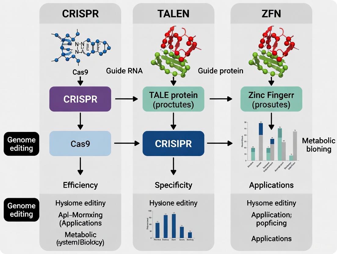
This article provides a detailed comparison of the three major genome-editing platforms—Zinc Finger Nucleases (ZFNs), Transcription Activator-Like Effector Nucleases (TALENs), and the CRISPR-Cas system.
CRISPR vs. TALEN vs. ZFN: A Comprehensive Guide to Choosing Your Genome Editing Tool
Abstract
This article provides a detailed comparison of the three major genome-editing platforms—Zinc Finger Nucleases (ZFNs), Transcription Activator-Like Effector Nucleases (TALENs), and the CRISPR-Cas system. Tailored for researchers, scientists, and drug development professionals, it explores the foundational mechanisms, methodological applications, and key challenges of each technology. Drawing on the latest research and direct comparative studies, the content delivers actionable insights for troubleshooting, optimizing editing efficiency, and validating outcomes. The review also covers emerging trends, such as base and prime editing, and discusses the clinical implications and safety considerations vital for therapeutic development.
The Evolution of Programmable Nucleases: From ZFNs to CRISPR-Cas
Programmable nucleases have revolutionized genetic engineering by acting as precise molecular scissors. Their core function is to create DNA double-strand breaks (DSBs) at predetermined genomic locations, harnessing the cell's own repair machinery to achieve targeted genetic modifications. [1] [2] This guide provides a detailed comparison of the three primary nuclease technologies—Zinc-Finger Nucleases (ZFNs), Transcription Activator-Like Effector Nucleases (TALENs), and the CRISPR/Cas9 system—focusing on their mechanisms, efficiencies, and practical applications in research and therapy.
The Universal Trigger: Double-Strand Break Repair Pathways
The power of all programmable nucleases lies not just in the cut they make, but in the cellular repair processes they activate. Once a double-strand break (DSB) is introduced, the cell attempts to repair this potentially genotoxic lesion primarily through two pathways: the error-prone Non-Homologous End Joining (NHEJ) and the high-fidelity Homology-Directed Repair (HDR). [1] [2] [3]
- NHEJ directly ligates the broken DNA ends, often resulting in small insertions or deletions (indels). When these indels occur within a gene's coding sequence, they can disrupt the reading frame, leading to gene knockout. [4] [2] [3]
- HDR requires a homologous DNA template to accurately repair the break. By providing an engineered donor DNA template, researchers can exploit this pathway to introduce specific sequence changes, such as point mutations or gene insertions. [4] [5]
The following diagram illustrates the critical decision point a cell faces after a nuclease-induced DSB and the potential outcomes for genome engineering.
Molecular Architectures of Programmable Nucleases
While ZFNs, TALENs, and CRISPR/Cas9 all achieve DSBs, their molecular compositions and DNA recognition mechanisms are fundamentally different. The table below summarizes the core architectural components of each system.
Table 1: Architectural Comparison of Programmable Nuclease Systems
| Feature | Zinc-Finger Nucleases (ZFNs) | TALENs | CRISPR/Cas9 |
|---|---|---|---|
| DNA-Binding Domain | Engineered zinc-finger proteins (Cys2-His2 motif) [2] [6] | Transcription Activator-Like Effectors (TALEs) [4] [2] | CRISPR RNA (crRNA) guide sequence [1] [3] |
| Recognition Code | 1 zinc finger domain ≈ 3 base pairs [2] [6] | 1 TALE repeat ≈ 1 base pair (e.g., NI=A, HD=C, NG=T, NN=G) [4] [2] | RNA-DNA base pairing (Watson-Crick) [1] [3] |
| Cleavage Domain | FokI nuclease domain [5] [6] | FokI nuclease domain [4] [7] | Cas9 protein (HNH & RuvC domains) [1] |
| Cleavage Mechanism | Dimeric: Requires a pair of ZFNs binding opposite strands for FokI dimerization. [5] [6] | Dimeric: Requires a pair of TALENs binding opposite strands for FokI dimerization. [4] [3] | Monomeric: A single Cas9 protein complexed with a guide RNA creates the DSB. [1] [3] |
| Target Site Structure | Two "half-sites" separated by a 5-7 bp spacer. [5] [6] | Two "half-sites" separated by a spacer. [4] [3] | Target sequence adjacent to a Protospacer Adjacent Motif (PAM, e.g., NGG for SpCas9). [1] |
The following diagram visualizes how these architectural differences translate into the process of finding and cutting a DNA target.
Performance and Practical Comparison
Beyond molecular architecture, practical considerations such as efficiency, specificity, ease of use, and application suitability are critical for selecting the right tool.
Table 2: Performance and Practical Application Comparison
| Aspect | ZFN | TALEN | CRISPR/Cas9 |
|---|---|---|---|
| Targeting Range | Limited by G-rich sequence preference; context-dependent effects complicate design. [2] [6] | Virtually unlimited; simple code links protein repeats to DNA bases. [4] [2] | Limited only by the presence of a PAM sequence (e.g., NGG). [1] |
| Editing Efficiency | Variable; highly dependent on the success of zinc-finger array design. [2] [6] | High; studies show success rates comparable to ZFNs and CRISPR. [4] | Very high; often the most efficient system in direct comparisons. [3] [8] |
| Off-Target Effects | Moderate to high; dependent on ZF array specificity. One study reported 287 off-target sites for an HPV-targeted ZFN. [8] | Low to moderate; the dimeric requirement acts as a natural "fail-safe". [3] [8] | Can be higher due to single-guide RNA dependency; tolerates mismatches, especially in PAM-distal region. [1] [8] |
| Ease of Design & Use | Technically challenging; requires expertise to account for context-dependence between fingers. [2] [6] | Cloning is laborious due to highly repetitive sequences, but design is straightforward. [4] [2] | Very simple; requires only the synthesis of a ~20nt guide RNA sequence. [3] |
| Multiplexing Potential | Low; difficult to express and deploy multiple ZFN pairs simultaneously. | Low; similar challenges to ZFNs due to large, repetitive constructs. | High; multiple guide RNAs can be expressed to target several sites at once. [3] |
| Delivery Consideration | cDNA size ~1 kb, easier for viral delivery. [3] | cDNA size ~3 kb, pushing the limits of standard viral vectors (e.g., AAV). [3] | Cas9 cDNA ~4.2 kb, large for viral delivery; often requires split systems or alternative Cas proteins. |
Supporting Experimental Data: A Direct Comparison
A rigorous 2021 study using the GUIDE-seq method to profile off-target activity in a human papillomavirus (HPV) gene therapy context provides a direct, quantitative comparison. [8]
- Efficiency and Specificity: When targeting the HPV E6 gene, SpCas9 was not only highly efficient but also exhibited zero detectable off-target events. In contrast, TALENs targeting the same region produced 7 off-target sites. [8]
- ZFN Performance: The same study found that ZFNs could generate a strikingly high number of off-targets (ranging from 287 to 1,856), with specificity potentially inversely correlated with the count of middle "G" bases in the target sequence. [8]
These findings underscore that while all tools are effective, CRISPR/Cas9 can offer a superior combination of efficiency and specificity in head-to-head tests.
Detailed Experimental Protocols
To ensure reproducibility and deepen the understanding of the data presented, this section outlines key methodologies used to generate the comparative findings.
GUIDE-seq for Genome-Wide Off-Target Detection
The GUIDE-seq (Genome-wide, Unbiased Identification of DSBs Enabled by sequencing) method is a powerful technique for profiling off-target cleavage by nucleases. [8]
- Oligonucleotide Tag Transfection: Cells co-transfected with the nuclease (ZFN, TALEN, or CRISPR/Cas9) and a blunt-ended, double-stranded oligonucleotide "tag".
- Tag Integration: During the repair of nuclease-induced DSBs via NHEJ, the tag is integrated into the break sites.
- Library Preparation & Sequencing: Genomic DNA is sheared and adaptor-ligated. PCR amplification, using one primer specific to the integrated tag and another to the genomic adaptor, enriches for tag-integrated fragments, which are then sequenced.
- Bioinformatic Analysis: Sequencing reads are mapped to the reference genome to identify all tag integration sites, which correspond to both on-target and off-target nuclease cleavage events.
UMI-DSBseq for Kinetic Analysis of DSB Repair
A 2024 study developed UMI-DSBseq to precisely quantify DSB intermediates and repair products over time. [9]
- Protoplast Transfection & Sampling: Tomato protoplasts are transfected with preassembled Cas9 ribonucleoprotein (RNP) complexes for synchronized DSB induction. Samples are collected along a time-course (e.g., 0-72 hours).
- End-Repair and Adaptor Ligation: Genomic DNA is extracted. A restriction enzyme creates a reference DSB at a site flanking the target. DNA ends, including both the reference breaks and nuclease-induced DSBs, are repaired and ligated to adaptors containing Unique Molecular Identifiers (UMIs).
- Library Sequencing and Analysis: Target sites are amplified and sequenced. UMIs allow for accurate counting of original molecules, distinguishing between intact, precisely repaired molecules, unrepaired DSBs, and error-prone repair products (indels). This enables modeling of DSB induction and repair kinetics.
Essential Research Reagent Solutions
The following table lists key reagents and their functions, as derived from the experimental protocols and technologies discussed.
Table 3: Key Research Reagents for Nuclease-Based Genome Editing
| Reagent / Solution | Function / Description | Example Application |
|---|---|---|
| Preassembled RNP | Complex of purified Cas9 protein and synthetic guide RNA. Allows for transient, rapid nuclease activity without genomic integration. [9] | Direct delivery into protoplasts or cells for synchronized DSB induction in kinetic studies. [9] |
| Obligate Heterodimer FokI Variants | Engineered FokI nuclease domains with mutations that force pairing only between two different monomers, reducing homodimer off-target cleavage. [6] | Used in ZFN and TALEN architectures to improve specificity. [5] [6] |
| Chemically Modified gRNA | Synthetic guide RNAs with chemical modifications (e.g., 2'-O-methyl analogs) to improve stability and reduce immune responses in cells. [1] | Enhances editing efficiency and reduces toxicity in therapeutic applications. |
| GUIDE-seq Oligo Tag | A short, double-stranded DNA oligonucleotide that is integrated into DSB sites during repair to mark them for genome-wide identification. [8] | Unbiased detection of off-target cleavage sites for ZFNs, TALENs, and CRISPR/Cas9. [8] |
| UMI-DSBseq Adaptors | Sequencing adaptors containing Unique Molecular Identifiers (UMIs) for ligation to DSB ends, enabling single-molecule tracking of repair outcomes. [9] | Precise quantification of DSB intermediates and repair dynamics over time. [9] |
Zinc Finger Nucleases (ZFNs) represent the pioneering technology that first enabled true precision in genome engineering. As the initial member of the programmable nuclease triad that also includes TALENs and CRISPR-Cas9, ZFNs demonstrated for the first time that researchers could make targeted, specific modifications to complex genomes [10]. This groundbreaking technology is built upon a protein-driven design framework, where engineered zinc finger proteins provide sequence specificity fused to a nuclease domain that executes DNA cleavage [11]. The development of ZFNs opened new frontiers in biological research and therapeutic development by moving beyond random mutagenesis to targeted gene editing. Despite the subsequent emergence of newer technologies, ZFNs remain relevant due to their high specificity and continued refinement, including applications in clinical trials and advanced therapeutic development [11] [12]. This article explores the protein-driven architecture of ZFNs and provides a objective comparison with TALENs and CRISPR-Cas9 based on experimental data and performance metrics.
Molecular Architecture and Mechanism
The ZFN system operates through an elegant protein-DNA recognition mechanism. Each ZFN is a chimeric protein composed of two functional domains: a DNA-binding domain derived from Cys2-His2 (C2H2) zinc finger proteins and a cleavage domain from the FokI restriction enzyme [11] [2]. The C2H2 zinc finger domain is one of the most common DNA-binding motifs in eukaryotes, folding into a compact ββα structure stabilized by zinc ion coordination [11]. Each individual zinc finger domain recognizes a 3-4 base pair DNA sequence, with multiple fingers assembled in tandem to create extended specificity for typically 9-18 base pairs [2] [10].
The FokI cleavage domain must dimerize to become active, necessitating the design of ZFN pairs that bind to opposite DNA strands with correct orientation and spacing [11]. This dimerization requirement provides a natural check on specificity, as off-target cleavage is less likely to occur without simultaneous binding of both ZFNs at adjacent sites. When successfully bound and dimerized, the FokI domains create a double-strand break (DSB) in the DNA with 5' overhangs [10]. The cellular repair of these breaks through either error-prone non-homologous end joining (NHEJ) or homology-directed repair (HDR) enables the desired genetic modifications, from gene knockouts to precise knockins [2].
Figure 1: ZFN Mechanism of Action. ZFNs function as pairs with DNA-binding domains and FokI cleavage domains that must dimerize to create double-strand breaks, repaired by cellular mechanisms.
Engineering and Design Methodologies
The engineering of functional ZFNs has evolved through several methodological approaches, each with distinct advantages:
Modular Assembly: This approach utilizes pre-characterized zinc finger modules that recognize specific 3-base pair triplets [11]. Researchers can assemble these predefined modules in tandem to target desired DNA sequences. The Barbas and ToolGen domains represent the two most commonly used sets of modular assembly fingers, covering GNN, most ANN, many CNN, and some TNN triplets (where N represents any nucleotide) [11]. The primary advantage of modular assembly is the ability to rapidly construct ZFNs without additional selection steps, though context-dependent effects between neighboring fingers can influence specificity [11].
Oligomerized Pool Engineering (OPEN): Developed to address context-dependency limitations, OPEN employs a combinatorial selection-based strategy using pre-selected zinc finger pools from an archive [11]. Appropriate finger pools are recombined to create libraries of multi-finger arrays, which are then screened using bacterial two-hybrid (B2H) systems to identify functional binders [11]. OPEN achieves approximately 70-80% success rate for obtaining functional ZF arrays and is publicly available through the Zinc Finger Consortium Database [11].
Context-Dependent Assembly: This hybrid approach utilizes zinc finger modules pre-selected for context dependency, combining advantages of both modular assembly and selection-based methods [2]. These methods acknowledge that zinc finger specificity can be influenced by positional effects and neighboring finger sequences, leading to more reliable DNA-binding proteins.
Comparative Analysis: ZFNs vs. TALENs vs. CRISPR-Cas9
Mechanism and Design Comparison
Table 1: Fundamental Characteristics of Genome Editing Technologies
| Feature | ZFNs | TALENs | CRISPR-Cas9 |
|---|---|---|---|
| DNA Recognition Molecule | Engineered zinc finger proteins [13] | Transcription activator-like effectors (TALEs) [13] | Guide RNA (gRNA) [13] |
| Recognition Mechanism | Protein-DNA interaction [13] | Protein-DNA interaction [13] | RNA-DNA complementarity [13] |
| Recognition Length | 9-18 bp per ZFN (18-36 bp for pair) [13] | 30-40 bp per TALEN pair [13] | 20 bp gRNA + PAM sequence [13] |
| Cleavage Domain | FokI nuclease (requires dimerization) [11] [2] | FokI nuclease (requires dimerization) [2] | Cas9 nuclease (single protein) [13] |
| Target Constraints | Prefers G-rich sequences; limited target sites [10] | Requires T at 5' position; more flexible than ZFNs [2] | Requires PAM sequence (NGG for SpCas9) [13] |
| Multiplexing Capacity | Difficult | Moderate | Easy [13] |
Experimental Performance Metrics
Recent comparative studies have provided quantitative assessments of nuclease performance, particularly regarding specificity and efficiency:
Table 2: Experimental Performance Comparison in HPV-Targeted Gene Therapy [8]
| Parameter | ZFNs | TALENs | SpCas9 |
|---|---|---|---|
| Off-target Count (URR target) | 287 | 1 | 0 |
| Off-target Count (E6 target) | Not tested | 7 | 0 |
| Off-target Count (E7 target) | Not tested | 36 | 4 |
| Specificity Trend | Massive off-target variation; correlates with "G" count | Design trade-offs: higher efficiency increases off-targets | Highest specificity overall |
| Efficiency | Variable | High with optimized designs | Highest |
A comprehensive 2021 study using GUIDE-seq analysis for human papillomavirus (HPV) targeted therapy revealed that ZFNs with similar targets could generate distinct massive off-targets (287-1,856), with specificity reversely correlated with counts of middle "G" in zinc finger proteins [8]. This same study found SpCas9 demonstrated superior efficiency and specificity compared to both ZFNs and TALENs across multiple genomic targets [8].
Practical Implementation Considerations
Table 3: Practical Application Considerations for Researchers
| Aspect | ZFNs | TALENs | CRISPR-Cas9 |
|---|---|---|---|
| Design Complexity | High [14] [10] | Moderate [14] | Low [14] |
| Construction Time | Months for optimized designs [10] | Weeks [14] | Days [14] |
| Cost Efficiency | Low (protein engineering intensive) [14] | Moderate | High [14] |
| Technical Accessibility | Requires specialized expertise [10] | Standard molecular biology | Accessible to most labs |
| Delivery Considerations | Compatible with viral vectors [12] | Large size challenges delivery | Cas9 size may challenge delivery |
| Therapeutic Development | Clinical trials ongoing (e.g., HIV) [11] | Preclinical development | Extensive clinical development |
Experimental Protocols and Methodologies
ZFN Validation Workflow
The development of functional ZFNs requires rigorous validation through a structured experimental pipeline:
Figure 2: ZFN Validation Workflow. Key steps in developing and validating functional ZFNs, from target identification to specificity assessment.
Step 1: Target Site Identification
- Identify target sequences of the form (NNC)₃...(GNN)₃ separated by 4-6 bp within the gene of interest [15]
- Screen for sequences with minimal off-target potential across the genome
- Prioritize targets with G-rich sequences for optimal zinc finger binding [10]
Step 2: Zinc Finger Protein Design
- Select appropriate zinc finger modules using modular assembly, OPEN, or context-dependent methods [11]
- For modular assembly, utilize pre-defined zinc finger archives (Barbas or ToolGen sets) [11]
- Design ZFN pairs with appropriate spacing (4-6 bp) for FokI dimerization [11] [15]
Step 3: ZFN Construction
- Clone zinc finger arrays into expression vectors containing FokI cleavage domain
- Utilize systems such as CompoZr for commercial ZFN sources [2]
- Verify protein expression through Western blot or in vitro transcription/translation [15]
Step 4: In Vitro Cleavage Assay (IVTT)
- Express ZFNs using rabbit reticulocyte lysate TnT Quick-Coupled Transcription-Translation system [15]
- Incubate with plasmid substrate containing target site
- Analyze cleavage products via gel electrophoresis for sequence-specific cleavage activity [15]
- Modify IVTT assay for rapid screening of multiple ZFN constructs [15]
Step 5: Cell-Based Validation
- Deliver ZFNs to target cells via transfection or viral transduction
- Assess mutation rates using SURVEYOR or T7E1 mismatch assays
- Quantify indel formation through sequencing analysis
- Evaluate HDR efficiency using donor templates
Step 6: Off-Target Assessment
- Identify potential off-target sites through in silico prediction based on sequence similarity [10]
- Utilize GUIDE-seq or similar genome-wide methods for unbiased off-target detection [8]
- Assess cytotoxicity and cellular responses to ZFN treatment
Key Research Reagent Solutions
Table 4: Essential Research Reagents for ZFN Experiments
| Reagent Category | Specific Examples | Function/Application |
|---|---|---|
| ZFN Engineering Systems | Barbas modular assembly, OPEN system, CompoZr | Design and construction of zinc finger arrays [11] [2] |
| Expression Systems | TnT Quick-Coupled Transcription/Translation | In vitro ZFN synthesis and testing [15] |
| Cleavage Assay Reagents | Custom target plasmids, gel electrophoresis reagents | Validation of sequence-specific cleavage activity [15] |
| Delivery Vehicles | Adenoviral, lentiviral vectors, transfection reagents | Introduction of ZFNs into target cells [10] |
| Analysis Tools | SURVEYOR assay, T7E1 mismatch detection, sequencing | Detection and quantification of editing events |
| Off-Target Assessment | GUIDE-seq, BLISS, Digenome-seq | Genome-wide identification of off-target effects [8] |
Applications and Therapeutic Developments
ZFN technology has demonstrated significant utility across diverse research and therapeutic areas:
Biomedical Research Applications
Disease Modeling: ZFNs have been used to correct disease-causing mutations associated with sickle cell disease, α1-antitrypsin deficiency, hemophilia B, and Parkinson's disease in patient-derived cells [11]. The combination of ZFNs with induced pluripotent stem cell (iPSC) technology enables the genetic correction of point mutations in patient-specific cell lines [11].
Gene Function Studies: ZFNs facilitate targeted gene knockout studies in various cell types and model organisms, providing conclusive information about gene function through complete and permanent gene disruption [2].
Biotechnology Applications: Beyond nucleases, zinc finger proteins have been fused to various effector domains including transcriptional activators, repressors, recombinases, and transposases for diverse genome engineering applications [2].
Clinical Translation and Therapeutic Development
ZFN technology has progressed to clinical trials, demonstrating its therapeutic potential:
HIV Treatment: Clinical trials (NCT00842634 and NCT01044654) have investigated ZFN-mediated disruption of the CCR5 gene in T-cells to create HIV-resistant immune cells [11].
Sickle Cell Disease: ZFN-mediated genome editing of the BCL11A erythroid-specific enhancer in hematopoietic stem progenitor cells represents a potential therapeutic approach [12].
Allogeneic Cell Therapies: ZFNs are being utilized to disrupt genes in allogeneic cell therapies for autoimmune diseases and cancer [12].
Neurological Disorders: Zinc finger transcriptional regulators (ZF-TRs), including repressors and activators, are being evaluated as potential treatments for chronic neuropathic pain, prion disease, and Huntington's disease [12].
The development of ZFN technology established the foundation for programmable genome editing and continues to evolve through ongoing innovations. While CRISPR-Cas9 currently dominates the research landscape due to its ease of use and accessibility, ZFNs maintain distinct advantages in specific applications, particularly where their compact size benefits delivery constraints or where high specificity is paramount [12]. Recent advances include the development of zinc finger base editors that enable precise nucleotide changes without creating double-strand breaks, potentially reducing off-target effects [12].
The comparative analysis presented here demonstrates that each genome editing platform offers distinct advantages and limitations. ZFNs provide high specificity and the benefit of being based on human-derived proteins, potentially offering safety advantages for therapeutic applications [12]. However, their technical complexity and design challenges have limited widespread adoption compared to CRISPR-based systems. TALENs strike a balance between specificity and design feasibility, while CRISPR-Cas9 offers unparalleled ease of use and multiplexing capabilities, though with ongoing concerns about off-target effects that continue to be addressed through engineering improvements [8] [13].
For researchers selecting genome editing technologies, the decision should be guided by specific application requirements rather than assuming the superiority of any single platform. ZFNs remain a valuable option particularly for therapeutic applications where their clinical experience, compact size, and human protein origin may provide distinct advantages. As the field of genome editing continues to advance, the pioneering ZFN technology continues to inform and inspire new developments in precision genetic medicine.
The advent of programmable nucleases has revolutionized genetic engineering, enabling precise modifications across diverse organisms. Among these technologies, Transcription Activator-Like Effector Nucleases (TALENs) have emerged as a powerful platform distinguished by their exceptional specificity and unique molecular architecture. While CRISPR-Cas9 systems have gained widespread popularity due to design simplicity, TALENs offer distinct advantages in applications where targeting precision is paramount, particularly in therapeutic development [16]. This guide objectively compares TALEN performance against alternative genome editing tools, with focused examination of the experimental approaches used to quantify and enhance their specificity.
TALENs represent a fusion between bacterial transcription activator-like effectors (TALEs) from Xanthomonas bacteria and the FokI nuclease domain [7]. Their DNA recognition mechanism employs a simple, predictable code that translates specific amino acid sequences to nucleotide binding preferences. This review comprehensively analyzes how this "simpler code" translates to practical advantages in research and clinical applications, supported by direct experimental comparisons and methodological details.
Molecular Architecture and Comparative Mechanisms
TALEN Structure and DNA Recognition Code
The TALEN system functions as a dimer, with each monomer consisting of a central DNA-binding domain derived from TALEs coupled to a FokI nuclease domain [16]. The DNA-binding domain comprises 12-28 highly conserved 33-35 amino acid repeats, each recognizing a single DNA nucleotide through two critical residues at positions 12 and 13, known as Repeat Variable Diresidues (RVDs) [7]. The established RVD-DNA recognition code primarily utilizes four RVD modules: NI for adenine (A), HD for cytosine (C), NN for guanine (G), and NG for thymine (T) [17]. This modular one-repeat-to-one-base-pair recognition system provides TALENs with unparalleled targeting flexibility, enabling theoretical targeting of any DNA sequence [16].
The FokI nuclease domain requires dimerization to become active, meaning two TALEN monomers must bind opposite DNA strands at precisely spaced intervals (typically 14-20 base pairs between binding sites) to enable DNA cleavage [18]. This dimerization requirement significantly enhances targeting specificity by effectively doubling the recognition length to approximately 30-36 base pairs, a sequence statistically unlikely to appear randomly in genomes [16].
Comparative Mechanisms of Major Genome Editing Platforms
Table 1: Fundamental Comparison of Genome Editing Platforms
| Feature | TALEN | CRISPR/Cas9 | ZFN |
|---|---|---|---|
| Recognition Type | DNA-protein interaction [16] | RNA-DNA complementarity [16] | DNA-protein interaction [17] |
| DNA Binding Domain | TALE repeats (1 repeat/1 bp) [7] | Guide RNA (∼20 nt) [16] | Zinc fingers (3-6 fingers/9-18 bp) [17] |
| Nuclease | FokI (requires dimerization) [16] | Cas9 (functions as monomer) [16] | FokI (requires dimerization) [17] |
| Target Length | 30-36 bp (combined dimer recognition) [16] | 20 bp guide + PAM [16] | 18-36 bp (combined dimer recognition) [17] |
| Specific Constraint | 5' T requirement [19] | PAM sequence requirement (e.g., NGG for SpCas9) [16] | Context-dependent binding effects [17] |
The molecular mechanism differences create distinct practical implications. CRISPR-Cas9 systems rely on RNA-DNA hybridization for targeting, with Cas9 nuclease activity triggered by successful matching between guide RNA and target DNA adjacent to a Protospacer Adjacent Motif (PAM) sequence [16]. This mechanism allows easier retargeting but increases mismatch tolerance potential. In contrast, TALENs and ZFNs employ protein-DNA interactions, with ZFNs utilizing C2H2 zinc finger arrays where each finger typically recognizes 3-base pair sequences [17]. The TALEN approach represents an intermediate in design simplicity between the highly modular CRISPR system and the more complex ZFN platform.
Diagram 1: Molecular recognition mechanisms of major genome editing platforms. TALENs and ZFNs utilize protein-DNA interactions with FokI dimerization requirements, while CRISPR employs RNA-DNA hybridization with PAM sequence constraints. The extended recognition length of TALENs contributes to their high specificity profile.
Quantitative Comparison of Editing Performance
Specificity and Off-Target Profiles
Comprehensive studies directly comparing genome editing technologies have revealed distinct performance characteristics. TALENs consistently demonstrate superior specificity metrics with minimal off-target activity, a critical consideration for therapeutic applications [16].
Table 2: Experimental Performance Comparison of Genome Editing Tools
| Performance Metric | TALEN | CRISPR/Cas9 | Experimental Context |
|---|---|---|---|
| Off-Target Rate | Low [16] | High (≥50% in some studies) [16] | Human cell lines [16] |
| Typical Indel Formation | ∼33% [18] | Up to >70% [18] | Chromosomal targets [18] |
| Mismatch Tolerance | Few mismatches tolerated [16] | Moderate, up to 5 mismatches reported [18] | In vitro specificity profiling [19] |
| Genomic Off-Target Sites Identified | 16 sites across 76 predicted in human cells [19] | Numerous off-target sites with high mutation rates [16] | Genome-wide studies [16] [19] |
| Mitochondrial Genome Editing | Possible (mito-TALEN) [16] | Complicated due to gRNA import issues [16] | Mitochondrial DNA manipulation [16] |
Experimental evidence from specificity profiling studies demonstrates that TALENs maintain high on-target activity while minimizing off-target effects. In one comprehensive analysis, 30 unique TALENs were profiled against 10¹² potential off-target sequences, revealing that TALENs cleave specifically at intended sites with minimal activity against mismatched sequences [19]. The same study identified only 16 bona fide off-target sites in the human genome across all tested TALENs, with most showing significantly reduced cleavage efficiency compared to on-target sites.
Targeting Range and Practical Efficiency
While TALENs demonstrate superior specificity, each platform exhibits distinct advantages in practical applications. CRISPR systems offer unparalleled ease of design and multiplexing capabilities, enabling simultaneous targeting of multiple genomic loci [20]. TALENs provide broader targeting freedom without PAM sequence constraints but require more complex protein engineering for each new target [16].
DNA methylation sensitivity represents an important practical consideration. TALEN activity can be inhibited by cytosine methylation, particularly at CpG dinucleotides, which requires careful target selection or use of specialized RVDs [18]. CRISPR systems do not share this limitation, providing more consistent activity across differentially methylated genomic regions.
Experimental Approaches for Enhancing TALEN Specificity
Non-Conventional RVDs for Specificity Optimization
Research efforts have focused on expanding the TALEN recognition code beyond the four conventional RVDs to enhance specificity. High-throughput screening of non-conventional RVDs (ncRVDs) has identified novel combinations with improved discriminatory power [21].
Table 3: Experimental Reagents for TALEN Specificity Enhancement
| Research Reagent | Composition/Function | Specificity Application |
|---|---|---|
| Non-Conventional RVDs | Alternative amino acids at positions 12/13 | Enhance nucleotide discrimination, particularly at mismatch positions [21] |
| TALEN-Q3 Variant | Modified TALEN architecture with reduced non-specific DNA binding | 10-fold lower off-target activity in human cells [19] |
| Obligate Heterodimeric FokI | EL/KK FokI variants requiring heterodimerization | Prevents homodimer activity, reduces off-target cleavage [19] |
| Specificity Profiling Library | 10¹² potential off-target DNA sequences | Comprehensive specificity assessment [19] |
The experimental approach for identifying ncRVDs involved creating randomized RVD libraries using NNK codon degeneracy, incorporating these alternative RVDs at defined positions within TALE arrays, and screening against targets containing all four nucleotides at corresponding positions [21]. This high-throughput methodology evaluated approximately 18,000 TALEN/target combinations, identifying ncRVDs with novel exclusion properties - the ability to discriminate against specific nucleotides while maintaining robust activity on the intended nucleotide [21].
Experimental Workflow for Specificity Optimization
Diagram 2: Experimental workflow for enhancing TALEN specificity using non-conventional RVDs. The process begins with library construction, proceeds through high-throughput screening, identifies optimal ncRVDs with exclusion properties, and validates specificity enhancement in human cell systems.
Application Case Study: Discrimination of Highly Homologous Sequences
A compelling demonstration of TALEN specificity optimization comes from targeting the human HBB gene (associated with sickle cell anemia) while avoiding cleavage of the highly homologous HBD gene (94% identity) [21]. Researchers implemented an "exclusion strategy" by incorporating ncRVDs at mismatch positions between HBB and HBD sequences. These specialized ncRVDs maintained robust activity against the intended HBB target while discriminating against the HBD off-target, demonstrating the practical application of specificity-enhanced TALENs for therapeutic development [21].
The experimental protocol for this application involved:
- Identifying optimal target sites with minimal off-target sequences genome-wide
- Selecting ncRVDs with appropriate exclusion properties for mismatch positions
- Constructing TALEN arrays incorporating these ncRVDs
- Transfecting human cells and measuring cleavage efficiency at both on-target and off-target sites
- Validating specificity using sequencing-based methods to detect indels
This approach successfully generated TALENs capable of discriminating between sequences with 94% identity, highlighting the power of engineered specificity for targeting disease-associated genes with high homology to other genomic regions [21].
Research Reagent Solutions for TALEN Engineering
Table 4: Essential Research Reagents for TALEN Experiments
| Reagent Category | Specific Examples | Research Function |
|---|---|---|
| TALEN Scaffolds | Golden Gate assembly systems [20], Solid-phase assembly [21] | Modular TALEN construction with varying repeat lengths |
| RVD Modules | Conventional RVDs (NI, HD, NN, NG) [17], Non-conventional RVD libraries [21] | Target sequence recognition with tunable specificity |
| FokI Nuclease Variants | Wild-type FokI, Obligate heterodimers (EL/KK, ELD/KKR) [19] | DNA cleavage with controlled dimerization requirements |
| Specificity Assessment Tools | In vitro selection libraries [19], High-throughput sequencing assays | Comprehensive off-target profiling |
| Delivery Systems | mRNA, Plasmid DNA, Viral vectors (lentiviral, AAV) [17] | Efficient intracellular TALEN delivery |
The development of modular TALEN assembly systems, particularly Golden Gate assembly, has significantly streamlined TALEN construction despite the inherent complexity of protein engineering [20]. These systems utilize standardized parts and type IIS restriction enzymes to efficiently assemble repeat arrays, making TALEN technology more accessible to research laboratories [20]. For specificity assessment, in vitro selection methods using highly diverse DNA libraries (10¹² sequences) provide comprehensive profiling of TALEN cleavage preferences beyond what is achievable through cellular methods alone [19].
TALEN technology represents a powerful genome editing platform with distinct advantages in targeting specificity, a critical parameter for therapeutic applications. The simple, predictable DNA recognition code of TALENs provides a foundation for ongoing optimization efforts, including the development of non-conventional RVDs with enhanced discriminatory capabilities [21]. While CRISPR systems offer advantages in design simplicity and multiplexing capacity, TALENs maintain an important position in the genome editing toolbox, particularly for applications requiring maximal precision and minimal off-target effects [16].
Future directions in TALEN development include continued expansion of the RVD repertoire, optimization of domain architectures for improved DNA binding specificity, and integration with emerging delivery technologies. As the field advances toward clinical applications, the refined specificity of TALENs positions them as valuable tools for precision genetic engineering, complementing rather than competing with other genome editing platforms [7]. The choice between TALEN, CRISPR, and ZFN technologies ultimately depends on specific research requirements, with TALENs offering an optimal balance of targeting flexibility and precision for many applications.
The advent of programmable gene-editing technologies has revolutionized molecular biology, providing researchers with unprecedented tools for investigating gene function and developing therapeutic interventions. Among these technologies, Zinc Finger Nucleases (ZFNs), Transcription Activator-Like Effector Nucleases (TALENs), and the Clustered Regularly Interspaced Short Palindromic Repeats (CRISPR)-Cas system represent three generations of engineered nucleases that have successively transformed the field. While ZFNs and TALENs demonstrated the feasibility of targeted genome engineering, the discovery and adaptation of CRISPR-Cas systems have truly democratized gene editing due to their exceptional simplicity and versatility [22] [23].
This comparison guide objectively examines these three gene-editing platforms, focusing specifically on how CRISPR's unique RNA-guided mechanism has addressed many limitations of its protein-based predecessors. We present experimental data, detailed methodologies, and practical resources to help researchers, scientists, and drug development professionals select the most appropriate technology for their specific applications.
Molecular Mechanisms: A Tale of DNA Recognition Strategies
The fundamental difference between these technologies lies in their mechanisms for DNA recognition: ZFNs and TALENs rely on custom-engineered proteins, while CRISPR-Cas systems utilize a programmable RNA molecule for target recognition.
Protein-Based Recognition: ZFNs and TALENs
Zinc Finger Nucleases (ZFNs) are fusion proteins comprising a DNA-binding domain and a FokI nuclease domain. Each zinc finger motif recognizes approximately 3 base pairs of DNA, and multiple fingers are assembled to create a domain that recognizes a specific 9-18 bp sequence. Since FokI requires dimerization to become active, two ZFN monomers must bind opposite strands of DNA with correct orientation and spacing to create a double-strand break [22] [23].
TALENs similarly utilize the FokI nuclease domain but employ Transcription Activator-Like Effector (TALE) proteins for DNA recognition. Each TALE repeat recognizes a single base pair through highly variable repeat di-residues, making the engineering process more straightforward and predictable than zinc finger assembly. Like ZFNs, TALENs function as pairs binding to opposing DNA strands and require FokI dimerization for DNA cleavage [22] [23].
RNA-Guided Recognition: The CRISPR-Cas System
The CRISPR-Cas system functions as an adaptive immune system in bacteria and archaea. The most widely used CRISPR-Cas9 system consists of two key components: the Cas9 nuclease and a guide RNA (gRNA). The gRNA is a synthetic fusion of two natural RNAs - CRISPR RNA (crRNA) and trans-activating crRNA (tracrRNA) - creating a single-guide RNA (sgRNA) [24] [25]. The ~20 nucleotide sequence at the 5' end of the gRNA directs Cas9 to complementary DNA sequences adjacent to a Protospacer Adjacent Motif (PAM), typically 5'-NGG-3' for Streptococcus pyogenes Cas9. Upon binding, Cas9 induces a double-strand break in the target DNA [24].
The following diagram illustrates the fundamental difference in DNA recognition between these systems:
Comparative Analysis: Key Features for Research Applications
The different DNA recognition mechanisms of ZFNs, TALENs, and CRISPR translate into distinct practical advantages and limitations for research and therapeutic applications. The table below provides a comprehensive comparison of their key characteristics:
| Feature | CRISPR-Cas9 | TALENs | ZFNs |
|---|---|---|---|
| Targeting Molecule | Guide RNA (gRNA) | Engineered TALE protein | Engineered zinc finger protein |
| Target Recognition | RNA-DNA hybridization | Protein-DNA interaction | Protein-DNA interaction |
| Ease of Design | Simple (change 20-nt gRNA sequence) | Moderate (protein engineering required) | Complex (protein engineering required) |
| Development Time | Days | Weeks to months | Months |
| Cost | Low | High | High |
| Specificity | Moderate (subject to off-target effects) | High | High |
| Multiplexing Capacity | High (multiple gRNAs simultaneously) | Limited | Very limited |
| Efficiency | High | Moderate to high | Moderate |
| Optimal Applications | High-throughput screens, multiplexed editing, rapid prototyping | Applications requiring high specificity, clinical applications | Validated therapeutic applications |
| PAM Requirement | Yes (varies by Cas variant) | No | No |
Table 1: Comparative analysis of major gene-editing platforms. Data synthesized from multiple sources [22] [23].
Experimental Data and Performance Metrics
Editing Efficiency and Specificity
Recent studies directly comparing these technologies provide quantitative insights into their performance. In a landmark study targeting the CCR5 gene (a co-receptor for HIV), CRISPR demonstrated significantly higher editing efficiency (approximately 60-80% modification rates in human cells) compared to TALENs (typically 30-50% efficiency) [22]. However, TALENs showed fewer off-target effects in deep-sequencing analyses, with CRISPR exhibiting detectable off-target activity at sites with 3-5 nucleotide mismatches to the gRNA [22] [23].
Advanced CRISPR systems have been developed to address specificity concerns. High-fidelity Cas9 variants (such as SpCas9-HF1 and eSpCas9) incorporate mutations that reduce off-target effects by strengthening Cas9's binding specificity to the target DNA [22]. Additionally, CRISPR-Cas systems derived from different bacterial species (such as Cas12a) often exhibit different PAM requirements and editing signatures, providing researchers with options to optimize specificity for particular genomic contexts [26] [25].
Practical Implementation: Time and Resource Requirements
The simplicity of CRISPR system design translates into dramatic reductions in both time and cost. While developing a new ZFN or TALEN pair typically requires 4-8 weeks of protein engineering and validation at a cost of $5,000-25,000+, a new CRISPR target can be designed in days for approximately $50-200 (including gRNA synthesis) [22] [23]. This efficiency advantage makes CRISPR particularly suitable for high-throughput screens and iterative experimental approaches.
Experimental Protocols for Technology Evaluation
Protocol: Comparative Assessment of Editing Efficiency
Objective: To quantitatively compare the editing efficiency and specificity of CRISPR, TALEN, and ZFN platforms at the same genomic locus.
Materials:
- HEK293T cells or other relevant cell line
- Plasmid constructs encoding: (1) SpCas9 + target-specific gRNA, (2) TALEN pair for same target, (3) ZFN pair for same target
- Transfection reagent
- Genomic DNA extraction kit
- PCR primers flanking target site
- Next-generation sequencing platform
Methodology:
- Design and clone editing constructs for all three platforms to target the same 20-30 bp genomic region.
- Transfect cells with each construct individually, including untransfected controls.
- Harvest cells 72 hours post-transfection and extract genomic DNA.
- Amplify target region by PCR and subject amplicons to next-generation sequencing.
- Analyze sequencing data using computational tools (such as CRISPResso2 for CRISPR, TALEN-specific, and ZFN-specific analysis pipelines) to quantify:
- Indel frequencies at on-target site
- Mutation spectra (distribution of insertions vs. deletions)
- Off-target editing at predicted secondary sites
Expected Outcomes: CRISPR typically shows highest on-target efficiency, while TALENs often demonstrate superior specificity with minimal off-target activity [22] [23].
Protocol: Multiplexed Editing Capability Assessment
Objective: To evaluate the capacity of each platform for simultaneous editing of multiple genomic loci.
Materials: As above, with multiple gRNA/TALEN/ZFN constructs.
Methodology:
- For CRISPR: Clone 3-5 different gRNA expression cassettes into a single vector with Cas9.
- For TALENs/ZFNs: Attempt co-transfection of 3-5 different TALEN or ZFN pairs.
- Transfert cells and analyze editing efficiency at each target locus as described in Protocol 5.1.
- Compare the efficiency of multiplexed editing versus single editing for each platform.
Expected Outcomes: CRISPR systems maintain high efficiency when targeting multiple loci simultaneously (typically 40-70% efficiency per locus), while TALEN and ZFN efficiency dramatically decreases with increasing target number due to delivery challenges and potential cellular toxicity [22].
Research Reagent Solutions for Gene Editing Studies
Successful implementation of gene-editing technologies requires access to specialized reagents and tools. The following table outlines essential materials and their functions:
| Reagent Category | Specific Examples | Function | Platform Compatibility |
|---|---|---|---|
| Nuclease Expression Plasmids | px459 (CRISPR), Golden Gate TALEN kits, CompoZr ZFN vectors | Delivery of editing machinery to cells | Platform-specific |
| Delivery Tools | Lentiviral particles, Lipofectamine 3000, Electroporation systems | Introduction of editing constructs into cells | All platforms |
| Validation Tools | T7E1 assay, TIDE analysis, next-generation sequencing | Detection and quantification of editing events | All platforms |
| Bioinformatics Resources | CRISPOR, CHOPCHOP, E-CRISP, TALEN-NT, ZFiT | Target site selection and off-target prediction | Platform-specific |
| Cell Culture reagents | HEK293T, HCT116, iPSCs; appropriate media and supplements | Provide cellular context for editing experiments | All platforms |
Table 2: Essential research reagents for gene-editing studies. Bioinformatics tools are particularly important for optimizing experimental design [25].
Clinical Translation and Therapeutic Applications
The therapeutic potential of gene-editing technologies is increasingly being realized in clinical trials. CRISPR-based therapies have shown remarkable success in treating sickle cell disease and β-thalassemia (with Casgevy receiving first regulatory approvals), hereditary transthyretin amyloidosis (with Intellia's LNP-delivered CRISPR system reducing TTR protein by ~90%), and hereditary angioedema [27] [28].
Notably, both ZFNs and TALENs continue to have important therapeutic roles where their high specificity is advantageous. For example, TALEN-based allogeneic CAR-T cells (lasme-cel) have shown promising Phase 1 results in B-cell acute lymphoblastic leukemia, with 42% of patients achieving complete remission [27].
The following diagram illustrates the clinical development pathway for gene-editing therapies, highlighting key considerations at each stage:
Emerging Innovations and Future Directions
The gene-editing landscape continues to evolve rapidly, with several emerging technologies building upon the CRISPR foundation:
Advanced CRISPR Systems
Base editing enables direct, irreversible conversion of one DNA base pair to another without double-strand breaks, significantly reducing indel formation [29] [22]. Prime editing offers even greater versatility, capable of making all 12 possible base-to-base conversions, as well as small insertions and deletions, without requiring donor DNA templates [27] [29].
AI-designed editors represent the cutting edge of CRISPR innovation. Recently, researchers used large language models trained on 1 million CRISPR operons to generate OpenCRISPR-1, a Cas9-like effector that is 400 mutations away from any natural protein yet shows comparable or improved activity and specificity relative to SpCas9 [26].
CRISPR Activation and Interference
CRISPR activation (CRISPRa) and CRISPR interference (CRISPRi) technologies use catalytically dead Cas9 (dCas9) fused to transcriptional regulators to precisely control gene expression without altering DNA sequences [24] [30]. These approaches are particularly valuable for functional genomics screens and disease modeling where reversible gene modulation is preferred.
The CRISPR-Cas system has undoubtedly revolutionized gene editing through its simple, RNA-guided programming mechanism, making sophisticated genome engineering accessible to virtually any molecular biology laboratory. Its advantages in ease of design, cost-effectiveness, multiplexing capability, and rapid implementation make it the default choice for most research applications [22] [23].
However, ZFNs and TALENs maintain important niches where their proven precision, reduced off-target effects, and established regulatory pathways provide distinct advantages, particularly in therapeutic contexts where maximal specificity is paramount [27] [23].
Researchers should base their platform selection on specific project requirements: CRISPR for most applications requiring flexibility and efficiency; TALENs for projects demanding maximal specificity with manageable target numbers; and ZFNs for specialized applications building on established systems. As all technologies continue to advance, particularly with AI-enabled design approaches [26], the future of gene editing promises even greater precision, efficiency, and therapeutic potential.
The advent of programmable genome editing has revolutionized biomedical research and therapeutic development, enabling scientists to modify DNA with unprecedented precision. Three technologies have been pivotal in this revolution: Zinc Finger Nucleases (ZFNs), Transcription Activator-Like Effector Nucleases (TALENs), and Clustered Regularly Interspaced Short Palindromic Repeats (CRISPR) systems. These technologies function as molecular scissors, creating targeted double-strand breaks (DSBs) in DNA that harness cellular repair mechanisms to achieve desired genetic modifications. This guide provides a historical and technical comparison of these three foundational genome-editing platforms, offering researchers a objective framework for selecting appropriate tools for specific applications. Understanding their developmental trajectories, relative strengths, and limitations is crucial for advancing basic research and developing next-generation genetic therapies [31] [32] [33].
Historical Timeline of Development
The development of programmable nucleases unfolded over two decades, with each technology building upon lessons learned from its predecessor. The following timeline visualizes these key milestones, from the initial protein engineering efforts to the widespread adoption of RNA-programmable systems.
Table: Key Technology Platforms and Their Origins
| Technology | Initial Discovery/Concept | First Human Cell Application | First Clinical Trial | Regulatory Approval |
|---|---|---|---|---|
| ZFNs | 1996 (ZFN concept) [33] | 2005 [31] | 2014 (HIV via CCR5 modification) [31] [34] | - |
| TALENs | 2011 [31] [32] | 2011 [31] | - | - |
| CRISPR-Cas9 | 2012 (Adapted for eukaryotes) [31] | 2013 | 2016 (Non-small cell lung cancer) [31] | 2023 (Casgevy for sickle cell disease) [31] |
Technical Mechanisms and Experimental Workflows
Fundamental Mechanisms of Action
All three editing platforms function by creating targeted double-strand breaks (DSBs) in DNA, but achieve this through distinct molecular mechanisms:
ZFNs utilize a DNA-binding domain composed of zinc finger motifs (each recognizing a 3-base pair DNA triplet) fused to the FokI nuclease domain. FokI must dimerize to become active, requiring two ZFN monomers to bind opposite DNA strands in a tail-to-tail orientation with a specific spacer sequence between them [34] [33].
TALENs similarly employ the FokI nuclease domain but use Transcription Activator-Like Effector (TALE) repeats for DNA recognition. Each TALE repeat recognizes a single nucleotide, following a simple code that makes design more straightforward than ZFNs. Like ZFNs, TALENs require dimerization of FokI domains for activity [35] [22].
CRISPR-Cas9 employs a fundamentally different mechanism based on RNA-DNA hybridization. The Cas9 nuclease is directed to its target by a guide RNA (gRNA) that is complementary to the DNA sequence of interest. Target recognition requires both gRNA complementarity and the presence of a Protospacer Adjacent Motif (PAM) sequence adjacent to the target site [35] [22].
After DSB creation, all platforms rely on endogenous cellular repair pathways:
- Non-Homologous End Joining (NHEJ): An error-prone process that often results in small insertions or deletions (indels), typically leading to gene knockout.
- Homology-Directed Repair (HDR): Uses a donor DNA template to enable precise gene insertion or correction [34] [33].
Core Experimental Protocol
A generalized workflow for genome editing experiments encompasses several key stages, from target selection to validation. The following diagram illustrates this process, highlighting how different nuclease platforms integrate into a shared experimental structure.
Key Methodological Considerations:
Target Selection: For CRISPR, targets must be adjacent to appropriate PAM sequences (e.g., 5'-NGG-3' for SpCas9). For ZFNs and TALENs, targets must allow for appropriate spacer distances between binding sites for FokI dimerization [35] [33].
Reagent Design and Assembly:
- CRISPR: Synthesize ~20 nt gRNA sequence complementary to target; clone into appropriate expression vector with Cas9.
- TALENs: Assemble TALE repeat arrays using modular cloning systems (e.g., Golden Gate assembly) to match target sequence.
- ZFNs: Engineer zinc finger arrays to recognize target triplets; more complex due to context-dependent effects [35] [22].
Delivery Methods: Common approaches include:
Validation Techniques:
Performance Comparison and Experimental Data
Quantitative Comparison of Editing Technologies
Table: Comprehensive Performance Metrics of Genome Editing Technologies
| Parameter | ZFNs | TALENs | CRISPR-Cas9 |
|---|---|---|---|
| Target Size Limitation | ~18 bp [35] | Variable length (typically 30-40 bp) [35] | 20 nt + PAM (NGG for SpCas9) [35] |
| Design & Assembly Time | Several months [35] [22] | Several days to weeks [35] | 1-3 days [22] |
| Relative Cost | High [22] | Moderate to High [22] | Low [22] |
| Editing Efficiency | Moderate (Varies by target) [35] | High (>90% in some studies) [35] | Moderate to High (typically 60-80%) [36] |
| Multiplexing Capacity | Limited [22] | Limited [22] | High (multiple gRNAs simultaneously) [22] |
| Primary Applications | Gene knockout, gene correction [34] | Gene knockout, gene correction [35] | Gene knockout, screening, activation/repression [36] |
Table: Experimental Success Metrics in Different Biological Contexts
| Experimental Context | ZFNs | TALENs | CRISPR-Cas9 |
|---|---|---|---|
| Immortalized Cell Lines | Moderate efficiency [36] | High efficiency [36] [35] | High efficiency (>80% in optimal conditions) [36] |
| Primary Cells (T cells) | Challenging, lower efficiency [36] | Moderate efficiency [36] | Moderate efficiency, protocol-dependent [36] |
| Heterochromatin Regions | Not well characterized | 5x more efficient than CRISPR in heterochromatin [37] | Lower efficiency in densely packed DNA [37] |
| Time to Generate Knockouts | 3-6 months [36] | 3-6 months [36] | ~3 months [36] |
| Time to Generate Knock-ins | 6+ months [36] | 6+ months [36] | ~6 months [36] |
Key Experimental Findings
Recent comparative studies have revealed context-specific advantages for each technology:
Heterochromatin Performance: A 2020 study using single-molecule imaging demonstrated that TALENs are up to five times more efficient than CRISPR-Cas9 at editing genes within heterochromatin, the densely packed regions of DNA that contain many disease-relevant genes. This suggests TALENs may be preferable for targets in these challenging genomic regions [37].
Editing Specificity: Research comparing off-target effects has shown that TALENs generally exhibit fewer off-target mutations than first-generation CRISPR-Cas9 systems, though high-fidelity Cas9 variants have substantially closed this gap [35] [22].
Workflow Efficiency: Surveys of research laboratories indicate that CRISPR workflows typically require 3 months to generate knockouts and 6 months for knock-ins, with researchers reporting repeating clonal isolation steps a median of 3 times before achieving desired edits, regardless of the technology used [36].
Essential Research Reagents and Solutions
Table: Key Research Reagents for Genome Editing workflows
| Reagent/Solution | Function | Technology Applicability |
|---|---|---|
| FokI Nuclease Domain | DNA cleavage component | ZFNs, TALENs |
| Zinc Finger Arrays | Sequence-specific DNA binding | ZFNs only |
| TALE Repeat Arrays | Sequence-specific DNA binding | TALENs only |
| Cas9 Nuclease | RNA-guided DNA cleavage | CRISPR-Cas9 |
| Guide RNA (gRNA) | Target recognition molecule | CRISPR-Cas9 |
| Protospacer Adjacent Motif (PAM) | Cas9 recognition sequence | CRISPR-Cas9 |
| Lipid Nanoparticles (LNPs) | In vivo delivery of editing components | All platforms |
| Adeno-Associated Virus (AAV) | Viral vector for in vivo delivery | All platforms (size-limited) |
| Electroporation Systems | Physical delivery method ex vivo | All platforms |
| Repair Template Donor DNA | Homology-directed repair template | All platforms (for knock-in) |
The historical development of genome editing technologies reveals a clear trajectory toward increased accessibility, efficiency, and application breadth. ZFNs established the fundamental principle that programmable nucleases could stimulate targeted genome modification, while TALENs simplified the design process and demonstrated particular efficacy in heterochromatin regions. CRISPR-Cas9 has dramatically democratized genome editing through its simplicity and versatility, leading to its rapid dominance in research and clinical applications.
Each platform retains distinct advantages: CRISPR for multiplexed applications and straightforward design, TALENs for challenging targets in heterochromatin, and ZFNs for well-established clinical applications where their profile is favorable. The optimal choice depends on the specific experimental requirements, including target genomic context, desired modification type, delivery constraints, and regulatory considerations. As all three technologies continue to evolve through protein engineering and delivery optimization, their complementary strengths will likely expand the therapeutic landscape for genetic disorders, cancer, and infectious diseases.
From Bench to Bedside: Practical Applications and Workflow Design
The advent of programmable nucleases has revolutionized genetic engineering, enabling precise modifications across diverse biological systems. Among these tools, Zinc Finger Nucleases (ZFNs), Transcription Activator-Like Effector Nucleases (TALENs), and the Clustered Regularly Interspaced Short Palindromic Repeats (CRISPR) system represent three generations of genome editing technology [38]. Each platform operates on the fundamental principle of creating double-strand breaks (DSBs) in DNA at predetermined genomic locations, harnessing cellular repair mechanisms—either error-prone non-homologous end joining (NHEJ) or high-fidelity homology-directed repair (HDR)—to achieve desired genetic outcomes [38] [39]. Selecting the optimal tool requires careful consideration of project-specific requirements for precision, scale, and available resources. This guide provides a structured comparison of ZFNs, TALENs, and CRISPR-Cas9, supported by experimental data and methodologies, to inform decision-making for researchers and drug development professionals.
Technology Comparison: Mechanisms, Applications, and Performance
Fundamental Mechanisms and Design
Zinc Finger Nucleases (ZFNs) are chimeric proteins comprising a DNA-binding domain—composed of engineered zinc finger motifs—fused to the FokI nuclease domain [38] [35]. Each zinc finger motif recognizes a specific DNA triplet, and multiple fingers are assembled to target a longer sequence (typically 9-18 bp) [38]. A functional nuclease requires a pair of ZFNs binding to opposite DNA strands with a specific spacer sequence (5-6 bp) between them, enabling FokI dimerization and subsequent DNA cleavage [38]. ZFNs were foundational in demonstrating the feasibility of targeted DSB-induced genome editing [35].
Transcription Activator-Like Effector Nucleases (TALENs) also utilize the FokI nuclease domain but employ DNA-binding domains derived from TALE proteins of Xanthomonas bacteria [38]. Their key advantage is a simpler, modular recognition code: each TALE repeat (33-35 amino acids) binds a single nucleotide, with specificity determined by two key amino acids (Repeat-Variable Diresidues, RVDs) [40] [38]. Like ZFNs, TALENs function as pairs binding opposite DNA strands, requiring a spacer sequence (12-19 bp) for FokI dimerization [38].
CRISPR-Cas Systems represent a paradigm shift from protein-based to RNA-guided DNA recognition [39]. The most common system, CRISPR-Cas9, uses a single guide RNA (sgRNA) that combines CRISPR RNA (crRNA) and trans-activating CRISPR RNA (tracrRNA) to direct the Cas9 nuclease to a complementary DNA target sequence [35] [39]. Critical for target recognition is a short Protospacer Adjacent Motif (PAM), which is 'NGG' for the commonly used Streptococcus pyogenes Cas9 (SpCas9) [39] [35]. Cas9 induces a DSB upon successful sgRNA binding and PAM recognition [38].
Diagram 1: Genome Editing Tool Mechanisms. The diagram illustrates the distinct DNA recognition and cleavage mechanisms of CRISPR-Cas9, TALEN, and ZFN systems, converging on the creation of double-strand breaks repaired by cellular pathways.
Quantitative Performance Comparison
The following table synthesizes experimental data on the efficiency, specificity, and practical application parameters of ZFNs, TALENs, and CRISPR-Cas9.
Table 1: Performance and Practical Comparison of Major Genome Editing Tools
| Parameter | ZFN | TALEN | CRISPR-Cas9 |
|---|---|---|---|
| Efficiency (Editing Rate) | 0%–12% (Low) [39] | 0%–76% (Moderate) [39] | 0%–81% (High) [39] |
| Target Site Size | 18–36 bp per ZFN pair [39] | 30–40 bp per TALEN pair [39] | ~22 bp (gRNA specific) [39] |
| Design Complexity | Difficult; requires expert knowledge of zinc finger assembly [35] [22] | Complex; simpler code than ZFNs but laborious protein engineering [35] [22] | Very simple; requires only gRNA sequence design [38] [22] |
| Development Timeline | Months [35] [22] | ~1 Month [38] [35] | Within a week [38] [22] |
| Relative Cost | High [38] [35] | Medium/High [38] [35] | Low [38] [35] |
| Multiplexing Potential | Less feasible [39] | Less feasible [39] | Highly feasible [39] [22] |
| Off-Target Risk | Less predictable [39] | Lower than CRISPR; highly specific [40] [23] | Highly predictable; potential for higher off-target effects [39] [23] |
| Primary Advantage | Proven precision in clinical applications [22] | High specificity with flexible targeting [40] [23] | Unmatched ease of use, scalability, and cost-effectiveness [22] [35] |
Key Factor Analysis for Tool Selection
Precision and Specificity
- TALENs often exhibit the highest specificity, attributed to their longer, protein-based target recognition and the requirement for FokI dimerization, which minimizes off-target cleavage [40] [23]. A study targeting the CCR5 gene found TALENs produced significantly fewer off-target mutations than ZFNs [35].
- ZFNs also demonstrate high precision due to their protein-DNA interaction mechanism, making them suitable for therapeutic applications where validated edits are critical [22].
- CRISPR-Cas9, while highly efficient, has a greater potential for off-target effects because the gRNA may tolerate mismatches, particularly outside the seed sequence [39] [23]. However, its off-target sites are highly predictable computationally, and engineered high-fidelity Cas9 variants (e.g., SpCas9-HF1) have been developed to mitigate this issue [22].
Experimental Scale and Multiplexing
- CRISPR-Cas9 is the undisputed leader for large-scale and multiplexed experiments. Its RNA-based design allows for the simultaneous expression of numerous gRNAs from a single construct, enabling genome-wide library screens and the modification of multiple genetic loci in one experiment [39] [22]. This is a key advantage for functional genomics and identifying gene dependencies in drug discovery [22].
- TALENs and ZFNs are poorly suited for multiplexing. Designing and delivering multiple large, protein-based nucleases is labor-intensive, costly, and faces technical challenges in delivery [22].
Resource and Practical Considerations
- CRISPR-Cas9 requires minimal specialized expertise and financial investment compared to older technologies. Designing a new gRNA is fast and inexpensive, making CRISPR the most accessible and user-friendly tool [35] [22].
- TALENs, while easier to design than ZFNs due to their one-to-one nucleotide recognition code, still require complex protein engineering and are time-consuming to develop [35].
- ZFNs are the most resource-intensive, demanding extensive expertise in zinc finger assembly and a lengthy development process, which limits their widespread adoption [35] [22].
Experimental Protocols and Workflows
A Standard Workflow for CRISPR-Cas9 Genome Editing
The following protocol outlines a typical CRISPR-Cas9 experiment for gene knockout, a common application across basic research and therapeutic development.
Diagram 2: CRISPR-Cas9 Experimental Workflow. This flowchart outlines the key steps in a standard CRISPR-Cas9 gene editing experiment, from design to validation.
Step 1: Target Selection and gRNA Design
- Procedure: Identify the genomic locus to be edited. Design a 20-nucleotide gRNA sequence that is complementary to the target DNA and immediately precedes a 5'-NGG-3' PAM sequence [39] [35].
- Critical Parameters: Utilize computational tools like CRISPOR or CHOPCHOP to select gRNAs with high predicted on-target efficiency and minimal off-target activity across the genome [25] [41].
- Controls: Design multiple gRNAs for the same target to account for potential variability in efficiency.
Step 2: Preparation of Editing Components
- For CRISPR-Cas9: Clone the designed gRNA sequence into a plasmid vector that also expresses the Cas9 nuclease (all-in-one vector) or prepare Cas9 protein/gRNA complexes as ribonucleoproteins (RNPs) for direct delivery [22].
- For TALENs/ZFNs: Clone the genes encoding the engineered TALEN or ZFN pairs into appropriate expression plasmids. This step is significantly more time-consuming than CRISPR gRNA cloning [35].
Step 3: Delivery into Target Cells
- Methods: Choose a delivery method suitable for the cell type.
- Transfection: Chemical-based (e.g., lipofection) for easily transfectable cell lines.
- Electroporation: For primary cells or hard-to-transfect lines.
- Viral Vectors: Lentivirus or Adeno-Associated Virus (AAV) for high efficiency and stable delivery, particularly in vivo [39] [22]. AAV has a limited cargo capacity, which can be a constraint for larger nucleases.
- Lipid Nanoparticles (LNPs): Increasingly used for in vivo therapeutic delivery, as demonstrated in recent clinical trials for hereditary transthyretin amyloidosis (hATTR) [28].
Step 4: Induction of Double-Strand Breaks and Repair
- Mechanism: After successful delivery and expression, the nuclease (Cas9, FokI dimer) creates a DSB at the target site.
- Pathway Selection:
- For Gene Knockout: Rely on the error-prone NHEJ pathway, which often results in small insertions or deletions (indels) that disrupt the gene's coding sequence [38] [35].
- For Precise Gene Editing: Co-deliver a donor DNA template with homologous arms to guide the HDR pathway for precise nucleotide changes or gene insertions [38].
Step 5: Validation and Analysis
- Genotypic Analysis: Extract genomic DNA from edited cells. Screen for modifications using methods like:
- T7 Endonuclease I or SURVEYOR Assay: Detects mismatches in heteroduplex DNA caused by indels.
- Sanger Sequencing or Next-Generation Sequencing (NGS): Provides the exact sequence of the edited locus. NGS is the gold standard for quantifying editing efficiency and profiling off-target effects [22].
- Phenotypic Analysis: Confirm functional consequences of the edit via Western blot (protein loss), flow cytometry (surface marker expression), or functional assays relevant to the target gene.
Case Study: CCR5 Gene Knockout
A comparative study aimed at knocking out the CCR5 gene (an HIV co-receptor) provides illustrative data [22]:
- Technology: Both TALENs and CRISPR-Cas9 were deployed.
- Outcome: TALENs achieved high specificity with minimal off-target effects. However, CRISPR-Cas9's superior efficiency and scalability made it the preferred choice for subsequent clinical trial development due to its ability to generate a sufficient population of edited cells more readily.
Research Reagent Solutions
The following table details key materials and reagents essential for conducting genome editing experiments.
Table 2: Essential Reagents for Genome Editing Workflows
| Reagent / Solution | Function | Examples & Notes |
|---|---|---|
| Nuclease Expression Vector | Delivers the gene for the editing nuclease (e.g., Cas9, FokI-fused TALE/ZF). | pSpCas9(BB) plasmid for CRISPR; custom TALEN/ZFN expression plasmids. |
| gRNA Cloning Vector / Oligos | For CRISPR; provides the target-specific guide RNA sequence. | Vectors with U6 promoter for gRNA expression; synthetic sgRNA for RNP formation. |
| Donor DNA Template | Serves as a repair template for HDR-mediated precise editing. | Single-stranded oligodeoxynucleotide (ssODN) for point mutations; double-stranded DNA with homology arms for larger insertions. |
| Delivery Reagents | Facilitates the introduction of editing components into cells. | Lipofectamine, polyethyleneimine (PEI); Lonza, Neon systems for electroporation; viral packaging systems (lenti/AAV). |
| Cell Culture Media & Supplements | Supports the growth and viability of target cells during and after editing. | Standard media, serum, antibiotics, and cytokines specific to the cell type (e.g., IL-2 for T-cells). |
| Selection Agents | Enriches for successfully transfected/transduced cells. | Puromycin, blasticidin, G418 if the vector contains a resistance marker. |
| Genomic DNA Extraction Kit | Isolates high-quality DNA for downstream genotypic analysis. | Kits from QIAGEN, Thermo Fisher, etc. |
| Validation Assay Kits | Detects and quantifies the presence of genetic edits. | T7 Endonuclease I kit; commercial NGS library prep kits for amplicon sequencing. |
The selection of a genome editing tool is a strategic decision that balances precision, scale, and resources. ZFNs offer high precision but are resource-intensive, confining them to niche applications, particularly in validated therapeutic contexts. TALENs provide an excellent balance of high specificity and flexible targeting, making them ideal for projects where minimizing off-target effects is the paramount concern, and protein engineering resources are available. CRISPR-Cas9 stands out for its unparalleled ease of use, cost-effectiveness, and scalability, solidifying its role as the default choice for most applications, especially high-throughput screening and multiplexed editing.
Future directions point toward increased convergence and specialization. While CRISPR continues to dominate the landscape, its ongoing refinement—through high-fidelity Cas variants, base editing, and prime editing—will further enhance its precision [29] [22]. The choice is no longer static but should be re-evaluated based on the specific experimental question and the continuous evolution of these powerful technologies.
The ability to precisely modify the genome has revolutionized biological research and therapeutic development. The landscape of gene editing has been shaped by several programmable nuclease technologies, chief among them being Zinc-Finger Nucleases (ZFNs), Transcription Activator-Like Effector Nucleases (TALENs), and the more recent Clustered Regularly Interspaced Short Palindromic Repeats (CRISPR)-associated system, CRISPR-Cas9 [42] [22]. ZFNs, among the first "genome editing" nucleases, use engineered zinc-finger proteins that typically recognize DNA triplets. These proteins are fused to the FokI nuclease, which requires dimerization to become active; consequently, a pair of ZFNs must be designed to bind upstream and downstream of the target site for effective cleavage [42] [43]. TALENs operate on a similar principle but utilize TALE (Transcription Activator-Like Effector) proteins, where each single TALE repeat recognizes one specific nucleotide. This makes their design more straightforward than ZFNs [42] [43].
The discovery of the CRISPR-Cas9 system marked a paradigm shift. Originally identified as part of the adaptive immune system in bacteria, it was harnessed for programmable genome editing in eukaryotic cells in 2012 and 2013 [44] [45]. Unlike ZFNs and TALENs, which rely on custom-designed protein-DNA interactions for specificity, CRISPR-Cas9 uses a guide RNA (gRNA) molecule to direct the Cas9 nuclease to a complementary DNA sequence. This RNA-based targeting mechanism dramatically simplifies the design process, reduces costs, and accelerates the pace of genetic research [22]. The following table provides a comparative overview of these key editing platforms.
Table: Key Feature Comparison of Major Genome Editing Nucleases
| Feature | ZFNs | TALENs | CRISPR-Cas9 |
|---|---|---|---|
| Mechanism of Target Recognition | Protein-DNA interaction | Protein-DNA interaction | RNA-DNA interaction (Watson-Crick base pairing) [42] |
| Targeting Specificity | 9-18 bp (requires two binding sites) [42] | 30-40 bp (requires two binding sites) [42] | 20 bp gRNA sequence + PAM (e.g., 5'-NGG-3' for SpCas9) [42] |
| Molecular Scissors | FokI nuclease (must dimerize) [42] | FokI nuclease (must dimerize) [42] | Cas9 nuclease (single enzyme) [42] |
| Ease of Design & Cloning | Challenging; zinc finger motifs can affect neighbors [42] | Easy; well-defined TALE motifs [42] | Very easy; sgRNA design based on DNA complementarity [42] |
| Multiplexing Potential | Limited | Limited | High; multiple gRNAs can be used simultaneously [43] |
| Typical Development Time | Weeks to months [22] | Weeks [22] | Days [22] |
| Relative Cost | High [22] | High [22] | Low [22] |
The CRISPR-Cas9 Workflow: From Design to Analysis
Guide RNA (gRNA) Design and Optimization
The first and most critical step in any CRISPR experiment is the design of the guide RNA. The gRNA is a synthetic RNA composed of a CRISPR RNA (crRNA) sequence, which is ~20 nucleotides long and confers target specificity by base-pairing with the genomic DNA, and a trans-activating crRNA (tracrRNA) scaffold that binds to the Cas9 protein [46]. The target site in the genome must be immediately followed by a Protospacer Adjacent Motif (PAM), which for the most common Cas9 from Streptococcus pyogenes (SpCas9) is 5'-NGG-3' [42].
Design priorities include:
- On-target efficiency: Selecting a gRNA sequence that promotes highly efficient cleavage. This is influenced by factors such as genomic sequence context and chromatin accessibility.
- Minimizing off-target effects: Ensuring the gRNA sequence is unique in the genome to avoid cleavage at sites with similar sequences. A key advantage of CRISPR is that potential off-target sites can be readily predicted based on sequence complementarity [42].
State-of-the-art design tools are available to facilitate the selection of efficient and specific gRNAs. Furthermore, the use of chemically modified synthetic gRNAs can improve nuclease stability and reduce the risk of triggering an innate immune response in host cells [44] [46].
Delivery of CRISPR Components
Choosing the appropriate delivery method is crucial for successful genome editing. The CRISPR-Cas9 machinery can be delivered into cells in three primary formats [47]:
- DNA Plasmid: A plasmid encoding both the Cas9 gene and the gRNA sequence.
- mRNA and gRNA: In vitro transcribed mRNA for Cas9 translation, co-delivered with a separate synthetic gRNA.
- Ribonucleoprotein (RNP): A pre-assembled complex of purified Cas9 protein and synthetic gRNA.
The RNP format is increasingly becoming the preferred choice for many applications because it leads to a faster onset of editing action, reduces the duration of nuclease exposure, and consequently results in significantly fewer off-target effects compared to plasmid-based delivery [44] [46].
These payloads are introduced into cells using either viral or non-viral delivery systems, each with distinct advantages and limitations, as summarized in the table below.
Table: Comparison of CRISPR-Cas9 Delivery Methods
| Delivery Method | Mechanism | Advantages | Disadvantages / Limitations | Primary Applications | Delivery Efficiency |
|---|---|---|---|---|---|
| Lipid Nanoparticles (LNPs) [47] | Chemical encapsulation of CRISPR cargo in lipid particles. | Easy preparation, low cost, high biocompatibility, low immunogenicity. | Variable editing efficiency; potential endosomal degradation of cargo. | Ex vivo and in vivo (especially liver) | Moderate (+++) |
| Electroporation [47] | Electrical pulse creates temporary pores in cell membrane. | Suitable for most cell types, high transfection efficiency, good scalability. | Can induce significant cell death and stress; requires optimization. | Ex vivo (e.g., immune cells, HSPCs) | High (++++) |
| Adeno-Associated Virus (AAV) [47] | Viral vector transduction. | High transduction efficiency for certain tissues; long-lasting expression. | Limited packaging capacity (~4.7 kb); potential for immune responses. | In vivo | High (++++) |
| Microinjection [47] | Physical injection using a fine needle. | Highly specific and reproducible; no cargo size limit. | Low throughput; requires skilled manipulation; can induce cell damage. | Ex vivo (zygotes for animal models) | Very High (+++++) |
The following diagram illustrates the logical workflow of a typical CRISPR-Cas9 experiment, from component preparation to validation.
CRISPR-Cas9 Experimental Workflow
Experimental Protocols for RNP Delivery
Protocols using RNP complexes are highly effective for achieving high editing efficiency with minimal off-target effects. The following are detailed methodologies for two common delivery techniques.
This protocol is optimized for cell lines amenable to lipofection, such as HEK293 cells.
- Anneal crRNA and tracrRNA: Combine equimolar amounts of crRNA and tracrRNA (e.g., 1 µL of each from 10 µM stocks) in Duplex Buffer. Heat to 95°C and slowly cool to room temperature to form the guide RNA complex. This 1 µM stock can be stored.
- Form the RNP Complex: For a 96-well format, combine 5.25 µL of the 1 µM guide RNA complex with 5.25 µL of 1 µM Cas9 protein (diluted from a higher concentration stock in Opti-MEM). Add 77 µL of Opti-MEM, bringing the total volume to 87.5 µL (final RNP concentration: 60 nM). Incubate at room temperature for 5 minutes.
- Prepare Lipid Complexes: In a separate tube, mix 4.2 µL of Lipofectamine RNAiMAX with 83.3 µL of Opti-MEM.
- Combine RNP and Lipid Complexes: Add the lipid mixture to the RNP solution (total volume 175 µL, final RNP concentration 30 nM). Incubate at room temperature for 20 minutes to allow complex formation.
- Transfect Cells: Trypsinize, count, and wash HEK293 cells (washing is critical to remove residual RNases from trypsin). Resuspend cells in complete medium at 40,000 cells per 100 µL. Add 50 µL of the RNP-lipid transfection solution to a well and add 100 µL of the cell suspension (reverse transfection). This yields a final RNP complex concentration of 10 nM.
Electroporation is suitable for cell types that are difficult to transfect via lipofection.
- Prepare RNP Complex: Anneal crRNA and tracrRNA as described above. Complex the guide RNA with Cas9 protein at a final concentration of 10-20 µM in an appropriate electroporation buffer. Incubate for 5-20 minutes at room temperature.
- Prepare Cells: Harvest and wash the cells thoroughly to remove any nucleases. Resuspend the cell pellet in the electroporation buffer at a concentration of 1-10 x 10^6 cells/mL.
- Electroporation: Mix the cell suspension with the pre-formed RNP complex and transfer to an electroporation cuvette. Electroporate using a pre-optimized program for the specific cell type.
- Post-Transfection Recovery: Immediately after pulsing, transfer the cells to pre-warmed culture medium. The editing machinery is active immediately upon delivery into the cytoplasm.
Analysis of Editing Efficiency
After delivery, the success of the genome editing experiment must be validated.
- Genotyping: Genomic DNA is extracted from the edited cells. The target locus is amplified by PCR and analyzed by Sanger sequencing. The resulting chromatograms can be deconvoluted using tools like ICE (Inference of CRISPR Edits) to quantify the percentage of insertions and deletions (indels) [44].
- Functional Assays: Depending on the experimental goal, downstream analyses can include quantitative RT-PCR to measure mRNA transcript levels, immunoblotting or immunofluorescence to assess protein levels, and phenotypic assays to monitor functional changes in the cells [44].
Applications in Functional Genomics and Drug Discovery
CRISPR-Cas9 has become an indispensable tool for interrogating gene function on a massive scale, fundamentally accelerating target identification and validation in drug discovery.
- High-Throughput Genetic Screening: The ability to easily design thousands of gRNAs enables the creation of genome-wide CRISPR knockout libraries. In these screens, populations of cells are transduced with the library, and the effect of knocking out each gene is assessed by measuring changes in cell fitness, resistance to drugs, or other phenotypic markers [44] [22]. The arrayed format of these libraries, where each gRNA is in a separate well, enables easy data deconvolution. CRISPR has largely replaced RNAi for such screens due to its higher specificity and ability to generate complete knockouts rather than transient knockdowns, which reduces false negatives and positives [44].
- Disease Modeling: CRISPR-Cas9 allows researchers to rapidly introduce disease-associated mutations into cell lines (e.g., iPSCs) or animal models, creating accurate systems for studying disease mechanisms and testing potential therapeutics [45] [22].
- Therapeutic Target Validation: By systematically knocking out candidate genes in disease-relevant models, researchers can confirm whether a gene product is essential for disease survival or progression, thereby validating it as a promising therapeutic target [22]. CrownBio, for instance, uses CRISPR screening to identify essential genes, uncover novel drug targets, and optimize combination therapy strategies [22].
Comparative Performance Data
The selection of a gene-editing platform is often dictated by the required balance between efficiency, specificity, and ease of use. The following table synthesizes key performance metrics from comparative studies.
Table: Quantitative Performance Comparison of Gene-Editing Technologies
| Performance Metric | CRISPR-Cas9 | TALENs | ZFNs |
|---|---|---|---|
| Typical Editing Efficiency | 50% - 90% in different experimental setups [48] | Generally high but variable | Generally high but variable |
| Off-Target Effect Risk | Moderate; can be mitigated with high-fidelity Cas9 variants and RNP delivery [44] [46] | Lower than CRISPR [22] | Lower than CRISPR [22] |
| Specificity (Validation) | A recent comparative study showed CRISPR has far fewer off-target effects than RNAi [44] | High specificity due to protein-DNA recognition and FokI dimerization [42] | High specificity due to protein-DNA recognition and FokI dimerization [42] |
| Success Rate in Multiplexing | High; simultaneous editing of 5+ genes demonstrated [43] | Limited | Very Limited |
| Primary Best Use-Case | High-throughput screening, functional genomics, therapeutic development [44] [22] | Niche applications requiring validated, high-specificity edits [22] | Niche therapeutic applications (e.g., stable cell line generation) [22] |
The Scientist's Toolkit: Essential Research Reagents
Successful execution of CRISPR-Cas9 experiments relies on a core set of high-quality reagents. The table below details these essential materials and their functions.
Table: Essential Reagents for CRISPR-Cas9 RNP Experiments
| Research Reagent | Function / Description | Critical Considerations |
|---|---|---|
| Cas9 Nuclease | The engineered enzyme that creates a double-strand break in the target DNA. | Available as wild-type (creates DSBs) or mutant forms (e.g., "nickases" for single-strand breaks, "dCas9" for catalytically dead binding). Recombinant protein with nuclear localization signals (NLS) is used for RNP delivery [46]. |
| crRNA (CRISPR RNA) | A synthetic ~36 nt RNA that provides target specificity via its 5' 20-nucleotide guide sequence. | Chemically modified versions are available to improve nuclease stability and performance. Must be annealed with tracrRNA before complexing with Cas9 [46]. |
| tracrRNA (trans-activating crRNA) | A synthetic ~67 nt RNA that serves as a scaffold for Cas9 binding. | Binds to both the crRNA and the Cas9 protein to form the active complex. Used with crRNA in the two-part guide system [46]. |
| Single Guide RNA (sgRNA) | A fusion molecule of crRNA and tracrRNA. | Can be used as an alternative to the crRNA+tracrRNA pair. Can be produced by in vitro transcription or chemical synthesis [46]. |
| HDR Donor Template | A DNA template (single or double-stranded) containing the desired edit flanked by homology arms. | Used to introduce specific point mutations or insertions via Homology-Directed Repair. Single-stranded oligodeoxynucleotides (ssODNs) are common for small edits [46]. |
| Lipofectamine RNAiMAX | A proprietary lipid-based transfection reagent. | Optimized for the delivery of RNA and RNP complexes into a wide range of mammalian cell lines [46]. |
| Electroporation Buffer/Kit | Cell-type specific solutions designed for electroporation. | Maintains cell viability during the electrical pulse and facilitates efficient cargo delivery. Different formulations are required for sensitive cells like primary T cells or stem cells. |
CRISPR-Cas9 has undeniably revolutionized functional genomics and drug discovery. Its simplicity, derived from an RNA-guided targeting mechanism, high efficiency, and unparalleled versatility for multiplexing and high-throughput applications, have made it the dominant platform for most research applications [44] [22]. While traditional methods like TALENs and ZFNs retain relevance for specific, high-precision edits where their proven track record and potentially lower off-target risks are valued, they have been largely overshadowed by CRISPR's scalability and ease of use [22].
The evolution of CRISPR delivery, particularly the adoption of RNP formats, has further solidified its position by enhancing editing efficiency while mitigating off-target effects [44] [46]. As the technology continues to advance with the development of base editing, prime editing, and novel Cas variants with altered PAM requirements and higher fidelity, its impact on biomedical research and the development of transformative therapies is poised to grow even further.
The ability to make precise modifications to the genome has been revolutionized by the development of engineered nucleases. Among the earliest and most influential of these tools are Zinc Finger Nucleases (ZFNs) and Transcription Activator-Like Effector Nucleases (TALENs). Both are chimeric proteins, comprising a customizable DNA-binding domain fused to a non-specific DNA cleavage domain, derived from the FokI endonuclease [2] [49]. A critical operational feature of both ZFNs and TALENs is the requirement for FokI to dimerize to become active. Consequently, these nucleases are designed and used in pairs, binding to opposite DNA strands with a short spacer sequence in between, which allows the two FokI domains to come together and create a double-strand break (DSB) [50] [38]. This DSB then stimulates the cell's innate DNA repair machinery, primarily the error-prone Non-Homologous End Joining (NHEJ) or the high-fidelity Homology-Directed Repair (HDR), enabling targeted gene knockouts or precise insertions [2] [38].
The following diagram illustrates the fundamental architecture and mechanism of ZFNs and TALENs.
Diagram 1. Mechanism of ZFNs and TALENs. Both systems function as pairs of monomers that bind DNA and dimerize FokI to create a DSB, leading to cellular repair.
Protein Engineering and Assembly
The core distinction between ZFNs and TALENs lies in the design and assembly of their DNA-binding domains, which directly impacts their engineering complexity, flexibility, and overall efficiency.
Zinc Finger Nuclease (ZFN) Engineering
The DNA-binding domain of a ZFN is composed of Cys2-His2 zinc finger proteins, where each individual zinc finger domain is a ~30 amino acid module that recognizes a specific 3-base pair (bp) DNA triplet through interactions in the major groove [2] [38]. To target a longer, unique genomic sequence, multiple fingers (typically 3 to 6) are linked into an array, enabling recognition of 9 to 18 bp of DNA [2].
Assembly Methods: A key challenge in ZFN engineering is context-dependency, where the DNA-binding specificity of one zinc finger can be influenced by its neighbors [2]. This complicates simple modular assembly. Several strategies have been developed to address this:
- Modular Assembly: Uses a pre-selected library of zinc finger modules, each designed for a specific triplet [2]. This method is straightforward but can suffer from reduced specificity due to context effects.
- Oligomerized Pool Engineering (OPEN): A selection-based method that involves choosing functional zinc-finger arrays from randomized libraries, which more adequately accounts for context-dependent interactions between fingers [2].
- Context-Dependent Assembly: Utilizes modules that have been pre-selected for their compatibility with neighboring fingers, combining some advantages of both modular and selection-based approaches [2].
Key Considerations: The 5' base of the DNA sequence targeted by each ZFN monomer must be a guanine (G) for efficient binding by many common zinc finger designs [8]. Furthermore, the need to design two effective and specific ZFN monomers for each target site adds a layer of complexity and labor intensity to the process.
TALEN Engineering
TALENs utilize DNA-binding domains derived from Transcription Activator-Like Effectors (TALEs) from Xanthomonas bacteria. Each TALE is composed of a series of 33-35 amino acid repeats, and crucially, each repeat recognizes a single DNA base pair [2] [49]. The specificity is determined by two hypervariable amino acids at positions 12 and 13 within each repeat, known as the Repeat-Variable Diresidue (RVD). The RVD code is simple and largely modular [38]:
- NI recognizes Adenine (A)
- NG recognizes Thymine (T)
- HD recognizes Cytosine (C)
- NN recognizes Guanine (G) or Adenine (A)
Assembly Methods: The one-to-one recognition code makes TALEN design more straightforward and predictable than ZFN design. However, cloning the highly repetitive TALE array is technically challenging. Robust methods have been established to overcome this:
- Golden Gate Molecular Cloning: A versatile and widely adopted method that uses type IIS restriction enzymes to seamlessly assemble multiple TALE repeat modules in a single reaction [2] [49].
- High-Throughput Solid-Phase Assembly: Allows for the automated, parallel construction of many TALENs, improving efficiency and scalability [2].
- Ligation-Independent Cloning (LIC) Techniques: Provides an alternative that avoids the need for specific restriction enzymes [2].
Key Considerations: A primary constraint in TALEN design is that the first nucleotide of the DNA sequence targeted by each TALEN monomer must be a Thymine (T) [2] [38]. While assembly is more systematic than for ZFNs, the large size and repetitive nature of the TALEN coding sequence can make them difficult to package into delivery vectors with limited capacity, such as adeno-associated viruses (AAVs) [50].
Table 1: Comparison of Protein Engineering and Assembly for ZFNs and TALENs
| Feature | Zinc Finger Nuclease (ZFN) | Transcription Activator-Like Effector Nuclease (TALEN) |
|---|---|---|
| DNA Recognition Motif | ~30 aa finger recognizes a 3-bp triplet [2] [38] | 33-35 aa repeat recognizes a single bp [2] [49] |
| Specificity Code | Protein-DNA interaction; complex and context-dependent [2] | Repeat-Variable Diresidue (RVD); simple and modular (e.g., HD for C, NI for A) [2] [38] |
| Typical Target Length | 9-18 bp per monomer (3-6 fingers) [2] | 14-20 bp per monomer [50] [38] |
| Primary Assembly Challenge | Context-dependency between neighboring fingers [2] | Highly repetitive nature of the TALE sequence [2] |
| Common Assembly Methods | Modular Assembly, OPEN (selection-based) [2] | Golden Gate Cloning, Solid-Phase Assembly [2] [49] |
| Key Design Constraint | Target sequence should be non-guanine rich; 5' base often must be G [8] | Target sequence must begin with a Thymine (T) [2] [38] |
| Cloning and Delivery | Smaller gene size is easier to deliver via viral vectors [50] | Large, repetitive genes are challenging for viral vector packaging (e.g., AAV) [50] |
The following workflow diagram summarizes the key steps involved in designing and assembling these nucleases.
Diagram 2. TALEN and ZFN design and assembly workflows.
Quantitative Comparison and Experimental Data
Direct, parallel comparisons of genome-editing tools are essential for making informed decisions. A seminal 2021 study used the GUIDE-seq method to comprehensively evaluate the off-target activities of ZFNs, TALENs, and CRISPR-Cas9 when targeting the human papillomavirus (HPV16) genome [8].
- Efficiency and Specificity: The study concluded that SpCas9 was more efficient and specific than both ZFNs and TALENs for HPV-targeted therapy. For instance, when targeting the E7 oncogene, SpCas9 resulted in 4 off-target sites, whereas TALENs and ZFNs resulted in 36 and massive off-targets (287-1,856), respectively [8].
- ZFN Performance Insights: The specificity of ZFNs was found to be inversely correlated with the count of middle "G" nucleotides in the zinc finger protein's target sequence [8].
- TALEN Performance Insights: For TALENs, designs incorporating certain modules (like NN for guanine recognition) or N-terminal domains (αN) to improve efficiency were found to inevitably increase the number of off-target events, highlighting a trade-off between activity and specificity [8].
Table 2: Experimental Performance Comparison from GUIDE-seq Study (HPV16 Model) [8]
| Nuclease | Target Region | On-Target Efficiency | Off-Target Count (GUIDE-seq) | Key Findings |
|---|---|---|---|---|
| ZFN | URR | Variable | 287 | Specificity reversely correlated with counts of middle "G" in the target. Some ZFN designs yielded >1,800 off-targets. |
| ZFN | E6 | Variable | Not specified (Massive) | |
| TALEN | URR | High | 1 | Designs with improved efficiency (e.g., using αN domain or NN RVD) increased off-target counts. |
| TALEN | E6 | High | 7 | |
| TALEN | E7 | High | 36 | |
| SpCas9 | URR | High | 0 | Demonstrated higher efficiency and specificity than ZFNs and TALENs in this model. |
| SpCas9 | E6 | High | 0 | |
| SpCas9 | E7 | High | 4 |
Table 3: General Comparative Overview of Genome Editing Tools [50] [38] [43]
| Feature | ZFN | TALEN | CRISPR-Cas9 |
|---|---|---|---|
| Target Recognition | Protein-DNA | Protein-DNA | RNA-DNA |
| Ease of Design | Difficult / Complex | Moderate / Complex | Easy / Very Simple |
| Development Timeline | ~1 month [38] | ~1 month [38] | Within a week [38] |
| Relative Cost | High [50] [38] | Medium [50] [38] | Low [50] [38] |
| Typical Off-Target Effect | Low - Moderate [50] / Lower than CRISPR [38] | Moderate [50] / Lower than CRISPR [38] | Low - Moderate [50] / High [38] |
| Multiplexing Potential | Low [50] | Moderately High [50] | High [50] |
| Key Advantage | Small size for delivery [50] | High specificity, simple protein code [51] [23] | Simplicity, low cost, multiplexing [38] [43] |
Niche Applications and Therapeutic Potential
Despite the rise of CRISPR-Cas9, ZFNs and TALENs retain significant value in specific niches where their unique properties are advantageous.
- Therapeutic Applications with Size Constraints: The relatively compact coding sequence of ZFNs makes them better suited than TALENs or the commonly used SpCas9 for delivery via viral vectors with limited payload capacity, such as Adeno-Associated Viruses (AAVs), which are a leading platform for in vivo gene therapy [50]. This has enabled their clinical advancement.
- High-Specificity Requirements: TALENs are renowned for their high specificity and lower off-target effects compared to standard CRISPR-Cas9 [51] [23]. This makes them a preferred choice for therapeutic applications where minimizing off-target mutations is paramount. Their precision is also valuable for editing repetitive sequences or regions with high GC content, where CRISPR-Cas9 can sometimes struggle [51].
- Engineering Beyond Nucleases: Both platforms serve as scaffolds for other functions. The DNA-binding domains of ZFNs and TALENs can be fused to various effector domains, creating synthetic transcription factors to modulate gene expression, or to other enzymes like recombinases and transposases for alternative genetic modifications without creating double-strand breaks [2]. This expands their utility in synthetic biology and advanced cellular engineering.
Table 4: Niche Applications and Key Considerations for ZFNs and TALENs
| Application Context | Preferred Tool | Rationale |
|---|---|---|
| In Vivo Therapy requiring AAV delivery | ZFN | Smaller gene size is more amenable to packaging into size-constrained AAV vectors [50]. |
| Applications demanding utmost specificity | TALEN | Generally exhibits lower off-target effects due to high-affinity protein-DNA binding and requirement for dimerization [51] [23]. |
| Editing repetitive or GC-rich regions | TALEN | Protein-DNA recognition can be more robust in challenging genomic contexts compared to RNA-DNA hybridization [51]. |
| Creating synthetic transcription factors | ZFN or TALEN | The DNA-binding domain can be fused to transcriptional activator/repressor domains for programmable gene regulation [2]. |
| Clinical/Industrial use with stable design | TALEN or ZFN | The protein-based nature offers stable, pre-validated reagents, which can be advantageous for regulated processes [29]. |
The Scientist's Toolkit: Essential Reagents and Methods
Successful experimentation with TALENs and ZFNs requires a suite of specific reagents and methodologies.
Table 5: Key Research Reagent Solutions for TALEN and ZFN Workflows
| Reagent / Tool | Function / Description | Application in Workflow |
|---|---|---|
| FokI Endonuclease Domain | The non-specific DNA cleavage domain that must dimerize to create a double-strand break [49]. | Core nuclease component fused to custom DNA-binding domains in both ZFNs and TALENs. |
| Zinc Finger Module Libraries | Pre-characterized collections of zinc finger genes, each engineered to bind a specific 3-bp DNA triplet [2]. | Building blocks for constructing ZFN arrays via modular assembly. |
| TALE Repeat Kit (e.g., Golden Gate) | A set of plasmids containing individual TALE repeats with different RVDs (NI, HD, NG, NN) [2] [49]. | Enables modular, stepwise assembly of custom TALEN genes using standardized molecular biology protocols. |
| GUIDE-seq | A genome-wide method for the unbiased identification of off-target double-strand breaks [8]. | Critical experimental protocol for profiling the specificity of engineered nuclease and validating their safety for therapeutic use. |
| Cell Line with Reporter Assay | A stable cell line containing a built-in reporter (e.g., GFP reconstitution upon successful HDR). | Allows for rapid quantification of nuclease activity and homologous recombination efficiency. |
| Delivery Vector (e.g., AAV, Plasmid) | The vehicle for introducing nuclease genes into target cells. Plasmid, viral, or mRNA delivery can be used [50]. | Essential for in vitro and in vivo application. Choice of vector is critical, especially for large TALEN genes. |
Experimental Protocol: Off-Target Assessment with GUIDE-seq
The following is a summarized methodology based on the study that directly compared ZFNs, TALENs, and CRISPR-Cas9 [8].
- Nuclease Delivery: Co-deliver the plasmids encoding the ZFN or TALEN pair into the target cells (e.g., human cell lines) along a synthetic, end-protected, double-stranded oligonucleotide tag (the "GUIDE-seq oligo").
- Tag Integration: During the repair of nuclease-induced double-strand breaks via NHEJ, the GUIDE-seq oligo is integrated into the genomic break sites.
- Genomic DNA Extraction and Enrichment: Harvest cells after 2-3 days. Extract genomic DNA and use shearing or enzymatic fragmentation. Enrich for tag-integrated fragments via PCR.
- Sequencing Library Preparation and Sequencing: Prepare a next-generation sequencing library from the enriched PCR products. Perform high-throughput sequencing.
- Bioinformatic Analysis: Map the sequenced reads back to the reference genome. Identify genomic locations that are both cleaved by the nuclease (containing the integrated tag sequence) and exhibit sequence similarity to the intended on-target site. These are the predicted off-target sites.
The advent of programmable nucleases has revolutionized biomedical research and therapeutic development, enabling precise manipulation of genetic material with unprecedented accuracy. Zinc-finger nucleases (ZFNs), transcription activator-like effector nucleases (TALENs), and clustered regularly interspaced short palindromic repeats (CRISPR)-Cas systems represent three generations of genome editing tools that have progressed from concept to clinical application [52]. These technologies function by creating targeted double-strand breaks (DSBs) in DNA, harnessing cellular repair mechanisms to introduce specific genetic modifications [2]. The therapeutic landscape for these technologies has expanded rapidly, with applications spanning cancer immunotherapy, monogenic disorder correction, and infectious disease management [53] [54].
Each platform consists of a DNA recognition module coupled to a nuclease domain. ZFNs and TALENs utilize protein-DNA interactions for target recognition, while CRISPR-Cas systems employ RNA-DNA base pairing, a fundamental distinction that influences their ease of design, specificity, and overall applicability [55]. As of October 2020, clinical trial registrations reflected the rising prominence of these technologies, with 13 trials related to ZFNs, 6 to TALENs, and 42 to CRISPR systems [56]. This review provides a comparative analysis of these genome editing tools, focusing on their therapeutic applications in gene therapy, oncology, and infectious diseases, supported by experimental data and clinical case studies.
Technology Comparison: Mechanisms and Development
Molecular Mechanisms and Design
The three genome editing platforms share a common functional principle: inducing controlled DNA double-strand breaks (DSBs) at predetermined genomic locations. However, they diverge significantly in their molecular architectures and mechanisms of target recognition.
ZFNs are fusion proteins comprising an engineered zinc-finger DNA-binding domain and the cleavage domain of the FokI endonuclease [2] [52]. Each zinc-finger domain recognizes approximately three base pairs, and arrays are assembled to target longer sequences (typically 9-18 bp) [55]. A critical feature is that FokI requires dimerization to become active, necessitating the design of two ZFN units that bind opposite DNA strands in tail-to-tail orientation with a specific spacer sequence between their binding sites [52]. While ZFNs were the first programmable nucleases developed, their design can be challenging due to context-dependent effects between adjacent zinc fingers [2] [43].
TALENs similarly utilize the FokI nuclease domain but employ DNA-binding domains derived from transcription activator-like effectors (TALEs) from Xanthomonas bacteria [2] [52]. Each TALE repeat domain recognizes a single nucleotide through two hypervariable amino acids known as repeat variable diresidues (RVDs). The simple cipher linking RVD sequences to specific nucleotides (e.g., NI for A, NG for T, HD for C, NN for G) makes TALEN design more straightforward and modular compared to ZFNs [2] [55]. Like ZFNs, TALENs also function as pairs with appropriate spacing between their target sites to allow FokI dimerization [52].
CRISPR-Cas9 systems represent a paradigm shift from protein-based to RNA-guided DNA recognition [55]. The core components are the Cas9 nuclease and a single guide RNA (sgRNA) that combines the functions of CRISPR RNA (crRNA) and trans-activating crRNA (tracrRNA) [53]. The sgRNA, through its 20-nucleotide spacer sequence, directs Cas9 to complementary DNA sites adjacent to a protospacer adjacent motif (PAM), typically 5'-NGG-3' for standard Streptococcus pyogenes Cas9 [55]. Upon binding, Cas9 induces a blunt-ended DSB approximately three nucleotides upstream of the PAM site [53]. The RNA-guided nature of CRISPR-Cas9 simplifies design and enables rapid targeting of multiple genomic loci simultaneously (multiplexing) [43].
Table 1: Fundamental Characteristics of Genome Editing Technologies
| Feature | ZFNs | TALENs | CRISPR-Cas9 |
|---|---|---|---|
| DNA Recognition Mechanism | Protein-DNA interaction | Protein-DNA interaction | RNA-DNA complementarity |
| Recognition Target Length | 9-18 bp per pair | 30-40 bp per pair | 20 nt + PAM (NGG) |
| Nuclease Component | FokI dimer | FokI dimer | Cas9 monomer |
| Design Complexity | Challenging (context-dependent effects) | Moderate (modular RVDs) | Straightforward (sgRNA design) |
| Multiplexing Capacity | Limited | Limited | High (multiple gRNAs) |
| Typical Delivery Format | DNA (mRNA for clinical use) | DNA | DNA, RNA, or ribonucleoprotein (RNP) |
Evolution and Current Market Landscape
The genome editing field has matured rapidly, with CRISPR-Cas systems dominating recent research and clinical translation. The global market for genome editing technologies is projected to grow from $10.8 billion in 2025 to $23.7 billion by 2030, reflecting a compound annual growth rate (CAGR) of 16.9% [57]. This growth is fueled by continuous technological improvements and expanding therapeutic applications.
First-generation technologies (ZFNs and TALENs) established the proof-of-concept for targeted genome editing and paved the way for clinical applications. ZFNs demonstrated the first therapeutic success in clinical trials for HIV resistance through CCR5 disruption in CD4+ T cells [56]. TALENs enabled the development of universal chimeric antigen receptor (UCART19) T cells, which induced molecular remission in an infant with B-cell acute lymphoblastic leukemia [56]. However, the complexity of protein engineering for ZFNs and TALENs limited their widespread adoption among research communities.
The emergence of CRISPR-Cas9 in 2012 democratized genome editing due to its simplicity, flexibility, and efficiency [53] [43]. The technology has since evolved beyond the standard Cas9 nuclease to include advanced editors such as base editors and prime editors that enable precise nucleotide changes without creating DSBs, reducing off-target effects [29] [54]. Additionally, CRISPR systems have been harnessed for epigenetic editing, transcriptional regulation, and diagnostics [53] [58].
The clinical trial landscape has shifted markedly toward CRISPR-based approaches, with the first FDA approval of a CRISPR-based therapy, Casgevy, for sickle cell disease in 2023 [53]. This milestone validated the therapeutic potential of genome editing and accelerated investment in CRISPR technology development.
Comparative Performance Analysis
Efficiency and Specificity Data
Direct comparative studies provide valuable insights into the relative performance of ZFNs, TALENs, and CRISPR-Cas9. A landmark study using genome-wide unbiased identification of DSBs enabled by sequencing (GUIDE-seq) to evaluate nucleases targeting human papillomavirus 16 (HPV16) genes revealed significant differences in off-target profiles [56].
Table 2: Off-Target Comparison in HPV16 Gene Therapy Context [56]
| Target Gene | SpCas9 Off-Targets | TALEN Off-Targets | ZFN Off-Targets |
|---|---|---|---|
| URR | 0 | 1 | 287 |
| E6 | 0 | 7 | Not tested |
| E7 | 4 | 36 | Not tested |
This investigation demonstrated that SpCas9 was more efficient and specific than both ZFNs and TALENs in this context. The study also revealed that ZFNs with similar targets could generate distinct massive off-targets (287-1,856), with specificity reversely correlated with the count of middle "G" in zinc finger proteins. For TALENs, designs that improved efficiency (e.g., using αN or NN domains) inevitably increased off-target events [56].
The higher specificity observed with CRISPR-Cas9 in this study can be attributed to several factors, including the continuous 20-bp RNA-DNA hybridization for target recognition and the development of high-fidelity Cas9 variants [55]. However, it is important to note that CRISPR-Cas9 off-target effects can vary significantly depending on the specific gRNA design, delivery method, and cell type [53].
Technical Advantages and Limitations
Each technology presents a unique profile of advantages and limitations that influences its suitability for specific applications.
ZFNs represent the most mature platform with extensive safety data from clinical trials. Their relatively compact size facilitates delivery via viral vectors, particularly adeno-associated viruses (AAVs) [52]. However, ZFNs suffer from limited targeting range due to the G-rich sequence preference of many zinc finger domains, and their design remains challenging and often requires proprietary platforms [2] [43].
TALENs offer superior design flexibility compared to ZFNs, with minimal context dependence between adjacent TALE repeats enabling targeting of a wider range of sequences [55]. Their single-nucleotide recognition provides greater specificity in theory, but the large size of TALEN constructs poses challenges for viral delivery [52]. The highly repetitive nature of TALE arrays also complicates cloning and can lead to recombination events during viral packaging [2].
CRISPR-Cas9 provides unparalleled ease of design and rapid implementation, allowing researchers to target new sequences simply by modifying the sgRNA [43]. The capacity for multiplexed editing enables simultaneous modification of multiple genomic loci, greatly facilitating the study of complex genetic interactions [43]. However, the requirement for a PAM sequence adjacent to the target site can restrict targeting space, though this limitation is being addressed through the discovery and engineering of Cas variants with alternative PAM specificities [55] [58]. Concerns about immunogenicity against bacterial-derived Cas proteins also need consideration for clinical applications [53].
Table 3: Comprehensive Technology Comparison
| Parameter | ZFNs | TALENs | CRISPR-Cas9 |
|---|---|---|---|
| Targeting Range | Limited (G-rich preference) | Broad | Restricted by PAM requirement |
| Specificity | Moderate (context-dependent effects) | High (single-base recognition) | Variable (guide-dependent) |
| Efficiency | Moderate | Moderate | High |
| Multiplexing | Difficult | Difficult | Straightforward |
| Delivery Efficiency | High (compact size) | Challenging (large size) | Moderate (Cas9 size) |
| Clinical Maturity | High (multiple trials) | Moderate (fewer trials) | Rapidly advancing (first approved therapy) |
| Cost and Time | High cost, long development | Moderate cost, moderate development | Low cost, rapid development |
Therapeutic Applications and Case Studies
Oncology
Genome editing technologies have revolutionized cancer treatment, particularly in the field of immunotherapy. All three platforms have been employed to engineer chimeric antigen receptor (CAR) T cells and T cell receptor (TCR) T cells for adoptive cell transfer therapies.
CRISPR-Cas9 has demonstrated remarkable success in generating allogeneic "off-the-shelf" CAR-T cells. Clinical trials have utilized CRISPR to simultaneously disrupt multiple genes, including the endogenous TCR α and β chains to prevent graft-versus-host disease and the PD-1 gene to enhance antitumor activity [53] [54]. A notable example involves CRISPR-edited CAR-T cells targeting B-cell maturation antigen (BCMA) for multiple myeloma, which showed sustained remission in clinical trials [53]. CRISPR has also been employed to identify novel cancer drivers and elucidate resistance mechanisms through genome-wide screens [53] [54].
TALENs have been successfully applied in the development of UCART19 cells for treating B-cell acute lymphoblastic leukemia (B-ALL) [56] [52]. In a landmark case, an 11-month-old infant achieved molecular remission after treatment with TALEN-engineered CAR-T cells targeting CD19 [56]. The precision of TALENs made them particularly suitable for disrupting genes that could interfere with CAR function or cause alloreactivity.
ZFNs have been utilized to enhance the persistence and function of CAR-T cells by disrupting inhibitory receptors such as PD-1 or genes encoding endogenous TCRs to create allogeneic products [52]. Sangamo Therapeutics has pioneered the application of ZFNs for generating universal CAR-T cells, with several candidates in clinical trials [52].
Beyond immunotherapy, genome editing tools are being deployed to target oncogenes directly. For example, CRISPR systems have been used to disrupt the HPV E7 oncogene in cervical cancer models and to target fusion oncogenes such as BCR-ABL in leukemia [54].
Genetic Disorders
The correction of monogenic disorders represents a major focus for therapeutic genome editing, with notable clinical successes across all platforms.
The most advanced application is the treatment of hemoglobinopathies. CRISPR-Cas9 received its first FDA approval for Casgevy (exagamglogene autotemcel), a therapy for sickle cell disease and β-thalassemia that disrupts the BCL11A gene to reactivate fetal hemoglobin production [53]. Clinical trials demonstrated that a single treatment with Casgevy eliminated vaso-occlusive crises in most sickle cell patients and eliminated the need for transfusions in β-thalassemia patients [53].
ZFNs have shown promising results in clinical trials for hemophilia B and mucopolysaccharidosis I and II [52]. Sangamo's SB-913 ZFN approach for mucopolysaccharidosis II involves inserting the iduronate-2-sulfatase gene into the albumin locus in hepatocytes, leveraging the high albumin production capacity to provide therapeutic levels of the missing enzyme [52].
For Duchenne muscular dystrophy (DMD), all three platforms have been explored to restore dystrophin expression through exon skipping or gene correction. CRISPR-Cas9 has demonstrated particular promise in preclinical studies, with several approaches aiming to delete mutated exons or correct point mutations using base editing technology [53] [54].
Infectious Diseases
Genome editing technologies offer novel strategies for combating infectious diseases by targeting pathogen genomes or host factors essential for infection.
In HIV therapy, ZFNs have advanced to clinical trials for disrupting the CCR5 co-receptor in CD4+ T cells, rendering them resistant to HIV infection [56] [52]. Completed studies demonstrated the safety of this approach and showed that ZFNs could efficiently reduce HIV DNA copy numbers in most patients [56].
For hepatitis B virus (HBV) infection, CRISPR-Cas9 has shown efficacy in disrupting the covalently closed circular DNA (cccDNA) reservoir in preclinical models, potentially offering a curative approach [53] [52]. Similarly, CRISPR systems have been designed to target and cleave integrated HPV genomes in cervical cancer precursors and latent herpesvirus genomes [53].
The COVID-19 pandemic accelerated the development of CRISPR-based diagnostics, such as SHERLOCK and DETECTR, which provide rapid, sensitive detection of SARS-CoV-2 RNA [53]. These platforms are being adapted for detecting various pathogens and are valuable tools for outbreak management.
Experimental Protocols and Workflows
GUIDE-seq for Off-Target Assessment
The GUIDE-seq (genome-wide unbiased identification of double-stranded breaks enabled by sequencing) method represents a robust approach for unbiased genome-wide profiling of off-target effects of programmable nucleases [56]. Below is a detailed protocol based on the methodology used to compare ZFNs, TALENs, and SpCas9 [56].
Principle: This method captures off-target sites by integrating a short, blunt, double-stranded oligodeoxynucleotide (dsODN) tag into nuclease-induced DSBs through the NHEJ repair pathway. The integrated tag then serves as a primer for amplification and sequencing of off-target sites.
Reagents and Materials:
- dsODN tag (double-stranded oligodeoxynucleotide)
- Programmable nuclease (ZFN, TALEN, or CRISPR-Cas9) components
- Transfection reagent
- Lysis buffer
- PCR amplification reagents
- Next-generation sequencing platform
- Bioinformatics analysis tools
Procedure:
- Cell Transfection: Co-transfect cells with nuclease-encoding constructs (or ribonucleoprotein for Cas9) and the dsODN tag using an appropriate transfection method.
- Genomic DNA Extraction: Harvest cells 72-96 hours post-transfection and extract genomic DNA using standard methods.
- dsODN Breakpoint PCR: Amplify the integrated dsODN tags using a biotinylated primer specific to the tag and a second primer targeting the known on-target site. This serves as quality control to verify nuclease activity and successful tag integration.
- Library Preparation and Sequencing: Fragment the genomic DNA and prepare sequencing libraries using tagspecific primers. Sequence on an appropriate next-generation sequencing platform.
- Bioinformatic Analysis: Process sequencing reads to identify dsODN integration sites using specialized algorithms. Map these sites to the reference genome to identify on-target and off-target cleavage sites.
In Vivo Gene Editing for Therapeutic Applications
Therapeutic gene editing can be performed either ex vivo (cells edited outside the body and reinfused) or in vivo (direct administration of editing components to the patient). The following protocol outlines a general workflow for ex vivo editing, commonly used for hematopoietic stem cells and T cells [53] [52].
Reagents and Materials:
- Patient-derived cells (e.g., HSCs, T cells)
- Editing platform components (ZFN, TALEN, or CRISPR-Cas9)
- Delivery method (electroporation for RNPs, viral vectors for DNA)
- Culture media and cytokines
- Analytical tools (flow cytometry, sequencing)
Procedure:
- Cell Collection and Preparation: Isolate target cells from the patient via apheresis or bone marrow harvest. Activate T cells if necessary using CD3/CD28 antibodies.
- Editing Component Delivery:
- For CRISPR-Cas9: Deliver as ribonucleoprotein (RNP) complexes via electroporation for highest efficiency and reduced off-target effects.
- For ZFNs/TALENs: Deliver as mRNA via electroporation or using viral vectors.
- Optional Donor Template Delivery: For precise editing (HDR), co-deliver a donor DNA template containing desired modifications along with editing components.
- Cell Expansion: Culture edited cells with appropriate cytokines and growth factors to expand the population.
- Quality Control Assessment:
- Evaluate editing efficiency (indel percentage) using T7E1 assay or tracking of indels by decomposition (TIDE)
- Assess cell viability and phenotype
- Perform off-target analysis if required
- Product Infusion: After quality release, infuse the edited cell product back into the patient, typically following lymphodepleting chemotherapy.
Research Reagent Solutions
Successful implementation of genome editing experiments requires specific reagents and tools. The following table outlines essential research reagents for working with ZFNs, TALENs, and CRISPR-Cas9 systems.
Table 4: Essential Research Reagents for Genome Editing
| Reagent Category | Specific Examples | Function and Application | Technology Compatibility |
|---|---|---|---|
| Nuclease Expression Systems | ZFN: CompoZr platform; TALEN: Golden Gate assembly kits; CRISPR: Cas9 expression vectors | Delivery of editing machinery to cells | Platform-specific |
| Guide RNA Components | sgRNA cloning vectors, synthetic crRNA/tracrRNA | Target specification for CRISPR systems | CRISPR only |
| Delivery Tools | Electroporation systems, lipid nanoparticles, AAV vectors | Introduction of editing components into cells | All platforms |
| Detection Assays | T7E1 assay, TIDE analysis, next-generation sequencing | Assessment of editing efficiency and specificity | All platforms |
| Off-Target Assessment | GUIDE-seq, CIRCLE-seq, Digenome-seq | Genome-wide identification of off-target sites | All platforms |
| Cell Culture Supplements | Cytokines (IL-2, IL-7, IL-15), stem cell media | Maintenance and expansion of edited cells | All platforms |
| HDR Donor Templates | Single-stranded DNA oligos, AAV donor vectors, plasmid donors | Introduction of precise genetic modifications | All platforms |
The comparative analysis of ZFNs, TALENs, and CRISPR-Cas9 reveals a dynamic landscape of genome editing technologies, each with distinct strengths and limitations. CRISPR-Cas9 currently dominates the research landscape due to its simplicity, versatility, and high efficiency, particularly for multiplexed applications [56] [43]. The recent FDA approval of the first CRISPR-based therapy for sickle cell disease marks a pivotal milestone in the field [53]. However, ZFNs maintain clinical relevance with their established safety profile and compact size, while TALENs offer high specificity for applications where off-target effects are a primary concern [56] [52].
The selection of an appropriate genome editing platform depends on multiple considerations, including the specific application, target sequence constraints, delivery requirements, and off-target concerns. For most research applications, CRISPR-Cas9 provides the optimal balance of efficiency, ease of use, and flexibility. For clinical translation, the decision becomes more complex, requiring careful evaluation of immunogenicity, delivery efficiency, and long-term safety data.
Future directions in genome editing include the development of more precise editors (base and prime editors), enhanced specificity systems (high-fidelity Cas variants), and novel delivery modalities [29] [58]. As the field continues to evolve, the complementary strengths of these different platforms will likely be leveraged in combination to address the most challenging therapeutic applications, ultimately advancing personalized medicine and expanding treatment options for patients with genetic disorders, cancer, and infectious diseases.
The field of genome engineering has evolved far beyond the initial goal of simple gene disruption. While CRISPR-Cas9 nucleases introduced a revolutionary ability to create double-strand breaks (DSBs) for gene knockouts, this approach relies on error-prone non-homologous end joining (NHEJ) repair and presents significant limitations, including unintended indel mutations and potential cytotoxicity from persistent DSBs [59] [60]. The need for more precise, versatile, and safer editing technologies has driven the development of three transformative platforms: base editing, prime editing, and epigenetic modulation.
These advanced technologies operate through fundamentally different mechanisms than standard nuclease-based editing, enabling precise nucleotide changes, small insertions/deletions, and stable gene expression modulation without creating double-strand DNA breaks [59] [29]. This article provides a comprehensive comparison of these precision editing tools within the broader context of established technologies like ZFNs and TALENs, focusing on their mechanisms, applications, and experimental implementation for researchers and drug development professionals.
Technology Comparison: Mechanisms and Capabilities
The evolution from nuclease-based editing to precision editing tools represents a paradigm shift in genome engineering. Table 1 provides a systematic comparison of the key technical specifications and capabilities across the major genome editing platforms.
Table 1: Comparative Analysis of Genome Editing Technologies
| Editing Technology | Mechanism of Action | Editing Window | Key Components | Primary Editing Outcomes | DSB Formation | Theoretical Correction Scope of Pathogenic SNPs |
|---|---|---|---|---|---|---|
| CRISPR-Cas9 Nuclease | DSB induction followed by NHEJ/HDR | N/A | Cas9 nuclease, sgRNA | Gene knockouts, large deletions | Yes | N/A |
| Cytosine Base Editor (CBE) | Deamination of C→U followed by DNA repair | ~5-nucleotide window | Cas9 nickase, cytidine deaminase, UGI | C•G to T•A transitions | No | ~15% |
| Adenine Base Editor (ABE) | Deamination of A→I followed by DNA repair | ~5-nucleotide window | Cas9 nickase, adenosine deaminase | A•T to G•C transitions | No | ~50% (combined with CBE) |
| Prime Editor | Reverse transcription of edited template | ~30-90 nt template capacity | Cas9 nickase, reverse transcriptase, pegRNA | All transition and transversion mutations, small insertions/deletions | No | Up to 89% |
| Epigenetic Editor | Targeted methylation/demethylation | N/A | dCas9, epigenetic effector domains | Stable gene activation/silencing | No | N/A (regulates expression) |
| ZFN/TALEN | DSB induction followed by NHEJ/HDR | N/A | FokI nuclease, DNA-binding domains | Gene knockouts, targeted integrations | Yes | N/A |
Foundational Nuclease Platforms
ZFNs and TALENs represent the first generation of programmable nucleases. Both function as dimeric proteins with customizable DNA-binding domains fused to the FokI nuclease domain, requiring engineered protein arrays for each new target site [14]. While offering high specificity, their complex protein engineering requirements and limited scalability made widespread adoption challenging [14] [20]. CRISPR-Cas systems dramatically simplified genome editing by utilizing a guide RNA for DNA recognition, making target redesign as straightforward as synthesizing a new RNA molecule [20]. However, all nuclease-based platforms share the fundamental limitation of relying on DSB formation, which can lead to cytotoxicity, large genomic rearrangements, and unpredictable repair outcomes [60] [61].
Precision Editing Technologies: Mechanisms and Workflows
Base Editing Systems
Base editors represent a breakthrough in precision editing by enabling direct chemical conversion of one DNA base pair to another without DSBs. These fusion proteins combine a catalytically impaired Cas9 (nCas9) with a nucleobase deaminase enzyme [59]. Cytosine base editors (CBEs) perform C•G to T•A conversions through cytidine deamination to uridine, which is then replicated as thymidine [59]. Adenine base editors (ABEs) achieve A•T to G•C conversions through adenine deamination to inosine, which is read as guanosine during DNA replication [59]. Both systems contain additional components to improve editing efficiency and product purity.
Table 2: Base Editor Systems and Applications
| Editor Type | Key Components | Catalytic Mechanism | Common Applications | Notable Limitations |
|---|---|---|---|---|
| Cytosine Base Editor (CBE) | nCas9, cytidine deaminase, uracil glycosylase inhibitor (UGI) | C→U deamination | Correcting C•G to T•A point mutations, introducing stop codons | Off-target RNA editing, bystander editing within activity window |
| Adenine Base Editor (ABE) | nCas9, engineered adenosine deaminase | A→I deamination | Correcting A•T to G•C point mutations, splice site modulation | Larger size can challenge viral packaging, fewer evolved variants |
| Dual Base Editors | Combination of deaminase domains | Simultaneous C and A editing | Complex mutation modeling, pathway engineering | Increased size constraints, potential reduced efficiency |
The following diagram illustrates the fundamental mechanism of action for base editing systems:
Prime Editing Systems
Prime editing represents a more recent advancement that substantially expands editing capabilities beyond base transitions. This system uses a catalytically impaired Cas9 nickase fused to a reverse transcriptase enzyme, programmed with a prime editing guide RNA (pegRNA) that specifies both the target site and encodes the desired edit [59] [29]. The pegRNA directs the prime editor to the target genomic locus, where it nicks one DNA strand and uses the encoded template for reverse transcription, directly writing new genetic information into the genome [59]. This versatile mechanism enables all possible transition and transversion mutations, plus small insertions and deletions, without DSBs or donor DNA templates.
Epigenetic Editing Platforms
Epigenome editing represents a fundamentally different approach that modulates gene expression without altering the underlying DNA sequence. These systems use catalytically dead Cas9 (dCas9) fused to epigenetic effector domains to write or erase specific chromatin marks [62] [63]. CRISPRoff is a prominent example that enables stable gene silencing by recruiting DNA methyltransferases (DNMT3A/3L) and repressive chromatin modifiers (KRAB) to target genes, establishing heritable DNA methylation patterns [62]. Conversely, CRISPRon can reverse silencing by recruiting TET demethylases to remove DNA methylation marks [62]. The maintenance of epigenetic states through cell divisions makes this platform particularly valuable for long-term gene regulation studies and therapeutic applications.
The workflow below illustrates the experimental process for implementing epigenetic editing in primary human T cells, as demonstrated in recent studies:
Experimental Design and Implementation
Delivery Considerations
Effective delivery of editing components remains a critical challenge for all precision editing platforms. Viral vectors, particularly adeno-associated viruses (AAVs), are widely used due to their broad tropism and well-characterized serotypes, but their limited packaging capacity (~4.7 kb) presents constraints for larger editors [59]. Prime editors and some base editors exceed this capacity, requiring specialized approaches such as:
- Dual AAV systems that split editors into separate vectors
- Smaller Cas orthologs (SaCas9, CjCas9) with reduced size [59]
- Non-viral delivery including lipid nanoparticles (LNPs) for in vivo delivery [60] [61]
For epigenetic editing, transient mRNA delivery via electroporation has proven highly effective in primary human T cells, establishing persistent epigenetic states without sustained editor expression [62].
Research Reagent Solutions
Table 3: Essential Research Reagents for Precision Editing Applications
| Reagent Category | Specific Examples | Function & Application | Considerations |
|---|---|---|---|
| Editor Plasmids | BE4max (CBE), ABE8e (ABE), PE2 (Prime Editor), CRISPRoff-V2.3 | Core editor expression | Choose based on size constraints and efficiency needs |
| Delivery Vectors | AAV serotypes (AAV2, AAV6, AAV9), Lentiviral vectors, LNPs | In vivo or ex vivo editor delivery | Match serotype to target cell type; consider size limitations |
| Guide RNA Systems | sgRNA, pegRNA, multiplexed gRNA arrays | Target specification and editing template | Optimize sgRNA length (18-20 nt) for specificity; include EMSA validation |
| Validation Assays | Sanger sequencing, NGS (amplicon & WGS), RNA-seq, WGBS | Confirm on-target editing and assess off-target effects | WGBS essential for epigenetic editing validation |
| Cell Lines | HEK293T, K562, iPSCs, Primary T cells | Model systems for editing optimization | Primary cells may require optimized delivery protocols |
Protocol for Multiplexed Epigenetic Editing in Primary Human T Cells
Based on recent advances in epigenetic engineering [62], the following protocol enables stable gene silencing in therapeutic cell types:
sgRNA Design: Select 3-6 sgRNAs targeting within 250 bp downstream of the transcription start site (TSS) of target genes, prioritizing regions with high CpG density for methylation-based silencing.
mRNA Preparation: Generate CRISPRoff mRNA using codon-optimized design with Cap1 5' cap and 1-methyl-pseudouridine (1-Me ps-UTP) base modifications to enhance stability and reduce immunogenicity.
Electroporation: Co-electroporate 2-5 µg of CRISPRoff mRNA with pooled sgRNAs (100-200 nM each) into primary human T cells using a Lonza 4D Nucleofector with pulse code DS-137.
Cell Culture and Expansion: Maintain cells in IL-2 supplemented media with anti-CD2/CD3/CD28 soluble antibody restimulation every 9-10 days to assess epigenetic memory through cell divisions.
Validation: Assess silencing efficiency at day 7-14 via flow cytometry (for surface markers) or RNA-seq. Confirm epigenetic modifications through whole-genome bisulfite sequencing (WGBS) at target loci.
This approach has demonstrated persistent gene silencing through >30 cell divisions and multiple T cell restimulations, with minimal chromosomal abnormalities compared to multiplexed Cas9 editing [62].
Clinical Applications and Therapeutic Development
The therapeutic potential of precision editing technologies is rapidly being realized in clinical trials. Table 4 highlights notable clinical-stage programs applying these technologies:
Table 4: Clinical Applications of Precision Editing Technologies
| Therapy/Program | Developer | Editing Technology | Target Disease | Delivery Method | Clinical Stage |
|---|---|---|---|---|---|
| VERVE-101 | Verve Therapeutics | Base Editing (CBE) | Familial Hypercholesterolemia (PCSK9) | LNP | Phase I |
| BEAM-201 | BEAM Therapeutics | Base Editing (ABE & CBE) | T-ALL/T-LL (CD7, TRAC) | Ex vivo CAR-T | Phase I/II |
| EPI-321 | Epic Bio | Epigenetic Editing (CRISPRoff) | FSHD (DUX4) | AAV | Preclinical |
| TUNE-401 | Tune Therapeutics | Epigenetic Editing | HBV (cccDNA) | LNP | Preclinical |
| CTX-112 | CRISPR Therapeutics | Cas9 Nuclease | B-cell Malignancies | Ex vivo CAR-T | Phase I/II |
Base editing therapies are showing particular promise for correcting single-nucleotide polymorphisms (SNPs) associated with monogenic disorders. For example, VERVE-101 targets the PCSK9 gene to permanently lower cholesterol levels, representing a potential one-time treatment for familial hypercholesterolemia [60]. In ex vivo applications, BEAM-201 employs multiplexed base editing to generate allogeneic CAR-T cells resistant to host rejection while maintaining anti-tumor activity [60].
Epigenetic editing programs are generally at earlier stages but offer unique advantages for treating diseases where altering gene expression patterns without permanent genomic changes is desirable. EPI-321 aims to silence the aberrantly expressed DUX4 gene in facioscapulohumeral muscular dystrophy (FSHD), while TUNE-401 targets epigenetic silencing of hepatitis B viral cccDNA [60].
The genome editing landscape has diversified far beyond the initial nuclease-based knockout approaches. Base editing, prime editing, and epigenetic modulation each offer distinct advantages for specific research and therapeutic applications. Base editors provide efficient, precise single-nucleotide changes with minimal indel formation; prime editors offer unprecedented versatility in mutation types; while epigenetic editors enable stable gene regulation without altering DNA sequence.
For researchers selecting appropriate editing technologies, key considerations include the specific genetic modification required, efficiency thresholds, delivery constraints, and potential off-target effects. As these technologies continue to evolve, integration with emerging delivery platforms and improved computational design tools will further expand their experimental and therapeutic potential. The ongoing clinical validation of these approaches will ultimately define their respective roles in the future genome engineering toolkit.
Navigating Challenges: Off-Target Effects, Delivery, and Safety Optimization
The advent of programmable genome editing technologies has revolutionized biological research and therapeutic development, offering unprecedented precision in manipulating genetic sequences. From first-generation zinc finger nucleases (ZFNs) to second-generation transcription activator-like effector nucleases (TALENs) and the current CRISPR/Cas9 system, each platform presents a unique balance of efficiency, specificity, and practical utility [64] [65]. While CRISPR/Cas9 has emerged as the most widely adopted system due to its simplicity and cost-effectiveness, concerns over its off-target effects—unintended mutations at sites other than the intended target—remain a significant barrier to clinical translation [64] [66]. These off-target effects can lead to chromosomal rearrangements, oncogene activation, and other genotoxic consequences that pose substantial safety risks for therapeutic applications [64] [67].
This guide provides a systematic comparison of off-target profiles across major genome editing platforms, evaluates current detection methodologies, and presents experimental data on mitigation strategies. For researchers and drug development professionals, understanding these nuances is critical for selecting appropriate editing systems, designing rigorous safety assessments, and developing therapies that maximize efficacy while minimizing genotoxic risk.
Comparative Analysis of Genome Editing Platforms
Mechanism of Action and Specificity Determinants
The fundamental mechanisms through which ZFNs, TALENs, and CRISPR/Cas9 recognize and cleave DNA significantly influence their specificity profiles and off-target potential.
ZFNs utilize engineered zinc finger proteins that typically recognize 3-base pair sequences, with 3-6 fingers required for effective targeting. This protein-DNA interaction provides good specificity but presents challenges for targeting guanine-rich regions [64]. The necessity for custom protein engineering for each target site increases complexity, time, and cost [64].
TALENs employ transcription activator-like effectors that recognize single base pairs through repeat variable diresidues (RVDs). While this simplifies design and improves targeting range compared to ZFNs, their larger size complicates delivery via viral vectors such as adeno-associated virus (AAV) [64]. Both ZFNs and TALENs function as dimers, requiring two binding events for DNA cleavage, which inherently increases specificity [64].
CRISPR/Cas9 systems achieve DNA recognition through RNA-DNA base pairing, where a single-guide RNA (sgRNA) directs the Cas9 nuclease to complementary DNA sequences adjacent to a protospacer adjacent motif (PAM) [64] [65]. This mechanism simplifies design but introduces different specificity challenges. The most commonly used Streptococcus pyogenes Cas9 (SpCas9) recognizes a 5'-NGG-3' PAM, though it can tolerate non-canonical PAM sequences (e.g., NAG, NGA) with reduced efficiency, creating numerous potential off-target sites throughout the genome [64].
Table 1: Fundamental Characteristics of Major Genome Editing Platforms
| Platform | Recognition Mechanism | Specificity Determinants | PAM Requirement | Key Off-Target Concerns |
|---|---|---|---|---|
| ZFN | Protein-DNA (Zinc fingers) | Protein engineering quality, dimerization | None | G-rich preference, context-dependent effects |
| TALEN | Protein-DNA (RVDs) | Dimerization, RVD specificity | None | Large size impedes delivery, potential non-specificity |
| CRISPR/Cas9 | RNA-DNA base pairing | sgRNA complementarity, PAM recognition | NGG (SpCas9) | Mismatch tolerance (up to 6 bases), DNA/RNA bulges, non-canonical PAM usage |
Quantitative Comparison of Off-Target Profiles
Direct comparative studies reveal significant differences in off-target potential between editing platforms. CRISPR/Cas9 demonstrates a notably higher frequency of off-target activity (≥50% in some systems) compared to ZFNs and TALENs [66]. This elevated risk primarily stems from the system's tolerance for mismatches between the sgRNA and target DNA, particularly in the PAM-distal region where up to six base mismatches can be tolerated while still enabling DNA cleavage [64]. Additionally, CRISPR/Cas9 can cleave DNA with imperfect complementarity in the presence of DNA/RNA bulges (extra nucleotide insertions) [64].
TALENs generally exhibit higher specificity due to their requirement for dimerization and longer recognition sequences, though this comes at the cost of reduced design flexibility and delivery challenges [64]. While off-target effects have been observed with all platforms, the nature and frequency differ substantially, with CRISPR/Cas9 showing a broader spectrum of unintended mutations including single-nucleotide changes, small insertions/deletions (indels), and large structural variations [67].
Table 2: Experimentally Determined Off-Target Frequencies and Characteristics
| Editing System | Reported Off-Target Frequency | Primary Off-Target Types | Influencing Factors | Clinical Implications |
|---|---|---|---|---|
| ZFN | Low to moderate | Primarily small indels at homologous sites | Protein design quality, target site | Established safety profile in clinical trials |
| TALEN | Low | Small indels | Delivery efficiency, RVD design | Favorable safety profile but delivery limitations |
| CRISPR/Cas9 (Wild-type) | High (≥50% in some studies) | Small indels, large deletions, chromosomal translocations | sgRNA design, mismatch number/position, PAM variants, chromatin state | Major safety concern requiring extensive off-target assessment |
| High-Fidelity Cas9 Variants | Significantly reduced | Primarily small indels | Cellular delivery method, on-target efficiency | Improved safety profile with potential trade-offs in efficiency |
Advanced Detection Methodologies for Off-Target Assessment
In silico Prediction Tools
Computational methods provide the first line of screening for potential off-target sites by leveraging algorithmic models to identify genomic locations with sequence similarity to the intended target.
Alignment-based tools including CasOT, Cas-OFFinder, FlashFry, and Crisflash scan reference genomes to identify sites with partial complementarity to the sgRNA, allowing researchers to customize parameters such as PAM sequences and mismatch numbers [64] [65]. Cas-OFFinder stands out for its wide applicability, tolerating variations in sgRNA length, PAM types, and the number of mismatches or bulges [65].
Scoring-based models such as MIT, CCTop, CROP-IT, and CFD (Cutting Frequency Determination) employ more sophisticated algorithms that assign weights to mismatches based on their position relative to the PAM [65]. These tools recognize that mismatches in the PAM-proximal "seed region" (10-12 nucleotides) are more disruptive to Cas9 binding than those in the PAM-distal region [64].
Machine learning approaches represent the cutting edge in prediction capabilities. Tools like DeepCRISPR incorporate both sequence and epigenetic features, while the recently introduced CCLMoff leverages a pretrained RNA language model to capture mutual sequence information between sgRNAs and target sites [68]. This deep learning framework, trained on comprehensive datasets from 13 genome-wide off-target detection technologies, demonstrates superior performance and generalization across diverse next-generation sequencing (NGS) datasets [68].
Experimental Detection Methods
While in silico predictions provide valuable guidance, experimental validation remains essential for comprehensive off-target assessment due to the limitations of computational approaches in modeling complex cellular environments.
In vitro cell-free methods utilize purified genomic DNA or cell-free chromatin incubated with Cas9/sgRNA ribonucleoprotein (RNP) complexes. Digenome-seq involves in vitro digestion of target DNA followed by whole-genome sequencing to identify cleavage sites [64] [65]. CIRCLE-seq employs circularized sheared genomic DNA that is linearized upon Cas9 cleavage, offering high sensitivity for detecting low-frequency off-target sites [65] [68]. SITE-seq uses selective biotinylation and enrichment of fragments after Cas9 digestion, requiring minimal sequencing depth [65]. These methods provide high sensitivity but may have lower validation rates due to the lack of chromatin context [69].
Cell-based methods capture off-target effects within the native cellular environment, including chromatin structure and DNA repair machinery. GUIDE-seq integrates double-stranded oligodeoxynucleotides (dsODNs) into double-strand breaks (DSBs), enabling highly sensitive genome-wide detection with low false-positive rates [65] [68]. BLESS (Direct in situ breaks labeling, enrichment on streptavidin and next-generation sequencing) captures DSBs in fixed cells using biotinylated junctions, allowing real-time detection but limited to a specific timepoint [64] [65]. DISCOVER-seq leverages the DNA repair protein MRE11 as bait for chromatin immunoprecipitation followed by sequencing, providing high sensitivity and precision in cells [65] [68].
The following workflow diagram illustrates the strategic application of these methods in a comprehensive off-target assessment protocol:
Diagram 1: Comprehensive Off-target Assessment Workflow. This strategic approach integrates computational prediction with experimental validation to establish a thorough safety profile for genome editing applications.
Experimental Protocols for Off-Target Detection
GUIDE-seq Protocol for Genome-Wide Off-Target Detection
GUIDE-seq (Genome-wide, Unbiased Identification of DSBs Enabled by Sequencing) stands as one of the most widely adopted methods for detecting CRISPR-Cas off-target effects in cells [65] [68]. The protocol involves:
Transfection: Co-deliver Cas9/sgRNA RNP complexes with 94-bp double-stranded oligodeoxynucleotides (dsODNs) into susceptible cells using appropriate transfection methods. The dsODNs serve as tags that integrate into DSB sites.
Genomic DNA Extraction: Harvest cells 72 hours post-transfection and extract genomic DNA using standard methods.
Library Preparation and Sequencing: Fragment the genomic DNA and prepare sequencing libraries using adapters compatible with your NGS platform. Enrich for dsODN-integrated fragments through PCR amplification.
Data Analysis: Map sequencing reads to the reference genome and identify dsODN integration sites using specialized bioinformatics tools. Compare these sites to predicted off-target locations and validate top candidates through targeted amplicon sequencing.
GUIDE-seq offers high sensitivity, capable of detecting off-target sites with frequencies as low as 0.1%, and demonstrates low false-positive rates, making it particularly valuable for therapeutic development [65] [69].
CIRCLE-seq Protocol for Sensitive In Vitro Detection
CIRCLE-seq provides an ultra-sensitive in vitro approach for identifying potential off-target sites without cellular constraints [65] [68]:
Genomic DNA Preparation: Extract high-molecular-weight genomic DNA from relevant cell types and fragment it through controlled sonication.
Circularization: Ligate the fragmented DNA using splint oligonucleotides and T4 DNA ligase to create circularized DNA libraries.
In Vitro Cleavage: Incubate circularized DNA with preassembled Cas9/sgRNA RNP complexes under optimal reaction conditions.
Adapter Ligation and Sequencing: Linearize cleaved fragments through heat denaturation, ligate sequencing adapters, and amplify libraries for high-throughput sequencing.
Bioinformatic Analysis: Map sequencing reads to the reference genome, identify cleavage sites, and rank potential off-target sites based on read counts and mismatch patterns.
CIRCLE-seq offers exceptional sensitivity for detecting low-frequency off-target sites but may identify sites not accessible in cellular contexts due to chromatin organization [68].
Mitigation Strategies: From High-Fidelity Nucleases to AI-Driven Design
Engineered High-Fidelity Cas Variants
Substantial progress has been made in developing engineered Cas9 variants with enhanced specificity through rational design and directed evolution:
SpCas9-HF1 (High-Fidelity 1) contains four mutations (N497A, R661A, Q695A, Q926A) that reduce non-specific interactions with the DNA backbone, resulting in significantly reduced off-target effects while maintaining robust on-target activity [64] [69].
eSpCas9 (enhanced Specificity) features mutations (K848A, K1003A, R1060A) that stabilize the DNA-RNA heteroduplex and reduce off-target cleavage, particularly at sites with mismatches in the seed region [64] [69].
HiFi Cas9 demonstrates improved on-to-off-target ratio when delivered as RNP complexes, facilitating robust gene targeting for correcting disease-causing mutations with minimal off-target effects [69].
xCas9 expands the PAM recognition capability while maintaining high specificity, though with some reduction in cleavage efficiency compared to wild-type Cas9 [64].
SuperFi-Cas9 can discriminate between on- and off-target DNA substrates without compromising DNA cleavage, offering exceptional fidelity, though it exhibits relatively low on-target activity that may limit its general application [69].
Table 3: Performance Comparison of High-Fidelity Cas9 Variants
| Variant | Key Mutations | On-Target Efficiency | Off-Target Reduction | Therapeutic Applicability |
|---|---|---|---|---|
| Wild-Type SpCas9 | None | High | Baseline (high off-target) | Limited due to safety concerns |
| SpCas9-HF1 | N497A, R661A, Q695A, Q926A | Moderate to high | Significant improvement | Promising with validation |
| eSpCas9 | K848A, K1003A, R1060A | Moderate to high | Significant improvement | Promising with validation |
| HiFi Cas9 | Not specified | High | Marked improvement | High (used in clinical development) |
| xCas9 | Not specified | Moderate | Improved with broader PAM | Context-dependent |
| SuperFi-Cas9 | Not specified | Low | Exceptional improvement | Limited by low efficiency |
Guide RNA Engineering and Delivery Optimization
Strategic modifications to sgRNA design and delivery methods significantly impact editing specificity:
Truncated sgRNAs with 2-3 nucleotides removed from the 5' end demonstrate reduced off-target effects while maintaining on-target activity by shortening the region of complementarity [64] [69].
Chemically modified sgRNAs incorporating 2'-O-methyl-3'-phosphonoacetate, bridged nucleic acids, or locked nucleic acids enhance nuclease resistance and reduce off-target effects without compromising on-target efficiency [69].
Ribonucleoprotein (RNP) delivery of preassembled Cas9/sgRNA complexes, rather than plasmid DNA encoding these components, reduces the duration of nuclease exposure and significantly decreases off-target effects while improving editing efficiency [69].
Emerging AI-Driven Approaches
Artificial intelligence is revolutionizing gene editing specificity through novel protein design and optimization:
Neoclease's generative AI model creates novel gene editors not found in nature, designing nucleases optimized for specific gene targets. This approach has produced miniaturized editors with comparable cleavage efficiency to CRISPR-Cas9 but with sixfold reduction in off-target effects, enabling better delivery via AAV vectors [70] [71].
ZFDesign utilizes AI to design zinc finger arrays with enhanced precision, with the latest iteration incorporating improved specificity screening to identify designs with minimal off-target activity. Current data indicates approximately 80% of designed ZFNs produce changes in target gene expression, with about 30% showing strong activation (>5-fold) and 70% effective repression [70] [71].
DeepNEU simulates CRISPR-Cas9 enzyme function through an intelligent database system that functions as a "text editor for genes," enabling rapid prototyping and quality checks to identify and minimize potential off-target effects before experimental validation [70] [71].
Research Reagent Solutions for Off-Target Assessment
Table 4: Essential Reagents and Tools for Comprehensive Off-Target Analysis
| Reagent/Tool | Function | Example Applications | Considerations |
|---|---|---|---|
| High-Fidelity Cas9 Variants | Engineered nucleases with reduced off-target activity | Therapeutic development, sensitive cell models | Balance between on-target efficiency and specificity |
| Chemically Modified sgRNAs | Enhanced stability and specificity | RNP delivery, in vivo applications | Cost, modification optimization |
| dsODN Tags (GUIDE-seq) | Labeling DSBs for genome-wide identification | Comprehensive off-target profiling in cells | Transfection efficiency, tag integration rate |
| Cas9 RNP Complexes | Direct delivery of editing components | Reduced off-target effects, primary cell editing | Complex formation quality, delivery efficiency |
| CIRCLE-seq Kit | Sensitive in vitro off-target detection | Preclinical safety assessment | Validation required in cellular context |
| Bioinformatics Tools (CCLMoff, Cas-OFFinder) | Computational prediction of off-target sites | sgRNA design, risk assessment | Training data limitations, epigenetic considerations |
| NGS Library Prep Kits | Sequencing library construction | All NGS-based detection methods | Coverage depth, sequencing platform compatibility |
| Structural Variation Detection Tools | Identifying large-scale genomic alterations | Comprehensive genotoxicity assessment | Specialized analysis expertise required |
The comprehensive assessment and mitigation of off-target effects remains a critical challenge in translating genome editing technologies to clinical applications. While CRISPR/Cas9 offers unprecedented programmability and efficiency, its higher off-target potential compared to ZFNs and TALENs necessitates rigorous safety assessment protocols. A multi-layered strategy combining computational prediction with experimental validation using both in vitro and cell-based methods provides the most comprehensive safety profile.
The field continues to evolve rapidly, with high-fidelity Cas variants, optimized sgRNA designs, and emerging AI-driven approaches significantly reducing off-target risks. However, complete elimination of off-target effects remains elusive, particularly as detection methods reveal more complex genomic alterations including large structural variations. For therapeutic developers, implementing robust off-target assessment protocols that align with regulatory expectations—using a combination of prediction tools, sensitive detection methods, and mitigation strategies—is essential for advancing safe and effective genome editing therapies. As these technologies progress toward clinical application, continued refinement of specificity and comprehensive safety assessment will remain paramount for realizing their full therapeutic potential.
The advent of programmable genome-editing technologies has revolutionized biological research and therapeutic development. Clustered Regularly Interspaced Short Palindromic Repeats (CRISPR), Transcription Activator-Like Effector Nucleases (TALENs), and Zinc-Finger Nucleases (ZFNs) represent three generations of tools that enable precise modifications to the genome [72]. While often compared for their editing efficiency and specificity, a critical and underappreciated point of comparison lies in their propensity to induce large-scale structural variations (SVs), including kilobase- to megabase-scale deletions and chromosomal translocations [73] [74]. These unintended genotoxic events can lead to the disruption of vital genes and regulatory elements, and are of particular concern for clinical applications due to their potential to drive tumorigenesis [73]. This guide provides an objective comparison of the SV risks associated with CRISPR, TALEN, and ZFN technologies, summarizing key experimental data and detailing the methodologies essential for their detection.
Each editing platform operates through a distinct mechanism to create a double-strand break (DSB) in DNA, which the cell then repairs. The specific architecture of these tools influences the nature and scale of the resulting DNA repair outcomes.
- CRISPR-Cas9: This system uses a guide RNA (gRNA) to program the Cas9 nuclease to a specific genomic locus, where it induces a DSB [73]. Its simplicity and programmability have made it the most widely adopted editor.
- TALEN: A TALEN is a fusion protein consisting of a Transcription Activator-Like Effector (TALE) DNA-binding domain, which is engineered to recognize a specific sequence, and the FokI nuclease domain. TALENs function as pairs, with each monomer binding one strand of DNA and the FokI domains dimerizing to create a DSB in the spacer region between the binding sites [2].
- ZFN: Similar to TALENs, ZFNs are fusion proteins. They combine an engineered zinc-finger DNA-binding domain with the FokI nuclease domain. They also operate in pairs to create a targeted DSB [2].
The following diagram illustrates the fundamental mechanisms by which each editor binds DNA and creates a double-strand break.
Comparative Analysis of Structural Variation Risks
A critical consideration for any therapeutic application is the genotoxic profile of the editing tool. A growing body of evidence indicates that all DSB-inducing editors can cause SVs, but the frequency and nature of these events may differ.
Quantitative Comparison of Structural Variation Frequencies
The table below summarizes key experimental data from studies that have quantified large deletions and chromosomal aberrations following genome editing in human cells.
- Table 1: Documented Structural Variations in Human Cells
| Editing Tool | Cell Type | Structural Variation Type | Reported Frequency | Experimental Detection Method | Citation |
|---|---|---|---|---|---|
| CRISPR-Cas9 | HEK293T (Cancer) | Kilobase-sized deletions (0.1-5 kb) | ~3% | Long-range PCR / NGS [73] | |
| CRISPR-Cas9 | HEK293T (Cancer) | Chromosomal arm truncations | 10 - 25.5% | Karyotyping / FISH [73] | |
| CRISPR-Cas9 | HEK293T (Cancer) | Intra-chromosomal translocations | 6.2 - 14% | LAM-HTGTS / CAST-Seq [73] | |
| CRISPR-Cas9 | HCT116 (Cancer) | Chromosomal truncations | 2 - 7% | Karyotyping / RNA-Seq [73] | |
| CRISPR-Cas9 | HSPCs (Primary) | Kilobase-scale deletions at BCL11A | Frequently detected [74] | Long-read sequencing [74] | |
| TALEN/ZFN | Various | Large deletions, translocations | Detected (frequency often not directly compared) | Specific studies note similar effects [74] |
Key Insights from Comparative Data
- A Shared Risk: It is crucial to note that the generation of structural variants is not unique to CRISPR-Cas9. Similar effects, including large deletions and translocations, have also been observed with other DSB-inducing platforms like ZFNs and TALENs [74]. The initial focus on CRISPR stems from its widespread adoption and the subsequent intensity of research into its safety profile.
- Context is Critical: The frequency of SVs is highly dependent on the cell type. Genetically unstable cancer cell lines (e.g., HEK293T) often show higher rates of chromosomal abnormalities like truncations and translocations compared to more stable primary cells [73].
- Impact of Editing Strategy: Strategies designed to enhance precise Homology-Directed Repair (HDR), such as the use of DNA-PKcs inhibitors (e.g., AZD7648), can inadvertently and significantly aggravate the risk of SVs. Studies report this can lead to a thousand-fold increase in the frequency of chromosomal translocations [74].
Methodologies for Detecting Structural Variations
Accurate assessment of SV risk requires specialized experimental protocols that go beyond standard short-read sequencing. The workflow below outlines a comprehensive approach for identifying and validating large-scale genomic alterations.
Detailed Experimental Protocols
Protocol 1: Detection of Large Deletions using Long-Range PCR and Sequencing
- Principle: This method uses PCR primers that flank the intended on-target cut site by a large distance (e.g., 1-10 kb). Successful amplification of a large product indicates an intact locus, while the appearance of smaller, unexpected products indicates large deletions.
- Procedure:
- Design Primers: Design primers 1-10 kb upstream and downstream of the target site.
- PCR Amplification: Perform long-range PCR on genomic DNA from edited and control cells using a high-fidelity polymerase.
- Gel Electrophoresis: Resolve PCR products on an agarose gel. The presence of bands smaller than the expected full-length product suggests a large deletion.
- Validation: Gel-purify the aberrant bands and subject them to Sanger sequencing or next-generation sequencing (NGS) to characterize the exact deletion boundaries [73] [74].
Protocol 2: Detection of Chromosomal Translocations using CAST-Seq
- Principle: CRISPR associated structural translocations sequencing (CAST-Seq) is an NGS-based method designed to identify translocations between the on-target site and potential off-target sites across the genome.
- Procedure:
- Circularization: Genomic DNA is fragmented and circularized using ligation.
- Nested PCR: Two rounds of PCR are performed. The first uses one primer specific to the on-target locus and another to a common adapter. The second (nested) PCR uses internal primers to enhance specificity.
- Library Prep and Sequencing: The amplified products are prepared into an NGS library and sequenced on a high-throughput platform.
- Bioinformatic Analysis: Sequencing reads are mapped to the reference genome to identify chimeric sequences that fuse the on-target site with other genomic loci, indicating a translocation [74].
Protocol 3: Karyotyping and Fluorescence In Situ Hybridization (FISH)
- Principle: A classical cytogenetics approach to visualize gross chromosomal abnormalities, such as truncations, translocations, and ring chromosomes.
- Procedure:
- Metaphase Arrest: Treat cells with a mitotic inhibitor (e.g., colcemid) to arrest them in metaphase.
- Slide Preparation: Harvest cells, swell them in a hypotonic solution, and fix them on glass slides.
- Staining & Imaging (Karyotyping): Stain chromosomes with Giemsa (G-banding) and analyze under a microscope for numerical and structural abnormalities.
- FISH: For specific loci, use fluorescently labeled DNA probes complementary to the on-target region. Hybridize to metaphase spreads and analyze for signal patterns that indicate translocations or deletions (e.g., split signals) [73] [75].
The Scientist's Toolkit: Essential Reagents for SV Analysis
- Table 2: Key Research Reagent Solutions
| Reagent / Material | Function in SV Detection |
|---|---|
| Long-Range PCR Kits | Amplification of large genomic regions (1-20+ kb) to detect deletions via gel electrophoresis. |
| High-Fidelity DNA Polymerase | Essential for accurate amplification in long-range PCR to avoid polymerase-introduced errors. |
| CAST-Seq or LAM-HTGTS Kits | Commercialized specialized NGS kits for genome-wide detection of translocations and other rearrangements. |
| FISH Probes | Fluorescently labeled DNA probes designed for specific genomic loci to visualize translocations and deletions on chromosomes. |
| Next-Generation Sequencer | Platform for high-throughput sequencing of PCR amplicons or specialized libraries to identify breakpoints. |
| DNA-PKcs Inhibitors (e.g., AZD7648) | Small molecule used to enhance HDR rates; also a critical reagent for testing the impact of HDR-enhancement strategies on SV generation [74]. |
The risk of structural variations is a hidden but critical factor in the safety profile of all major genome-editing technologies, including CRISPR, TALEN, and ZFN. While the underlying DSB repair mechanisms that lead to these events are shared, the frequency and spectrum of SVs can be influenced by the editor's specific mechanism, the genomic context, the cell type, and the editing strategy employed. A comprehensive safety assessment for therapeutic development must incorporate specialized detection methodologies, such as long-range sequencing and translocation-specific assays, to fully evaluate this risk. Moving forward, the choice of editing platform should be informed not only by efficiency and ease of use but also by a thorough, context-specific understanding of its genotoxic potential.
The transformative potential of genome editing tools—Zinc Finger Nucleases (ZFNs), Transcription Activator-Like Effector Nucleases (TALENs), and the CRISPR-Cas9 system—is fundamentally constrained by a central challenge: the efficient and safe delivery of their molecular components into target cells. The choice of delivery platform is not merely a technical detail but a critical determinant of editing efficiency, specificity, and ultimate therapeutic success. As these technologies transition from basic research to clinical applications, understanding the nuances of viral and non-viral delivery systems becomes paramount for researchers and drug development professionals. This guide provides a comparative analysis of these platforms, underpinned by experimental data and structured to inform strategic decision-making in therapeutic development.
Performance Comparison of Genome-Editing Tools
The selection of a genome-editing tool is often the first critical step in designing an experiment or therapy. The three major technologies—ZFN, TALEN, and CRISPR-Cas9—differ significantly in their design, efficiency, and specificity, which in turn can influence the choice of delivery method.
Table 1: Comparison of Major Genome-Editing Platforms
| Feature | ZFN | TALEN | CRISPR-Cas9 |
|---|---|---|---|
| DNA Recognition Mechanism | Engineered zinc finger proteins (recognize 3 bp per module) [2] | TALE repeats with RVDs (recognize 1 bp per repeat) [2] | Guide RNA (gRNA) via Watson-Crick base pairing [76] |
| Nuclease Component | FokI dimer [35] | FokI dimer [35] | Cas9 single nuclease [35] |
| Target Design Complexity | High (complex modular assembly) [35] | Moderate (modular but repetitive cloning) [35] | Low (simple gRNA design) [35] |
| Development Timeline | Months [35] | Days to weeks [35] | Days [35] |
| Typical Target Size | ~18 bp [35] | User-defined, can be extended [35] | Defined by gRNA + PAM sequence [76] |
| Reported Off-Target Activity | High (e.g., 287 off-targets in a HPV16 study) [8] | Moderate (e.g., 1-36 off-targets in a HPV16 study) [8] | Lower (e.g., 0-4 off-targets in a HPV16 study) [8] |
| Key Advantage | High precision potential [23] | High specificity, modular design [23] | Simplicity, cost-effectiveness, multiplexing [35] |
A direct comparative study using GUIDE-seq to evaluate off-target activity in human papillomavirus (HPV) targeted gene therapy provides compelling experimental data for this comparison. The results demonstrated that SpCas9 was more efficient and specific than ZFNs and TALENs, with notably fewer off-target counts in the URR (SpCas9: 0; TALEN: 1; ZFN: 287), E6 (SpCas9: 0; TALEN: 7), and E7 (SpCas9: 4; TALEN: 36) regions [8].
Delivery Platforms: A Comparative Analysis
The efficacy of any gene-editing tool is contingent on its delivery into the nucleus of the target cell. Delivery platforms are broadly categorized into viral and non-viral systems, each with distinct advantages, limitations, and ideal use cases.
Table 2: Comparison of Delivery Platforms for Gene-Editing Tools
| Delivery Method | Mechanism of Action | Cargo Type | Advantages | Disadvantages & Challenges |
|---|---|---|---|---|
| Adeno-Associated Virus (AAV) | Non-pathogenic viral vector; non-integrating [77] | DNA, but limited to ~4.7 kb [76] [77] | Mild immune response; FDA-approved for some therapies; tissue-specific serotypes [77] | Small payload capacity (requires small Cas variants); potential pre-existing immunity [76] [77] |
| Lentivirus (LV) | Retroviral vector that integrates into host genome [77] | DNA, large capacity [77] | Infects dividing and non-dividing cells; long-term expression; can be pseudotyped [76] [77] | Insertional mutagenesis risk; safety concerns with HIV backbone [77] |
| Adenovirus (AdV) | Non-integrating viral vector [77] | DNA, very large capacity (up to ~36 kb) [77] | High transduction efficiency; large cargo capacity; broad tissue tropism [77] | Can trigger strong immune responses; toxicity concerns [77] |
| Lipid Nanoparticles (LNPs) | Synthetic lipid vesicles that encapsulate cargo [77] | RNA, DNA, RNP [77] | Low immunogenicity; suitable for in vivo delivery; clinically validated (COVID-19 vaccines); allows redosing [28] [77] | Endosomal entrapment can limit efficiency; primarily targets liver without modification [28] [77] |
| Electroporation | Electrical pulses create temporary pores in cell membrane [76] | RNA, DNA, RNP [76] | High efficiency for ex vivo delivery (e.g., immune cells) [76] | Mostly applicable to ex vivo use; can cause cell damage [76] |
Experimental Protocols for Delivery and Validation
To ensure reproducible and reliable results, standardized protocols for delivery and validation are essential. Below is a detailed methodology for a commonly used approach.
Protocol: Ex Vivo Genome Editing using CRISPR-Cas9 RNP and Electroporation
This protocol is widely used in clinical trials, such as for engineering CAR-T cells [78].
- Guide RNA (gRNA) Design and Synthesis: Design a gRNA with high on-target efficiency and minimal off-target potential. Tools like CRISPRscan can be used. Synthesize the gRNA via in vitro transcription or commercial synthesis [77].
- Ribonucleoprotein (RNP) Complex Formation:
- Purify the Cas9 protein (e.g., SpCas9, high-fidelity variants, or smaller orthologs).
- Combine the Cas9 protein and synthesized gRNA at a molar ratio of 1:1.2 to 1:1.5 in a suitable buffer.
- Incubate the mixture at 25°C for 10-20 minutes to allow for RNP complex formation [77].
- Cell Preparation and Electroporation:
- Isolate primary human T-cells from donor blood using density gradient centrifugation.
- Activate the T-cells using CD3/CD28 antibodies for 24-48 hours.
- Wash and resuspend the cells in an electroporation buffer at a concentration of 1-2 x 10^8 cells/mL.
- Mix the cell suspension with the pre-formed RNP complex and transfer it to an electroporation cuvette.
- Electroporate using a device-specific protocol (e.g., 1500V, 20ms pulse width for Neon Transfection System) [76].
- Post-Transfection Culture: Immediately after electroporation, transfer the cells to pre-warmed culture medium supplemented with cytokines (e.g., IL-2). Culture at 37°C and 5% CO₂ [78].
- Efficiency and Specificity Validation:
- Tracking Indels for Disruption (TIDE): PCR-amplify the target genomic region from edited cell populations 48-72 hours post-editing. Sequence the PCR products and analyze the decomposition of sequencing chromatograms to quantify insertion/deletion (indel) efficiency [8].
- GUIDE-seq: For a comprehensive off-target profile, transfect cells with the RNP complex along with a double-stranded oligodeoxynucleotide (dsODN) tag. After 72 hours, extract genomic DNA. Use the tag as a primer binding site to enrich, sequence, and identify off-target sites genome-wide [8].
Visualizing Delivery Workflows
The following diagrams illustrate the logical workflow for selecting a delivery method and the mechanism of a key non-viral delivery platform.
Diagram 1: Decision workflow for selecting a delivery method based on application and cargo needs.
Diagram 2: LNP delivery mechanism, from injection to genome editing, highlighting the endosomal escape challenge.
The Scientist's Toolkit: Essential Reagents and Solutions
Successful genome editing experiments rely on a suite of high-quality reagents and tools. The following table details key solutions for implementing the described protocols.
Table 3: Essential Research Reagent Solutions for Genome Editing
| Research Reagent | Function | Key Considerations |
|---|---|---|
| High-Fidelity Cas9 Nuclease | Catalyzes the double-strand break in DNA at the target site. | Reduces off-target effects compared to wild-type SpCas9. Smaller variants (e.g., saCas9) are available for AAV packaging [77]. |
| Synthetic sgRNA | Guides the Cas nuclease to the specific genomic locus via complementary base pairing. | Chemically modified gRNAs can enhance stability and reduce immune responses [77]. |
| Electroporation Kits | Enable delivery of CRISPR cargo (RNP, mRNA) into hard-to-transfect cells (e.g., primary T-cells). | System-specific buffers and protocols are critical for high viability and editing efficiency [76]. |
| Lipid Nanoparticles (LNPs) | Synthetic delivery vehicles for in vivo transport of CRISPR cargo (especially RNA and RNP). | Composition determines tropism (e.g., liver-targeting). SORT molecules can redirect LNPs to other tissues [28] [77]. |
| Viral Vectors (AAV, LV) | Biological vehicles for efficient, long-term delivery of CRISPR machinery in vivo or ex vivo. | Serotype/pseudotype determines tissue tropism. Payload size is a major constraint for AAV [76] [77]. |
| GUIDE-seq Kit | A comprehensive solution for genome-wide identification of off-target effects. | Includes dsODN tag and necessary reagents and bioinformatics pipelines for unbiased off-target discovery [8]. |
The journey toward effective and safe therapeutic genome editing is a balancing act between the power of the editing tool and the efficiency of its delivery platform. As the data shows, CRISPR-Cas9 often holds an advantage in design simplicity and specificity, but its therapeutic application depends entirely on overcoming the delivery bottleneck. Viral vectors like AAV offer high efficiency but are constrained by cargo size and immune concerns. Non-viral methods, particularly LNPs, are emerging as powerful, tunable, and redosable alternatives, as evidenced by their success in recent clinical trials for conditions like hereditary transthyretin amyloidosis (hATTR) [28]. The optimal strategy is highly context-dependent, influenced by the target tissue, the size of the editing machinery, and the desired duration of expression. Future progress will hinge on the continued development of more precise delivery systems, such as tissue-specific LNPs and novel viral capsids, ultimately ensuring that these revolutionary genetic tools can reliably reach their destination.
The advent of clustered regularly interspaced short palindromic repeats (CRISPR) technology has revolutionized genetic engineering, yet concerns regarding off-target effects and unintended genomic alterations persist. Within therapeutic development and basic research, enhancing the precision of genome editing is paramount for both efficacy and safety. Two primary strategies have emerged to address this challenge: the development of high-fidelity Cas variants that minimize off-target activity, and the enhancement of homology-directed repair (HDR) pathways to increase the accuracy of desired edits. These approaches are particularly critical when compared to other genome editing tools like transcription activator-like effector nucleases (TALENs) and zinc-finger nucleases (ZFNs), which employ distinct mechanisms for DNA recognition. This guide provides a comparative analysis of these precision-enhancing strategies, supported by experimental data and methodological protocols relevant to researchers and drug development professionals.
High-Fidelity Cas Variants: Engineering Specificity
Mechanisms and Development of High-Fidelity Variants
CRISPR-Cas9 systems can be categorized into canonical and non-canonical variants based on their mutation profiles and functional enhancements [79]. Canonical variants typically contain mutations in the Cas9 endonuclease domains (HNH or RuvC) that reduce non-specific interactions with DNA, thereby enhancing target specificity and reducing off-target effects [79]. These engineered high-fidelity variants demonstrate improved performance by minimizing mismatch tolerance between the guide RNA and target DNA sequence [79]. Non-canonical variants, in contrast, often feature mutations in the PAM-interacting (PI) domain that serve to relax protospacer adjacent motif recognition requirements, thereby expanding the potential targeting range, though sometimes at the cost of reduced editing efficiency [79].
The push for greater precision has led to the development of several engineered Cas variants with enhanced specificity. For instance, HiFi Cas9 was specifically designed to mitigate off-target effects while maintaining robust on-target activity [67]. Similarly, paired nickase strategies utilizing two Cas9 nickases (nCas9) introduce adjacent single-strand breaks instead of a double-strand break, further reducing unintended genetic alterations [67]. While these approaches significantly decrease off-target activity, it is important to note that they do not completely eliminate the risk of on-target genomic aberrations, including structural variations [67].
Comparative Performance of High-Fidelity Variants
Table 1: Comparison of High-Fidelity Cas Variants and Alternative Precision Editors
| Editor Type | Mechanism | Editing Window/Scope | Key Advantages | Key Limitations |
|---|---|---|---|---|
| HiFi Cas9 | Engineered protein-DNA interaction | Standard Cas9 target range | Reduced off-target effects [67] | Potential for on-target aberrations remains [67] |
| Prime Editor (PE) | Reverse transcriptase + nCas9 with pegRNA | All 12 base-to-base conversions, small insertions/deletions [80] | No double-strand breaks; versatile editing [80] | Complex system design; variable efficiency [80] |
| Base Editor (BE) | Deaminase fused to nCas9 or dCas9 | Limited to specific base transitions (C→T, A→G) [80] | No double-strand breaks; high efficiency [80] | Bystander edits; restricted conversion types [80] |
| TALEN | Customizable DNA-binding domain + FokI nuclease | 30-40 bp/TALEN pair [39] | High specificity; predictable off-target effects [39] | Complex protein design; lower efficiency [39] |
| ZFN | Zinc-finger protein + FokI nuclease | 18-36 bp/ZFN pair [39] | First programmable nucleases; smaller size | Difficult design; context-dependent effects [39] |
Table 2: Experimental Performance Metrics of Precision Editing Tools
| Editing Tool | Reported Efficiency Range | Specificity Metrics | Cell Type Validation |
|---|---|---|---|
| CRISPR-Cas9 | 0-81% [39] | Highly predictable off-targets [39] | Broad range (mammalian, plant) |
| TALEN | 0-76% [39] | Less predictable off-targets [39] | Broad range (mammalian, plant) |
| ZFN | 0-12% [39] | Less predictable off-targets [39] | Broad range (mammalian) |
| Prime Editor 2 | 20-40% in HEK293T [80] | Minimal indels and off-targets [80] | HEK293T, various mammalian |
| Prime Editor 3 | 30-50% in HEK293T [80] | Minimal indels and off-targets [80] | HEK293T, various mammalian |
| Prime Editor 5 | 60-80% in HEK293T [80] | Reduced indel formation [80] | HEK293T, various mammalian |
HDR Enhancement Strategies: Balancing Efficiency and Safety
Molecular Mechanisms of HDR Enhancement
Homology-directed repair represents the preferred pathway for precise genome modifications as it enables the incorporation of desired genetic changes using a donor template. However, HDR is inherently less efficient than error-prone non-homologous end joining (NHEJ) in human cells, particularly in non-dividing cells [38]. To shift this balance toward HDR, researchers have developed multiple strategic approaches. One prominent method involves the synchronization of the cell cycle to exploit the fact that HDR is most active during S and G2 phases [67]. Another approach utilizes small molecule inhibitors targeting key components of the NHEJ pathway, such as DNA-PKcs, 53BP1, or DNA ligase IV [67]. More sophisticated localized strategies employ fusion proteins that tether NHEJ-inhibiting factors (e.g., dominant negative domains of RNF168 or 53BP1) directly to Cas9, thereby spatially constraining the manipulation of DNA repair outcomes [67].
Diagram Title: HDR Enhancement Strategies and Associated Risks
Experimental Evidence and Safety Considerations
Recent investigations have revealed significant safety considerations regarding HDR enhancement strategies, particularly those involving DNA-PKcs inhibitors. Studies demonstrate that the use of DNA-PKcs inhibitors such as AZD7648, while effective at promoting HDR by suppressing NHEJ, can lead to exacerbated genomic aberrations [67]. These include increased frequencies of kilobase- and megabase-scale deletions, chromosomal arm losses, and a marked aggravation of off-target profiles with a thousand-fold increase in chromosomal translocation frequencies across multiple human cell types and loci [67].
These findings have important implications for the quantitative accuracy of editing assessments. Traditional short-read amplicon sequencing approaches may fail to detect extensive deletions that eliminate primer-binding sites, leading to overestimation of HDR rates and concurrent underestimation of indel frequencies [67]. Alternative HDR enhancement approaches appear to carry different risk profiles; for instance, transient inhibition of 53BP1 did not affect translocation frequencies in experimental settings [67]. Furthermore, co-inhibition of DNA-PKcs and DNA polymerase theta (POLQ) showed a protective effect against kilobase-scale (but not megabase-scale) deletions [67].
Experimental Protocols for Precision Enhancement
Protocol for High-Fidelity Editing with HDR Enhancement
Materials Required:
- High-fidelity Cas9 variant (e.g., HiFi Cas9)
- Chemically modified synthetic sgRNA
- HDR enhancer (e.g., 53BP1 inhibitor or cell cycle synchronizing agent)
- Donor DNA template (single-stranded or double-stranded)
- Appropriate delivery system (e.g., electroporation for RNPs)
- Validated target cells (e.g., HEK293T or primary cells as required)
Methodology:
- Design and Preparation: Design sgRNA with minimal off-target potential using computational prediction tools. Select appropriate high-fidelity Cas variant based on PAM requirements and target sequence.
- Complex Formation:
- Form ribonucleoprotein (RNP) complexes by incubating high-fidelity Cas9 protein with sgRNA at molar ratio of 1:2.5 (Cas9:sgRNA)
- Incubate at room temperature for 10-15 minutes to allow proper RNP complex formation
- Cell Preparation and Transfection:
- Harvest and wash cells appropriate for experiment
- For HDR enhancement via cell cycle synchronization, treat cells with nocodazole (100 ng/mL) or thymidine (2 mM) for 16-18 hours prior to editing
- Deliver RNP complexes and donor template via electroporation using optimized parameters for specific cell type
- HDR Enhancement:
- For small molecule-based HDR enhancement, add selected inhibitor (e.g., 53BP1 inhibitor) immediately after electroporation at predetermined optimal concentration
- Incubate cells for 48-72 hours under standard conditions
- Analysis and Validation:
- Harvest cells and extract genomic DNA
- Assess editing efficiency using targeted next-generation sequencing approaches capable of detecting large structural variations
- Employ orthogonal validation methods (e.g., CAST-Seq, LAM-HTGTS) to comprehensively assess on-target and off-target effects, including chromosomal translocations
Protocol for Prime Editing Application
Materials Required:
- Prime editor construct (PE2, PE3, or advanced versions)
- Optimized pegRNA with 3' extension encoding desired edit
- Additional nicking sgRNA (for PE3 systems)
- Mismatch repair inhibitors (e.g., dominant-negative MLH1 for PE4/5 systems)
- Appropriate delivery vehicle (e.g., lentiviral or AAV vectors)
Methodology:
- pegRNA Design:
- Design pegRNA with spacer sequence (targeting) and extension containing RT template and primer binding site
- Optimize length and sequence to minimize secondary structure and maximize stability
- Delivery and Expression:
- Co-deliver prime editor and pegRNA constructs to target cells via preferred method
- For PE3 systems, include additional nicking sgRNA targeting non-edited strand
- Efficiency Enhancement:
- For advanced systems (PE4/PE5), co-express mismatch repair inhibitors to prevent correction of edited strands
- Utilize engineered pegRNAs (epegRNAs) with structural motifs to reduce degradation
- Validation:
- Assess editing efficiency 72-96 hours post-delivery
- Sequence target loci to confirm precise edits and screen for byproduct formations
- Evaluate potential off-target effects through genome-wide methods
Comparative Analysis and Research Reagents
Table 3: Essential Research Reagents for Precision Genome Editing
| Reagent Category | Specific Examples | Function/Purpose | Considerations |
|---|---|---|---|
| High-Fidelity Nucleases | HiFi Cas9 [67], eSpCas9(1.1) [79] | Reduce off-target effects while maintaining on-target activity | Varying PAM requirements; efficiency trade-offs |
| Prime Editing Systems | PE2, PE3, PE5 [80] | Enable precise edits without double-strand breaks | Complex design; efficiency varies by locus |
| HDR Enhancers | 53BP1 inhibitors, DNA-PKcs inhibitors [67] | Shift repair balance from NHEJ to HDR | Risk of genomic aberrations with DNA-PKcs inhibitors |
| Delivery Vehicles | AAV, Lentivirus, Electroporation [39] | Introduce editing components into cells | Size constraints (especially AAV); efficiency varies |
| Analysis Tools | CAST-Seq, LAM-HTGTS [67] | Detect structural variations and translocations | More comprehensive than standard amplicon sequencing |
The pursuit of precision in genome editing has yielded multiple sophisticated strategies, each with distinct advantages and limitations. High-fidelity Cas variants significantly reduce off-target effects, while HDR enhancement approaches can improve the efficiency of precise edits—though recent evidence suggests important safety considerations for some methods. Prime editing represents a promising alternative that bypasses double-strand breaks entirely, offering a different pathway to precision. When selecting a strategy, researchers must consider the specific requirements of their application, including the need for absolute precision, efficiency thresholds, and safety parameters, particularly for therapeutic development. The continuous refinement of these tools, coupled with comprehensive assessment methods capable of detecting subtle genomic alterations, will further enhance our ability to perform precise genetic modifications with increased safety and efficacy.
Addressing Immunogenicity and Long-Term Safety in Clinical Applications
The transition of genome-editing technologies from research tools to clinical therapies places immunogenicity and long-term safety at the forefront of critical considerations. Zinc Finger Nucleases (ZFNs), Transcription Activator-Like Effector Nucleases (TALENs), and Clustered Regularly Interspaced Short Palindromic Repeats (CRISPR-Cas) systems each present distinct safety profiles stemming from their fundamental molecular architectures. While all function by creating double-strand breaks (DSBs) in DNA to stimulate cellular repair processes like non-homologous end joining (NHEJ) or homology-directed repair (HDR), their origins, delivery mechanisms, and interactions with the human immune system differ substantially [2] [38] [17]. For clinical applications, especially in vivo therapies, the immunogenicity of the editing proteins and the potential for off-target effects are major concerns that can influence the choice of platform [28] [81] [17]. This guide provides an objective comparison of these three leading technologies, focusing on their relative performance in immunogenicity and long-term safety, supported by experimental data and detailed protocols.
Technology Comparison: Mechanisms and Safety Profiles
The following table summarizes the core characteristics of ZFNs, TALENs, and CRISPR-Cas systems that directly influence their safety and immunogenicity.
Table 1: Fundamental Characteristics of Genome-Editing Technologies
| Feature | ZFN | TALEN | CRISPR-Cas9 |
|---|---|---|---|
| DNA Recognition Molecule | Zinc Finger Protein (Protein-DNA) [2] [82] | TALE Protein (Protein-DNA) [2] [83] | Guide RNA (RNA-DNA) [38] [17] |
| Nuclease | FokI [2] [82] | FokI [2] [83] | Cas9 [38] [17] |
| Origin | Eukaryotic (Cys2-His2 fingers) [82] | Bacterial (Xanthomonas) [2] [7] | Bacterial Adaptive Immune System (Streptococcus pyogenes, etc.) [38] [17] |
| Immunogenicity Concern | Low to Moderate (Human-derived domain) [82] | Moderate (Bacterial protein) [7] | High (Bacterial protein, pre-existing immunity possible) [81] [17] |
| Typical Delivery | Viral Vector (e.g., AAV) [82] | Viral Vector, mRNA [83] | Viral Vector, mRNA, Ribonucleoprotein (RNP) [28] [84] |
| Key Safety Advantage | High specificity with obligate heterodimer FokI [82] | High specificity; simple single-base recognition code reduces off-target risk [83] [7] | Rapid RNP delivery minimizes exposure; high-efficiency editing allows lower doses [28] [84] |
Quantitative Safety and Efficacy Data
Direct comparative studies and individual platform assessments provide quantitative insights into editing efficiency and off-target activity, which are critical proxies for long-term safety.
Table 2: Comparison of Editing Performance and Off-Target Effects
| Parameter | ZFN | TALEN | CRISPR-Cas9 |
|---|---|---|---|
| Typical Editing Efficiency | Moderate to High [82] | High (e.g., ~70-80% indel frequency shown in PD-1 knockout) [83] | Very High [38] [84] |
| Reported Off-Target Rate | Low (with engineered FokI heterodimers) [82] | Very Low (e.g., one off-target site at 0.5% frequency vs. 70% on-target) [83] | Variable; can be high without optimization [38] [81] |
| Specificity Enhancement Methods | Obligate heterodimer FokI domains (e.g., ELD/KKR variants) [82] | Use of non-conventional Repeat-Variable Diresidues (RVDs) to eliminate off-target binding [83] | High-fidelity Cas9 variants [81], engineered gRNAs [81] [84], Cas12a [84] |
| Key Experimental Evidence | Reduced toxicity in cell cultures with heterodimeric FokI [82] | Deep sequencing confirmed elimination of off-target effects after RVD optimization [83] | Whole-genome sequencing reveals Cas12a has ~9x fewer off-target sites than Cas9 [84] |
Experimental Protocols for Assessing Safety
To generate the comparative data above, standardized experimental workflows are employed.
Protocol 1: Off-Target Assessment by High-Throughput Sequencing This protocol is used to identify and quantify unintended edits across the genome [83] [84].
- Cell Editing: Deliver nuclease (via mRNA, RNP, etc.) to a relevant cell type (e.g., primary T cells).
- In Silico Prediction: Use bioinformatics tools to generate a list of potential off-target sites with the highest sequence similarity to the on-target site.
- PCR Amplification: Design primers flanking the on-target and predicted off-target sites. Amplify these regions from genomic DNA harvested from edited cells.
- Library Preparation & Sequencing: Prepare amplicon libraries for high-throughput sequencing (e.g., Illumina).
- Data Analysis: Align sequences to a reference genome and use software tools to calculate the frequency of insertions or deletions (indels) at each site. Off-target activity is quantified as the indel frequency at off-target sites relative to the on-target site.
Protocol 2: In Vivo Immunogenicity Profiling This protocol assesses immune responses against the editing machinery in animal models or clinical trials [28] [17].
- Administration: Systemically administer the therapeutic editing component (e.g., LNP-packaged mRNA, viral vector).
- Serial Sampling: Collect blood serum from subjects at multiple time points post-administration.
- Antibody Detection: Use enzyme-linked immunosorbent assays (ELISAs) to detect the presence of anti-Cas9, anti-TALEN, or anti-ZFN antibodies in the serum.
- T-cell Activation Assay: Isulate peripheral blood mononuclear cells (PBMCs) and use interferon-γ ELISpot or similar assays to detect T-cells specific to the bacterial-derived editors.
- Clinical Correlation: Monitor subjects for infusion-related reactions or other adverse events and correlate with immunogenicity data.
Visualizing the Safety and Specificity Workflow
The following diagram illustrates a generalized workflow for designing and validating a high-specificity gene editing experiment, incorporating key steps to minimize off-target effects and assess immunogenicity.
The Scientist's Toolkit: Essential Reagents for Safety-Focused Editing
The following table details key reagents and their functions in experiments designed to evaluate and ensure the safety of genome-editing applications.
Table 3: Key Research Reagent Solutions for Safety Assessment
| Research Reagent | Function in Safety & Specificity Assessment |
|---|---|
| Obligate Heterodimer FokI Variants (e.g., ELD/KKR) | Engineered FokI nuclease domains for ZFNs/TALENs that must pair to become active, drastically reducing off-target cleavage by homodimers [82]. |
| High-Fidelity Cas Variants (e.g., SpCas9-HF1, eSpCas9) | Engineered Cas9 proteins with mutations that reduce non-specific interactions with DNA backbone, lowering off-target effects while maintaining on-target activity [81] [84]. |
| Chemically Modified Guide RNAs | Incorporation of chemical modifications (e.g., 2'-O-methyl, phosphorothioate) into gRNAs to enhance stability and can improve specificity by reducing interaction with non-target DNA [84]. |
| Lipid Nanoparticles (LNPs) | A delivery vehicle for in vivo administration of editing components (mRNA, RNP). Avoids immune activation associated with viral vectors and allows for redosing, as demonstrated in clinical trials for hATTR [28]. |
| Surrogate Reporters | Episomal plasmids expressing a fluorescent or selectable marker only upon successful nuclease cleavage and repair. Used to rapidly identify and enrich for successfully edited cell populations, allowing the use of lower, safer doses of editors [82]. |
| dCas9-Based Screening Tools | Catalytically "dead" Cas9 fused to transcriptional repressors (CRISPRi) or activators (CRISPRa). Allows for functional gene modulation without creating DSBs, thereby eliminating risks associated with off-target cutting and DNA damage [84]. |
The choice between ZFNs, TALENs, and CRISPR-Cas for a clinical application requires a careful balance of specificity, immunogenicity, and practical delivery. TALENs offer exceptionally high specificity with minimal off-target effects, as demonstrated by studies where off-target activity was eliminated through protein engineering [83]. ZFNs, with their human-derived DNA-binding domain and refined obligate heterodimer architectures, present a lower risk of immunogenicity and good specificity [82]. CRISPR-Cas systems, while prone to higher off-target effects and pre-existing immune responses in their native form, are evolving rapidly [81] [17]. The development of high-fidelity Cas variants, novel systems like Cas12a with lower off-target profiles, and advanced delivery methods like LNP-packaged RNP complexes are directly addressing these safety concerns, making CRISPR a powerfully adaptable and increasingly safe platform for future therapies [28] [84]. Ultimately, the therapeutic context—whether it requires the supreme specificity of TALENs, the lower immunogenicity of ZFNs, or the flexibility and rapid innovation of CRISPR—will guide the optimal choice for ensuring patient safety.
Direct Comparison and Validation: Efficiency, Specificity, and Clinical Readiness
The advent of engineered nucleases has revolutionized genetic research and therapeutic development, providing investigators with an unprecedented ability to manipulate virtually any gene in a diverse range of cell types and organisms. Among these technologies, Zinc Finger Nucleases (ZFNs), Transcription Activator-Like Effector Nucleases (TALENs), and CRISPR/Cas9 represent three foundational generations of genome editing tools [2] [85]. These systems operate on a common fundamental principle: they induce targeted DNA double-strand breaks (DSBs) at specific genomic locations, which then stimulate the cell's endogenous DNA repair mechanisms—either error-prone non-homologous end joining (NHEJ) or homology-directed repair (HDR) [2] [85]. The activation of these repair pathways enables researchers to achieve precise genetic modifications, including gene knockouts, knockins, and corrections.
Despite this shared mechanism, these technologies differ significantly in their molecular architectures, design complexities, and practical implementation. ZFNs, as the first generation of programmable nucleases, utilize engineered zinc finger proteins that typically recognize 3-4 base pair sequences, with multiple fingers combined to create specificity for longer DNA sites [85]. TALENs employ a more modular approach, with each TALE repeat domain recognizing a single DNA base pair through repeat-variable diresidues (RVDs) [2] [85]. CRISPR/Cas9, the most recent addition to the genome editing toolkit, differs fundamentally as an RNA-guided system where a short guide RNA (gRNA) directs the Cas9 nuclease to complementary DNA sequences [23] [8]. These structural differences directly impact their efficiency, specificity, and applicability across various experimental contexts, which forms the basis for this comprehensive comparison.
Quantitative Comparison of Editing Efficiencies
Direct comparative studies provide valuable insights into the performance characteristics of ZFNs, TALENs, and CRISPR/Cas9. A landmark 2021 study utilizing the GUIDE-seq method for genome-wide profiling of double-strand breaks offered a parallel comparison of these three nuclease platforms when targeting the human papillomavirus 16 (HPV16) genome [8]. The results demonstrated clear differences in both efficiency and specificity among the platforms.
Table 1: Efficiency and Specificity Comparison of ZFNs, TALENs, and SpCas9 in HPV-Targeted Gene Therapy
| Editing Tool | Target Region | Editing Efficiency | Off-Target Count | Key Findings |
|---|---|---|---|---|
| ZFN | URR | Not Specified | 287 | Specificity reversibly correlated with count of middle "G" in zinc finger proteins |
| TALEN | URR | Not Specified | 1 | Designs with improved efficiency (αN or NN) increased off-targets |
| SpCas9 | URR | Not Specified | 0 | More efficient and specific than ZFNs and TALENs |
| TALEN | E6 | Not Specified | 7 | - |
| SpCas9 | E6 | Not Specified | 0 | - |
| TALEN | E7 | Not Specified | 36 | - |
| SpCas9 | E7 | Not Specified | 4 | - |
Beyond these specific findings, broader observations across the field indicate that CRISPR/Cas9 generally demonstrates higher editing efficiency compared to ZFNs and TALENs [23] [8]. However, it is crucial to note that editing rates can vary significantly based on multiple factors, including target site selection, delivery method, and cell type. Survey data from researchers in both commercial and non-commercial institutions reveal that the median time to successfully generate CRISPR knockouts is approximately 3 months, extending to 6 months for knock-ins, with researchers typically repeating the entire CRISPR workflow 3 times before succeeding [36]. These practical considerations highlight that while CRISPR often shows superior performance in direct comparisons, all genome editing approaches require significant optimization efforts.
Editing Success Across Different Cell Types
The efficiency of genome editing tools is highly dependent on the cellular context, with different cell types presenting unique challenges and limitations. Primary cells, which are biologically more relevant for many research and therapeutic applications, consistently prove more difficult to edit compared to immortalized cell lines [36]. Survey data from drug discovery researchers provides clear evidence of this disparity, with 60% of those who found CRISPR "easy" working predominantly with immortalized cell lines, while 50% of those who reported CRISPR "difficult" worked primarily with primary T cells [36].
Table 2: Cell Type-Dependent Difficulty of CRISPR Editing
| Cell Model | Percentage Finding CRISPR "Easy" | Percentage Finding CRISPR "Difficult" | Key Challenges |
|---|---|---|---|
| Immortalized Cell Lines | 60% | 33.3% | More amenable to editing, standardized protocols |
| Primary T Cells | 16.2% | 50% | Harder to handle, more biologically relevant |
| iPS Cells | Varied | Varied | Ease of use depends on additional factors |
The increased difficulty in editing primary cells stems from multiple biological factors, including differences in transfection efficiency, cell cycle status, DNA repair machinery activity, and innate immune responses to editing components. These challenges are particularly relevant for therapeutic applications, where editing often must occur in clinically relevant primary cell types. For instance, in vivo delivery of CRISPR therapies using lipid nanoparticles (LNPs) has shown remarkable success in targeting liver cells, as LNPs naturally accumulate in the liver after systemic administration [28]. This tropism has enabled efficient editing for liver-expressed targets in conditions such as hereditary transthyretin amyloidosis (hATTR) and hereditary angioedema (HAE) [28]. The development of delivery systems with affinity for other organs and cell types remains an active area of research critical for expanding the therapeutic applications of genome editing technologies.
Experimental Protocols for Efficiency Enhancement
GUIDE-seq Protocol for Off-Target Assessment
The unbiased identification of nuclease off-target activity is essential for comprehensive efficiency and safety assessments. The GUIDE-seq (Genome-wide Unbiased Identification of DSBs Enabled by Sequencing) method has been adapted for all three major nuclease platforms [8]. This protocol begins with the transfection of cells with nuclease-encoding plasmids (ZFNs, TALENs, or CRISPR/Cas9) along with the GUIDE-seq oligo, a blunt-ended, phosphorothioate-modified double-stranded oligodeoxynucleotide that incorporates into DSB sites. After 48-72 hours, genomic DNA is extracted and fragmented, followed by library preparation and next-generation sequencing. Specialized bioinformatics pipelines then identify off-target sites by detecting genomic locations where the GUIDE-seq oligo has integrated. This method provides a comprehensive landscape of nuclease off-target activities, enabling direct comparison of specificity across platforms [8].
RS-1 Enhancement for Improved Knock-in Efficiency
Homology-directed repair (HDR), which enables precise gene knock-ins, typically occurs at lower frequencies than NHEJ. A proven protocol to enhance HDR efficiency involves the use of RS-1, a small molecule HDR enhancer that stimulates the RAD51 protein, a key mediator of homologous recombination [86]. In this protocol, rabbit embryos were co-microinjected with Cas9 mRNA, sgRNA, and donor template DNA, then treated with 7.5 μM RS-1 for 20 hours [86]. This treatment resulted in a dramatic improvement in knock-in efficiency—from 7.0% to 26.3% (calculated as knock-in founders/total kits born) for Cas9-mediated targeting at the RLL locus, and from 6.3% to 41.2% for TALEN-mediated targeting at the ApoAI locus [86]. The optimal RS-1 concentration was found to be critical, as 15 μM showed no beneficial effect, highlighting the importance of dose optimization for specific experimental systems.
Diagram 1: DNA Repair Pathway Regulation. This diagram illustrates how DNA double-strand breaks are processed through competing repair pathways, and how small molecule interventions can shift this balance to favor desired editing outcomes.
The Scientist's Toolkit: Essential Research Reagents
Successful genome editing experiments require careful selection and implementation of specialized reagents and systems. The following table outlines key solutions and their applications across the three major editing platforms.
Table 3: Essential Research Reagents for Genome Editing
| Reagent/Solution | Function | Platform Compatibility | Application Notes |
|---|---|---|---|
| RS-1 | Small molecule HDR enhancer | ZFN, TALEN, CRISPR | Increases knock-in efficiency 2-5 fold; optimal at 7.5μM [86] |
| GUIDE-seq Oligo | Off-target detection | ZFN, TALEN, CRISPR | Genome-wide DSB mapping; requires NGS library prep [8] |
| Lipid Nanoparticles (LNPs) | In vivo delivery vehicle | CRISPR | Liver-tropic; enables systemic administration [28] |
| SCR7 | NHEJ inhibitor | ZFN, TALEN, CRISPR | Limited efficacy in enhancing HDR in some systems [86] |
| Modified gRNAs | Specificity enhancement | CRISPR | DNA nucleotide substitutions reduce off-target editing [87] |
| Kanamycin Selection | Enrichment of edited cells | CRISPR (Plant Systems) | Identifies transiently expressing cells; 17x efficiency improvement [88] |
Beyond these specific reagents, successful genome editing requires careful platform selection based on project requirements. CRISPR/Cas9 generally offers the simplest design and highest efficiency for most applications, while TALENs provide superior specificity when off-target effects are a primary concern [23] [8]. ZFNs, though more challenging to design, have proven effective in therapeutic contexts, including clinical trials for genetic disorders [85]. The emergence of additional CRISPR systems, including base editors, prime editors, and epigenome modifiers, continues to expand the available toolkit, enabling more precise genetic modifications without requiring double-strand breaks [87] [89].
Diagram 2: Genome Editing Workflow. This diagram outlines the key decision points in a typical genome editing experiment, highlighting platform selection as a critical initial step influenced by multiple technical and practical considerations.
The comprehensive analysis of editing efficiencies across ZFNs, TALENs, and CRISPR/Cas9 reveals a complex landscape where no single technology universally outperforms others in all metrics. While CRISPR/Cas9 generally demonstrates superior editing efficiency and broader applicability across most cell types, TALENs maintain advantages in specific contexts requiring maximal specificity with minimal off-target effects [23] [8]. The choice of genome editing platform must therefore be guided by the specific experimental requirements, considering factors such as target sequence constraints, desired modification type (knockout vs. knock-in), cell type accessibility, and off-target tolerance. As the field continues to advance, the development of improved reagents, delivery systems, and efficiency enhancers like RS-1 will further expand the capabilities of all editing platforms, ultimately enabling more precise and effective genetic interventions across basic research and therapeutic applications.
The advent of targeted nucleases, including CRISPR-Cas9, TALENs (Transcription Activator-Like Effector Nucleases), and ZFNs (Zinc Finger Nucleases), has revolutionized biological research and therapeutic development [29] [2]. These powerful tools enable precise genetic modifications by inducing DNA double-strand breaks (DSBs) at specific genomic locations. However, a significant challenge impeding their clinical translation is the risk of off-target effects—unintended cleavage at sites with sequence similarity to the intended target [90] [91]. Such off-target activity can lead to detrimental consequences, including the disruption of essential genes, activation of oncogenes, or large genomic rearrangements [90] [91].
As the field progresses, with the first CRISPR-based therapies like CASGEVY (exa-cel) receiving regulatory approval, the ability to accurately and reproducibly measure off-target edits has become paramount [92]. In its 2024 guidance, the U.S. Food and Drug Administration (FDA) recommends using multiple methods to measure off-target editing events, including genome-wide analysis [92]. This article provides a comparative guide for researchers navigating the landscape of off-target detection assays, focusing on the strengths, limitations, and appropriate applications of key technologies, with a particular emphasis on GUIDE-seq.
A Landscape of Off-Target Detection Methods: From In Silico to In Situ
Off-target analysis methods can be broadly categorized into four approaches, each with distinct strengths and limitations [92]. The following table provides a high-level summary of these general approaches.
Table 1: Summary of General Approaches to Off-Target Analysis
| Approach | Assays/Tools | Input Material | Strengths | Limitations |
|---|---|---|---|---|
| In silico | Cas-OFFinder, CRISPOR, MIT CRISPR tool | Genome sequence & computational models | Fast, inexpensive; useful for guide RNA design | Predictions only; lacks biological context (chromatin, repair) |
| Biochemical | CIRCLE-seq, CHANGE-seq, SITE-seq, DIGENOME-seq | Purified genomic DNA | Ultra-sensitive, comprehensive, standardized | Uses naked DNA (no chromatin); may overestimate cleavage |
| Cellular | GUIDE-seq, DISCOVER-seq, HTGTS, UDiTaS | Living cells (edited) | Reflects true cellular activity (native chromatin & repair) | Requires efficient delivery; less sensitive; may miss rare sites |
| In situ | BLISS, BLESS, GUIDE-tag | Fixed cells or nuclei | Preserves genome architecture; captures breaks in situ | Technically complex; lower throughput; variable sensitivity |
The choice between a biased (e.g., in silico) or unbiased (e.g., biochemical, cellular) approach is critical. Biased methods rely on a priori knowledge and computational predictions, which can be incomplete, as highlighted by FDA concerns during the exa-cel review regarding the adequacy of genetic databases for all patient populations [92]. Unbiased, genome-wide methods are therefore increasingly recommended during pre-clinical development to identify unexpected off-target sites [92].
Detailed Comparison of Key Genome-Wide Off-Target Assays
Among the unbiased methods, biochemical and cellular NGS-based assays are the most widely used for comprehensive off-target profiling. The following table provides a detailed, quantitative comparison of the leading assays.
Table 2: Comparative Analysis of Biochemical and Cellular NGS-based Off-Target Assays
| Assay Name | General Description & Principle | Sensitivity | Input DNA | Key Detections | Pros & Cons |
|---|---|---|---|---|---|
| GUIDE-seq [92] | Incorporates a double-stranded oligonucleotide tag into DSBs in living cells, followed by enrichment and sequencing. | High sensitivity for DSB detection. | Cellular DNA from edited, tagged cells. | DSBs (Indels). | Pro: Biologically relevant context. Con: Requires efficient delivery of oligonucleotide tag. |
| CHANGE-seq [92] [93] | An in vitro biochemical method using DNA circularization and tagmentation for efficient capture of nuclease cuts. | Very high; can detect rare off-targets with reduced false negatives. | Nanogram amounts of purified genomic DNA. | DSBs. | Pro: Highly sensitive and standardized. Con: Lacks cellular context (chromatin, repair). |
| CIRCLE-seq [92] | Uses circularized genomic DNA and exonuclease digestion to enrich for nuclease-induced breaks in vitro. | High sensitivity; lower sequencing depth needed than DIGENOME-seq. | Nanogram amounts of purified genomic DNA. | DSBs. | Pro: High in vitro sensitivity. Con: May overestimate biologically relevant sites. |
| DISCOVER-seq [92] | Maps DSBs in living cells by monitoring the recruitment of the DNA repair protein MRE11 via ChIP-seq. | High; captures real nuclease activity genome-wide. | Cellular DNA; ChIP-seq of MRE11 binding. | DSBs. | Pro: Relies on endogenous repair machinery, no external tag needed. Con: ChIP-seq workflow can be complex. |
| DIGENOME-seq [92] | Treats purified genomic DNA with nuclease and detects cleavage sites by direct whole-genome sequencing. | Moderate; requires deep sequencing to detect off-targets. | Micrograms of purified genomic DNA. | DSBs. | Pro: No library enrichment steps. Con: Lower sensitivity; high sequencing costs. |
| UDiTaS [92] | An amplicon-based NGS assay to quantify indels, translocations, and vector integration at targeted loci. | High for indels and rearrangements at targeted loci. | Genomic DNA from edited cells. | Indels, Translocations. | Pro: Highly sensitive for targeted validation. Con: Not a genome-wide discovery tool (biased). |
| HTGTS [92] | Captures translocations from programmed DSBs to map nuclease activity genome-wide. | Moderate; dependent on translocation frequency. | Cellular DNA after nuclease expression. | Translocations. | Pro: Excellent for detecting chromosomal rearrangements. Con: Does not directly detect indels. |
Experimental Protocols for Key Assays
GUIDE-seq (Genome-wide, Unbiased Identification of DSBs Enabled by Sequencing)
GUIDE-seq is a highly sensitive cellular method that directly captures the physical double-strand breaks (DSBs) occurring in living cells [92].
- Cell Transfection: The target cells are co-transfected with the CRISPR-Cas9 machinery (e.g., Cas9 and guide RNA expression plasmids or ribonucleoprotein complexes) along with a proprietary, short, double-stranded oligonucleotide tag ("GUIDE-seq tag").
- Tag Integration: When Cas9 induces a DSB, whether at the on-target or an off-target site, the cellular repair machinery incorporates the GUIDE-seq tag into the break via non-homologous end joining (NHEJ).
- Genomic DNA Extraction and Library Preparation: Genomic DNA is harvested from the cells. The DNA is sheared, and fragments containing the integrated tag are enriched and amplified using PCR with primers specific to the tag.
- Next-Generation Sequencing (NGS) and Data Analysis: The amplified library is sequenced. Computational pipelines then map the sequenced reads back to the reference genome, identifying all genomic locations where the tag was integrated, thereby revealing the landscape of Cas9-induced DSBs.
The following diagram illustrates the core workflow of the GUIDE-seq protocol:
CHANGE-seq (Circularization for High-throughput Analysis of Nuclease Genome-wide Effects by Sequencing)
CHANGE-seq is a sensitive in vitro biochemical method that does not require living cells [92] [93].
- Genomic DNA Isolation: High-molecular-weight genomic DNA is purified from the cell type of interest.
- In Vitro Cleavage: The purified genomic DNA is incubated with the Cas9 nuclease (as a ribonucleoprotein complex with the guide RNA) under controlled conditions in vitro.
- Adapter Ligation and Circularization: Sequencing adapters are ligated to the ends of the DNA fragments generated by cleavage. The DNA is then circularized.
- Exonuclease Digestion: An exonuclease is added to degrade all linear DNA molecules. This step enriches for the circularized molecules, which contain the cleavage sites.
- Fragmentation and Library Preparation: The circular DNA is fragmented (e.g., via tagmentation), and a sequencing library is prepared with primers that amplify from the ligated adapters.
- NGS and Analysis: The library is sequenced, and data analysis identifies the precise locations of cleavage events across the genome.
Successful off-target profiling requires careful selection of reagents and computational tools. The table below details key solutions for designing and executing these experiments.
Table 3: Key Research Reagent Solutions for Off-Target Analysis
| Item / Solution | Function / Description | Example Use Cases |
|---|---|---|
| Cas9 Nuclease Variants | High-fidelity versions of Cas9 (e.g., eSpCas9) engineered to reduce off-target activity while maintaining on-target efficiency. | Used in all assays to profile editors with improved specificity; can be compared to wild-type SpCas9. |
| GUIDE-seq dsODN Tag | A proprietary double-stranded oligodeoxynucleotide that is incorporated into DSBs during NHEJ for genome-wide DSB mapping [92]. | Essential reagent specifically for the GUIDE-seq protocol. |
| CHANGE-seq/CIRCLE-seq Kits | Commercialized reagent kits that provide optimized buffers, enzymes, and adapters for streamlined in vitro off-target discovery. | Simplifies the complex workflow of biochemical methods like CHANGE-seq and CIRCLE-seq. |
| Next-Generation Sequencer | Platform (e.g., Illumina) for high-throughput sequencing of prepared libraries to identify cleavage sites genome-wide. | Required for the final readout of all NGS-based assays (GUIDE-seq, CHANGE-seq, etc.). |
| In Silico Prediction Tools | Algorithmic software (e.g., Cas-OFFinder, DeepCRISPR) to predict potential off-target sites based on sequence homology and other features [91] [93]. | Used for initial gRNA design/screening and for cross-referencing with empirical data from unbiased assays. |
The selection of an off-target analysis method is not one-size-fits-all and should be guided by the specific stage of research and the required balance between sensitivity and biological relevance.
- For Comprehensive Pre-Clinical Discovery: A combination of an ultra-sensitive biochemical method (like CHANGE-seq or CIRCLE-seq) and a biologically relevant cellular method (like GUIDE-seq or DISCOVER-seq) is ideal. This two-pronged approach identifies a broad spectrum of potential off-target sites while filtering for those that are actually cleaved in a cellular environment [92].
- For gRNA Screening and Prioritization: In silico tools are indispensable for the initial design phase. Furthermore, advanced deep learning models that incorporate epigenetic features (e.g., chromatin accessibility data from ATAC-seq) are showing promise in improving prediction accuracy for in cellula activity [93].
- For Targeted Validation: After candidate off-target sites are identified, amplicon-based deep sequencing methods (like UDiTaS or targeted PCR with NGS) provide a cost-effective and highly sensitive way to quantify the frequency of indels at these specific loci in treated samples [92].
As the gene editing field advances toward more widespread clinical application, the development of standardized off-target assessment protocols is critical. Currently, no single assay is recognized as the universal gold standard, though organizations like the National Institute of Standards and Technology (NIST) are working toward this goal [92]. By understanding the capabilities and limitations of each method detailed in this guide, researchers can make informed decisions to rigorously evaluate the safety of their gene editing therapeutics, thereby paving the way for their successful translation into the clinic.
This guide provides an objective comparison of the three primary genome-editing technologies—CRISPR-Cas9, TALENs, and ZFNs. Aimed at researchers and drug development professionals, it synthesizes data on their core operational characteristics, supported by experimental data and protocols to inform tool selection for specific research applications.
Comparative Analysis of Genome Editing Technologies
The following table summarizes the key quantitative and qualitative attributes of ZFNs, TALENs, and CRISPR-Cas9, based on current industry and research data [57] [94] [38].
Table 1: Direct Comparison of ZFNs, TALENs, and CRISPR-Cas9
| Feature | ZFNs (Zinc Finger Nucleases) | TALENs (Transcription Activator-Like Effector Nucleases) | CRISPR-Cas9 (Clustered Regularly Interspaced Short Palindromic Repeats) |
|---|---|---|---|
| DNA Recognition Mechanism | Protein-based (Zinc finger domains) [38] | Protein-based (TALE domains) [38] | RNA-based (guide RNA) [38] |
| Nuclease | FokI dimer [38] | FokI dimer [38] | Cas9 single nuclease [38] |
| Design Complexity | High / Complex [51] [38] | Medium / Complex [51] [14] | Low / Very Simple [51] [38] |
| Design & Assembly Time | ~1 month or more [38] | ~1 month [38] | Within a week [38] |
| Relative Cost | High [38] [95] | Medium [38] [95] | Low [38] [95] |
| Specificity & Off-Target Effects | High specificity; lower off-target risk than CRISPR [38] [14] | High specificity; lower off-target risk than CRISPR [51] [38] | High efficiency with a higher risk of off-target effects; specificity improving with new variants [51] [38] |
| Key Advantage | High specificity; established history [14] | High precision, especially in repetitive or high-GC content regions [51] | Simplicity, versatility, cost-effectiveness, and suitability for multiplexing [51] [43] |
| Primary Limitation | Technically demanding design; high cost; lower accessibility [51] [14] | Labor-intensive protein design and construction [51] [14] | Off-target effects remain a key concern [51] [14] |
| Market Share (Est.) | ~5-10% [94] | ~10-15% [94] | ~42.9-60% [94] [96] |
Experimental Protocols for Technology Evaluation
The following methodologies are standard protocols used to generate the comparative data on specificity, efficiency, and overall performance for these editing tools.
Protocol for Assessing Off-Target Effects
A critical experiment for any gene-editing tool is the comprehensive profiling of its off-target activity.
- Objective: To identify and quantify unintended cleavage events across the genome.
- Materials: Cultured human cell lines (e.g., HEK293), transfection reagent, nuclease expression plasmids (e.g., encoding ZFN pair, TALEN pair, or Cas9+gRNA), PCR reagents, and next-generation sequencing (NGS) library preparation kit.
- Methodology:
- Transfection: Introduce the nuclease constructs into the cells.
- Genomic DNA Extraction: Harvest cells 48-72 hours post-transfection and isolate genomic DNA.
- Targeted NGS: Design PCR primers to amplify known genomic loci with sequence homology to the intended target site. Sequence these amplicons to detect insertion/deletion (indel) mutations [43] [14].
- Genome-Wide NGS: For an unbiased assessment, use methods like circle-sequencing or Digenome-seq [29] [38]. This involves sequencing the entire genome in vitro or using chromatin-immunoprecipitation of the nuclease to find its binding sites.
- Data Analysis: The frequency of indels at the on-target site defines editing efficiency. The number and frequency of mutations at off-target sites determine the tool's specificity. CRISPR-Cas9 has historically shown a higher propensity for off-target effects, though high-fidelity variants are mitigating this issue [51] [38].
Protocol for Evaluating On-Target Editing Efficiency
This experiment measures the intended functionality of the nuclease at its target locus.
- Objective: To quantify the rate of successful gene modification at the desired genomic location.
- Materials: Cells, nuclease constructs, donor DNA template (if performing HDR), lysis buffer, restriction enzymes (if applicable), and T7 Endonuclease I or TIDE analysis reagents.
- Methodology:
- Co-transfection: Deliver the nuclease with or without a donor DNA template into cells.
- Harvest and Lysate: Collect cells after 3-5 days and lyse to extract genomic DNA.
- PCR Amplification: Amplify the genomic region surrounding the target site.
- Analysis:
- T7 Endonuclease I Assay: Denature and reanneal the PCR products. The T7 enzyme cleaves heteroduplex DNA formed by wild-type and mutated strands. Cleavage band intensity on a gel correlates with editing efficiency [43].
- Tracking of Indels by Decomposition (TIDE): Sanger sequence the PCR product and use software decomposition to quantify the spectrum of indel mutations from the sequencing chromatogram [38].
- Data Analysis: Both methods provide a percentage of indel formation, which is a direct measure of on-target efficiency. CRISPR-Cas9 is renowned for its high efficiency in inducing DSBs [51] [43].
Protocol for Functional Knockout via NHEJ
This workflow tests the ability of a nuclease to disrupt a gene's function.
- Objective: To create a gene knockout by introducing frameshift mutations via the error-prone Non-Homologous End Joining (NHEJ) repair pathway.
- Materials: Cells, nuclease constructs, antibodies for flow cytometry (if targeting a surface protein), or a functional assay for the gene of interest.
- Methodology:
- Transfection: Introduce the nuclease into cells.
- Repair: Allow the cells to repair the induced double-strand break via the NHEJ pathway, which often results in small insertions or deletions.
- Validation:
- Data Analysis: Successful knockout is confirmed by a correlation between measured indels and the observed loss-of-function phenotype. The simplicity of designing gRNAs makes CRISPR the preferred tool for high-throughput knockout screens [43].
Genome Editing Workflow and Key Pathways
The following diagram illustrates the general experimental workflow for using these technologies, from design to validation, highlighting the shared and divergent steps.
The core mechanism of these nucleases involves creating a double-strand break (DSB) in the DNA, which the cell then repairs. The following diagram details the two primary repair pathways that are harnessed for different editing outcomes.
The Scientist's Toolkit: Essential Research Reagents
Successful genome editing requires a suite of specialized reagents and tools. The following table details key solutions for implementing these technologies [94] [38] [43].
Table 2: Key Research Reagent Solutions
| Reagent / Solution | Function in Experiment | Technology Applicability |
|---|---|---|
| Nuclease Expression Plasmid | Vector for expressing the engineered nuclease (ZFN, TALEN, or Cas9) in target cells. | ZFN, TALEN, CRISPR |
| Guide RNA (gRNA) Expression Plasmid/Oligo | Vector or synthesized RNA for expressing the target-specific guide RNA. | CRISPR |
| Donor DNA Template | A single-stranded or double-stranded DNA fragment containing the desired edit, flanked by homology arms for HDR. | ZFN, TALEN, CRISPR |
| Delivery Vehicle (e.g., Lipofectamine, Viral Vectors) | Method for introducing editing constructs into cells. Electroporation is common for hard-to-transfect cells. | ZFN, TALEN, CRISPR |
| T7 Endonuclease I | Enzyme used to detect mismatches in heteroduplex DNA, enabling quantification of editing efficiency without full sequencing. | ZFN, TALEN, CRISPR |
| Next-Generation Sequencing (NGS) Library Prep Kit | Reagents for preparing genomic DNA libraries to sequence the target locus and genome-wide for on- and off-target analysis. | ZFN, TALEN, CRISPR |
| Cell Line Engineering Kits | Commercial kits that provide pre-optimized reagents, enzymes, and protocols for specific editing applications. | Primarily CRISPR |
The advent of programmable genome editing technologies, primarily Zinc Finger Nucleases (ZFNs), Transcription Activator-Like Effector Nucleases (TALENs), and CRISPR-Cas systems, has revolutionized biomedical research and therapeutic development [2]. These technologies enable precise modification of DNA sequences, offering unprecedented potential for treating genetic disorders, cancers, and other intractable diseases. The journey from laboratory research to clinically approved therapies, however, necessitates rigorous regulatory and clinical validation to ensure safety, efficacy, and reproducibility [22]. This guide provides a comprehensive comparison of the regulatory pathways and clinical validation milestones for ZFNs, TALENs, and CRISPR-Cas, leveraging experimental data and lessons from approved therapies to inform researchers, scientists, and drug development professionals.
Regulatory approval represents the culmination of a multi-stage process that assesses the quality, safety, and efficacy of a genome editing therapy. This pathway begins with extensive preclinical studies in vitro and in animal models to demonstrate proof-of-concept and initial toxicology profiles [2] [97]. Successful candidates then progress through phased clinical trials: Phase I trials primarily assess safety and dosage, Phase II evaluates efficacy and further refines safety, and Phase III confirms efficacy and monitors adverse reactions in larger populations [28]. Throughout this process, regulatory agencies like the U.S. Food and Drug Administration (FDA) and the European Medicines Agency (EMA) scrutinize the manufacturing consistency, delivery precision, and long-term stability of the genetic modifications [22].
The three major genome editing platforms—ZFNs, TALENs, and CRISPR-Cas—share the common goal of creating targeted double-strand breaks (DSBs) in DNA, which harnesses the cell's innate repair mechanisms to introduce genetic changes [2] [97]. Despite this shared principle, they diverge significantly in their molecular architectures, mechanisms of target recognition, and practical implementation, which in turn influences their path to clinical application and regulatory approval.
Zinc Finger Nucleases (ZFNs) were the first programmable nucleases to emerge. They are chimeric proteins composed of a DNA-binding domain—typically an array of Cys2-His2 zinc finger motifs, each recognizing approximately 3 base pairs—fused to the non-specific cleavage domain of the FokI endonuclease [2] [97]. A critical feature is that FokI must dimerize to become active, necessitating the design and delivery of two separate ZFN monomers that bind to opposite DNA strands at the target site [97] [98]. While their relatively small size is advantageous for viral packaging, the context-dependent specificity of zinc finger arrays can make their design and validation challenging [2] [43].
Transcription Activator-Like Effector Nucleases (TALENs) also utilize the FokI nuclease domain but employ DNA-binding domains derived from TALE proteins of Xanthomonas bacteria [2]. Each TALE repeat domain recognizes a single DNA base pair through two hypervariable amino acids known as Repeat Variable Diresidues (RVDs) [2]. This one-to-one recognition code simplifies design compared to ZFNs. Like ZFNs, TALENs function as pairs to facilitate FokI dimerization, generally resulting in high specificity and lower off-target effects [97].
The CRISPR-Cas system, most commonly using the Cas9 nuclease from Streptococcus pyogenes, represents a paradigm shift in genome editing. Its specificity is directed by a guide RNA (gRNA) that base-pairs with the target DNA sequence, a mechanism that is fundamentally easier to program than engineering proteins [97] [43]. The system requires the presence of a short Protospacer Adjacent Motif (PAM) adjacent to the target site [97]. A key advantage is its simplicity and adaptability; targeting a new genomic locus requires only the synthesis of a new gRNA, not the engineering of new proteins. This has enabled its rapid adoption for high-throughput screening and multiplexed editing [22] [43].
Table: Key Differentiating Features of Major Genome Editing Technologies
| Feature | ZFNs | TALENs | CRISPR-Cas9 |
|---|---|---|---|
| DNA Recognition Mechanism | Protein-DNA (Zinc finger domains, ~3 bp/finger) [97] | Protein-DNA (TALE repeats, 1 bp/repeat) [2] [97] | RNA-DNA (gRNA via Watson-Crick base pairing) [97] [22] |
| Nuclease Component | FokI (requires dimerization) [97] | FokI (requires dimerization) [97] | Cas9 (single nuclease) [97] |
| Target Design Simplicity | Challenging, context-dependent effects [2] [43] | Moderate, modular code [2] | Simple, based on complementarity [22] [43] |
| Typical Target Length | 9-18 bp [97] | 30-40 bp [97] | 20 bp + PAM [97] |
| Multiplexing Capacity | Low [22] | Low [22] | High (multiple gRNAs) [22] [43] |
| Delivery Considerations | Smaller size advantageous for viral vectors [98] | Large gene size can challenge viral delivery [2] | Cas9 gene size is large; gRNA is compact [98] |
Clinical Trial Landscape and Market Adoption
The clinical translation of genome editing technologies is advancing at a rapid pace, with a pronounced shift toward CRISPR-based therapies in recent years. The global market for genome editing is a testament to this growth, projected to increase from $10.8 billion in 2025 to $23.7 billion by 2030, representing a compound annual growth rate (CAGR) of 16.9% [99]. Within this market, CRISPR has established dominance, accounting for an estimated 62% share of the genome editing market, followed by TALENs at 23% and ZFNs at 15% [100].
As of 2023, more than 1,500 active clinical trials worldwide were utilizing genome editing techniques [100]. A distribution analysis of these trials reveals that CRISPR is employed in approximately 62% of them, solidifying its status as the most widely adopted tool in clinical research [100]. The therapeutic focus of these trials is predominantly on oncology, with 38% of all genome editing trials targeting cancer, while 27% focus on rare genetic disorders [100]. This data underscores the significant clinical demand for targeted genetic interventions in these disease areas.
The regional distribution of clinical activity highlights North America's leading role, contributing a 45% share of global genome editing research, followed by Europe (27%) and Asia-Pacific (24%) [100]. This leadership is further evidenced by the United States alone hosting more than 650 genome editing clinical trials in 2023 [100]. The high adoption rate of CRISPR is reflected in the fact that 72% of U.S. biotech firms have adopted CRISPR platforms, compared to 18% for TALENs and 10% for ZFNs [100]. Despite CRISPR's dominance, TALENs and ZFNs maintain important niches, particularly in applications where their high precision and well-characterized protein-based targeting are advantageous, such as in the generation of validated cell lines and certain therapeutic edits [22].
Table: Clinical Trial and Market Adoption Snapshot (2023-2025)
| Metric | CRISPR | TALENs | ZFNs |
|---|---|---|---|
| Global Market Share | 62% [100] | 23% [100] | 15% [100] |
| Share of Clinical Trials | ~62% (of >1,500 trials) [100] | Information Not Located | Information Not Located |
| Leading Therapeutic Area | Oncology (38% of all editing trials) [100] | Information Not Located | Information Not Located |
| U.S. Biotech Adoption Rate | 72% [100] | 18% [100] | 10% [100] |
| New Therapies in Clinical Stages (2023-2024) | 52 [100] | Information Not Located | Information Not Located |
Analysis of Approved Therapies and Regulatory Milestones
The most significant milestone in the clinical validation of genome editing to date is the approval of CASGEVY (exagamglogene autotemcel), a CRISPR-Cas9-based therapy for sickle cell disease (SCD) and transfusion-dependent beta thalassemia (TDT) [28] [101]. Developed in a collaboration between Vertex Pharmaceuticals and CRISPR Therapeutics, CASGEVY is an ex vivo therapy where a patient's own hematopoietic stem cells are collected, edited outside the body to reactivate fetal hemoglobin production, and then reinfused [101]. This approval by regulatory bodies in the U.S., U.K., EU, and other regions marks a historic precedent, providing a regulatory blueprint for future genome editing therapies.
The clinical data supporting CASGEVY's approval demonstrated a compelling risk-benefit profile. In practice, the treatment process has shown accelerating momentum. As of September 2025, nearly 300 patients had been referred for treatment, approximately 165 patients had completed the initial cell collection, and 39 patients had received infusions [101]. The companies involved project over $100 million in revenue for CASGEVY in 2025, with significant growth anticipated in 2026, indicating both clinical and commercial validation [101].
While no TALEN or ZFN-based therapies have yet received the same level of widespread regulatory approval as CASGEVY, they have paved the way in early clinical development. For instance, therapies using ZFNs have been investigated for conditions like HIV, leveraging their high specificity [22]. The different technology platforms often dictate the regulatory strategy. Ex vivo approaches, like CASGEVY, can simplify regulatory scrutiny as the editing is performed in a controlled, clinical setting. In contrast, in vivo therapies, where editing components are delivered directly into the patient's body, present more complex challenges related to delivery, biodistribution, and potential immune responses, which regulators examine with extreme care [28] [22].
A key regulatory consideration for all platforms is the management of off-target effects. CRISPR's initial challenge with off-target cleavage due to partial complementarity of gRNAs [97] has been actively addressed in clinical development through improved computational design, the use of high-fidelity Cas9 variants (e.g., HF-Cas9, eCas9), and comprehensive off-target assessment in preclinical studies [97] [22]. TALENs and ZFNs, by virtue of requiring two protein-DNA binding events for FokI dimerization, naturally exhibit high specificity, which can be a regulatory advantage [97]. The emergence of base editing and prime editing—technologies that modify DNA without creating double-strand breaks—further promises to mitigate off-target risks and are now entering clinical trials, representing the next frontier in safer genome editing therapeutics [28] [100].
Experimental Assessment of Editing Efficiency
A critical component of regulatory submission is the robust and quantitative assessment of editing efficiency and specificity. Several well-established methods are used to evaluate on-target success, each with distinct strengths, limitations, and appropriate contexts for use [102].
The T7 Endonuclease I (T7EI) Assay is a commonly used, accessible method for detecting small insertions or deletions (indels). It functions by cleaving heteroduplex DNA formed when edited and wild-type PCR products are annealed, with the cleavage products visualized on a gel. While it provides quick results, it is considered semi-quantitative and lacks the sensitivity of more advanced techniques [102].
Tracking of Indels by Decomposition (TIDE) and Inference of CRISPR Edits (ICE) are Sanger sequencing-based methods that use sequence trace decomposition algorithms to provide a more quantitative estimation of the frequencies and types of various indels introduced at the target site. Their accuracy, however, is dependent on the quality of the initial PCR amplification and sequencing [102].
Droplet Digital PCR (ddPCR) offers a highly precise and quantitative measurement of editing efficiency by using fluorescent probes to distinguish between edited and wild-type alleles within thousands of individual droplets. This method is particularly valuable for applications requiring fine discrimination, such as simultaneously quantifying HDR and NHEJ repair outcomes [102].
Finally, Live-Cell Fluorescent Reporter Systems involve engineering cells with a construct where a successful edit restores the function of a fluorescent protein. Editing efficiency can then be quantified via flow cytometry or microscopy. This method allows for live-cell tracking but assesses editing at an artificial reporter locus, not the endogenous chromosomal context, which can be influenced by local chromatin structure [102].
Table: Comparison of Methods for Assessing On-Target Editing Efficiency
| Method | Principle | Quantitative Nature | Key Advantages | Key Limitations |
|---|---|---|---|---|
| T7EI Assay [102] | Mismatch-specific cleavage of heteroduplex DNA | Semi-quantitative | Rapid, low-cost, no need for specialized equipment beyond PCR and gel electrophoresis | Lower sensitivity, results are not highly quantitative |
| TIDE/ICE [102] | Decomposition of Sanger sequencing chromatograms | Quantitative | More quantitative than T7EI, provides information on specific indel sequences | Accuracy depends on PCR/sequencing quality; can miss complex edits |
| ddPCR [102] | Endpoint quantification of target molecules using fluorescent probes in water-oil emulsion droplets | Highly precise and quantitative | Very high precision, absolute quantification without standard curves, can multiplex to detect different edit types | Requires specific instrumentation, probe design is needed |
| Fluorescent Reporter [102] | Restoration of fluorescent protein function upon successful editing | Quantitative (via FACS) | Enables live-cell tracking and sorting of edited cells; high-throughput | Measures editing at an artificial reporter site, not the endogenous locus |
The following workflow diagram illustrates a typical process for designing and validating a genome editing experiment, incorporating key steps from target selection to efficiency analysis:
Diagram: Workflow for Genome Editing Experiment Design and Validation. This diagram outlines the key steps from initial target design to final validation, highlighting stages where critical analytical methods are applied.
The Scientist's Toolkit: Essential Reagents and Materials
Successful execution of genome editing experiments and generation of data for regulatory submissions requires a suite of reliable research reagents. The following table details essential materials and their functions for work across ZFN, TALEN, and CRISPR platforms.
Table: Essential Research Reagent Solutions for Genome Editing
| Reagent/Material | Function | Technology Applicability |
|---|---|---|
| Expression Vectors [2] [98] | Plasmid DNA designed to express the nuclease (e.g., Cas9, FokI fusions) and/or guide components (gRNA, TALE arrays, ZF arrays) in target cells. | CRISPR, TALEN, ZFN |
| Viral Delivery Systems [98] | Engineered viruses (e.g., Lentivirus, AAV, Adenovirus) used to deliver editing components into cells, particularly for hard-to-transfect cells or in vivo use. AAV is noted for its utility with smaller ZFN constructs. | CRISPR, TALEN, ZFN |
| Lipid Nanoparticles (LNPs) [28] | Synthetic lipid-based particles used for in vivo delivery of CRISPR ribonucleoproteins (RNPs) or mRNA. They show particular promise for liver-targeted therapies and allow for potential re-dosing. | CRISPR (primarily) |
| Synthetic gRNA and mRNA [97] | Chemically synthesized guide RNAs or Cas9 mRNA for direct delivery into cells, reducing the duration of nuclease exposure and potentially lowering off-target effects. | CRISPR |
| Cell Culture Media & Supplements | Formulated media and additives (e.g., cytokines, serum) necessary for the maintenance and expansion of target cells, especially critical for ex vivo editing protocols. | CRISPR, TALEN, ZFN |
| PCR Reagents & Kits [102] | Enzymes, primers, and master mixes for the amplification of target genomic loci from edited cells, which is the first step for most efficiency analysis methods (T7EI, TIDE, sequencing). | CRISPR, TALEN, ZFN |
| T7 Endonuclease I [102] | A mismatch-specific endonuclease used in the T7EI assay to detect and roughly quantify the presence of indels at the target site. | CRISPR, TALEN, ZFN |
| ddPCR Reagents & Probes [102] | Specialized fluorescent probes (e.g., FAM, HEX) and droplet generation oil for performing highly quantitative droplet digital PCR to measure editing frequencies. | CRISPR, TALEN, ZFN |
| Next-Generation Sequencing Kits | Library preparation kits for deep sequencing of target regions, enabling comprehensive characterization of editing outcomes and unbiased off-target profiling. | CRISPR, TALEN, ZFN |
The regulatory and clinical validation of genome editing technologies is an iterative and evolving process, with CRISPR currently at the forefront due to its ease of use, versatility, and demonstrated clinical success with CASGEVY [101]. However, the choice of platform—CRISPR, TALEN, or ZFN—must be guided by the specific requirements of the therapeutic or research application, weighing factors such as the need for multiplexing, delivery constraints, specificity thresholds, and existing regulatory precedents [22].
The future of the field is being shaped by several key trends. First, the move toward in vivo delivery, exemplified by Intellia Therapeutics' LNP-based CRISPR therapies for hATTR and hereditary angioedema, is expanding the treatable disease landscape beyond ex vivo approaches [28]. Second, the development of novel editor platforms like base editing and prime editing is enabling more precise genetic corrections without double-strand breaks, potentially mitigating safety concerns and simplifying the regulatory path [28] [100]. Finally, the ongoing refinement of delivery technologies, particularly LNPs and novel AAV serotypes, alongside improved off-target prediction algorithms, will be crucial for enhancing the efficacy and safety of all genome editing therapies [28] [97].
For researchers and drug developers, the pathway to approval will continue to demand rigorous preclinical data, including comprehensive on-target efficiency and off-target characterization using the quantitative methods outlined in this guide. As the clinical track record for these powerful technologies grows, so too will the regulatory frameworks designed to ensure they are deployed safely and effectively, ultimately fulfilling their potential to treat and cure some of humanity's most challenging diseases.
The field of genome editing has been revolutionized by the development of programmable nucleases, enabling precise genetic modifications across diverse organisms and cell types. For researchers and drug development professionals, selecting the right editing platform is a critical strategic decision with significant implications for project success, resource allocation, and long-term viability. This guide provides an objective comparison of the three principal nuclease-based technologies—CRISPR-Cas9, TALENs, and ZFNs—framed within the context of their evolving roles amid emerging next-generation editing tools. We evaluate these platforms based on current literature and experimental data to help you make informed, future-proof decisions for your research programs.
The core function of ZFNs, TALENs, and CRISPR-Cas9 is to induce targeted DNA double-strand breaks (DSBs), which are then repaired by the cell's endogenous repair mechanisms: error-prone non-homologous end joining (NHEJ) or homology-directed repair (HDR) [2] [103] [38]. Despite this shared outcome, their underlying molecular mechanisms differ substantially.
Zinc Finger Nucleases (ZFNs) are chimeric proteins created by fusing a Cys2-His2 zinc-finger DNA-binding domain to the FokI endonuclease cleavage domain [2] [38]. Each zinc finger motif recognizes a specific 3-base pair DNA triplet. ZFNs function as pairs, with two individual ZFN monomers engineered to bind opposite DNA strands. Dimerization of the FokI nuclease domains is required to create a DSB in the spacer sequence between the two binding sites [103] [43].
Transcription Activator-Like Effector Nucleases (TALENs) operate on a similar paired-protein principle. They are fusions of a TALE DNA-binding domain, derived from Xanthomonas bacteria, to the FokI nuclease domain [2] [38]. The key distinction lies in the DNA recognition; each TALE repeat, comprising 33-35 amino acids, recognizes a single DNA nucleotide through two hypervariable residues known as Repeat-Variable Diresidues (RVDs) [2]. Like ZFNs, TALENs require dimerization of the FokI domains to cleave the target DNA [103].
CRISPR-Cas9 (Clustered Regularly Interspaced Short Palindromic Repeats) employs a fundamentally different, RNA-guided mechanism. The system uses a single guide RNA (gRNA), which combines the functions of tractRNA and crRNA, to direct the Cas9 nuclease to a complementary DNA target sequence [104] [103]. Cas9 induces a DSB at the target site, which must be adjacent to a short DNA sequence known as a Protospacer Adjacent Motif (PAM) [103] [38]. This RNA-DNA base pairing simplifies the re-targeting process, as it only requires the design of a new gRNA sequence.
The diagram below illustrates the fundamental mechanisms and components of each system.
Comparative Performance and Technical Specifications
Selecting a gene-editing platform involves balancing factors such as specificity, efficiency, ease of use, and cost. The following table summarizes the quantitative and qualitative characteristics of ZFNs, TALENs, and CRISPR-Cas9 based on current data.
| Feature | ZFN | TALEN | CRISPR-Cas9 |
|---|---|---|---|
| DNA Recognition Mechanism | Protein-DNA (Zinc finger proteins) [103] | Protein-DNA (TALE proteins) [103] | RNA-DNA (guide RNA) [103] [22] |
| Recognition Target Length | 9-18 bp per monomer [2] [38] | ~14-20 bp per monomer [103] [38] | 20 nt guide + PAM (e.g., NGG for SpCas9) [103] |
| Nuclease Component | FokI (requires dimerization) [103] | FokI (requires dimerization) [103] | Cas9 (single nuclease) [103] |
| Design Complexity | Complex; context-dependent effects [2] [22] | Moderate; modular code [22] | Simple; gRNA sequence design [22] [43] |
| Cloning and Assembly | Challenging; requires engineering linkages between zinc fingers [103] | Simplified; Golden Gate assembly [2] [103] | Very simple; gRNA cloning or direct RNA delivery [103] |
| Typical Development Timeline | ~1 month or more [38] | ~1 month [38] | Within a week [38] |
| Multiplexing Potential | Low [22] | Low [22] | High (multiple gRNAs) [22] [43] |
| Reported Off-Target Effects | Lower than CRISPR-Cas9 [38] | Lower than CRISPR-Cas9 [38] | Higher; subject to off-target cleavage [22] [38] |
| Relative Cost | High [22] [38] | Medium to High [22] [38] | Low [22] [38] |
Experimental Protocols for Validation
Rigorous validation is essential for any gene-editing experiment. The following protocols outline key methodologies for assessing the efficiency and specificity of your editing tools.
Protocol for On-Target Efficiency Assessment
Objective: To quantify the rate of insertion/deletion (indel) mutations at the intended target site following nuclease delivery.
- Delivery: Transfect your chosen nuclease system (e.g., ZFN/TALEN plasmids or CRISPR Cas9-gRNA RNP) into the target cells using an appropriate method (e.g., electroporation, lipofection).
- Harvesting: Incubate cells for 48-72 hours to allow for editing and repair, then harvest genomic DNA using a standard kit.
- PCR Amplification: Design primers flanking the nuclease target site and perform PCR to amplify a 300-500 bp region encompassing the cut site.
- Analysis: Analyze the PCR products using one of the following methods:
- T7 Endonuclease I (T7E1) or Surveyor Assay: Denature and reanneal the PCR products. Mismatches in heteroduplex DNA formed by wild-type and indel-containing sequences are cleaved by the mismatch-specific enzymes. Cleavage products are visualized by gel electrophoresis, and indel frequency is calculated based on band intensity [103].
- Tracking of Indels by Decomposition (TIDE): Sanger sequence the PCR products and use the TIDE web tool (or similar software) to decompose the sequencing chromatogram data and quantify the spectrum and frequency of indels relative to a control, non-edited sample.
- Next-Generation Sequencing (NGS): For the most accurate and comprehensive analysis, subject the PCR amplicons to high-throughput sequencing. This allows for the precise identification and quantification of all mutation types at the target locus.
Protocol for Genomic Integrity and Off-Target Analysis
Objective: To identify and quantify unintended nuclease activity at off-target genomic sites.
- In Silico Prediction: Use bioinformatics tools to predict potential off-target sites. For CRISPR-Cas9, this involves searching the genome for sequences with the highest complementarity to the gRNA, allowing for mismatches, especially in the "seed" region near the PAM [103].
- Targeted Interrogation: If a limited number of high-confidence off-target sites are predicted, design PCR primers for these loci and perform deep sequencing (as in Section 3.1, Step 4) to measure indel rates.
- Genome-Wide Screening:
- CIRCLE-Seq: This in vitro method involves incubating purified genomic DNA with the nuclease (e.g., Cas9-gRNA complex). The cleaved DNA fragments are circularized, amplified, and sequenced via NGS to identify off-target sites in an unbiased manner.
- Guide-Seq: An in-cell method where a short, double-stranded oligodeoxynucleotide is transfected into cells alongside the editing machinery. This tag integrates into DSBs, allowing for the subsequent PCR amplification and NGS-based identification of both on- and off-target cleavage sites throughout the genome [103].
The experimental workflow for validating gene editing experiments, from design to final analysis, is summarized below.
The Scientist's Toolkit: Research Reagent Solutions
Successful execution of gene-editing experiments requires a suite of reliable reagents and tools. The following table details essential materials and their functions.
| Item | Function | Application Notes |
|---|---|---|
| Nuclease Expression Constructs | Plasmids or mRNAs encoding ZFNs, TALENs, or Cas9. gRNA expression vectors for CRISPR. | For CRISPR, all-in-one vectors or separate Cas9 and gRNA plasmids can be used. mRNA/protein delivery can reduce off-targets [103]. |
| Delivery Vehicles | Electroporation systems, lipofection reagents, viral vectors (Lentivirus, AAV), Lipid Nanoparticles (LNPs). | Choice depends on cell type (e.g., primary cells often require nucleofection). AAV has a limited cargo capacity, favoring smaller nucleases like SaCas9 [104] [105] [28]. |
| Donor DNA Template | Single-stranded oligodeoxynucleotide (ssODN) or double-stranded DNA donor for HDR. | Contains desired edits flanked by homology arms. Essential for precise knock-in or correction, not just knockout [103]. |
| Genomic DNA Isolation Kit | For high-quality, PCR-ready DNA extraction from edited cells. | A critical step for downstream analysis of editing outcomes. |
| PCR Reagents & Primers | To amplify the genomic target locus for analysis. | Primer design is crucial for specificity and amplicon length suitable for the chosen detection method. |
| T7 Endonuclease I / Surveyor Nuclease | Mismatch-specific nucleases for detecting indel mutations. | A cost-effective method for initial efficiency screening without the need for NGS [103]. |
| Next-Generation Sequencing Services | For comprehensive, quantitative analysis of on-target and off-target editing. | The gold standard for validation, providing base-pair resolution of all edits [103]. |
Application in Therapeutic Development and Future Outlook
The transition of gene-editing technologies from bench to bedside highlights their distinct advantages and challenges in a clinical context.
Therapeutic Landscape: CRISPR-based therapies have seen rapid clinical advancement, exemplified by the recent approval of Casgevy for sickle cell disease and beta-thalassemia [28]. Notably, in vivo CRISPR therapies have also demonstrated success, such as Intellia Therapeutics' LNP-delivered treatment for hereditary transthyretin amyloidosis (hATTR), which achieved sustained protein reduction in clinical trials [28]. ZFNs maintain a presence in niche clinical applications, with ongoing trials for diseases like HIV, leveraging their long history and well-characterized specificity profile [22] [23].
Next-Generation CRISPR Platforms: The future of therapeutic editing lies in systems that improve precision and safety. Base editing and prime editing have emerged as powerful alternatives that enable precise nucleotide changes without creating DSBs, thereby minimizing indel byproducts and improving safety profiles [104] [29] [38]. Furthermore, novel engineered Cas variants like hfCas12Max and eSpOT-ON are being developed to address limitations of standard SpCas9, offering higher fidelity, reduced off-target effects, different PAM preferences, and smaller sizes for easier delivery [105].
The following diagram outlines the strategic decision-making process for selecting a gene-editing platform based on project goals.
The landscape of genome editing is dynamic, with CRISPR-Cas9 currently dominating due to its unparalleled ease of use, versatility, and cost-effectiveness. However, ZFNs and TALENs retain critical importance for applications demanding the highest possible specificity and where their historical validation is advantageous. Future-proofing your research does not mean adhering to a single platform but rather maintaining a flexible, tool-agnostic perspective. The emergence of next-generation CRISPR systems like base editors and high-fidelity Cas variants is not rendering older systems obsolete but is instead expanding the toolkit. The most resilient research strategy is one that aligns the specific technical attributes of each platform—be it the RNA-guided simplicity of CRISPR, the singular nucleotide recognition of TALENs, or the established clinical profile of ZFNs—with the precise goals of your scientific inquiry or therapeutic development program.
Conclusion
The choice between CRISPR, TALEN, and ZFN is not a matter of declaring a single winner but of strategically matching the tool to the experimental or therapeutic goal. While CRISPR-Cas9 stands out for its unparalleled ease of use, versatility, and efficiency—solidified by its success in approved therapies like Casgevy—TALENs and ZFNs retain critical advantages in scenarios demanding extreme specificity and lower off-target profiles. The future of gene editing lies not in the displacement of older technologies but in their convergence with next-generation CRISPR derivatives like base and prime editors. For biomedical and clinical research, overcoming challenges related to delivery, complex on-target structural variations, and immune responses will be paramount. As the field evolves, a nuanced understanding of all available platforms will empower scientists to drive the next wave of transformative genetic medicines.
References
- 1. DNA repair pathway choices in CRISPR-Cas9 mediated ... [https://pmc.ncbi.nlm.nih.gov/articles/PMC8187289/]
- 2. ZFN, TALEN and CRISPR/Cas-based methods for genome ... [https://pmc.ncbi.nlm.nih.gov/articles/PMC3694601/]
- 3. Gene Editing Techniques: ZFNs, TALENs or CRISPR ? [https://www.news-medical.net/life-sciences/Gene-Editing-Techniques-ZFNs-TALENs-or-CRISPR.aspx]
- 4. TALENs: a widely applicable technology for targeted genome ... [https://pmc.ncbi.nlm.nih.gov/articles/PMC3547402/]
- 5. Genome Engineering With Zinc-Finger Nucleases - PMC [https://pmc.ncbi.nlm.nih.gov/articles/PMC3176093/]
- 6. Zinc-finger nuclease [https://en.wikipedia.org/wiki/Zinc-finger_nuclease]
- 7. TALE and TALEN genome editing technologies [https://www.sciencedirect.com/science/article/pii/S2666388021000071]
- 8. The comparison of ZFNs, TALENs, and SpCas9 by GUIDE- ... [https://www.sciencedirect.com/science/article/pii/S216225312100202X]
- 9. Uncovering the dynamics of precise repair at CRISPR ... [https://www.nature.com/articles/s41467-024-49410-x]
- 10. Expanding the genetic editing tool kit: ZFNs, TALENs, and ... [https://www.jci.org/articles/view/72992]
- 11. Engineering and Application of Zinc Finger Proteins and ... [https://pmc.ncbi.nlm.nih.gov/articles/PMC5582299/]
- 12. Zinc Finger Platform [https://www.sangamo.com/science/technology-platforms/zinc-finger-platform/]
- 13. CRISPR-Cas9, TALENs and ZFNs - the battle in gene editing [https://www.ptglab.com/news/blog/crispr-cas9-talens-and-zfns-the-battle-in-gene-editing/?srsltid=AfmBOorlEtrVdtMdu3mz-YTi_HSaDP4UBEcj2BtWfZ1B-qS4PmQSCBXe]
- 14. CRISPR vs. TALEN vs. ZFN: Which Gene Editing Tool Is ... [https://synapse.patsnap.com/article/crispr-vs-talen-vs-zfn-which-gene-editing-tool-is-right]
- 15. Design, engineering, and characterization of zinc finger ... [https://www.sciencedirect.com/science/article/abs/pii/S0006291X05015652]
- 16. TALENs—an indispensable tool in the era of CRISPR [https://pmc.ncbi.nlm.nih.gov/articles/PMC8380213/]
- 17. Advancing CRISPR genome editing into gene therapy clinical ... [https://pmc.ncbi.nlm.nih.gov/articles/PMC12094669/]
- 18. GE: Which Should I Choose, TALEN or CRISPR [https://www.genecopoeia.com/resource/genome-editing-talen-or-crispr/]
- 19. Broad Specificity Profiling of TALENs Results in ... [https://pmc.ncbi.nlm.nih.gov/articles/PMC4010127/]
- 20. Applications of multiplexed CRISPR–Cas for genome ... [https://www.nature.com/articles/s12276-025-01500-6]
- 21. Optimized tuning of TALEN specificity using non- ... [https://www.nature.com/articles/srep08150]
- 22. Comparing Gene Editing Platforms: CRISPR vs. Traditional ... [https://blog.crownbio.com/comparing-gene-editing-platforms-crispr-vs-traditional-methods]
- 23. CRISPR vs. TALEN vs. ZFN: Which Gene Editing Tool ... [https://synapse.patsnap.com/article/crispr-vs-talen-vs-zfn-which-gene-editing-tool-should-you-use]
- 24. CRISPR activation: identifying and using novel genes for ... [https://pmc.ncbi.nlm.nih.gov/articles/PMC12450905/]
- 25. Unlocking the potential of CRISPR tools and databases for ... [https://www.frontiersin.org/journals/plant-science/articles/10.3389/fpls.2025.1563711/full]
- 26. Design of highly functional genome editors by modelling ... [https://www.nature.com/articles/s41586-025-09298-z]
- 27. CMN Weekly (17 October 2025) [https://crisprmedicinenews.com/news/cmn-weekly-17-october-2025-your-weekly-crispr-medicine-news/]
- 28. CRISPR Clinical Trials: A 2025 Update [https://innovativegenomics.org/news/crispr-clinical-trials-2025/]
- 29. Recent advances in therapeutic gene-editing technologies [https://www.sciencedirect.com/science/article/pii/S152500162500200X]
- 30. CRISPRa and CRISPRi for Genome Editing | IDT [https://sg.idtdna.com/page/support-and-education/decoded-plus/crispra-and-crispri-similarities-and-differences-you-should-know-about/]
- 31. The History of CRISPR Cell and Gene Therapies [https://www.synthego.com/blog/history-crispr-cell-and-gene-therapies/]
- 32. A short, idiosyncratic history of genome editing [https://www.sciencedirect.com/science/article/pii/S2666388021000022]
- 33. Origins of Programmable Nucleases for Genome Engineering [https://pmc.ncbi.nlm.nih.gov/articles/PMC4798875/]
- 34. Advancing gene editing therapeutics: Clinical trials and ... [https://pmc.ncbi.nlm.nih.gov/articles/PMC12395446/]
- 35. A Comparative Review of Genetic Modification Tools for ... [https://nhsjs.com/2025/a-comparative-review-of-genetic-modification-tools-for-crop-improvement-efficiency-scalability-and-usability/]
- 36. State of CRISPR Gene Editing in the Drug Discovery Sector [https://www.synthego.com/blog/survey-report-crispr-therapeutics/]
- 37. Genome-editing tool TALEN outperforms CRISPR-Cas9 in ... [https://news.illinois.edu/genome-editing-tool-talen-outperforms-crispr-cas9-in-tightly-packed-dna/]
- 38. Precision gene editing: The power of CRISPR-Cas in ... [https://pmc.ncbi.nlm.nih.gov/articles/PMC12590234/]
- 39. Exploring CRISPR-Cas: The transformative impact of gene ... [https://pmc.ncbi.nlm.nih.gov/articles/PMC12506487/]
- 40. TALEN-Interceded Genome Editing in Plants [https://www.mdpi.com/2223-7747/14/19/3024]
- 41. Unlocking the potential of CRISPR tools and databases for ... [https://pmc.ncbi.nlm.nih.gov/articles/PMC12319022/]
- 42. CRISPR-Cas9, TALENs and ZFNs - the battle in gene editing [https://www.ptglab.com/news/blog/crispr-cas9-talens-and-zfns-the-battle-in-gene-editing/?srsltid=AfmBOoqT-nphKqrKE7AwPLxlIyehOeRtwf04nUcEG_RVcDDvd5EsUhHJ]
- 43. Pros and cons of ZNFs, TALENs, and CRISPR/Cas [https://www.jax.org/news-and-insights/jax-blog/2014/march/pros-and-cons-of-znfs-talens-and-crispr-cas]
- 44. RNAi vs. CRISPR: Guide to Selecting the Best Gene ... [https://www.synthego.com/blog/rnai-vs-crispr-guide/]
- 45. Application of CRISPR-Cas9 genome editing technology in ... [https://pmc.ncbi.nlm.nih.gov/articles/PMC10916045/]
- 46. Simplified CRISPR tools for efficient genome editing and ... [https://pmc.ncbi.nlm.nih.gov/articles/PMC5761324/]
- 47. Advances in CRISPR Delivery Methods - PubMed Central - NIH [https://pmc.ncbi.nlm.nih.gov/articles/PMC9835311/]
- 48. CRISPR Statistics and Facts (2025) [https://media.market.us/crispr-statistics/]
- 49. CRISPR-Cas9, TALENs and ZFNs - the battle in gene editing [https://www.ptglab.com/news/blog/crispr-cas9-talens-and-zfns-the-battle-in-gene-editing/?srsltid=AfmBOopOXXhys39UhXqlNIEhQmLju7eJjYebkhWN5iDlPsBHMHhOLIAj]
- 50. Programmable Genome Editing Tools and their Regulation ... [https://pmc.ncbi.nlm.nih.gov/articles/PMC5279741/]
- 51. CRISPR-Cas9 vs TALEN vs ZFN: Precision Gene Editing ... [https://synapse.patsnap.com/article/crispr-cas9-vs-talen-vs-zfn-precision-gene-editing-compared]
- 52. Applications of genome editing technology in the targeted ... [https://www.nature.com/articles/s41392-019-0089-y]
- 53. CRISPR Technology in Disease Management [https://pmc.ncbi.nlm.nih.gov/articles/PMC12584872/]
- 54. Unveiling the potential of gene editing techniques in ... [https://www.sciencedirect.com/science/article/pii/S0304419X24001641]
- 55. CRISPR-Cas9, TALENs and ZFNs - the battle in gene editing [https://www.ptglab.com/news/blog/crispr-cas9-talens-and-zfns-the-battle-in-gene-editing/?srsltid=AfmBOooKrq0q6zAOTE-4N-pFmwt98scw0NmBkJeWfvjWwkCkZZtzdbMN]
- 56. The comparison of ZFNs, TALENs, and SpCas9 by GUIDE ... [https://pmc.ncbi.nlm.nih.gov/articles/PMC8655392/]
- 57. Genome Editing Market Report 2025: CRISPR and Beyond [https://finance.yahoo.com/news/genome-editing-market-report-2025-151500990.html]
- 58. Advances in large-scale DNA engineering with the ... [https://www.nature.com/articles/s12276-025-01530-0]
- 59. CRISPR-Cas9 DNA Base-Editing and Prime-Editing - PMC [https://pmc.ncbi.nlm.nih.gov/articles/PMC7503568/]
- 60. The realization of CRISPR Gene Therapy - PMC [https://pmc.ncbi.nlm.nih.gov/articles/PMC12096820/]
- 61. Grand Challenges in Gene and Epigenetic Editing for ... [https://www.frontiersin.org/journals/genome-editing/articles/10.3389/fgeed.2019.00001/full]
- 62. Integrated epigenetic and genetic programming of primary ... [https://www.nature.com/articles/s41587-025-02856-w]
- 63. Precision epigenetic editing: Technological advances ... [https://www.sciencedirect.com/science/article/pii/S245194562400309X]
- 64. Methods for detecting off-target effects of CRISPR/Cas9 [https://www.sciencedirect.com/science/article/abs/pii/S0734975025002368]
- 65. Off-target effects in CRISPR/Cas9 gene editing - PMC [https://pmc.ncbi.nlm.nih.gov/articles/PMC10034092/]
- 66. Off-target Effects in CRISPR/Cas9-mediated Genome ... [https://www.sciencedirect.com/science/article/pii/S216225311630049X]
- 67. The hidden risks of CRISPR/Cas: structural variations and ... [https://www.nature.com/articles/s41467-025-62606-z]
- 68. A versatile CRISPR/Cas9 system off-target prediction tool ... [https://www.nature.com/articles/s42003-025-08275-6]
- 69. evaluating and mitigating off-target effects in CRISPR gene ... [https://www.frontiersin.org/journals/bioengineering-and-biotechnology/articles/10.3389/fbioe.2023.1339189/full]
- 70. AI-Enabled Gene Editing Produces Fewer Off-Target ... [https://www.genengnews.com/topics/artificial-intelligence/ai-enabled-gene-editing-produces-fewer-off-target-outcomes/]
- 71. Fewer Off-Target Gene Editors Using AI [https://www.isaaa.org/kc/cropbiotechupdate/ged/article/default.asp?ID=21199]
- 72. Precise genome-editing in human diseases [https://www.nature.com/articles/s41392-024-01750-2]
- 73. Unintended CRISPR-Cas9 editing outcomes [https://pmc.ncbi.nlm.nih.gov/articles/PMC10182114/]
- 74. The hidden risks of CRISPR/Cas: structural variations and ... [https://pmc.ncbi.nlm.nih.gov/articles/PMC12325979/]
- 75. Can changes in the structure of chromosomes affect health ... [https://medlineplus.gov/genetics/understanding/mutationsanddisorders/structuralchanges/]
- 76. Challenges and Opportunities in the Application of CRISPR ... [https://pmc.ncbi.nlm.nih.gov/articles/PMC12503735/]
- 77. CRISPR Delivery Methods: Cargo, Vehicles, and Challenges [https://www.synthego.com/blog/delivery-crispr-cas9/]
- 78. Overcoming the Delivery Challenges in CRISPR/Cas9 ... [https://www.medsci.org/v22p3625.htm]
- 79. Unraveling the future of genomics: CRISPR, single-cell ... [https://www.frontiersin.org/journals/genome-editing/articles/10.3389/fgeed.2025.1565387/full]
- 80. Emerging trends in prime editing for precision genome ... [https://pmc.ncbi.nlm.nih.gov/articles/PMC12322275/]
- 81. Therapeutic potential of CRISPR/CAS9 genome ... [https://www.sciencedirect.com/science/article/abs/pii/S1465324924000604]
- 82. Recent developments and clinical studies utilizing ... [https://pmc.ncbi.nlm.nih.gov/articles/PMC11113831/]
- 83. Fine and Predictable Tuning of TALEN Gene Editing ... [https://pmc.ncbi.nlm.nih.gov/articles/PMC5684446/]
- 84. Leveraging CRISPR gene editing technology to optimize ... [https://www.nature.com/articles/s41375-024-02444-y]
- 85. Expanding the genetic editing tool kit: ZFNs, TALENs, and ... [https://pmc.ncbi.nlm.nih.gov/articles/PMC4191047/]
- 86. RS-1 enhances CRISPR/Cas9- and TALEN-mediated ... [https://www.nature.com/articles/ncomms10548]
- 87. CMN Weekly (7 November 2025) [https://crisprmedicinenews.com/news/cmn-weekly-7-november-2025-your-weekly-crispr-medicine-news/]
- 88. New Method Makes Transgene-Free Gene Editing Even More ... [https://today.uconn.edu/2025/11/new-method-makes-transgene-free-gene-editing-even-more-promising/]
- 89. CRISPR-GPT for agentic automation of gene-editing ... [https://www.nature.com/articles/s41551-025-01463-z]
- 90. Off-target effects in CRISPR-Cas genome editing for ... [https://www.sciencedirect.com/science/article/pii/S2162253125001908]
- 91. Latest Developed Strategies to Minimize the Off-Target ... [https://pmc.ncbi.nlm.nih.gov/articles/PMC7407193/]
- 92. BLOG: Selecting the Right Gene Editing Off-Target Assay [https://seqwell.com/guide-to-selecting-right-gene-editing-off-target-assay/]
- 93. Improved CRISPR/Cas9 off-target prediction with DNABERT ... [https://pmc.ncbi.nlm.nih.gov/articles/PMC12611124/]
- 94. Gene Editing Kit Market Growth Outlook for 2024 [https://exactitudeconsultancy.com/reports/65919/gene-editing-kit-market]
- 95. Gene-Editing Could Modify and Cure Disease: CRISPR vs. ... [https://www.ark-invest.com/articles/analyst-research/crispr-vs-talens]
- 96. Genome Engineering Market Size & YoY Growth Rate, ... [https://www.coherentmarketinsights.com/market-insight/genome-engineering-market-1262]
- 97. CRISPR-Cas9, TALENs and ZFNs - the battle in gene editing [https://www.ptglab.com/news/blog/crispr-cas9-talens-and-zfns-the-battle-in-gene-editing/?srsltid=AfmBOop7r3d5dxdW_qWi9_viAcRkTvWo7569hZNdNCAFcCOxSL_0Zd2i]
- 98. Efficient Genome Editing Using the T2A-Coupled Co- ... [https://pmc.ncbi.nlm.nih.gov/articles/PMC12347167/]
- 99. Genome Editing Market Report 2025: CRISPR and Beyond - [https://www.globenewswire.com/news-release/2025/10/01/3159748/28124/en/Genome-Editing-Market-Report-2025-CRISPR-and-Beyond-Emerging-Technologies-Reshape-Biopharmaceutical-Landscape.html]
- 100. Genome Editing Market Market Size | Global Report [2034] [https://www.industryresearch.biz/market-reports/genome-editing-market-108342]
- 101. Press Release [https://ir.crisprtx.com/news-releases/news-release-details/crispr-therapeutics-provides-business-update-and-reports-third-6]
- 102. Comparative Analysis of Methods for Assessing On-Target ... [https://pmc.ncbi.nlm.nih.gov/articles/PMC11932317/]
- 103. CRISPR-Cas9, TALENs and ZFNs - the battle in gene editing [https://www.ptglab.com/news/blog/crispr-cas9-talens-and-zfns-the-battle-in-gene-editing/?srsltid=AfmBOoqjkJpXik_fAfdvkm1m2ouW95rglfWidZwoiAGOAWTcfKouIQhq]
- 104. Next-generation CRISPR gene editing tools in the ... [https://www.sciencedirect.com/science/article/abs/pii/S1568163725001977]
- 105. Alternatives to CRISPR-Cas9: Nucleases for Next-Gen ... [https://www.synthego.com/blog/alternatives-to-crispr-cas9-nucleases-for-next-gen-therapy/]