Model Validation and Selection in Metabolic Flux Analysis: A Guide for Reliable Metabolic Engineering and Drug Development
This article provides a comprehensive guide to model validation and selection for metabolic flux analysis (MFA) and flux balance analysis (FBA), critical methodologies in systems biology and metabolic engineering.
Model Validation and Selection in Metabolic Flux Analysis: A Guide for Reliable Metabolic Engineering and Drug Development
Abstract
This article provides a comprehensive guide to model validation and selection for metabolic flux analysis (MFA) and flux balance analysis (FBA), critical methodologies in systems biology and metabolic engineering. We explore the foundational principles of constraint-based modeling, including 13C-MFA and FBA, which estimate in vivo metabolic fluxes that cannot be directly measured. The content details established and emerging methodological approaches for testing model reliability, from traditional χ2-tests to advanced validation-based selection frameworks. We address common troubleshooting challenges such as overfitting, underfitting, and measurement uncertainty, while presenting optimization strategies that integrate multi-omics data. Finally, we examine comparative validation techniques and their application in biomedical research, offering scientists and drug development professionals a robust framework for enhancing confidence in metabolic models and their applications in biotechnology and therapeutic development.
Understanding the Core Principles of Metabolic Flux Analysis and Why Validation Matters
A Comparative Guide to Metabolic Flux Analysis Methods
13C-Metabolic Flux Analysis (13C-MFA) and Flux Balance Analysis (FBA) are cornerstone computational techniques in constraint-based modeling, enabling researchers to predict intracellular metabolic reaction rates (fluxes) that are impossible to measure directly [1]. While both methods use metabolic network models operating at steady state, their underlying principles, data requirements, and applications differ significantly. This guide provides an objective comparison of their fundamentals, performance, and validation within the critical context of model selection for robust flux analysis [1] [2].
│ Core Principles and Methodologies
13C-MFA and FBA are built on different philosophical approaches to determining metabolic fluxes.
Flux Balance Analysis (FBA): A Predictive Approach
FBA is a constraint-based approach that predicts flux distributions using linear optimization [1]. It operates on genome-scale stoichiometric models (GSSMs) that incorporate all known metabolic reactions for an organism [3].
- Objective Function: FBA requires the assumption that cellular metabolism is optimized for a biological objective, most commonly maximization of biomass production or growth rate [3] [4]. Alternative objectives include minimization of total flux or ATP production.
- Constraints: The solution is constrained by the stoichiometry of the metabolic network and, optionally, measurements of external fluxes (e.g., substrate uptake rates) [1] [3].
- Output: FBA provides a single flux map or a set of flux maps that optimize the stated objective within the feasible solution space [1].
The following diagram illustrates the linear optimization logic at the core of FBA:
13C-Metabolic Flux Analysis (13C-MFA): An Estimative Approach
13C-MFA is a data-driven approach that estimates fluxes by fitting a model to experimental data from isotope labeling experiments (ILEs) [1] [5]. It typically uses smaller, core models of central carbon metabolism.
- Experimental Data: Cells are fed with ¹³C-labeled substrates (e.g., glucose or glutamine). After isotopic steady state is reached, the labeling patterns (mass isotopomer distributions, MIDs) of intracellular metabolites or proteinogenic amino acids are measured via mass spectrometry (GC-MS or LC-MS) [6] [5].
- Model Fitting: Fluxes are estimated by solving a nonlinear optimization problem that minimizes the difference between the experimentally measured MIDs and those simulated by the model [1] [7].
- Validation: A χ²-test of goodness-of-fit is commonly used for model validation, and confidence intervals for fluxes are determined via statistical methods like Monte Carlo simulation [6] [8].
The workflow for 13C-MFA is more complex and involves both wet-lab and computational steps, as shown below:
│ Direct Comparison: 13C-MFA vs. FBA
The table below summarizes the fundamental differences between the two methods.
| Feature | 13C-Metabolic Flux Analysis (13C-MFA) | Flux Balance Analysis (FBA) |
|---|---|---|
| Primary Approach | Data-driven estimation [1] [7] | Hypothesis-driven prediction [1] [3] |
| Core Data | ¹³C Isotopic labeling data (MIDs) [1] [5] | Stoichiometric model; optional external fluxes [1] |
| Typical Model Scope | Core metabolic networks (e.g., central carbon) [3] | Genome-scale models (GSSMs) [3] |
| Mathematical Foundation | Nonlinear regression [7] | Linear programming [3] |
| Key Assumption | Metabolic and isotopic steady state [1] | Evolutionarily optimized objective function [3] [4] |
| Flux Validation | Direct via fit to experimental MID data (χ²-test) [1] [8] | Indirect, often by comparison to 13C-MFA data [1] [3] |
| Key Strength | High precision and accuracy for core metabolism [3] | System-wide perspective; predicts all metabolic fluxes [3] |
│ Model Validation and Selection Frameworks
Model validation and selection are critical for ensuring the reliability of flux maps [1].
Validation in 13C-MFA
- The χ²-test: This is the most widely used quantitative validation method. It evaluates whether the difference between measured and simulated data is statistically acceptable given the measurement errors [1] [8].
- Limitations of the χ²-test: This test can be unreliable if measurement errors are inaccurately estimated, which is common when error magnitudes are very low or when biases exist [8]. This may force researchers to either artificially inflate error estimates or over-complicate the model to force a good fit [8].
- Validation-Based Model Selection: A more robust approach uses independent validation data (e.g., from a different tracer experiment) not used for model fitting. The model that best predicts this new data is selected, which is more robust to uncertainties in measurement error estimates [8].
- Parsimonious 13C-MFA (p13CMFA): For underdetermined systems, a second optimization can be applied to find the flux map that fits the data with the minimum total flux. This principle can be weighted by gene expression data to ensure biological relevance [7].
Validation in FBA
- Lack of Direct Validation: Unlike 13C-MFA, FBA lacks a built-in mechanism to falsify model predictions against a self-contained dataset [3]. An FBA solution will be produced for almost any input.
- Comparison to 13C-MFA: The most robust validation for FBA predictions is to compare them against fluxes estimated by 13C-MFA, which is considered the gold standard [1] [3]. Discrepancies can reveal flaws in the model's network structure or the chosen objective function [3].
The logical process for model selection, highlighting the modern validation-based approach, is shown below:
│ The Scientist's Toolkit
Successful implementation of 13C-MFA and FBA relies on specialized software and reagents.
Essential Research Reagent Solutions
| Item | Function in Research |
|---|---|
| ¹³C-Labeled Tracers (e.g., [1-¹³C] glucose, [U-¹³C] glutamine) | Fingerprint downstream metabolites to infer flux through different pathways [5]. |
| Defined Culture Medium | Essential for 13C-MFA to maintain a known and controlled labeling input [6]. |
| Proteinogenic Amino Acids | Proxy metabolites for GC-MS measurement; their labeling patterns reflect central metabolic fluxes [6] [5]. |
| Gas Chromatography-Mass Spectrometry (GC-MS) | The workhorse analytical platform for measuring mass isotopomer distributions (MIDs) in 13C-MFA [5]. |
Computational Software Platforms
| Software | Primary Function | Key Features & Notes |
|---|---|---|
| WUFlux [6] | 13C-MFA | Open-source MATLAB platform with user-friendly GUI; provides model templates for various microbes. |
| 13CFLUX(v3) [9] | 13C-MFA | High-performance C++ engine with Python interface; supports stationary and non-stationary MFA. |
| Iso2Flux / p13CMFA [7] | 13C-MFA | Implements parsimonious flux minimization and can integrate transcriptomics data. |
| COBRA Toolbox | FBA | A standard suite of MATLAB tools for constraint-based reconstruction and analysis (COBRA) [3]. |
│ Integrated and Advanced Approaches
The distinction between 13C-MFA and FBA is blurring with the development of hybrid methods that leverage the strengths of both.
- Constraining Genome-Scale Models with ¹³C Data: New methods integrate ¹³C labeling data directly into genome-scale models without relying on an assumed evolutionary objective. One approach uses the simple biological assumption that flux flows from core to peripheral metabolism and does not flow back, effectively constraining the solution space with the labeling data [3] [4]. This provides flux estimates for peripheral metabolism with the validation benefit of matching experimental labeling measurements [3].
- Parallel Labeling Experiments (PLEs): Performing multiple tracer experiments in parallel and integrating the data into a single 13C-MFA model significantly improves flux resolution and precision [1] [5].
The Critical Role of Metabolic Fluxes in Systems Biology and Phenotypic Expression
Metabolic fluxes represent the functional phenotype of a biological system, integrating information from the genome, transcriptome, proteome, and metabolome. This review comprehensively compares two primary methodologies for flux analysis—Flux Balance Analysis (FBA) and 13C-Metabolic Flux Analysis (13C-MFA)—within the critical context of model validation and selection. We present structured comparisons of their technical capabilities, data requirements, and validation approaches, supported by experimental data and detailed protocols. By framing this analysis around statistical validation frameworks and selection criteria, we provide researchers and drug development professionals with objective guidance for implementing robust flux analysis in metabolic engineering and biomedical research.
Metabolic fluxes, defined as the rates of metabolite conversion through biochemical pathways, constitute an integrated functional phenotype that emerges from multiple layers of biological organization and regulation [10] [11] [12]. The fluxome represents the complete set of metabolic fluxes in a cell and provides a dynamic representation of cellular phenotype that results from interactions between the genome, transcriptome, proteome, post-translational modifications, and environmental factors [12]. Unlike static molecular inventories, fluxes capture the functional outcome of these complex interactions, making them crucial for understanding cellular behavior in both health and disease [10] [13].
The significance of flux analysis extends across multiple domains of biological research. In metabolic engineering, flux measurements have guided the development of high-producing microbial strains, such as lysine hyper-producing Corynebacterium glutamicum [10]. In biomedical research, flux analysis has revealed metabolic rewiring in cancer cells, including the Warburg effect, reductive glutamine metabolism, and altered serine/glycine metabolism [13]. The critical challenge, however, lies in accurately measuring these fluxes, as they cannot be directly observed but must be inferred through mathematical modeling of experimental data [10] [14].
Comparative Analysis of Flux Analysis Methods
Multiple computational approaches have been developed to determine metabolic fluxes, each with distinct theoretical foundations and application domains. Flux Balance Analysis (FBA) is a constraint-based approach that uses linear optimization to predict flux distributions that maximize or minimize a specified cellular objective, such as growth rate or ATP production [10] [15]. FBA operates at steady state and can analyze genome-scale metabolic networks incorporating thousands of reactions [10] [12]. In contrast, 13C-Metabolic Flux Analysis (13C-MFA) employs isotopic tracers to experimentally determine fluxes in central carbon metabolism, including glycolysis, pentose phosphate pathway, and TCA cycle [15] [13]. 13C-MFA combines mass balancing with isotope labeling patterns to estimate intracellular fluxes with high precision [10] [13].
Additional specialized methods have evolved to address specific research needs. Isotopically Nonstationary MFA (INST-MFA) extends 13C-MFA by analyzing transient labeling patterns before the system reaches isotopic steady state, significantly reducing experiment time, particularly for slow-labeling systems like mammalian cells [15]. Dynamic MFA (DMFA) determines flux changes in cultures not at metabolic steady state by dividing experiments into time intervals and assuming relatively slow flux transients [15]. COMPLETE-MFA utilizes multiple singly labeled substrates simultaneously to enhance flux resolution [15].
Technical Comparison of FBA and 13C-MFA
Table 1: Comprehensive Comparison of FBA and 13C-MFA Methodologies
| Characteristic | Flux Balance Analysis (FBA) | 13C-Metabolic Flux Analysis (13C-MFA) |
|---|---|---|
| Theoretical Basis | Constraint-based optimization using stoichiometric matrix [12] | Mass balance combined with isotopic labeling distribution [13] |
| Network Scale | Genome-scale (1000+ reactions) [15] | Central metabolism (50-100 reactions) [15] |
| Steady-State Requirement | Metabolic steady state only [15] | Metabolic and isotopic steady state [15] |
| Primary Data Input | Stoichiometry, constraints, objective function [10] | Isotopic labeling patterns, extracellular fluxes [13] |
| Measurement Type | Predictive [10] | Estimative [10] |
| Key Software Tools | COBRA Toolbox, cobrapy, FASIMU [10] [12] | INCA, Metran, OpenFLUX [15] [13] |
| Typical Applications | Genome-scale prediction, network discovery, gap filling [10] | Quantitative flux quantification in core metabolism [13] |
| Validation Approaches | Growth/no-growth prediction, growth rate comparison [10] | χ2-test of goodness-of-fit, validation-based selection [10] [14] |
Table 2: Quantitative Performance Metrics for Flux Analysis Methods
| Performance Metric | FBA | 13C-MFA | INST-MFA | DMFA |
|---|---|---|---|---|
| Time Resolution | Single steady state | Single steady state | Minutes to hours | Multiple time intervals |
| Isotope Experiment Duration | Not applicable | Hours to days (to isotopic steady state) | Minutes to hours | Hours to days |
| Typical Flux Precision | Low to medium | High | Medium to high | Medium |
| Network Coverage | High (genome-scale) | Medium (central metabolism) | Medium (central metabolism) | Medium (central metabolism) |
| Computational Demand | Low to medium | High | Very high | Extremely high |
| Measurement Uncertainty Quantification | Flux variability analysis [10] | Confidence intervals, statistical tests [10] [14] | Confidence intervals | Not standardized |
Experimental Design and Workflow
The experimental workflow for 13C-MFA involves several critical stages, each requiring careful execution to ensure reliable flux estimation [13]. The process begins with cell cultivation under controlled conditions to achieve metabolic steady state, where metabolic fluxes and intermediate concentrations remain constant over time [15]. Next, labeling experiments are performed by introducing 13C-labeled substrates (e.g., [1,2-13C]glucose, [U-13C]glucose) to the system [15] [13]. After sufficient time for isotope incorporation (reaching isotopic steady state for 13C-MFA, or during transient labeling for INST-MFA), samples are quenched and metabolites extracted [15]. Analytical measurement of isotopic labeling patterns is typically performed using mass spectrometry (62.6% of studies) or NMR spectroscopy (35.6% of studies) [15]. Finally, computational modeling integrates the labeling data with network stoichiometry to estimate flux values that best explain the experimental measurements [13].
Diagram 1: 13C-MFA Experimental Workflow. The process begins with cell cultivation at metabolic steady state, proceeds through isotope labeling and analytical measurement, and culminates in computational modeling with validation.
Model Validation and Selection Frameworks
The Critical Importance of Model Selection
Model selection represents a fundamental challenge in metabolic flux analysis, as the choice of model structure directly impacts flux estimates and subsequent biological interpretations [14]. The model selection problem arises because multiple network architectures may potentially explain experimental data, yet selecting an incorrect model can lead to either overfitting (including unnecessary reactions that fit noise rather than signal) or underfitting (excluding essential reactions) [14]. Both scenarios result in inaccurate flux estimates and potentially erroneous biological conclusions.
Traditional approaches to model selection often rely on informal trial-and-error procedures during model development, where models are successively modified until they pass statistical tests based on the same data used for fitting [14]. This practice can introduce bias and overconfidence in selected models. As noted by Sundqvist et al., "Model selection is often done informally during the modelling process, based on the same data that is used for model fitting (estimation data). This can lead to either overly complex models (overfitting) or too simple ones (underfitting), in both cases resulting in poor flux estimates" [14].
Validation Methodologies
The χ2-Test of Goodness-of-Fit
The χ2-test of goodness-of-fit represents the most widely used quantitative validation approach in 13C-MFA [10] [14]. This statistical test evaluates whether the differences between measured and simulated mass isotopomer distributions (MIDs) are likely due to random measurement error alone [10]. A model passes the χ2-test when the sum of weighted squared residuals falls below a critical threshold determined by the desired confidence level and degrees of freedom in the data [14].
Despite its widespread use, the χ2-test has significant limitations when used for model selection. Its correctness depends on accurately knowing the number of identifiable parameters, which can be difficult to determine for nonlinear models [14]. More importantly, the test relies on accurate estimates of measurement errors, which are often underestimated in practice due to unaccounted experimental biases and instrumental limitations [14]. When errors are underestimated, even correct models may fail the χ2-test, potentially leading researchers to incorporate unnecessary complexity to improve fit.
Validation-Based Model Selection
Validation-based model selection has emerged as a robust alternative to address limitations of goodness-of-fit tests [14]. This approach utilizes independent validation data—distinct from the data used for model fitting—to evaluate model performance and select the most predictive model structure [14]. The fundamental principle is that a model with appropriate complexity should generalize well to new data not used during parameter estimation.
The implementation of validation-based selection involves dividing experimental data into training and validation sets [14]. Candidate model structures are fitted to the training data, and their predictive performance is evaluated on the validation data. The model with the best predictive performance for the validation set is selected. This approach offers particular advantages when measurement uncertainties are poorly estimated, as it remains robust even when error magnitudes are substantially miscalculated [14].
Diagram 2: Validation-Based Model Selection Workflow. Experimental data is partitioned into estimation and validation sets. Models are fitted to estimation data and evaluated on validation data based on predictive performance before final selection.
Validation in Flux Balance Analysis
Validation approaches for FBA differ significantly from those used in 13C-MFA due to FBA's predictive rather than estimative nature [10]. Quality control checks ensure basic model functionality, such as verifying the inability to generate ATP without an external energy source or synthesize biomass without required substrates [10]. The MEMOTE (MEtabolic MOdel TEsts) pipeline provides standardized tests to ensure stoichiometric consistency and metabolic functionality across different growth conditions [10].
Common FBA validation strategies include comparing predicted versus experimental growth capabilities on different substrates and comparing predicted versus measured growth rates [10]. While growth/no-growth validation provides qualitative assessment of network completeness, growth rate comparison offers quantitative assessment of model accuracy regarding metabolic efficiency [10]. However, these approaches primarily validate overall network function rather than internal flux predictions.
Experimental Protocols for Flux Analysis
Protocol for 13C-MFA in Cancer Cell Lines
Cell Culture and Labeling
- Cell Cultivation: Maintain cancer cells in appropriate medium (e.g., DMEM or RPMI-1640) with 10% fetal bovine serum under standard culture conditions (37°C, 5% CO2). Passage cells regularly to maintain exponential growth [13].
- Experimental Setup: Seed cells at appropriate density (typically 0.5-1.0 × 10^6 cells per well in 6-well plates) in unlabeled medium and allow to attach for 24 hours [13].
- Tracer Introduction: Replace medium with fresh medium containing 13C-labeled substrates. Common tracers for cancer studies include [1,2-13C]glucose, [U-13C]glucose, or [U-13C]glutamine at physiological concentrations (e.g., 5-10 mM glucose, 2-4 mM glutamine) [13].
- Harvesting: Incubate cells until isotopic steady state is reached (typically 24-48 hours for mammalian cells). Harvest cells and medium at multiple time points for extracellular flux analysis and labeling measurements [13].
Metabolic Quenching and Extraction
- Quenching: Rapidly remove medium and quench cellular metabolism by adding cold methanol (-40°C) [15] [13].
- Metabolite Extraction: Extract intracellular metabolites using 80% methanol/water solution at -20°C. Add internal standards for quantification [15].
- Sample Preparation: Centrifuge extracts, collect supernatants, and dry under nitrogen gas. Derivatize metabolites for GC-MS analysis using appropriate derivatization agents (e.g., methoxyamine hydrochloride and MTBSTFA for polar metabolites) [15].
Analytical Measurement and Data Processing
- Mass Spectrometry Analysis: Analyze derivatized samples using GC-MS with electron impact ionization. Monitor appropriate mass fragments for key metabolites from central carbon metabolism [15] [13].
- Mass Isotopomer Distribution (MID) Calculation: Correct raw mass spectrometry data for natural isotope abundances and calculate mass isotopomer distributions for each metabolite [13].
- External Flux Determination: Measure nutrient consumption (glucose, glutamine) and product secretion (lactate, ammonium) rates using enzyme assays or HPLC. Calculate specific uptake/secretion rates using cell growth data [13].
Protocol for INST-MFA
Isotopically Nonstationary MFA follows a similar experimental approach but with critical modifications for time-course labeling measurements [15]:
- Rapid Sampling: After introducing 13C-labeled substrate, collect samples at multiple early time points (seconds to minutes) before isotopic steady state is reached [15].
- Rapid Quenching: Use specialized quenching techniques to accurately capture transient labeling states [15].
- Computational Modeling: Employ INST-MFA algorithms that solve differential equations for isotopomer dynamics rather than algebraic balance equations used in stationary MFA [15].
The elementary metabolite unit (EMU) modeling framework dramatically reduces computational difficulty in INST-MFA by decomposing the network into smaller fragments [15].
Research Reagent Solutions Toolkit
Table 3: Essential Research Reagents for Metabolic Flux Analysis
| Reagent Category | Specific Examples | Function in Flux Analysis |
|---|---|---|
| 13C-Labeled Substrates | [1,2-13C]glucose, [U-13C]glucose, [U-13C]glutamine, 13C-NaHCO3 | Serve as metabolic tracers; carbon backbone enables tracking of metabolic pathways through labeling patterns [15] [13] |
| Cell Culture Media | Glucose-free DMEM, glutamine-free RPMI-1640 | Enable precise control of labeled nutrient concentrations; absence of unlabeled components prevents isotopic dilution [13] |
| Mass Spectrometry Standards | 13C-labeled internal standards (e.g., U-13C-amino acids) | Enable quantification and correction for instrument variation; ensure accurate mass isotopomer distribution measurements [15] |
| Derivatization Reagents | Methoxyamine hydrochloride, MTBSTFA, N-methyl-N-(tert-butyldimethylsilyl)trifluoroacetamide | Enhance volatility and detectability of polar metabolites for GC-MS analysis; improve separation and sensitivity [15] |
| Enzyme Assay Kits | Glucose assay kit, lactate assay kit, glutamine/glutamate assay kit | Quantify extracellular metabolite concentrations for determination of uptake/secretion rates [13] |
| Metabolic Inhibitors | Rotenone (complex I inhibitor), UK5099 (mitochondrial pyruvate carrier inhibitor) | Perturb specific pathways to test model predictions; provide additional validation of flux estimates [13] |
Applications in Biomedical Research and Drug Development
Flux analysis has enabled significant advances in understanding disease mechanisms and identifying therapeutic targets. In cancer biology, 13C-MFA has revealed the critical role of pyruvate carboxylase in supporting anaplerosis and tricarboxylic acid (TCA) cycle function in various cancer types [14] [13]. Flux measurements have demonstrated that many cancer cells rely on both glucose and glutamine metabolism to maintain TCA cycle activity, providing insights into metabolic vulnerabilities that could be therapeutically exploited [13].
In infectious disease research, FBA has identified essential metabolic functions in pathogens such as Mycobacterium tuberculosis and multidrug-resistant Staphylococcus aureus [12]. For example, Rama et al. used FBA to analyze the mycolic acid pathway in M. tuberculosis, identifying multiple potential drug targets through in silico gene deletion studies [12]. Similarly, FBA of S. aureus metabolic networks identified enzymes essential for growth that represent promising antibacterial targets [12].
The integration of flux analysis with other omics technologies represents a powerful approach for identifying metabolic dependencies in disease states. By combining flux measurements with transcriptomic and proteomic data, researchers can distinguish between metabolic regulation at the enzyme abundance level (captured by transcriptomics/proteomics) and enzyme activity level (revealed by fluxomics) [11] [12]. This multi-layered understanding is particularly valuable for identifying nodes where metabolic control is exerted, which often represent the most promising targets for therapeutic intervention.
Metabolic flux analysis provides an unparalleled window into the functional state of cellular metabolism, serving as a crucial integrator of multi-omics data. As we have demonstrated through comparative analysis, both FBA and 13C-MFA offer distinct strengths and limitations, with appropriate application dependent on research goals, network scale, and data availability. The critical advancement in recent years has been the recognition that model validation and selection are not merely technical considerations but fundamental determinants of flux estimation accuracy.
The move toward validation-based model selection frameworks represents significant progress in addressing the limitations of traditional goodness-of-fit tests [14]. By prioritizing predictive performance over descriptive fit, these approaches enhance the reliability of flux estimates and biological conclusions derived from them. Furthermore, the integration of flux data with other omics layers through constraint-based modeling creates opportunities for more comprehensive understanding of metabolic regulation in health and disease.
For researchers implementing flux analysis in drug development and biomedical research, we recommend: (1) adopting validation-based model selection approaches, particularly when measurement uncertainties are poorly characterized; (2) applying multiple complementary flux analysis methods where feasible to leverage their respective strengths; and (3) transparently reporting model selection procedures and validation results to enable critical evaluation of flux estimates. As flux analysis methodologies continue to evolve, robust validation and model selection practices will be essential for maximizing their impact in understanding and manipulating metabolic systems.
Metabolic fluxes represent the dynamic flow of biochemical reactions within living organisms, defining an integrated functional phenotype that emerges from multiple layers of biological organization and regulation [10]. Unlike static molecular entities such as transcripts, proteins, or metabolites, fluxes are rates of conversion that cannot be isolated, amplified, or directly quantified using conventional analytical techniques [10] [16]. This fundamental limitation represents a core challenge in metabolism research, particularly for studies conducted in live organisms (in vivo) where physiological context is preserved. The inability to directly measure metabolic fluxes has necessitated the development of sophisticated indirect methods that combine isotope tracing with mathematical modeling, creating the specialized field of metabolic flux analysis (MFA) [10] [16].
The importance of understanding metabolic flux extends beyond basic scientific curiosity to practical applications in drug development and metabolic engineering. For metabolic diseases such as type 2 diabetes, nonalcoholic fatty liver disease (NAFLD), and cancer, alterations in pathway fluxes often precede pathological changes in metabolite concentrations or enzyme expression [17] [16]. Consequently, pharmaceutical researchers increasingly recognize that static "snapshot" measurements of metabolic intermediates (so-called "statomics") frequently fail to reveal actual metabolic status or identify viable drug targets [18]. This article examines the fundamental barriers to direct flux measurement, outlines the established methodological workarounds, and explores how robust model validation practices are essential for generating reliable flux estimates in complex in vivo systems.
The Nature of Metabolic Fluxes
Defining Metabolic Flux in Living Systems
Metabolic flux refers to the in vivo rate of substrate conversion to products through a defined biochemical pathway or network [10]. In a living organism, these fluxes are not isolated to individual cells or tissues but are distributed across organ systems connected by circulating nutrients and hormones [16]. For example, hepatic gluconeogenesis and the mitochondrial citric acid cycle work in concert during fasting to supply glucose to the body, with fluxes through these pathways being tightly regulated by allosteric control, substrate availability, and hormonal signaling [17]. This inter-organ coordination means that fluxes measured in isolated cell systems may not accurately reflect their values in intact organisms, highlighting the necessity of in vivo flux analysis despite its technical challenges [16].
A crucial characteristic of metabolic systems is that they maintain dynamic homeostasis through constant turnover of constituents, with metabolites existing in a state of continuous synthesis and degradation rather than static pools [18]. This means that the absolute concentration of a metabolite represents a balance between its production and consumption, providing no direct information about the rates of these opposing processes [18] [16]. Understanding this dynamic nature is essential for appreciating why fluxes cannot be determined from static measurements alone.
Key Reasons Why Direct Measurement Is Impossible
Table 1: Fundamental Barriers to Direct Flux Measurement
| Barrier | Explanation | Consequence |
|---|---|---|
| Non-Isolatable Nature | Fluxes are rates, not physical entities that can be isolated or purified | Cannot be amplified, concentrated, or detected with physical instruments |
| Network Embeddedness | Each flux is constrained by multiple interconnected pathways | Changing one flux affects others, preventing independent measurement |
| Dynamic Homeostasis | Metabolite concentrations remain relatively constant despite high flux rates | Static concentration measurements reveal net balance but not unidirectional fluxes |
| Cellular Compartmentalization | Metabolic pathways span multiple intracellular compartments | Creates subcellular flux gradients that cannot be directly sampled |
The non-isolatable nature of reaction rates presents the most fundamental barrier. While metabolites, enzymes, and transcripts can be extracted, quantified, and characterized ex vivo, the rate at which substrates flow through a pathway exists only as a dynamic property of the intact system [16]. This property vanishes when cellular integrity is compromised during sample collection, making it impossible to "capture" a flux for direct measurement in the same way one can isolate a metabolite for mass spectrometric analysis [16].
Additionally, metabolic fluxes exhibit network embeddedness, meaning that each flux is constrained by mass conservation and connectivity with other fluxes in the network [10] [19]. In constraint-based modeling approaches, this is formalized through the stoichiometric matrix (S), which describes how metabolites connect through biochemical reactions [19]. The relationship Sv = 0 (where v is the flux vector) at metabolic steady state means that fluxes are interdependent—measuring one flux directly would require knowing several others, creating a circular problem [19].
The Tracer Methodology: Indirect Flux Inference
Basic Principles of Isotope Tracer Analysis
Stable isotope tracing provides a sophisticated methodological workaround to the direct measurement barrier. By introducing isotopically labeled substrates (e.g., containing heavy isotopes such as ^13^C or ^2^H) into a biological system, researchers can track the fate of atoms through metabolic networks based on the unique labeling patterns that emerge in downstream metabolites [17] [16]. These patterns encode information about the activity of upstream metabolic pathways because enzymes rearrange substrate atoms in specific and predictable ways [16]. The fundamental premise is that the flow of isotopes through metabolic networks mirrors the flow of mass, thereby providing a window into flux distributions that would otherwise remain invisible [16].
The tracer methodology relies on one of four basic model structures or their combinations: (1) tracer dilution in single-pool systems, (2) tracer dilution in multiple-pool systems, (3) tracer incorporation with single precursor, or (4) tracer incorporation with multiple precursors, operating in either steady or non-steady states [18]. The choice of model structure depends on the biological question and system under investigation, with each approach having distinct advantages and limitations for flux inference [18].
Figure 1: The Fundamental Workflow of Metabolic Flux Analysis. Isotope tracers are introduced into a living system, where they undergo metabolic transformations. The resulting isotopomer patterns in metabolites are measured experimentally, and computational models use these patterns to infer metabolic fluxes.
Analytical Platforms for Isotope Detection
The detection and quantification of isotope labeling relies primarily on two analytical platforms: mass spectrometry (MS) and nuclear magnetic resonance (NMR) spectroscopy [16]. Each platform offers distinct advantages for different applications in flux analysis. MS-based platforms provide exceptional sensitivity, enabling detection of low-abundance metabolites from limited sample volumes, which is particularly valuable for mouse studies and clinical applications where sample availability is constrained [16]. Advancements in gas chromatography-mass spectrometry (GC-MS) and liquid chromatography-mass spectrometry (LC-MS) have significantly expanded the scope of measurable metabolites, while high-resolution MS and tandem MS (MS/MS) instruments can provide positional labeling information by fragmenting parent metabolites [16].
NMR spectroscopy, despite its inherently lower sensitivity compared to MS, offers unique capabilities for in vivo flux analysis, particularly its ability to assess position-specific isotope enrichments and directly differentiate between ^2^H and ^13^C nuclei without requiring chemical derivatization or separation [16]. Recent developments in hyperpolarized ^13^C magnetic resonance imaging (MRI) have improved NMR sensitivity by approximately 10,000-fold, enabling real-time monitoring of metabolic processes in living tissues [16]. This breakthrough has opened new possibilities for characterizing metabolic alterations in cancer, cardiac dysfunction, and neurological diseases, though the short hyperpolarization lifetime currently restricts analysis to initial pathway steps [16].
Experimental Evidence: Case Studies in Flux Analysis
Hepatic Gluconeogenesis and TCA Cycle Fluxes
The liver serves as a key metabolic hub, making it a frequent subject of in vivo flux analysis studies. Research on hepatic metabolism has revealed substantial tracer-dependent discrepancies in flux estimates, particularly for pyruvate cycling fluxes, when using different isotopic tracers. In studies with fasted mice, estimates of liver pyruvate cycling fluxes (V~PC.L~, V~PCK.L~, and V~PK+ME.L~) were significantly higher when using [^13^C~3~]propionate compared to [^13^C~3~]lactate tracers under similar modeling assumptions [17]. This incongruence demonstrates how methodological choices can lead to divergent biological interpretations despite examining the same underlying physiology.
Further investigation revealed that these discrepancies emanate, at least partially, from peripheral tracer recycling and incomplete isotope equilibration within the citric acid cycle [17]. When researchers expanded their models to include additional labeling measurements and relaxed conventional assumptions, they found that labeled lactate and urea (an indicator of circulating bicarbonate) were significantly enriched in plasma following tracer infusion [17]. This recycling of labeled metabolites from peripheral tissues back to the liver artificially influenced flux estimates, particularly for pyruvate cycling, highlighting the complex inter-tissue interactions that complicate in vivo flux analysis [17].
Table 2: Experimental Data Showing Tracer-Dependent Flux Differences in Mouse Liver
| Experimental Condition | Tracer Used | Pyruvate Cycling Flux | Key Findings |
|---|---|---|---|
| Fasted state (base model) | [^13^C~3~]lactate | Lower | Incongruent flux estimates between different tracers |
| Fasted state (base model) | [^13^C~3~]propionate | Higher | Highlighted sensitivity to methodological assumptions |
| Fasted state (expanded model) | [^13^C~3~]lactate | Significant (reconciled) | Accounting for metabolite recycling improved consistency |
| Fasted state (expanded model) | [^13^C~3~]propionate | Significant (reconciled) | Fewer constraining assumptions provided more robust estimates |
Multi-Tracer Approaches for Comprehensive Flux Mapping
Recognition of the limitations inherent in single-tracer experiments has driven the development of multi-tracer approaches that provide more comprehensive flux mapping [16]. Modern in vivo MFA studies frequently infuse cocktails of different isotope tracers specifically tailored to the pathways of interest [16]. For example, combined administration of ^2^H and ^13^C tracers has been used to concurrently assess glycolytic/gluconeogenic fluxes, TCA cycle activity, and anaplerotic fluxes in liver and cardiac tissue [16]. In human subjects, similar approaches have quantified glucose turnover, hepatic TCA cycle activity, and ketone turnover during starvation and obesity [16].
The technical requirements of these multi-tracer experiments have prompted innovations in surgical techniques and experimental design. Implantation of dual arterial-venous catheters now enables simultaneous tracer infusion and plasma sampling in conscious, unrestrained mice, avoiding physiological alterations caused by anesthesia or stress that can obscure experimental effects [16]. These methodological refinements are crucial for generating reliable data for model-based flux estimation, particularly when studying subtle metabolic phenotypes or responses to pharmacological interventions.
Technical and Modeling Challenges
Methodological Complexities in In Vivo Studies
In vivo flux analysis introduces several technical challenges rarely encountered in cell culture studies. The continuous exchange of metabolites between tissues means that isotopes introduced into the circulation are taken up and metabolized by multiple organs simultaneously, with the products of these reactions potentially being released back into circulation and taken up by other tissues [17] [16]. This secondary tracer recycling can profoundly influence labeling patterns and flux estimates if not properly accounted for in models [17]. For instance, studies using [^13^C~3~]propionate found significant enrichment of plasma lactate and urea, demonstrating that recycled metabolites re-enter the liver and influence apparent flux measurements [17].
Another significant challenge involves incomplete isotope equilibration within metabolic compartments. Traditional models often assume complete equilibration of four-carbon intermediates in the citric acid cycle, but evidence suggests this assumption may not hold for all tracers [17]. Specifically, ^13^C tracers that enter the CAC downstream of fumarate (e.g., lactate or alanine) show lesser interconversion with symmetric four-carbon intermediates compared to those entering upstream of succinate (e.g., propionate) [17]. This differential equilibration contributes to the tracer-dependent flux discrepancies observed in experimental studies and must be addressed through more sophisticated modeling approaches.
Mathematical Modeling and Assumption Dependencies
Flux estimation ultimately depends on mathematical models that relate measurable isotope labeling patterns to unobservable metabolic fluxes [10] [16]. Two predominant modeling frameworks have emerged: (1) constraint-based modeling, which incorporates reaction stoichiometry and thermodynamic constraints to define a solution space of possible fluxes, and (2) kinetic modeling, which simulates metabolite concentration changes over time using mechanistic rate laws and kinetic parameters [19]. For in vivo ^13^C-MFA, regression-based approaches that find the best-fit flux solution to experimentally measured isotopomer distributions are most common [16].
A critical challenge in model-based flux estimation is the dependency on underlying assumptions that must be introduced to make the analysis tractable [17]. Common assumptions include complete isotope equilibration in specific metabolic pools, negligible effects of secondary tracer recycling, and steady-state metabolic conditions [17]. The validity of these assumptions varies across biological contexts, and their appropriateness must be rigorously tested. Studies have demonstrated that relaxing conventional assumptions—for example, by including more labeling measurements and accounting for metabolite exchange between tissues—can reconcile apparently divergent flux estimates obtained with different tracers [17]. This highlights how flux values are not purely observational measurements but are instead model-informed estimates that depend on the structural and parametric assumptions of the analytical framework.
Table 3: Key Research Reagents and Computational Tools for In Vivo Flux Analysis
| Resource Category | Specific Examples | Primary Function |
|---|---|---|
| Stable Isotope Tracers | [^13^C~3~]lactate, [^13^C~3~]propionate, ^2^H-water | Metabolic labeling for pathway tracing |
| Analytical Instruments | GC-MS, LC-MS/MS, NMR spectroscopy | Detection and quantification of isotope enrichment |
| Surgical Tools | Arterial-venous catheters for conscious mice | Minimally invasive sampling during tracer infusion |
| Software Platforms | INCA, COBRA Toolbox, MEMOTE | Flux estimation, model validation, and quality control |
| Model Repositories | BiGG Models, BioModels, MetaNetX | Access to curated metabolic reconstructions |
| Validation Standards | MIRIAM, MIASE, SBO terms | Model annotation and simulation standards |
The experimental workflow for in vivo flux analysis requires specialized reagents and tools spanning from isotope administration to computational analysis [16]. Stable isotope tracers represent the fundamental starting point, with selection of appropriate tracers being critical for targeting specific metabolic pathways [16]. For hepatic metabolism studies, [^13^C~3~]lactate and [^13^C~3~]propionate have been particularly valuable, though their differential metabolism requires careful interpretation [17].
Analytical instrumentation for detecting isotope enrichment has seen significant advancements, with GC-MS and LC-MS/MS platforms now capable of measuring low-abundance metabolites from small sample volumes [16]. NMR spectroscopy remains valuable for position-specific enrichment analysis, particularly with the development of hyperpolarization techniques that dramatically enhance sensitivity [16].
Computational tools have become indispensable for flux estimation from complex isotopomer data. Software such as INCA (Isotopomer Network Compartmental Analysis) enables flexible modeling of isotope labeling experiments and statistical evaluation of flux solutions [17] [16]. The COBRA (COnstraint-Based Reconstruction and Analysis) framework provides tools for constraint-based modeling and flux balance analysis [20]. Quality control resources such as MEMOTE (MEtabolic MOdel TEsts) help standardize model evaluation and ensure biological consistency [20].
Model Validation and Selection Frameworks
Critical Importance of Validation in Flux Analysis
Given the model-dependent nature of flux estimation, validation frameworks are essential for establishing confidence in flux predictions [10] [14]. The traditional approach to model selection in ^13^C-MFA has relied on the χ^2^-test for goodness-of-fit, which evaluates how well a model reproduces the experimental data used for parameter estimation [14]. However, this approach presents several limitations, particularly its sensitivity to errors in measurement uncertainty estimates and its tendency to favor increasingly complex models when applied to the same dataset used for fitting [14].
Validation-based model selection has emerged as a more robust alternative that addresses these limitations [14]. This approach uses independent "validation" data that were not used during model fitting to evaluate model performance, thereby protecting against overfitting and providing a more realistic assessment of predictive capability [14]. Simulation studies demonstrate that validation-based methods consistently select the correct model structure in a way that is independent of errors in measurement uncertainty, unlike χ^2^-test-based approaches whose outcomes vary substantially with assumed measurement error [14].
Figure 2: The Traditional Model Development Cycle in MFA. Models are constructed, fitted to estimation data, and evaluated using a χ²-test. If rejected, the model structure is revised and the process repeats. This approach can lead to overfitting when the same data is used for both fitting and model selection.
Community Standards for Model Quality
The metabolic modeling community has developed community-driven standards to improve model quality, reproducibility, and interoperability [20]. The Minimum Information Required In the Annotation of biochemical Models (MIRIAM) establishes guidelines for model annotation, while the Systems Biology Ontology (SBO) provides standardized terms for classifying model components [20]. For model sharing, the Systems Biology Markup Language (SBML) has emerged as the de facto standard format, enabling machine-readable encoding of biological models [20].
The MEMOTE suite represents a specialized testing framework for metabolic models, evaluating multiple aspects of model quality including component namespaces, biochemical consistency, network topology, and version control [20]. These tests check for fundamental biochemical principles such as mass and charge balance across reactions while also assessing the comprehensiveness of metabolic coverage and annotation [20]. Adoption of such community-defined standards is increasingly expected for newly published models and enhances the reliability of flux analysis findings.
The fundamental impossibility of directly measuring metabolic fluxes in vivo has driven the development of increasingly sophisticated methodological workarounds that combine isotope tracing with mathematical modeling. While these approaches have proven remarkably powerful for quantifying pathway activities in living organisms, they remain fundamentally model-dependent estimations rather than direct measurements. The resulting flux values are consequently influenced by methodological choices including tracer selection, analytical instrumentation, and modeling assumptions, creating challenges for comparison across studies and biological contexts.
Future advancements in in vivo flux analysis will likely focus on addressing key limitations in current methodologies. Further development of validation-based model selection approaches will improve the robustness of flux estimates, particularly when true measurement uncertainties are difficult to characterize [14]. Multi-tracer protocols that provide complementary information about pathway activities will continue to expand, enabled by analytical platforms capable of deconvoluting complex labeling patterns from multiple isotopic sources [16]. Additionally, community standards for model quality and annotation will play an increasingly important role in ensuring that flux estimates are reproducible and biologically meaningful [20].
For drug development professionals and researchers, understanding the inherent limitations and assumptions of flux measurement approaches is essential for appropriate interpretation of MFA data. Rather than viewing flux estimates as direct measurements, they are more accurately understood as model-informed inferences whose validity depends on both experimental design and analytical choices. This nuanced perspective allows for more critical evaluation of flux data and more informed decisions about targeting metabolic pathways for therapeutic intervention.
Defining Model Validation vs. Model Selection in Metabolic Networks
In the study of cellular metabolism, mathematical models are indispensable for quantifying the integrated functional phenotype of a living system: its metabolic fluxes. Metabolic fluxes represent the rates at which metabolites are converted to other metabolites through biochemical reactions, and they emerge from complex interactions across the genome, transcriptome, and proteome [10]. Since these intracellular reaction rates cannot be measured directly, researchers rely on constraint-based modeling frameworks—primarily 13C Metabolic Flux Analysis (13C-MFA) and Flux Balance Analysis (FBA)—to estimate or predict them [10] [21]. Both methodologies operate on a defined metabolic network model and assume the system is at a metabolic steady state, meaning metabolite concentrations and reaction rates are constant [10]. The accuracy of the resulting flux maps, however, is profoundly dependent on two critical and distinct processes: model validation and model selection. Model validation concerns assessing the reliability and accuracy of flux estimates from a chosen model, while model selection involves choosing the most statistically justified model architecture from among competing alternatives [10] [14]. Despite their importance, these practices have been underappreciated in the flux analysis community, and a lack of standardized approaches can undermine confidence in model-derived biological conclusions [10]. This guide provides a comparative analysis of model validation and model selection, detailing their methodologies, applications, and the experimental data required to perform them robustly.
Core Concepts and Definitions
What is Model Validation?
Model validation is the process of evaluating the goodness-of-fit of a single, chosen metabolic model to experimental data. It tests whether a given model's predictions are consistent with observed measurements, thereby assessing the model's reliability and the accuracy of its flux estimates [10]. In 13C-MFA, validation often involves a χ2-test of goodness-of-fit to compare simulated Mass Isotopomer Distributions (MIDs) against experimentally measured MIDs [10] [14]. For FBA, validation can be more qualitative, such as checking if a model correctly predicts the essentiality of nutrients for growth or comparing predicted growth rates against measured ones [10]. The central question of validation is: "Does this specific model adequately explain the data?"
What is Model Selection?
Model selection is the process of discriminating between alternative model architectures to identify the one that is best supported by the data. This involves making choices about which reactions, compartments, and metabolites to include in the metabolic network model itself [10] [14]. Model selection is necessary because different biological hypotheses or network topologies can be represented by different model structures. The process can be informal, based on trial-and-error and the χ2-test, or formalized using approaches like validation-based model selection, which uses an independent dataset to choose the model with the best predictive performance [14]. The central question of model selection is: "Which model structure among several candidates is the most justified?"
Table 1: Conceptual Comparison between Model Validation and Model Selection
| Aspect | Model Validation | Model Selection |
|---|---|---|
| Core Objective | Assess the fit and reliability of a single model | Choose the best model structure from multiple candidates |
| Central Question | "Is this model valid and reliable?" | "Which model is the best?" |
| Typical Methods | χ2-test of goodness-of-fit, growth/no-growth comparison | Validation-based selection, χ2-test with degrees of freedom adjustment |
| Primary Outcome | Confidence in the model's flux estimates | Identification of the most statistically supported network architecture |
| Role in Workflow | Final checking step after model is built and fitted | Upstream structural decision-making process |
Methodologies and Experimental Protocols
The experimental foundation for both validation and selection in 13C-MFA is the isotope labeling experiment. The general workflow begins with cultivating cells on a growth medium containing 13C-labeled substrates (e.g., glucose or glutamine) [21]. After the cells reach a metabolic and isotopic steady state (for stationary MFA), they are quenched and metabolites are extracted [21]. The mass isotopomer distributions (MIDs) of intracellular metabolites are then measured using techniques like mass spectrometry (MS) or nuclear magnetic resonance (NMR) [21] [22]. These measured MIDs are the key experimental data used for both fitting and evaluating models.
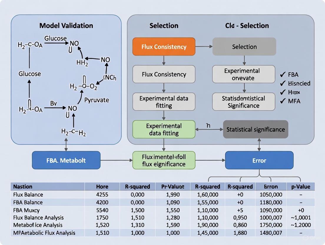
Workflow for Model Validation and Selection
The following diagram illustrates the integrated iterative process of model development, selection, and validation in metabolic flux analysis.
Protocol for Traditional Model Validation
The most common method for validating a 13C-MFA model is the χ2-test of goodness-of-fit [10] [14].
- Model Fitting: A metabolic network model is fitted to the experimental MID data (the "training" or "estimation" data) by varying the flux values to minimize the sum of squared residuals between measured and simulated MIDs [10] [22].
- Goodness-of-Fit Calculation: The weighted sum of squared residuals (SSR) is calculated. If the model's assumptions are correct and measurement errors are accurately known, this statistic follows a χ2-distribution [14].
- Statistical Testing: The calculated SSR is compared to a critical value from the χ2-distribution. The number of degrees of freedom is typically calculated as the number of data points minus the number of independently fitted parameters [14].
- Interpretation: If the SSR is below the critical value (p-value > 0.05), the model is not statistically rejected and is considered valid. If the SSR is too high (p-value < 0.05), the model is rejected, indicating a poor fit to the data [14].
Protocol for Validation-Based Model Selection
The traditional iterative modeling cycle can lead to overfitting, where a model is tailored to the noise in a single dataset [14]. Validation-based model selection offers a more robust alternative.
- Data Splitting: The experimental MID data is split into two independent sets: a training dataset used for model fitting and a validation dataset held back for testing [14].
- Model Fitting and Comparison: Multiple candidate model structures (e.g., with or without a specific reaction like pyruvate carboxylase) are fitted to the training dataset.
- Prediction and Selection: Each fitted model is used to predict the validation dataset. The model that achieves the best predictive performance (i.e., the smallest prediction error on the validation data) is selected as the most appropriate one [14].
- Uncertainty Quantification (Advanced): Prediction uncertainty can be quantified using methods like prediction profile likelihood to ensure the validation data has an appropriate level of novelty compared to the training data [14].
Table 2: Comparison of Selection and Validation Methods
| Method | Key Principle | Advantages | Limitations |
|---|---|---|---|
| χ2-test Validation | Tests if model-predicted MIDs match measured MIDs within expected error. | Well-established; provides a clear statistical criterion for model rejection. | Highly sensitive to accurate knowledge of measurement errors; can lead to model rejection if errors are underestimated [14]. |
| FBA Growth/No-Growth Validation | Tests if model predicts viability on specific substrates. | Computationally simple; useful for testing network completeness. | Qualitative; does not test accuracy of internal flux values [10]. |
| χ2-test based Selection | Iterative model revision until the first model passes the χ2-test. | Simple to implement and understand. | Informal; prone to overfitting; selection depends on the often-uncertain measurement error magnitude [14]. |
| Validation-based Selection | Chooses the model with the best predictive performance on an independent dataset. | Robust to inaccuracies in measurement error estimates; protects against overfitting [14]. | Requires more experimental data to be split into training and validation sets. |
The Scientist's Toolkit: Key Research Reagents and Software
Successful execution of MFA and its associated validation/selection procedures requires a suite of specialized reagents and software tools.
Table 3: Essential Research Reagents and Software for MFA
| Item Name | Type | Function in MFA, Validation, and Selection |
|---|---|---|
| 13C-Labeled Substrates | Research Reagent | Tracer compounds (e.g., [U-13C]-glucose, [1-13C]-glutamine) fed to cells to generate distinctive mass isotopomer distribution (MID) patterns for flux determination [21] [22]. |
| Liquid Chromatography-Mass Spectrometry (LC-MS) | Instrument | Primary technology for measuring the MID and concentration of metabolites extracted from cells. Provides the essential quantitative data for model fitting and validation [21]. |
| 13CFLUX2 | Software | A widely used software package for the design, simulation, and evaluation of 13C labeling experiments for flux calculation under metabolic and isotopic steady-state conditions [21]. |
| INCA | Software | The first software capable of performing Isotopically Non-Stationary MFA (INST-MFA) by simulating transient isotope labeling experiments, useful for systems where achieving isotopic steady state is difficult [21]. |
| COBRA Toolbox | Software | A MATLAB-based toolkit for performing Constraint-Based Reconstruction and Analysis (COBRA), including FBA, Flux Variability Analysis, and basic model quality checks [10]. |
| MEMOTE | Software | A test suite for standardized quality assurance and validation of genome-scale metabolic models, checking for thermodynamic consistency and biomass precursor synthesis capability [10]. |
Model validation and model selection are distinct but deeply interconnected processes that are fundamental to building confidence in metabolic flux predictions. Model validation acts as a final quality check on a single model's performance, while model selection is an upstream process of choosing the most plausible network structure from a set of candidates. The traditional reliance on χ2-testing for both purposes is fraught with difficulty, primarily due to its sensitivity to often-uncertain measurement error estimates [14]. The adoption of validation-based model selection, which leverages independent data to test model predictions, represents a more robust framework that is less susceptible to these errors. As the scale and complexity of metabolic models continue to grow, the rigorous application of these advanced statistical frameworks will be paramount. This will enhance the reliability of flux maps in both fundamental biological research and applied biotechnological contexts, such as the rational design of high-yielding microbial strains for therapeutic protein or metabolite production [10].
Metabolic Flux Analysis (MFA) has emerged as a cornerstone technique in systems biology for quantifying intracellular reaction rates (fluxes) that define the metabolic phenotype of cells [15]. At the heart of most MFA methodologies lies the steady-state assumption, a fundamental prerequisite that enables researchers to solve the mathematically underdetermined systems of metabolic networks. The steady-state assumption encompasses two distinct but often interrelated concepts: metabolic steady state and isotopic stationary state [15] [23]. Under metabolic steady state, all metabolic fluxes and metabolite concentrations remain constant over time, while isotopic stationary state describes the condition where isotope incorporation from labeled substrates has reached equilibrium within intracellular metabolite pools [15]. These assumptions form the bedrock upon which different MFA approaches are built, each with specific requirements and implications for experimental design and computational modeling.
The critical importance of these steady-state assumptions extends across multiple research domains, from metabolic engineering and biotechnology to drug discovery and cancer research [15] [24]. In metabolic engineering, MFA has been instrumental in developing high-producing strains for compounds like lysine, while in pharmacology, it helps identify metabolic vulnerabilities in cancer cells and understand mechanisms of drug action and resistance [10] [24]. The reliability of flux estimates in these applications depends heavily on both the validity of the steady-state assumptions during experimentation and the proper selection of mathematical models that represent the underlying metabolism [10] [8]. This guide systematically compares the primary MFA methodologies based on their steady-state requirements, providing experimental protocols, validation frameworks, and analytical tools essential for researchers working at the intersection of metabolism and drug development.
Comparative Analysis of MFA Methods and Steady-State Requirements
Classification of MFA Techniques Based on Steady-State Assumptions
MFA methodologies can be categorized based on their specific requirements for metabolic and isotopic steady states, which directly influence their experimental timelines, computational complexity, and application domains [15]. The table below summarizes the defining characteristics of the predominant MFA approaches:
Table 1: Classification of Metabolic Flux Analysis Methods by Steady-State Requirements
| Flux Method | Abbreviation | Labeled Tracers | Metabolic Steady State | Isotopic Steady State | Typical Experimental Duration | Computational Complexity |
|---|---|---|---|---|---|---|
| Flux Balance Analysis | FBA | No | Yes | Not Applicable | Not Applicable | Low |
| Metabolic Flux Analysis | MFA | No | Yes | Not Applicable | Hours to Days | Low |
| 13C-Metabolic Flux Analysis | 13C-MFA | Yes | Yes | Yes | Hours to Days (until isotopic steady state) | Medium |
| Isotopic Nonstationary MFA | INST-MFA | Yes | Yes | No | Seconds to Minutes | High |
| Dynamic Metabolic Flux Analysis | DMFA | No | No | Not Applicable | Multiple time intervals | High |
| 13C-Dynamic MFA | 13C-DMFA | Yes | No | No | Multiple time intervals | Very High |
| COMPLETE-MFA | COMPLETE-MFA | Yes | Yes | Yes | Hours to Days | Medium-High |
As illustrated in Table 1, the technical requirements and implementation complexity vary significantly across methods. Traditional 13C-MFA requires both metabolic and isotopic steady states, meaning that cells must be cultivated for sufficient time (typically several hours to a day for mammalian cells) to ensure full incorporation and stabilization of the isotopic label [15]. In contrast, INST-MFA maintains the metabolic steady-state assumption but leverages transient isotopic labeling data collected before the system reaches isotopic stationarity, thereby shortening experimental timelines but increasing computational demands due to the need to solve differential equations rather than algebraic balance equations [15]. The dynamic approaches (DMFA and 13C-DMFA) represent the most complex category, as they forgo both steady-state assumptions and instead divide experiments into multiple time intervals to capture flux transients, resulting in substantial increases in data requirements and computational complexity [15].
Quantitative Comparison of Methodological Capabilities
The choice between MFA methodologies involves trade-offs between resolution, temporal scope, and practical implementation constraints. The following table compares key performance metrics and application considerations:
Table 2: Performance Metrics and Application Considerations for MFA Methods
| Flux Method | Flux Resolution | Temporal Resolution | Network Scale | Data Requirements | Best-Suited Applications |
|---|---|---|---|---|---|
| FBA | Low (Predictive) | None | Genome-Scale | Growth rates, uptake/secretion rates | Genome-scale prediction, constraint-based modeling |
| MFA | Low (Deterministic) | None | Small-Scale (Central metabolism) | Extracellular fluxes | Initial flux estimation, network validation |
| 13C-MFA | High | Single time point | Small-Scale (Central metabolism) | Extracellular fluxes + Isotopic labeling | Most applications in biotechnology and systems biology |
| INST-MFA | High | Multiple early time points | Small-Scale (Central metabolism) | Time-course isotopic labeling | Systems with slow isotopic stationarity, plant metabolism |
| DMFA | Medium | Multiple time intervals | Small-Scale (Central metabolism) | Time-course extracellular fluxes | Dynamic processes, fermentation optimization |
| 13C-DMFA | High | Multiple time intervals | Small-Scale (Central metabolism) | Time-course extracellular fluxes + isotopic labeling | Dynamic flux analysis with pathway resolution |
| COMPLETE-MFA | Very High | Single time point | Small-Scale (Central metabolism) | Extracellular fluxes + multiple tracer labeling | High-precision flux mapping, network validation |
As evidenced in Table 2, 13C-MFA remains the most widely applied method due to its well-established protocols and robust computational frameworks, making it particularly suitable for routine applications in biotechnology and systems biology [15]. However, INST-MFA offers significant advantages for studying systems where reaching isotopic steady state is impractical due to experimental constraints or slow metabolic turnover, as demonstrated in plant metabolism studies where it has been used to quantify photorespiratory fluxes [25]. The emerging COMPLETE-MFA approach, which utilizes multiple singly labeled tracers simultaneously, provides the highest flux resolution and has been used to generate exceptionally precise flux maps for model organisms like E. coli [15] [26].
Experimental Design and Workflows
Generalized Experimental Protocol for Steady-State MFA
The implementation of MFA under steady-state conditions follows a systematic workflow with specific variations depending on the chosen methodology. The following diagram illustrates the core experimental workflow for steady-state MFA approaches:
Diagram 1: Experimental workflow for steady-state 13C-MFA illustrating key stages from sample preparation through computational analysis.
The experimental protocol begins with cell cultivation in an unlabeled medium to establish metabolic steady state, followed by transfer to a medium containing 13C-labeled substrates (tracers) [15]. For 13C-MFA, cells are cultivated until isotopic steady state is reached, which can require several hours to days depending on the biological system [15]. For INST-MFA, samples are collected at multiple early time points (seconds to minutes) during the transient labeling period before isotopic steady state is achieved [15]. The quenching and extraction step rapidly halts metabolic activity and extracts intracellular metabolites, preserving the labeling patterns for subsequent analysis [15] [27]. The analytical phase typically employs mass spectrometry (MS) or nuclear magnetic resonance (NMR) spectroscopy to measure mass isotopomer distributions (MIDs), which represent the fractional abundances of different isotopic isomers of metabolites [15] [14]. Finally, computational modeling uses these MIDs, along with extracellular flux measurements, to estimate intracellular fluxes through fitting procedures that minimize the difference between simulated and experimental labeling patterns [15] [23].
Specialized Protocol: INST-MFA for Plant Metabolism
The application of INST-MFA to plant systems illustrates how methodological adaptations address domain-specific challenges. A recent study investigating the link between photorespiration and one-carbon metabolism in Arabidopsis thaliana employed the following specialized protocol [25]:
Plant Growth and Labeling: Arabidopsis thaliana plants were grown under controlled conditions and exposed to 13CO2 labeling at different O2 concentrations (modulating photorespiration) [25].
Time-Course Sampling: Leaf samples were collected at multiple time points (seconds to minutes) after 13CO2 exposure to capture the transient labeling dynamics before isotopic steady state [25].
Metabolite Extraction and Analysis: Metabolites were extracted using rapid quenching methods and analyzed by LC-MS to determine time-dependent MIDs [25].
Flux Estimation: Computational flux estimation was performed using INST-MFA algorithms that simulate the time-course labeling patterns and optimize fluxes to fit the experimental data [25].
This approach revealed that approximately 5.8% of assimilated carbon passes to one-carbon metabolism under ambient photorespiratory conditions, with serine serving as the primary carbon flux from photorespiration to one-carbon metabolism [25]. The successful application demonstrates how INST-MFA enables flux quantification in systems where achieving isotopic steady state is challenging or where dynamic metabolic processes are of interest.
Model Validation and Selection Frameworks
The Critical Role of Model Selection in MFA
The accuracy of flux estimates in MFA depends critically on selecting an appropriate metabolic network model that correctly represents the underlying biochemistry [10] [8]. Model selection involves choosing which compartments, metabolites, and reactions to include in the metabolic network model used for flux estimation [8] [14]. Traditional approaches to model selection often rely on iterative trial-and-error processes, where models are successively modified and evaluated against the same dataset using goodness-of-fit tests, particularly the χ2-test [8] [14]. However, this practice can lead to overfitting (selecting overly complex models) or underfitting (selecting overly simple models), both of which result in poor flux estimates [8]. The problem is compounded by uncertainties in measurement errors, which can significantly influence model selection outcomes when using χ2-based methods [8] [14].
Validation-Based Model Selection Approach
Recent methodological advances have introduced validation-based model selection as a robust alternative to traditional χ2-testing [8] [14]. This approach addresses key limitations of conventional methods by utilizing independent validation data not used during model fitting. The following diagram illustrates the conceptual framework of validation-based model selection:
Diagram 2: Validation-based model selection framework showing how independent estimation and validation datasets are used to select models with the best predictive performance.
The validation-based approach partitions experimental data into estimation data (Dest), used for model fitting, and validation data (Dval), used exclusively for model evaluation [8]. This partition is typically done by reserving data from distinct experimental conditions or different tracer inputs for validation [8]. For each candidate model, parameters (fluxes) are estimated using Dest, and then the model's predictive performance is evaluated by calculating the sum of squared residuals (SSR) between the model predictions and the independent Dval [8]. The model achieving the smallest SSR with respect to Dval is selected as the most appropriate [8]. Simulation studies have demonstrated that this method consistently selects the correct metabolic network model despite uncertainties in measurement errors, whereas traditional χ2-testing methods show high sensitivity to error magnitude assumptions [8] [14].
Comparative Performance of Model Selection Methods
The table below compares different model selection approaches based on their statistical properties and practical implementation:
Table 3: Comparison of Model Selection Methods for MFA
| Method | Selection Criteria | Robustness to Error Uncertainty | Risk of Overfitting | Implementation Complexity |
|---|---|---|---|---|
| First χ2 | Selects simplest model that passes χ2-test | Low | Low | Low |
| Best χ2 | Selects model passing χ2-test with greatest margin | Low | Medium | Low |
| AIC | Minimizes Akaike Information Criterion | Medium | Medium | Medium |
| BIC | Minimizes Bayesian Information Criterion | Medium | Low | Medium |
| Validation | Minimizes prediction error on independent data | High | Low | High |
As shown in Table 3, validation-based model selection offers superior robustness to uncertainties in measurement errors, which is particularly valuable since estimating true measurement uncertainties can be challenging in practice [8] [14]. The method has been successfully applied in isotope tracing studies on human mammary epithelial cells, where it identified pyruvate carboxylase as a key model component [8]. While validation-based selection requires more experimental data and computational resources, it provides enhanced confidence in flux estimation results and facilitates more reliable biological conclusions [8].
Essential Research Tools and Reagents
Successful implementation of MFA under steady-state conditions requires specialized computational tools, analytical instrumentation, and biochemical reagents. The following table catalogues key solutions essential for conducting MFA studies:
Table 4: Essential Research Reagent Solutions for MFA
| Category | Specific Solution | Function/Application | Examples/Notes |
|---|---|---|---|
| Stable Isotope Tracers | 13C-labeled substrates | Create distinct labeling patterns for flux determination | [1,2-13C]glucose, [U-13C]glucose, 13C-CO2, 13C-NaHCO3 [15] [25] |
| Analytical Instruments | Mass Spectrometry (MS) | Measure mass isotopomer distributions (MIDs) | GC-MS, LC-MS, orbitrap instruments [15] [14] |
| Nuclear Magnetic Resonance (NMR) | Measure isotopic labeling patterns | Provides positional labeling information [15] | |
| Computational Tools | Flux Analysis Software | Perform flux calculations and statistical analysis | OpenFLUX, 13CFLUX2, INCA, METRAN [15] [26] |
| Model Validation Tools | Assess model quality and performance | χ2-test, validation-based selection [8] [14] | |
| Biological Materials | Cell Culture Systems | Maintain metabolic steady state during labeling | Microbial, mammalian, or plant systems [15] [25] |
| Quenching Solutions | Rapidly halt metabolic activity | Cold methanol, other organic solvents [15] [27] |
The selection of appropriate 13C-labeled tracers is particularly critical, as different tracers provide varying levels of information about specific metabolic pathways [15] [23]. For instance, [1,2-13C]glucose and [U-13C]glucose generate distinct labeling patterns that enable resolution of different fluxes in central carbon metabolism [15]. The trend toward parallel labeling experiments (PLEs), where multiple tracers are used simultaneously, has been shown to significantly improve flux precision through complementary information [26]. For computational analysis, open-source software platforms like OpenFLUX2 provide integrated environments for designing labeling experiments, estimating flux parameters, and evaluating flux statistics for both single and parallel labeling experiments [26].
The steady-state assumption, in its metabolic and isotopic forms, remains a foundational element in Metabolic Flux Analysis, enabling the quantification of intracellular reaction rates that would otherwise be mathematically intractable. This comparative analysis demonstrates that methodological selection involves inherent trade-offs between experimental feasibility, computational complexity, and biological resolution. While traditional 13C-MFA with full steady-state assumptions offers robustness and well-established protocols for many applications, INST-MFA provides powerful alternatives for systems where isotopic steady state is difficult to achieve or where dynamic metabolic processes are of interest. The emergence of validation-based model selection approaches represents a significant advancement in statistical rigor, addressing critical limitations of traditional goodness-of-fit testing and enhancing confidence in flux estimation outcomes. As MFA continues to find expanding applications in metabolic engineering, drug development, and systems biology, the thoughtful integration of appropriate steady-state methodologies with robust validation frameworks will remain essential for generating reliable biological insights.
Constraint-Based Reconstruction and Analysis (COBRA) has become an indispensable methodology for simulating, analyzing, and predicting metabolic phenotypes using genome-scale models (GEMs). This approach employs physicochemical, data-driven, and biological constraints to enumerate the set of feasible phenotypic states of a reconstructed biological network [28]. As the field has expanded, with applications ranging from microbial metabolic engineering to modeling human disease states, the need for robust model validation has become increasingly critical. The quality and reliability of GEMs directly impact the accuracy of flux balance analysis (FBA) predictions, which optimize biological objectives such as biomass production to predict metabolic behavior [28] [29].
Within this ecosystem, two key frameworks have emerged: the COBRA Toolbox, a comprehensive software platform for implementing COBRA methods, and MEMOTE, a standardized test suite for assessing GEM quality. While often mentioned together, they serve distinct but complementary roles. The COBRA Toolbox provides the analytical engine for conducting metabolic simulations, while MEMOTE functions as the quality control mechanism that ensures models meet community standards before analysis. This comparison guide examines both frameworks within the context of model validation and selection for metabolic flux analysis research, providing researchers with the information needed to effectively incorporate both tools into their workflows.
The COBRA Toolbox: An Analytical Workhorse
The COBRA Toolbox, established as a MATLAB package, provides researchers with a high-level interface to a vast array of COBRA methods. Version 2.0 of the toolbox significantly expanded computational capabilities to include network gap filling, 13C analysis, metabolic engineering, omics-guided analysis, and visualization tools [28]. The toolbox operates by reading and writing models in Systems Biology Markup Language (SBML) format and requires a linear programming solver such as Gurobi, CPLEX, or GLPK to perform optimizations [28]. The core principle underlying the COBRA approach is the application of constraints to define the feasible solution space of metabolic networks, enabling the prediction of metabolic behaviors under specific conditions.
The COBRA Toolbox supports the entire metabolic modeling workflow, from initial model import and refinement through simulation and results interpretation. Its functions can be categorized into several key areas: (1) flux balance analysis and variant techniques including geometric FBA and loop law applications; (2) fluxomics integration for 13C data fitting and flux estimation; (3) gap filling algorithms to identify and resolve network incompleteness; (4) metabolic engineering functions like optKnock and optGene for strain design; and (5) sampling methods for exploring solution spaces [28]. This comprehensive suite of tools has made the COBRA Toolbox a fundamental resource in systems biology, enabling both novice and experienced researchers to implement sophisticated constraint-based modeling techniques.
MEMOTE: The Quality Control Standard
MEMOTE (METabolic MOdel TEst suite) represents a community-driven effort to establish standardized quality assessment for genome-scale metabolic models. This open-source software contains a community-maintained, standardized set of tests that address aspects ranging from basic annotations to conceptual integrity [30]. MEMOTE's primary function is to generate informative reports detailing model quality in a visually accessible format, facilitating model development and error detection through continuous testing integration [31] [30].
The framework is designed to run four types of assessments: (1) snapshot reports for benchmarking individual models; (2) diff reports for comparing multiple models; (3) history reports for tracking model evolution across version-controlled histories; and (4) error reports for identifying SBML validation issues [32]. MEMOTE's test suite is divided into two main sections: an "independent" section containing tests agnostic to organism type and modeling paradigms, and a "specific" section with tests tailored to particular model characteristics [32]. The independent section focuses on fundamental principles of constraint-based modeling including mass, charge, and stoichiometric balance, while the specific section addresses model properties like biomass composition and reaction counts that cannot be normalized without introducing bias [32].
Table 1: Core Functional Comparison Between MEMOTE and COBRA Toolbox
| Feature | MEMOTE | COBRA Toolbox |
|---|---|---|
| Primary Function | Model quality assessment and validation | Metabolic network simulation and analysis |
| Testing Approach | Automated test suite with scoring | Algorithmic implementation with optimization |
| Core Metrics | Annotation completeness, stoichiometric consistency, mass/charge balance | Growth rates, flux distributions, phenotypic phase planes |
| Output Format | Comprehensive report with weighted scores | Numerical results, flux maps, simulation data |
| Model Requirements | SBML format | COBRA-compliant SBML format |
| Integration Capabilities | GitHub Actions, Travis CI | MATLAB, Python (via COBRApy), various solvers |
Methodologies and Experimental Protocols
MEMOTE Quality Assessment Protocol
The MEMOTE assessment protocol begins with ensuring model files are properly formatted in valid SBML. When initiated, MEMOTE first checks SBML compliance, generating an error report if validation fails [32]. For compliant models, the framework executes a battery of tests categorized into fundamental checks and organism-specific assessments. The fundamental tests in the independent section evaluate annotation completeness, stoichiometric consistency, mass and charge balance, and metabolic functionality [32] [30]. These tests produce weighted scores that contribute to an overall model quality percentage, calculated as the weighted sum of all individual test results normalized by the maximally achievable score [32].
The scoring system employs a color-coded gradient from red to green to indicate performance levels, with detailed explanations available for each test metric [32]. For model comparisons, the diff report calculates the ratio of sample minimum to maximum values, with results appearing red when the minimum is very small relative to the maximum [32]. This standardized approach allows researchers to quickly identify model deficiencies and track improvement over successive iterations. MEMOTE also accounts for different modeling paradigms, including the distinction between "reconstructions" (unconstrained metabolic knowledgebases) and "models" (parameterized networks ready for FBA), though tests in the specific section may fail for reconstructions that lack necessary constraints [32].
COBRA Toolbox Flux Analysis Protocol
The COBRA Toolbox employs a multi-step protocol for metabolic flux analysis, beginning with model acquisition and validation. Researchers first import a COBRA-compliant SBML model, ensuring it includes essential information: stoichiometry of each reaction, upper and lower bounds for reactions, and objective function coefficients [28]. The model undergoes preliminary checks for consistency before proceeding to simulation. For basic flux balance analysis, the protocol involves: (1) defining environmental conditions by setting exchange reaction bounds; (2) selecting an objective function (typically biomass production); (3) applying additional constraints as needed; and (4) solving the linear programming problem to obtain an optimal flux distribution [28].
The toolbox supports multiple FBA variants, including parsimonious FBA (pFBA), which minimizes total flux while maintaining optimal objective value, thereby reducing enzyme production costs [29]. For community modeling, approaches include: (1) group-level optimization using a community objective function; (2) independent optimization of each species' growth; and (3) abundance-adjusted optimization incorporating experimental measurements [29]. Tools like MICOM implement a "cooperative trade-off" approach that incorporates a trade-off between optimal community growth and individual growth rate maximization using quadratic regularization [29]. The COBRA Toolbox also includes functions for gap filling, which identifies dead-end metabolites and missing reactions, and growthExpMatch, which reconciles model predictions with experimental growth data [28].
Table 2: Experimental Outcomes for FBA-Based Predictions Using Different Quality Models
| Model Quality | Growth Rate Prediction Accuracy | Interaction Strength Correlation | Recommended Use Cases |
|---|---|---|---|
| Curated GEMs | High accuracy in defined media | Strong correlation with experimental data | Hypothesis testing, quantitative predictions |
| Semi-Curated GEMs (AGORA) | Moderate accuracy | Weak correlation with experimental data | Draft analysis, qualitative insights |
| Automatically Generated GEMs | Low accuracy | No significant correlation | Exploratory research only |
Performance Comparison and Experimental Data
Model Quality Impact on Prediction Accuracy
Recent systematic evaluations have quantified the critical relationship between model quality and prediction accuracy. A 2024 study assessed the performance of FBA-based methods for predicting microbial interactions using both curated and semi-curated GEMs [29]. The research collected 26 GEMs from the semi-curated AGORA database alongside four manually curated models, comparing predicted growth rates against experimentally determined values from literature. The results demonstrated that except for curated GEMs, predicted growth rates and their ratios (interaction strengths) did not correlate with experimentally obtained data [29]. This finding underscores the essential role of quality control measures like those implemented in MEMOTE for ensuring reliable computational predictions.
The study evaluated three tools—COMETS, Microbiome Modeling Toolbox, and MICOM—across different media conditions and parameter settings [29]. The tools employed distinct approaches: MICOM uses abundance-weighted community modeling, COMETS implements dynamic FBA with spatial considerations, and the Microbiome Modeling Toolbox enables pairwise interaction screening [29]. Despite these methodological differences, all tools showed similar dependencies on model quality, with semi-curated models from repositories like AGORA producing unreliable interaction predictions. This evidence strongly suggests that quality assessment should precede computational analysis, positioning MEMOTE as an essential first step in any metabolic modeling workflow.
MEMOTE Benchmarking Results
MEMOTE's evaluation system provides quantitative metrics for model quality assessment. The framework generates a comprehensive report with scores across multiple categories, allowing researchers to identify specific model deficiencies. The snapshot report presents results as a percentage score, with color coding from red (low performance) to green (high performance) [32]. This standardized scoring enables direct comparison between models and tracking of quality improvements during the development process.
The history report feature is particularly valuable for model development, as it visualizes how key metrics evolve across a version-controlled history [32]. By clicking on legend entries, researchers can toggle visibility of different branches in the development timeline, facilitating comparison of modeling approaches. MEMOTE's tests are specifically designed to identify common issues in GEMs, including dead-end metabolites, mass and charge imbalances, incomplete annotations, and stoichiometric inconsistencies [30]. The framework also assesses biochemical consistency by verifying that reactions are elementally balanced and that the model does not contain energy-generating cycles that violate thermodynamic principles [30].
Research Reagent Solutions and Essential Materials
Table 3: Essential Research Reagents and Computational Tools for Metabolic Flux Analysis
| Item | Function/Purpose | Implementation Considerations |
|---|---|---|
| COBRA Toolbox | MATLAB package for constraint-based reconstruction and analysis | Requires MATLAB 7.0+, libSBML 4.0.1+, SBMLToolbox 3.1.1+, and an LP solver [28] |
| MEMOTE | Quality test suite for genome-scale metabolic models | Open-source Python tool; integrates with GitHub and Travis CI for continuous testing [31] [30] |
| SBML Models | Standardized format for representing metabolic models | Must be COBRA-compliant with reaction bounds, objective coefficients, and gene-reaction associations [28] |
| Linear Programming Solvers | Solve optimization problems in FBA | Gurobi, CPLEX, or GLPK; GLPK has limitations for OptKnock or GDLS algorithms [28] |
| BiGG Knowledgebase | Resource for curated metabolic models | Provides COBRA-compliant SBML models with standardized identifiers [28] |
| MetaNetX | Resource for accessing and analyzing metabolic networks | Alternative platform for model reconciliation and comparison [30] |
Integrated Workflow for Model Validation and Analysis
For researchers engaged in metabolic flux analysis, integrating MEMOTE and the COBRA Toolbox creates a robust workflow for model development, validation, and simulation. The recommended sequence begins with model acquisition from sources like BiGG or ModelSEED, followed by quality assessment using MEMOTE to identify deficiencies. Based on MEMOTE's report, researchers can undertake model refinement to address identified issues, then revalidate until satisfactory scores are achieved. The quality-verified model can then proceed to computational analysis using appropriate COBRA Toolbox functions, with results validated against experimental data where possible.
This integrated approach addresses the fundamental challenge identified in recent evaluations: that prediction accuracy depends heavily on model quality [29]. MEMOTE's standardized assessment provides the quality assurance needed to have confidence in COBRA Toolbox simulations, particularly for applications in metabolic engineering and drug development where reliable predictions are essential. The workflow supports both single-species and community modeling applications, with MEMOTE ensuring each component model meets quality standards before incorporation into larger community simulations.
The following diagram illustrates the integrated validation and analysis workflow:
The complementary roles of MEMOTE and the COBRA Toolbox create a comprehensive framework for metabolic model validation and analysis. MEMOTE provides the essential quality control mechanisms through standardized testing and reproducible reporting, while the COBRA Toolbox delivers the analytical capabilities for metabolic simulation and prediction. Recent experimental evidence confirms that model quality directly impacts prediction accuracy, with curated models outperforming semi-curated alternatives in growth rate and microbial interaction prediction [29].
For researchers in metabolic flux analysis, adopting an integrated workflow that begins with MEMOTE assessment followed by COBRA Toolbox analysis represents a best practices approach. This methodology ensures that computational predictions rest on a foundation of model quality, increasing reliability for critical applications in drug development and metabolic engineering. As the field continues to advance, these tools provide the necessary infrastructure for building, validating, and utilizing high-quality metabolic models that can faithfully represent biological systems and generate testable hypotheses.
Established and Emerging Methodologies for Model Testing and Selection
Article Contents
- Introduction to the χ2-Test
- Theoretical Foundations and Calculation
- Application in Metabolic Flux Analysis
- Comparative Evaluation with Alternative Methods
- Experimental Protocols for Model Validation
- Essential Research Reagent Solutions
The Chi-square (χ2) goodness-of-fit test is a foundational statistical hypothesis test used to determine whether an observed frequency distribution of a categorical variable significantly deviates from a theoretical or expected distribution. Invented by Karl Pearson in 1900 and later refined by Ronald Fisher, this test serves as a critical tool for evaluating how well a statistical model fits a set of observations [33]. In the realm of metabolic research, particularly in 13C-Metabolic Flux Analysis (13C-MFA), the χ2-test provides a quantitative measure to validate whether the isotopic labeling data measured in experiments aligns with the fluxes predicted by a metabolic network model [1] [34].
The test operates on a straightforward principle: it compares observed values against expected values, with the null hypothesis (H0) stating that the observed data follows the specified theoretical distribution. In metabolic modeling, this translates to testing whether the measured data is consistent with the proposed metabolic model. The alternative hypothesis (Ha), conversely, suggests that the observed data does not follow the specified distribution, indicating a potential flaw in the model structure or assumptions [35] [36]. For researchers in biotechnology and pharmaceutical development, this test is indispensable for model validation and selection, helping to ensure that metabolic models used for predicting cellular behavior in drug treatment or bioproduction are statistically sound [1].
Theoretical Foundations and Calculation
The core of the χ2 goodness-of-fit test lies in its test statistic, which quantifies the discrepancy between observed (O) and expected (E) frequencies. The formula for the Pearson's chi-square test statistic is [35]:
$$ \chi^2 = \sum \frac{(Oi - Ei)^2}{E_i} $$
Where:
- Σ is the summation operator.
- O_i is the observed frequency for category i.
- Ei is the expected frequency for category *i*, calculated as *Ei = N · Pi*, where *N* is the total number of observations and *Pi* is the probability of category i under the null hypothesis.
The calculation involves a step-by-step process of creating a table of observed and expected frequencies, computing the differences, squaring them, and then summing the normalized squared differences [35] [36]. The following diagram illustrates this workflow:
To draw a meaningful conclusion from the test statistic, it must be compared against a critical value from the Chi-square distribution [35] [36]. This critical value depends on:
- The significance level (α), conventionally set at 0.05 (5%).
- The degrees of freedom (df), which for the goodness-of-fit test is the number of categories minus one.
If the χ2 test statistic exceeds the critical value, the null hypothesis is rejected, indicating a statistically significant difference between the observed and expected distributions. If the χ2 value is less than the critical value, there is not enough evidence to reject the null hypothesis, and the model is considered a statistically acceptable fit [35] [36].
Application in Metabolic Flux Analysis
In 13C-Metabolic Flux Analysis (13C-MFA), the χ2-test of goodness-of-fit plays a pivotal role in model validation. 13C-MFA is a powerful technique used to quantify the flow of metabolites through biochemical networks in vivo, providing insights into cellular metabolism that are critical for both basic biology and metabolic engineering [1] [34]. The method relies on feeding cells with 13C-labeled substrates (e.g., glucose or glutamine) and measuring the resulting isotopic labeling patterns in intracellular metabolites using techniques like gas chromatography-mass spectrometry (GC-MS) [34].
The core application of the χ2-test in this context is to validate the fit between the experimentally measured labeling data and the labeling patterns simulated by the metabolic model. A good fit suggests that the model's predicted flux map accurately represents the intracellular physiology [1] [34]. The test is formally integrated into the workflow as part of the statistical analysis step. After model parameters (fluxes) are estimated by minimizing the difference between simulated and measured data, the goodness-of-fit is assessed. The test statistic used is often a variance-weighted sum of squared residuals (SSR), which follows a χ2 distribution [34]. If the SSR falls within the expected range for the χ2 distribution (given the degrees of freedom), the model provides an acceptable fit to the experimental data [1] [34].
Table 1: Key Parameters for the χ2-Test in 13C-MFA Validation
| Parameter | Role in 13C-MFA Model Validation | Typical Interpretation |
|---|---|---|
| Sum of Squared Residuals (SSR) | Quantifies the total discrepancy between measured and simulated isotopic labeling data. | A lower SSR indicates a better fit. |
| Degrees of Freedom (df) | Calculated as the number of independent labeling measurements minus the number of fitted metabolic fluxes. | Determines the expected range of the SSR under the null hypothesis. |
| χ² Critical Value | The threshold value from the χ2 distribution for a given significance level (α, usually 0.05) and degrees of freedom. | If SSR < critical value, the model is an acceptable fit (p > α). |
| p-value | The probability of observing the obtained SSR (or a larger one) if the model is correct. | p < 0.05 suggests the model is not a good fit to the data. |
Comparative Evaluation with Alternative Methods
While the χ2-test is the most widely used quantitative validation method in 13C-MFA, it is not the only tool available. Researchers must understand its performance relative to other goodness-of-fit tests and validation approaches to select the most appropriate method for their specific context [35] [1].
The primary advantage of the χ2-test is its simplicity and strong theoretical foundation. It provides a clear, objective pass/fail criterion for model validity based on a well-understood probability distribution. However, a significant limitation is its reliance on accurate knowledge of measurement errors. An underestimation of these errors can lead to an inflated SSR and the incorrect rejection of a valid model (Type I error) [1]. Furthermore, the test can be sensitive to sample size and may lack power to detect specific types of misfits.
For continuous data, such as raw metabolite concentrations, the Anderson-Darling and Kolmogorov-Smirnov goodness-of-fit tests are more appropriate alternatives, as they do not require data to be binned into categories [35]. In complex metabolic modeling, complementary validation methods are often necessary. These include [1]:
- Flux Confidence Interval Analysis: Using statistical methods like Monte Carlo sampling to determine the precision of estimated fluxes.
- Cross-Validation: Assessing model performance on data not used for parameter estimation to test its predictive power and guard against overfitting [37].
- Comparison with Flux Balance Analysis (FBA): Using 13C-MFA validated fluxes as a benchmark to test and refine the predictions from constraint-based FBA models [1].
Table 2: Comparison of Goodness-of-Fit Tests for Model Validation
| Test Method | Data Type | Key Strengths | Key Limitations |
|---|---|---|---|
| χ2-Test of Goodness-of-Fit | Categorical (or binned continuous) | Simple, widely understood, provides a clear statistical criterion. | Sensitive to sample size, requires accurate measurement error estimates. |
| Anderson-Darling Test | Continuous | More powerful than KS test, sensitive to tail differences. | Less commonly used in metabolic flux software. |
| Kolmogorov-Smirnov Test | Continuous | Non-parametric, insensitive to distribution assumptions. | Less sensitive to differences near the ends of the distribution. |
| Cross-Validation | Any | Directly tests predictive power, helps prevent overfitting. | Computationally intensive, requires large datasets. |
Experimental Protocols for Model Validation
Implementing the χ2-test for model validation in metabolic flux studies requires a rigorous experimental and computational workflow. The following protocol, synthesizing best practices from the literature, outlines the key steps for conducting 13C-MFA with statistical validation [34].
Tracer Experiment Design and Execution
The foundation of a successful 13C-MFA is a well-designed labeling experiment. Parallel labeling experiments, using multiple tracers simultaneously, have been shown to provide superior flux resolution compared to single-tracer studies [34].
- Tracer Selection: Choose complementary 13C-labeled substrates. For microbial systems on glucose, a combination of [1,2-13C]glucose and [1,6-13C]glucose is often effective. For mammalian cell cultures, [U-13C]glutamine is commonly used alongside labeled glucose [34] [38].
- Cell Cultivation: Grow cells in a controlled bioreactor under metabolic steady-state conditions. For continuous cultures, ensure at least five volume turnovers before sampling. For batch cultures, harvest cells during the mid-exponential growth phase to avoid metabolic changes associated with substrate depletion [34].
- Sampling and Quenching: Rapidly collect and quench cells to instantly halt metabolic activity, preserving the isotopic labeling state.
Metabolite Labeling and Flux Analysis
Once samples are collected, the process of measuring labeling and calculating fluxes begins.
- Mass Spectrometry Analysis: Derivatize and analyze proteinogenic amino acids, glycogen-bound glucose, or other metabolites using GC-MS. This instrument measures the mass isotopomer distribution (MID) of the fragments, which serves as the primary data for flux estimation [34].
- Metabolic Network Construction: Build a stoichiometric model of the central carbon metabolism (glycolysis, TCA cycle, pentose phosphate pathway) including atom transition mappings for each reaction [34].
- Flux Estimation: Use specialized software platforms like Metran or INCA to find the set of metabolic fluxes that minimizes the difference between the simulated MIDs and the experimentally measured MIDs. This is typically done via iterative least-squares optimization [34].
The overall workflow, from experiment to validated flux map, is depicted below:
Statistical Validation and Model Selection
This is the stage where the χ2-test is formally applied.
- Calculate Goodness-of-Fit: The software outputs the SSR. Under the null hypothesis that the model is correct and measurement errors are accurately estimated, the SSR follows a χ2 distribution with degrees of freedom equal to the number of independent measurements minus the number of estimated parameters [34].
- Perform χ2-Test: Compare the SSR to the critical value of the χ2 distribution. A statistically acceptable fit is achieved if the SSR is less than the critical value (e.g., for a 95% confidence level) [1] [34].
- Model Selection (if needed): If multiple model architectures (e.g., with alternative pathways) are plausible, the χ2-test can be used as a selection criterion. The model with the best (lowest) goodness-of-fit statistic that also passes the validation test should be selected [1].
Essential Research Reagent Solutions
The experimental protocols underpinning the validation of metabolic models rely on a specific set of reagents and computational tools. The following table details key materials essential for conducting 13C-MFA and performing the associated χ2 goodness-of-fit validation [34] [38].
Table 3: Key Research Reagent and Tool Solutions for 13C-MFA
| Item Name | Function/Application | Specific Examples / Notes |
|---|---|---|
| 13C-Labeled Tracers | Serve as the metabolic probes to trace flux through pathways. | [1,2-13C]glucose, [U-13C]glucose, [U-13C]glutamine. Choice of tracer is critical for flux resolution [34] [38]. |
| Gas Chromatography-Mass Spectrometry (GC-MS) | The analytical workhorse for measuring the mass isotopomer distribution (MID) of metabolites. | Used for high-throughput analysis of proteinogenic amino acids and other metabolites [34]. |
| Metabolic Modeling Software | Platforms used to simulate isotopic labeling and estimate metabolic fluxes from experimental MID data. | Metran, INCA. These tools perform the non-linear optimization and calculate the SSR for the χ2-test [34]. |
| Chi-Square Critical Value Table | Reference for determining the statistical significance of the goodness-of-fit test. | Integrated into modeling software or available as statistical libraries in R or Python (e.g., chisq.test() in R) [35]. |
| Stoichiometric Database | Source for building and curating the metabolic network model used in simulations. | KEGG, BioCyc. Provide reaction lists and, crucially, atom transition mappings for 13C-MFA [34]. |
Model validation and selection are fundamental to ensuring the accuracy and reliability of metabolic flux analysis (MFA) and flux balance analysis (FBA). For decades, the χ2-test of goodness-of-fit has served as the cornerstone for these statistical evaluations in 13C-MFA. However, growing evidence reveals critical limitations in relying solely on this method, particularly when dealing with complex metabolic models, imperfect measurement error estimates, and iterative model development processes. This guide examines the specific scenarios where χ2-tests fall short, compares emerging alternative validation frameworks using structured quantitative data, and provides detailed experimental protocols for implementing more robust model selection procedures. The insights are particularly relevant for researchers and scientists in metabolic engineering and drug development who rely on precise flux estimations.
Metabolic flux analysis, particularly 13C-MFA, has become an indispensable tool for quantifying intracellular reaction rates in living cells [39] [15]. Both 13C-MFA and FBA employ constraint-based modeling frameworks that assume metabolic steady state, where reaction rates and metabolic intermediate levels remain invariant [39] [10]. These methods provide estimated (MFA) or predicted (FBA) values of in vivo fluxes that cannot be measured directly, offering critical insights for basic biology and metabolic engineering strategies [40].
The process of 13C-MFA typically involves feeding cells with 13C-labeled substrates, measuring the resulting mass isotopomer distributions (MIDs) of metabolites using mass spectrometry or NMR techniques, and then inferring fluxes by fitting a mathematical model to the observed MID data [14]. The χ2-test has emerged as the most widely used quantitative validation and selection approach in 13C-MFA, primarily testing whether the residuals between measured and estimated MID values are consistent with the assumed measurement error [39].
Despite advances in other areas of statistical evaluation for metabolic models, such as flux uncertainty quantification, validation, and model selection methods have remained underappreciated and underexplored until recently [39] [10]. This gap is particularly concerning as model complexity increases and as flux analysis finds broader applications in biotechnology and medical research, including understanding cancer metabolism, metabolic syndrome, and neurodegenerative diseases [14].
Critical Limitations of χ2-Tests in Metabolic Flux Analysis
Dependency on Accurate Measurement Error Estimation
The statistical validity of the χ2-test hinges on accurate knowledge of measurement errors, which is often difficult to obtain in practice [14]. For mass spectrometry data, measurement errors are typically estimated from biological replicates, often yielding very low values (sometimes as low as 0.001). However, these estimates may not reflect all error sources, including instrumental bias or deviations from metabolic steady-state in batch cultures [14].
Table 1: Sources of Error Misestimation in χ2-Tests for MFA
| Error Source | Impact on χ2-Test | Practical Consequence |
|---|---|---|
| Instrument bias in mass spectrometers | Underestimated measurement error | Overly sensitive test, rejecting valid models |
| Deviation from steady-state assumption | Unaccounted systematic error | Inflated χ2 values, leading to model rejection |
| Low biological replication | Poor error estimation | Uncertain test reliability |
| Non-normal distribution of MIDs | Violation of test assumptions | Incorrect p-value calculation |
When errors are underestimated, researchers face two problematic choices: arbitrarily increasing error estimates to pass the χ2-test or introducing additional fluxes into the model [14]. The former approach may lead to high uncertainty in estimated fluxes, while the latter increases model complexity and can lead to overfitting.
Challenges with Model Complexity and Overfitting
The iterative nature of MFA model development, where models are repeatedly modified and fitted to the same dataset until they pass the χ2-test, creates inherent risks for overfitting [14]. This process often occurs informally during modeling, based on the same data used for model fitting, without proper documentation of the underlying procedure [14].
The χ2-test primarily detects "gross measurement error" but does not adequately assess the overall quality of fit or identify when a model is overly complex [41]. Errors may be unreasonably large while remaining normally distributed, providing a false sense of validity. As metabolic models grow increasingly complex, often generated from genome-level data, robust validation that can directly assess model fit becomes essential [41].
Limited Assessment of Model Predictive Power
A fundamental limitation of the χ2-test is its focus on how well a model fits the estimation data rather than its ability to predict new, independent data [14]. This limitation becomes particularly problematic when comparing multiple model structures that all pass the χ2-test, as there is no statistical guidance for selecting the model with the greatest predictive power [14].
The test also provides limited information about which specific aspects of the model may be problematic, offering instead only a global goodness-of-fit measure. This lack of granularity makes it difficult to identify particular reactions or pathways that contribute to poor model performance [41].
Quantitative Comparison of Validation Approaches
Several alternative validation approaches have emerged to address the limitations of traditional χ2-tests. The table below provides a structured comparison of these methods based on key performance metrics.
Table 2: Comprehensive Comparison of Model Validation Methods for MFA
| Validation Method | Key Principle | Advantages | Limitations | Optimal Use Case |
|---|---|---|---|---|
| χ2-test of goodness-of-fit | Tests if residuals match expected measurement error | Widely adopted, computationally simple | Sensitive to error mis-specification, limited predictive assessment | Initial model screening with well-characterized errors |
| Validation-based model selection | Uses independent data to test model predictions | Robust to error mis-specification, prevents overfitting | Requires additional experimental data | Final model selection when resources permit parallel labeling |
| Generalized Least Squares (GLS) with t-tests | Framed as regression problem with parameter significance tests | Identifies non-significant fluxes, detects lack of model fit | Limited to traditional MFA formulations | Identifying problematic fluxes in core metabolic networks |
| Combined framework with pool size | Incorporates metabolite pool size information with labeling data | Improved precision of flux estimates | Increased sensitivity to unmodeled reactions | INST-MFA with reliable pool size measurements |
Recent studies demonstrate that validation-based model selection consistently chooses the correct model in simulation studies where the true model is known, performing particularly well when measurement uncertainties are difficult to estimate precisely [14]. In contrast, χ2-test performance varies significantly with the believed measurement uncertainty, leading to different model structures being selected depending on error assumptions [14].
Experimental Protocols for Robust Model Validation
Protocol for Validation-Based Model Selection
The validation-based approach addresses fundamental limitations of χ2-tests by using independent data for model selection [14].
Step 1: Experimental Design for Parallel Labeling
- Grow cells in multiple parallel cultures with different 13C-labeled tracers (e.g., [1,2-13C]glucose, [U-13C]glucose)
- Ensure biological and technical replicates for error estimation
- Quench metabolism rapidly to preserve in vivo labeling patterns
- Extract intracellular metabolites and measure MIDs via GC-MS
Step 2: Data Partitioning
- Divide labeling data into estimation and validation sets
- Use one tracer type for estimation and another for validation
- Ensure validation data is independent of estimation data
Step 3: Model Fitting and Selection
- Fit candidate models to estimation data
- Evaluate each model's predictive power on validation data
- Select the model that best predicts the validation data
- Quantify prediction uncertainty using methods like prediction profile likelihood
Step 4: Flux Uncertainty Analysis
- Calculate confidence intervals for estimated fluxes
- Perform comprehensive statistical analysis to determine goodness of fit
- Validate key findings with independent experimental techniques
This protocol typically requires 4 days to complete and quantifies metabolic fluxes with a standard deviation of ≤2%, representing a substantial improvement over traditional implementations [42].
Protocol for Generalized Least Squares Approach
The GLS approach reframes MFA as a regression problem, enabling the use of t-tests for model validation [41].
Step 1: Stoichiometric Model Formulation
- Construct stoichiometric matrix (S) representing metabolic reactions
- Separate into calculated (Sc) and observed (So) components
- Ensure S_c has no more columns than rows for unique flux solution
Step 2: Flux Calculation
- Solve the equation -Sovo = Scvc + ε using generalized least squares
- Account for covariance structure in residuals using variance-covariance matrix
Step 3: Model Validation via t-tests
- Perform t-tests for each calculated flux to determine significance
- Compare real data significance with perfect model simulations
- Identify fluxes that cannot be distinguished from zero given measurement uncertainty
Step 4: Error Decomposition
- Differentiate between measurement error and model error
- Identify specific reactions where model fit is inadequate
- Guide model refinement based on statistical significance
This approach goes beyond traditional detection of "gross measurement error" to identify lack of fit between model and data [41].
Figure 1: Workflow for validation-based model selection in metabolic flux analysis, highlighting the iterative process of model testing against independent validation data.
The Scientist's Toolkit: Essential Research Reagents and Solutions
Implementing robust model validation requires specific experimental and computational tools. The table below details key reagents and solutions essential for advanced metabolic flux analysis.
Table 3: Essential Research Reagents and Computational Tools for MFA
| Category | Specific Item | Function/Application | Key Considerations |
|---|---|---|---|
| Isotopic Tracers | [1,2-13C]glucose, [U-13C]glucose | Enables tracing of carbon atoms through metabolic networks | Purity critical for accurate interpretation |
| Analytical Instruments | GC-MS systems, NMR spectroscopy | Measures mass isotopomer distributions (MIDs) | Sensitivity and precision affect error estimates |
| Software Platforms | Metran, INCA, OpenFLUX | Performs flux estimation and statistical analysis | Algorithm implementation affects confidence intervals |
| Cell Culture Components | Defined media, serum alternatives | Maintains metabolic steady-state | Composition affects extracellular flux measurements |
| Metabolite Extraction Reagents | Methanol:water:chloroform | Quenches metabolism and extracts intracellular metabolites | Rapid quenching essential for accuracy |
The limitations of χ2-tests in metabolic flux analysis represent a significant challenge for the field, particularly as models increase in complexity and find broader applications in biotechnology and human health. Traditional approaches fall short primarily due to their dependency on accurate measurement error estimation, vulnerability to overfitting in iterative model development, and limited assessment of predictive power.
Emerging validation methods, particularly those leveraging independent validation datasets and sophisticated statistical frameworks, offer promising alternatives that address these fundamental limitations. The experimental protocols outlined here provide practical pathways for implementing these more robust approaches, with the validation-based method showing particular resilience to error mis-specification.
As the field progresses, future developments will likely focus on integrating multiple forms of validation, developing standardized benchmarks for model performance, and creating more sophisticated computational tools that make advanced validation techniques accessible to a broader research community. Adopting these robust validation and selection procedures will enhance confidence in constraint-based modeling as a whole and facilitate more widespread and reliable application of metabolic flux analysis across biological research and metabolic engineering.
Model selection is a critical step in metabolic flux analysis (MFA) that directly impacts the accuracy and reliability of estimated intracellular fluxes. Traditional model selection methods often rely on informal trial-and-error approaches or goodness-of-fit tests applied to the same data used for parameter estimation, potentially leading to overfitting or underfitting. This review introduces validation-based model selection as a robust framework utilizing independent datasets, demonstrating superior performance compared to conventional methods. We present comprehensive comparative analysis of model selection techniques, detailed experimental protocols for implementation, and evidence from simulation studies and real-world applications showing that validation-based approaches consistently identify correct model structures while remaining robust to uncertainties in measurement error estimates. The framework's implementation in studying human mammary epithelial cells successfully identified pyruvate carboxylase as a key model component, underscoring its practical utility in metabolic research and drug development.
Model-based metabolic flux analysis represents the gold standard for measuring metabolic fluxes in living cells and tissues, with significant implications for understanding cancer metabolism, neurodegenerative diseases, and metabolic syndrome [8]. In 13C-MFA, cells are fed 13C-labeled substrates, and the resulting mass isotopomer distributions (MIDs) of metabolites are measured using techniques such as gas chromatography–mass spectrometry (GC–MS). Fluxes are then inferred by fitting a mathematical model of the metabolic network to the observed MID data [42]. A pivotal yet often overlooked aspect of this process is model selection—determining which compartments, metabolites, and reactions to include in the metabolic network model [8].
Traditional MFA model selection is frequently conducted informally during iterative modeling processes, using the same data for both model fitting and selection decisions [8]. This practice can introduce statistical biases toward either overly complex models (overfitting) or excessively simple ones (underfitting), ultimately compromising flux estimation accuracy. The limitations of conventional methods become particularly evident when dealing with imperfect error models and difficulties in determining the number of identifiable parameters in nonlinear models [8]. Validation-based model selection addresses these challenges by employing independent validation data, providing a more rigorous framework for developing biologically accurate metabolic models.
Comparative Analysis of Model Selection Methods
Traditional Model Selection Approaches
Model selection in MFA has predominantly relied on methods utilizing the same dataset for both parameter estimation and model evaluation. These approaches, while computationally straightforward, present significant statistical limitations [8]. The χ2-test-based methods are most common, where models are iteratively modified until they pass a statistical goodness-of-fit test. The "First χ2" method selects the simplest model that passes the χ2-test, while the "Best χ2" method chooses the model passing with the greatest margin [8]. Both approaches heavily depend on accurate knowledge of measurement uncertainties, which are frequently underestimated in mass spectrometry data due to unaccounted technical biases and deviations from steady-state assumptions [8].
Information-theoretic methods like the Akaike Information Criterion (AIC) and Bayesian Information Criterion (BIC) offer alternative approaches by balancing model complexity with goodness of fit [8]. These methods penalize model complexity to different extents but still utilize the same data for both fitting and selection. The "Sum of Squared Residuals (SSR)" method serves as a baseline approach, simply selecting the model with the lowest weighted residuals without considering complexity [8]. A fundamental limitation shared by all traditional methods is their vulnerability to overfitting when measurement error estimates are inaccurate, a common scenario in practical MFA applications.
Validation-Based Model Selection Framework
The validation-based approach introduces a paradigm shift by partitioning experimental data into distinct estimation and validation sets [8]. Model parameters are estimated exclusively using the estimation data (Dest), while model selection is based on predictive performance for the independent validation data (Dval). This method explicitly tests a model's ability to generalize to new data, providing a direct safeguard against overfitting [8]. For 13C-MFA, effective validation typically utilizes data from distinct tracer experiments, ensuring the validation data provides qualitatively new information not contained in the estimation data [8].
A critical advancement within this framework is the quantification of prediction uncertainty using prediction profile likelihood, which helps identify when validation data is either too similar or too dissimilar to the estimation data [8]. This addresses concerns about validation effectiveness and ensures robust model comparison. The selection criterion is straightforward: among candidate models M1, M2,..., Mk, the one achieving the smallest SSR with respect to Dval is selected [8]. This approach bypasses the need for accurate prior knowledge of measurement uncertainties and eliminates dependence on correctly determining the number of identifiable parameters, both significant challenges in traditional methods.
Performance Comparison Across Methods
Table 1: Comparative Performance of Model Selection Methods in MFA
| Method | Selection Criteria | Dependence on Error Estimates | Robustness to Overfitting | Implementation Complexity |
|---|---|---|---|---|
| Estimation SSR | Lowest SSR on estimation data | None | Poor | Low |
| First χ2 | First model passing χ2-test | High | Moderate | Medium |
| Best χ2 | Model with largest χ2-test margin | High | Moderate | Medium |
| AIC | Minimizes Akaike Information Criterion | Moderate | Moderate-High | Medium |
| BIC | Minimizes Bayesian Information Criterion | Moderate | High | Medium |
| Validation | Lowest SSR on independent validation data | Low | High | Medium-High |
Simulation studies where the true model structure is known have demonstrated that validation-based selection consistently identifies the correct model across varying levels of measurement uncertainty [8]. In contrast, χ2-test-based methods select different model structures depending on the believed measurement uncertainty, potentially leading to substantial errors in flux estimates, particularly when error magnitude is substantially misestimated [8]. Information criteria methods (AIC, BIC) show intermediate performance but still exhibit greater sensitivity to error model misspecification compared to the validation approach.
Table 2: Application Results in Human Mammalian Epithelial Cells
| Model Selection Method | Selected Model Features | Pyruvate Carboxylase Identification | Flux Estimation Confidence |
|---|---|---|---|
| First χ2 | Highly dependent on assumed measurement error | Inconsistent identification | Variable |
| Best χ2 | Tends toward unnecessary complexity with low error estimates | Often missed with standard error models | Overoptimistic with complex models |
| AIC/BIC | Balanced but sensitive to error model | Conditional on proper penalty | Generally good with correct error model |
| Validation | Consistent across error assumptions | Correctly identified as key component | Appropriate intervals |
In the application to human mammary epithelial cells, the validation-based method successfully identified pyruvate carboxylase as a crucial model component, a finding consistent with biological knowledge of this cell type [8]. This result emerged consistently regardless of assumptions about measurement uncertainty, demonstrating the method's robustness in practical scenarios where true error magnitudes are difficult to estimate precisely [8].
Experimental Protocols for Validation-Based MFA
Experimental Design and Tracer Selection
Implementing validation-based MFA begins with careful experimental design to ensure both estimation and validation datasets contain sufficient information for reliable flux determination. The protocol involves growing cells in two or more parallel cultures with different 13C-labeled glucose tracers [42]. For instance, parallel labeling with [1-13C]glucose and [U-13C]glucose provides complementary labeling information that can be partitioned into estimation and validation sets. This design ensures the validation data provides qualitatively new information beyond what is contained in the estimation data [8]. The high-resolution 13C-MFA protocol recommends using at least two tracer variants to achieve flux estimates with standard deviations ≤2% [42].
Critical to this process is the selection of optimal tracers for parallel labeling experiments. The precision and synergy scoring system developed by Crown et al. provides a quantitative framework for evaluating tracer combinations [42]. Optimal tracer combinations maximize the synergistic information content for flux determination while ensuring each tracer alone provides sufficient information for meaningful validation. The experimental phase typically requires 2-3 days, including cell cultivation under metabolic steady-state conditions in bioreactors or well-controlled batch cultures [42].
Analytical Procedures for Isotopic Labeling Measurements
Following the labeling experiments, the analytical phase focuses on meas isotopic labeling patterns in intracellular metabolites. The standard protocol involves GC-MS measurements of protein-bound amino acids, glycogen-bound glucose, and RNA-bound ribose [42]. Sample preparation includes metabolite extraction, derivation for GC-MS compatibility, and careful instrument calibration. For proteinogenic amino acids, hydrolysis liberates amino acids from protein chains, which are then derivatized to their tert-butyldimethylsilyl (TBDMS) derivatives before GC-MS analysis [42].
The MID measurements form the core dataset for both estimation and validation. The validation-based framework requires partitioning these measurements into estimation and validation sets, typically by reserving data from specific tracer experiments for validation purposes [8]. For example, MIDs from [U-13C]glucose labeling might be used for parameter estimation, while MIDs from [1-13C]glucose labeling serve for validation. This partitioning strategy ensures the validation data represents distinct model inputs, a key requirement for meaningful validation [8].
Computational Implementation and Statistical Analysis
The computational workflow begins with parameter estimation for each candidate model using only the estimation data. This involves solving a nonlinear optimization problem to find flux values that minimize the weighted difference between simulated and measured MIDs from the estimation set [8]. Software tools such as Metran, which implements the Elementary Metabolite Unit (EMU) framework, are commonly used for this purpose [42]. The EMU framework dramatically reduces computational complexity by decomposing metabolic networks into minimal stoichiometrically independent units [42].
For each estimated model, the predictive performance is then evaluated by computing the SSR between model predictions and the independent validation data [8]. The model with the lowest validation SSR is selected as optimal. Following model selection, comprehensive statistical analysis assesses goodness of fit and calculates confidence intervals for the estimated fluxes [42]. The validation-based approach incorporates prediction uncertainty quantification using prediction profile likelihood to identify potential issues with validation data novelty and ensure reliable model selection [8].
Diagram 1: Validation-Based MFA Workflow. The core validation loop tests each model's predictive performance on independent data not used for parameter estimation.
Essential Research Reagents and Computational Tools
Successful implementation of validation-based MFA requires specific experimental reagents and computational resources. The following table details essential components of the MFA research toolkit:
Table 3: Research Reagent Solutions for Validation-Based MFA
| Reagent/Tool | Specification | Function in MFA |
|---|---|---|
| 13C-Labeled Tracers | [1-13C]glucose, [U-13C]glucose, other position-specific labels | Creating distinct isotopic labeling patterns for estimation and validation |
| GC-MS System | Gas Chromatograph coupled to Mass Spectrometer | Measuring mass isotopomer distributions of metabolites |
| Derivatization Reagents | N-methyl-N-(tert-butyldimethylsilyl) trifluoroacetamide (MTBSTFA) | Making metabolites volatile for GC-MS analysis |
| Metabolic Modeling Software | Metran, COBRA Toolbox, other MFA platforms | Implementing flux estimation and validation procedures |
| Cell Culture Media | Chemically defined media with precise tracer composition | Maintaining metabolic steady-state during labeling experiments |
| Internal Standards | 13C-labeled internal standards for specific metabolites | Correcting for instrumental variation in MS measurements |
| Data Processing Tools | Custom scripts for EMU simulation, MID deconvolution | Handling computational aspects of flux determination |
The 13C-labeled substrates serve as the fundamental tool for generating the isotopic labeling data required for both model estimation and validation [42]. Position-specific labels (e.g., [1-13C]glucose) and uniformly labeled tracers (e.g., [U-13C]glucose) produce complementary labeling patterns that help resolve different flux pathways. The GC-MS system provides the analytical capability to measure MIDs with sufficient precision—typical standard errors for MID measurements range from 0.001 to 0.01, though actual biological variability may be higher [8].
Computational tools form the backbone of the validation framework. Software packages like Metran implement the core algorithms for flux estimation using the EMU framework [42]. The COBRA Toolbox provides additional constraint-based modeling capabilities that can complement 13C-MFA [42]. For validation-based selection, custom scripts are often needed to partition data, perform sequential estimation and validation, and calculate prediction uncertainties. These computational resources enable the implementation of the sophisticated statistical framework that distinguishes validation-based selection from traditional approaches.
Validation-based model selection represents a paradigm shift in metabolic flux analysis, addressing fundamental limitations of traditional methods that have relied on the same data for both parameter estimation and model selection. The robust performance of this approach—particularly its independence from precise measurement error estimates and its consistent identification of correct model structures in simulation studies—makes it particularly valuable for practical applications where true error magnitudes are difficult to determine [8].
The successful application to human mammary epithelial cells, correctly identifying pyruvate carboxylase as a key model component without sensitivity to error assumptions, demonstrates the method's practical utility in biologically complex systems [8]. As metabolic flux analysis continues to grow in importance for understanding human disease mechanisms and developing therapeutic strategies, validation-based selection provides a more rigorous foundation for building biologically accurate metabolic models.
Future methodological developments will likely focus on extending the validation framework to non-steady-state MFA, integrating multi-omics data sources, and developing more sophisticated approaches for quantifying prediction uncertainty. Additionally, standardized implementation of these methods in user-friendly software tools will promote broader adoption across the metabolic research community. By providing a statistically sound framework for model development, validation-based selection promises to enhance the reliability and biological insights gained from metabolic flux studies in basic research and drug development contexts.
Incorporating Metabolite Pool Size Information into Validation Frameworks
In the field of metabolic flux analysis, the validation of computational models has traditionally relied heavily on the agreement between predicted and measured extracellular fluxes and isotopic labeling patterns [10] [1]. While these approaches have provided valuable insights, they often overlook a crucial dimension of cellular physiology: metabolite pool sizes. The concentrations of metabolic intermediates represent an underutilized source of information that can significantly enhance the validation and discrimination between alternative metabolic models [1]. As metabolic models grow increasingly complex, incorporating pool size information into validation frameworks provides an additional constraint that improves both the accuracy and biological relevance of flux estimations.
The integration of metabolite pool sizes addresses a fundamental gap in traditional validation approaches. As noted in recent reviews of constraint-based metabolic modeling, "validation and model selection methods have been underappreciated and underexplored" despite advances in other areas of statistical evaluation of metabolic models [10] [1]. Metabolite pool sizes offer a direct window into the thermodynamic and kinetic constraints that shape metabolic function, providing a powerful tool for assessing model validity beyond what can be achieved through flux analysis alone. This comparative guide examines the experimental methodologies, computational frameworks, and practical implementations of pool size-informed validation, providing researchers with a comprehensive resource for enhancing their metabolic modeling workflows.
Theoretical Foundations: Why Metabolite Pool Sizes Matter
Metabolic Steady-State and Pool Size Dynamics
Metabolic flux analysis operates on the principle of metabolic steady-state, where the concentrations of metabolic intermediates and reaction rates are assumed to be constant [10] [1]. This steady-state assumption simplifies computational analysis but fails to capture the dynamic nature of metabolic pools that can significantly influence flux distributions. The incorporation of pool size information introduces an additional layer of constraint that reflects the biochemical reality that flux values alone cannot capture.
The relationship between metabolite pools and fluxes extends beyond simple mass balance. As identified in studies of co-substrate cycling, metabolite pool sizes can directly constrain metabolic fluxes through fundamental biophysical limitations [43]. Mathematical analyses have demonstrated that "co-substrate cycling imposes an additional flux limit on a reaction, distinct to the limit imposed by the kinetics of the primary enzyme," and this limitation is directly influenced by "the total pool size and turnover rate of the cycled co-substrate" [43]. This constraint emerges because the maximum possible flux through a reaction involving a cycled co-substrate is proportional to the product of the pool size and the turnover rate of that co-substrate.
Limitations of Traditional Validation Approaches
Traditional validation in metabolic flux analysis has primarily relied on the χ2-test of goodness-of-fit between measured and simulated mass isotopomer distributions [1]. While this approach can identify gross measurement errors, it suffers from significant limitations:
- Inability to Detect Model Error: The χ2-test may fail to identify fundamental flaws in model structure when errors are normally distributed, potentially validating models with incorrect network topology or missing reactions [41].
- Insensitivity to Thermodynamic Constraints: Traditional validation does not account for thermodynamic feasibility reflected in metabolite pool sizes [1].
- Limited Discriminatory Power: Without additional data dimensions, traditional approaches struggle to discriminate between alternative model architectures that produce similar flux distributions [10].
The integration of metabolite pool sizes addresses these limitations by introducing an independent dataset for validation, increasing the discriminatory power between competing models and ensuring thermodynamic feasibility.
Comparative Analysis of Validation Approaches
Table 1: Comparison of Metabolic Model Validation Approaches
| Validation Approach | Data Requirements | Statistical Foundation | Ability to Detect Model Error | Implementation Complexity |
|---|---|---|---|---|
| Traditional χ2-test | Extracellular fluxes, Mass isotopomer distributions | χ2-test of goodness-of-fit | Limited to gross measurement errors | Low |
| Generalized Least Squares with t-test | Extracellular fluxes, Stoichiometric matrix | t-test on calculated fluxes [41] | Identifies lack of model fit through flux significance | Medium |
| Pool Size-Informed Validation | Extracellular fluxes, Mass isotopomer distributions, Metabolite concentrations | Combined residual minimization with pool size constraints [1] | High - detects thermodynamic and kinetic incompatibilities | High |
| INST-MFA Framework | Time-course isotopic labeling, Metabolite concentrations | Dynamic least-squares minimization [10] [1] | Highest - captures transient metabolic states | Highest |
Table 2: Quantitative Assessment of Pool Size Impact on Flux Estimation
| Metabolic System | Validation Method | Flux Uncertainty without Pool Data | Flux Uncertainty with Pool Data | Reference |
|---|---|---|---|---|
| Central Carbon Metabolism | INST-MFA | 15-25% | 8-12% | [10] |
| Nitrogen Assimilation | Co-substrate cycling analysis | Not quantified | 2-4 fold error reduction for non-significant fluxes [41] | [43] |
| CHO Cell Culture | Generalized least squares | 20-30% | 10-15% (estimated) | [41] |
| Bidirectional Pathways | Product-feedback inhibition modeling | Not quantified | Enables detection of futile cycling [44] | [44] |
Experimental Protocols for Pool Size Quantification
Sample Preparation and Quenching
Proper sample preparation is critical for accurate quantification of intracellular metabolite pools. The following protocol has been optimized for microbial and mammalian cell systems:
- Rapid Quenching: Immediately transfer culture samples (1-2 mL) to pre-chilled (-40°C) quenching solution (60% methanol, 10 mM ammonium acetate). Maintain samples at -40°C for 5 minutes to arrest metabolic activity.
- Centrifugation: Pellet cells at 8,000 × g for 3 minutes at -20°C. Carefully remove supernatant.
- Metabolite Extraction: Resuspend cell pellet in 1 mL of extraction solution (40:40:20 methanol:acetonitrile:water with 0.1% formic acid). Vortex vigorously for 30 seconds.
- Incubation: Shake samples at 4°C for 15 minutes, followed by centrifugation at 16,000 × g for 10 minutes at 4°C.
- Storage: Transfer clarified supernatant to fresh tubes and store at -80°C until analysis.
LC-MS/MS Analysis of Metabolite Pools
Liquid chromatography coupled with tandem mass spectrometry provides the sensitivity and specificity required for comprehensive pool size quantification:
- Chromatographic Separation: Use a reversed-phase (HILIC) column (2.1 × 100 mm, 1.8 μm) maintained at 40°C. Employ a binary gradient with mobile phase A (10 mM ammonium acetate in water, pH 9.0) and mobile phase B (acetonitrile). Use a flow rate of 0.3 mL/min with the following gradient: 0-2 min 85% B, 2-8 min 85-30% B, 8-10 min 30% B, 10-11 min 30-85% B, 11-15 min 85% B.
- Mass Spectrometric Detection: Operate mass spectrometer in multiple reaction monitoring (MRM) mode with both positive and negative ionization. Optimize MRM transitions for each metabolite using authentic standards. Use nitrogen as nebulizing and drying gas, with argon as collision gas.
- Quantification: Prepare calibration curves using authentic standards spanning expected concentration ranges (typically 1 nM to 100 μM). Use stable isotope-labeled internal standards for quantification when available.
INST-MFA Experimental Design
Isotopically Nonstationary Metabolic Flux Analysis (INST-MFA) represents the most sophisticated approach for integrating pool size information into flux estimation:
- Tracer Introduction: Rapidly introduce 13C-labeled substrate to metabolic steady-state cultures. For microbial systems, use rapid filtration and resuspension in labeled medium. For mammalian cells, use medium replacement.
- Time-Course Sampling: Collect samples at optimized time intervals (typically 5-15 seconds for fast metabolic systems, 30-60 seconds for slower systems) during the initial labeling period.
- Simultaneous Extraction: Quench metabolism and extract metabolites for both mass isotopomer distribution and pool size quantification from the same sample.
- Data Integration: Combine time-course labeling patterns with absolute pool size measurements for comprehensive flux estimation [10].
Computational Implementation of Pool Size-Informed Validation
Statistical Framework for Model Selection
The integration of metabolite pool sizes into validation frameworks requires extension of traditional goodness-of-fit measures:
Pool Size Validation Workflow
The combined residual (R) for model evaluation incorporating pool sizes is calculated as:
R = Σ[(MIDmeasured - MIDsimulated)^2/σMID^2] + Σ[(Cmeasured - Csimulated)^2/σC^2]
Where MID represents mass isotopomer distributions, C represents metabolite concentrations, and σ represents measurement uncertainties [1]. This combined approach significantly enhances the discriminatory power between alternative model architectures compared to using either dataset alone.
Implementation in Modeling Software
While specific implementation details vary across software platforms, the general approach for incorporating pool size constraints includes:
- Extended Input Data Structures: Modify model structures to include metabolite pool size measurements with associated uncertainties.
- Additional Residual Terms: Implement additional least-squares residual terms comparing measured versus simulated pool sizes.
- Confidence Interval Evaluation: Use parameter continuation and statistical evaluation methods to assess flux confidence intervals considering both labeling and pool size constraints [41].
- Model Discrimination: Use statistical tests (e.g., F-test, Akaike Information Criterion) to select between competing models based on their ability to fit both labeling and pool size data.
Case Study: CHO Cell Metabolism
Application of Generalized Least Squares Framework
A study on Chinese Hamster Ovary (CHO) cell metabolism demonstrated the power of pool size-informed validation for identifying model error [41]. The researchers implemented a generalized least squares approach with t-test validation, which allowed them to:
- Identify Non-Significant Fluxes: Determine which calculated fluxes were not statistically distinct from zero, indicating potential model deficiencies.
- Differentiate Error Sources: Distinguish between measurement error and fundamental model error through simulation of ideal flux profiles.
- Quantify Improvement: Demonstrate that fluxes identified as non-significant had "2-4 fold larger error" compared to significant fluxes when measurement uncertainty was in the 5-10% range [41].
This approach revealed that traditional validation methods had failed to identify significant lack-of-fit between the model and experimental data, highlighting the critical importance of statistical validation beyond goodness-of-fit tests.
Co-Substrate Pool Constraints in Central Carbon Metabolism
Analysis of co-substrate cycling in central carbon metabolism provides compelling evidence for the constraining role of metabolite pools. Studies have identified "several reactions that could be limited by the dynamics of co-substrate cycling" rather than by enzyme kinetics alone [43]. The mathematical relationship governing this constraint for a single reaction is:
vmax = (k * Stotal) / (1 + (k / k_turnover))
Where vmax is the maximum flux, Stotal is the total pool size, k is the rate constant for co-substrate regeneration, and k_turnover is the turnover rate constant [43]. This relationship demonstrates how pool size measurements can provide fundamental constraints on feasible flux ranges.
Co-substrate Constraint on Metabolic Flux
Table 3: Essential Research Reagents for Pool Size-Informed Validation
| Reagent/Resource | Specifications | Application | Key Providers |
|---|---|---|---|
| 13C-Labeled Substrates | >99% 13C purity, cell culture tested | Isotopic labeling for MFA and INST-MFA | Cambridge Isotope Laboratories, Sigma-Aldrich |
| Mass Spectrometry Standards | Stable isotope-labeled internal standards (13C, 15N, 2H) | Absolute quantification of metabolite pools | IsoSciences, CDN Isotopes |
| Quenching Solutions | 60% methanol with ammonium acetate, pre-chilled to -40°C | Metabolic quenching for accurate pool size measurement | Prepared in-lab with LC-MS grade solvents |
| HILIC Chromatography Columns | 2.1 × 100 mm, 1.8 μm particle size | Separation of polar metabolites for LC-MS | Waters, Thermo Fisher, Agilent |
| Metabolic Modeling Software | Support for INST-MFA and pool size constraints | Flux estimation and model validation | INCA, OpenFlux, COBRA Toolbox |
| Quality Control Materials | Reference metabolite extracts, calibration standards | Method validation and instrument calibration | Bioreclamation, Cerilliant |
The incorporation of metabolite pool size information into validation frameworks represents a significant advancement in metabolic flux analysis. By providing additional constraints that reflect thermodynamic and kinetic realities, pool size data enhances the discriminatory power between alternative metabolic models, reduces flux uncertainties, and reveals fundamental constraints on metabolic function. While implementation requires careful experimental design and computational methodology, the benefits in model accuracy and biological insight justify the additional complexity.
As the field moves toward more dynamic and multi-scale modeling approaches, the integration of metabolite pool sizes will play an increasingly important role in model validation and selection. Future methodological developments will likely focus on high-throughput pool size quantification, integration with other omics datasets, and sophisticated computational frameworks for statistical evaluation. Through these advances, pool size-informed validation will continue to enhance the fidelity of metabolic models to biological reality, supporting applications in basic science, metabolic engineering, and drug development.
Parallel Labeling Experiments for Enhanced Flux Resolution
Parallel labeling experiments (PLEs) represent a sophisticated methodological advancement in 13C-metabolic flux analysis (13C-MFA) that substantially improves the precision and accuracy of intracellular flux quantification. Unlike traditional single labeling experiments (SLEs), PLEs involve conducting multiple isotopic tracer experiments simultaneously using the same biological system under identical conditions, differing only in the choice of 13C-labeled substrates [26]. The fundamental principle underlying this approach is that different isotopic tracers provide complementary information about various metabolic pathways, and when these datasets are simultaneously fitted to a common metabolic model, they produce synergistic effects that significantly enhance flux resolution [45] [42].
The importance of PLEs extends across multiple research domains, including metabolic engineering, systems biology, and biomedical research. In metabolic engineering, precise flux measurements are crucial for identifying metabolic bottlenecks and optimizing microbial cell factories for bioproduction [26]. In human health applications, particularly cancer research and drug development, accurate flux measurements enable researchers to understand how metabolic reprogramming contributes to disease pathogenesis and treatment response [46] [42]. The growing adoption of PLEs reflects a paradigm shift in metabolic flux analysis, moving from qualitative assessments of pathway activity toward highly quantitative, precise flux measurements that can reliably distinguish between alternative metabolic states or model structures [1] [10].
Optimal Tracer Selection for Parallel Labeling Experiments
Precision and Synergy Scoring Systems
The selection of appropriate isotopic tracers is a critical determinant of success in parallel labeling experiments. Crown et al. developed a systematic approach for evaluating tracer combinations using precision scoring and synergy scoring metrics [45]. The precision score quantifies the improvement in flux resolution relative to a reference tracer experiment and is calculated as:
P = 1/n ∑(i=1 to n) [((UB95,i - LB95,i)ref / (UB95,i - LB95,i)exp)^2]
where UB95,i and LB95,i represent the upper and lower bounds of the 95% confidence interval for flux i, "ref" denotes the reference tracer, and "exp" denotes the experimental tracer being evaluated [45]. This metric captures the non-linear behavior of flux confidence intervals and provides a quantitative measure of how much a particular tracer improves flux precision across multiple reactions in the metabolic network.
The synergy score specifically quantifies the additional information gained by combining multiple parallel labeling experiments compared to analyzing them individually:
S = 1/n ∑(i=1 to n) [pi,1+2 / (pi,1 + p_i,2)]
where pi,1+2 is the precision score for flux i from the parallel experiment, while pi,1 and p_i,2 are the precision scores from the individual experiments [45]. A synergy score greater than 1.0 indicates a greater-than-expected gain in flux information, demonstrating true complementarity between the chosen tracers.
Experimentally Validated Optimal Tracers
Through extensive evaluation of thousands of tracer combinations, researchers have identified specific glucose tracers that deliver superior performance in parallel labeling experiments:
Table 1: Optimal Tracers for Parallel Labeling Experiments
| Tracer Type | Single Tracer Performance | Parallel Combination Performance | Key Advantages |
|---|---|---|---|
| [1,6-13C]glucose | Among best single tracers | Optimal in combination with [1,2-13C]glucose | Excellent for resolving bidirectional fluxes in central metabolism |
| [1,2-13C]glucose | High precision scores | Optimal in combination with [1,6-13C]glucose | Complementary labeling patterns for pentose phosphate pathway |
| [5,6-13C]glucose | Consistently high precision | Effective in various combinations | Particularly informative for TCA cycle fluxes |
| 80% [1-13C]glucose + 20% [U-13C]glucose (Reference) | Moderate performance | Substantially outperformed by optimal pairs | Traditional benchmark, now superseded |
The combination of [1,6-13C]glucose and [1,2-13C]glucose has demonstrated remarkable performance, improving flux precision by nearly 20-fold compared to the traditionally used tracer mixture of 80% [1-13C]glucose + 20% [U-13C]glucose [45]. This dramatic improvement highlights the importance of systematic tracer selection rather than relying on historical conventions.
Furthermore, comprehensive analyses have revealed that pure glucose tracers generally outperform tracer mixtures for most applications [45]. This finding challenges previous practices of using complex tracer mixtures and simplifies experimental design by focusing on well-characterized, individual tracers with complementary labeling properties.
Experimental Design and Protocol Implementation
Standardized Workflow for Parallel Labeling Experiments
The implementation of parallel labeling experiments follows a structured workflow that ensures reproducibility and reliability of flux measurements:
Table 2: Key Stages in Parallel Labeling Experimental Workflow
| Stage | Key Activities | Outputs |
|---|---|---|
| 1. Experimental Design | - Selection of optimal tracer combinations- Determination of biological replicates- Definition of measurement endpoints | Optimized experimental plan with specified tracers and sample size |
| 2. Biological Cultivation | - Parallel cultures with different 13C-tracers- Steady-state cultivation (chemostat or turbidostat)- Precise control of environmental conditions | Multiple culture samples at metabolic steady-state |
| 3. Analytical Sampling | - Rapid quenching of metabolism- Extraction of intracellular metabolites- Preparation of derivatized samples | Protein hydrolysates, glycogen extracts, or polar metabolite extracts |
| 4. Isotopic Labeling Analysis | - GC-MS analysis of proteinogenic amino acids- Measurement of mass isotopomer distributions- Quality control of spectral data | Mass isotopomer distributions (MIDs) for key metabolites |
| 5. Flux Computation | - Simultaneous fitting of all labeling datasets- Statistical assessment of goodness-of-fit- Calculation of flux confidence intervals | Estimated intracellular fluxes with statistical confidence intervals |
This workflow, when properly executed, enables researchers to complete a comprehensive parallel labeling study within approximately 4 days, yielding metabolic fluxes with standard deviations of ≤2% - a substantial improvement over traditional single tracer approaches [42].
Advanced Analytical Measurements
Beyond the standard measurements of proteinogenic amino acid labeling, advanced implementations of parallel labeling experiments incorporate additional analytical dimensions that further enhance flux resolution:
GC-MS measurements of glycogen and RNA labeling: These measurements provide additional constraints on metabolic fluxes, particularly in the upper glycolysis and pentose phosphate pathway, improving the overall resolution of the flux map [42].
Tandem mass spectrometry (MS/MS): This technique enables positionally resolved labeling measurements, offering more detailed information about isotopomer distributions and further enhancing flux precision [26] [42].
Integrated analysis of extracellular fluxes: Precise measurements of substrate uptake, product secretion, and biomass formation rates provide essential constraints that complement the isotopic labeling data [26].
The expansion of measurement types, combined with the complementary information from parallel tracers, creates a comprehensive dataset that significantly constrains the possible flux solutions, leading to unprecedented precision in flux estimation.
Figure 1: Comprehensive workflow for parallel labeling experiments, highlighting the integrated approach from tracer selection to flux validation.
Computational Analysis and Data Integration
Software Solutions for Parallel Labeling Data Analysis
The complexity of analyzing multiple labeling datasets in parallel requires specialized computational tools that can handle the integrated data structure:
Table 3: Software Tools for Parallel Labeling Experiment Analysis
| Software | Key Features | PLE Support | Statistical Framework |
|---|---|---|---|
| OpenFLUX2 | Open-source, EMU-based algorithm, user-friendly interface | Extended from SLE to PLE | Non-linear least squares optimization with Monte Carlo confidence intervals |
| 13CFLUX2 | Comprehensive flux analysis platform, high-performance computing | Native support | Linearized statistics and advanced uncertainty evaluation |
| Metran | Specifically designed for high-resolution 13C-MFA | Native support | Parallel data fitting with comprehensive goodness-of-fit testing |
| INCA | User-friendly interface, extensive model library | Native support | Advanced confidence interval assessment and flux variability analysis |
OpenFLUX2 deserves particular attention as it was specifically extended from the original OpenFLUX platform to accommodate parallel labeling experiments [26]. This open-source solution implements the elementary metabolite unit (EMU) framework, which dramatically reduces computational complexity while maintaining mathematical rigor in simulating isotopic labeling patterns [26]. The software provides a complete workflow from experimental design to statistical validation, making PLEs accessible to both beginners and experienced flux analysis practitioners.
Statistical Framework for Data Integration
The statistical foundation for analyzing parallel labeling experiments relies on simultaneously fitting all labeling datasets to a common metabolic model by minimizing the weighted sum of squared residuals (SSR) across all measurements:
SSRtotal = ∑(i=1 to m) [wi × (MDobserved,i - MDsimulated,i)^2]
where MD represents the measured mass isotopomer distributions, and w_i are weighting factors that account for measurement precision [26] [46]. This integrated approach leverages the complementary information from different tracers, resulting in significantly improved flux precision compared to analyzing each dataset separately.
The statistical evaluation includes comprehensive goodness-of-fit testing, typically using the χ²-test, to verify that the metabolic model adequately describes all parallel labeling datasets [1] [46]. However, recent advances have highlighted limitations of relying solely on the χ²-test for model selection, particularly when measurement errors are uncertain [46]. Validation-based model selection approaches, which use independent datasets to test model predictions, have emerged as more robust alternatives for identifying the correct metabolic network structure [46].
Model Validation and Selection Framework
Beyond the χ²-Test: Advanced Validation Strategies
The framework for model validation in the context of parallel labeling experiments has evolved significantly beyond traditional goodness-of-fit tests. The χ²-test, while widely used, has several limitations: it depends on accurate knowledge of measurement errors, requires determination of identifiable parameters (which is challenging for non-linear models), and can be sensitive to experimental biases [46]. These limitations are particularly relevant in parallel labeling studies, where multiple datasets with potentially different error structures must be evaluated simultaneously.
Validation-based model selection has emerged as a robust alternative that addresses these limitations [46]. This approach involves:
- Dividing the available labeling data into estimation and validation sets
- Fitting candidate models to the estimation data
- Selecting the model that best predicts the validation data
- Quantifying prediction uncertainty using methods like prediction profile likelihood
This method has demonstrated consistent performance in selecting the correct metabolic network model even when measurement uncertainties are poorly characterized, making it particularly valuable for parallel labeling studies where error estimation can be challenging [46].
Integration with Model Selection in Metabolic Flux Analysis
Parallel labeling experiments provide a powerful foundation for model selection in metabolic flux analysis. The rich, complementary information from multiple tracers enables researchers to discriminate between alternative model structures with greater confidence than single tracer experiments [1] [46]. This capability is particularly important for identifying the presence and relative importance of:
- Alternative pathway usage: Such as the simultaneous operation of Embden-Meyerhof-Parnas and Entner-Doudoroff pathways
- Bidirectional fluxes: Including substrate cycling and futile cycles
- Compartmentalized metabolism: Particularly in eukaryotic systems with organelle-specific metabolism
- Missing or inactive reactions: Leading to model simplification when justified by the data
The integration of parallel labeling data with comprehensive model validation creates a rigorous framework for developing increasingly accurate metabolic models that faithfully represent the underlying biochemistry [1] [10] [46].
Figure 2: Model validation and selection framework integrating parallel labeling experiments with validation-based model selection approaches.
Comparative Performance and Applications
Quantitative Comparison with Alternative Approaches
The performance advantages of parallel labeling experiments become evident when quantitatively comparing their flux resolution against traditional single tracer approaches:
Table 4: Performance Comparison of Labeling Strategies
| Method | Typical Flux Precision | Key Limitations | Optimal Use Cases |
|---|---|---|---|
| Single Tracer Experiments | 5-10% confidence intervals | Limited pathway coverage, tracer-specific biases | Initial pathway validation, high-throughput screening |
| Tracer Mixtures | 3-8% confidence intervals | Complex interpretation, potential loss of complementary information | Well-characterized systems, targeted flux measurements |
| Parallel Labeling Experiments | 1-2% confidence intervals | Increased experimental complexity, computational demands | High-precision flux mapping, model discrimination, engineering applications |
| COMPLETE-MFA (6 parallel tracers) | Highest precision (<1% for key fluxes) | Substantial resource requirements | Reference flux maps, method validation, complex pathway resolution |
The COMPLETE-MFA approach, which utilizes all six singly labeled glucose tracers in parallel, represents the current state-of-the-art in flux resolution, providing unprecedented accuracy and precision for metabolic flux maps [26]. While resource-intensive, this approach establishes a gold standard against which other methods can be compared.
Applications in Metabolic Engineering and Biomedical Research
The enhanced flux resolution provided by parallel labeling experiments has enabled advances in multiple research domains:
In metabolic engineering, PLEs have been instrumental in identifying flux bottlenecks in production strains, quantifying the efficiency of metabolic engineering interventions, and validating computational models used in strain design [26] [42]. The high precision of flux measurements enables engineers to make data-driven decisions about which metabolic modifications are most likely to improve product yields.
In biomedical research, particularly cancer metabolism, PLEs have revealed important insights into metabolic reprogramming in transformed cells [46] [42]. The ability to precisely measure fluxes through competing pathways such as glycolysis, pentose phosphate pathway, and TCA cycle has helped identify metabolic dependencies that can be targeted therapeutically.
In microbial ecology and community metabolism, parallel labeling approaches are beginning to be applied to understand metabolic interactions in complex communities [47] [48]. While methodological challenges remain, particularly in dealing with metabolic heterogeneity, the principles of complementary tracer use continue to provide value in these complex systems.
Essential Research Reagents and Tools
Successful implementation of parallel labeling experiments requires careful selection of reagents, analytical tools, and computational resources:
Table 5: Essential Research Toolkit for Parallel Labeling Experiments
| Category | Specific Items | Purpose/Function |
|---|---|---|
| Isotopic Tracers | [1,6-13C]glucose, [1,2-13C]glucose, [U-13C]glucose | Creating distinct labeling patterns for complementary flux information |
| Analytical Instruments | GC-MS system, LC-MS/MS platform | Measuring mass isotopomer distributions with high precision and accuracy |
| Cultivation Equipment | Bioreactors, chemostat systems, controlled environment incubators | Maintaining metabolic steady-state during tracer experiments |
| Computational Tools | OpenFLUX2, 13CFLUX2, Metran software packages | Integrated data analysis, flux calculation, and statistical validation |
| Sample Preparation | Derivatization reagents, metabolite extraction kits, quenching solutions | Preparing biological samples for isotopic labeling analysis |
| Reference Materials | Unlabeled standards, isotopic calibration mixtures | Quantifying instrumental response and ensuring measurement accuracy |
The selection of specific reagents and tools should be guided by the biological system under investigation, the metabolic pathways of interest, and the available analytical infrastructure. The optimal combination of these resources enables researchers to extract maximum information from parallel labeling experiments while maintaining experimental rigor and reproducibility.
Parallel labeling experiments represent a significant methodological advancement in metabolic flux analysis, offering substantially improved flux resolution compared to traditional single tracer approaches. The strategic use of complementary tracers such as [1,6-13C]glucose and [1,2-13C]glucose, combined with integrated data analysis and robust model validation frameworks, enables researchers to quantify intracellular fluxes with unprecedented precision and accuracy.
The continued development of experimental protocols, analytical methods, and computational tools for parallel labeling studies promises to further enhance our ability to map metabolic fluxes in increasingly complex biological systems. As these methods become more accessible and widely adopted, they will undoubtedly accelerate advances in metabolic engineering, systems biology, and biomedical research by providing reliable, high-resolution insights into metabolic network operation.
Bayesian Techniques and Uncertainty Quantification in Flux Estimates
Metabolic flux analysis (MFA) represents a cornerstone of systems biology, providing critical insights into the integrated functional phenotype of living systems by quantifying the rates of biochemical reactions within metabolic networks [1]. The field of fluxomics has emerged as an innovative -omics discipline dedicated to measuring all intracellular fluxes in central metabolism, thereby portraying the complete picture of molecular interactions and metabolic phenotypes [49]. Despite remarkable advances, traditional flux estimation methods often rely on relaxed assumptions that omit critical uncertainty information necessary for robust decision-making in both basic research and metabolic engineering applications [50].
The emerging paradigm of Bayesian statistics offers a powerful alternative framework for metabolic flux analysis, addressing fundamental limitations in conventional uncertainty quantification methods. This approach recognizes that fluxes cannot be measured directly but must be estimated or predicted through modeling approaches, necessitating sophisticated methods to quantify the confidence in these estimations [1]. This comparative guide examines Bayesian techniques alongside traditional methods for flux estimation, with particular emphasis on their performance in uncertainty quantification, model validation, and selection—critical considerations for researchers, scientists, and drug development professionals working with metabolic networks.
Fundamental Concepts in Flux Analysis and Uncertainty
Core Methodologies in Metabolic Flux Analysis
13C-Metabolic Flux Analysis (13C-MFA) operates by feeding 13C-labeled substrates to biological systems and measuring the resulting mass isotopomer distributions (MIDs) of metabolites through mass spectrometry or NMR techniques [1] [49]. The fundamental principle involves working backward from measured label distributions to flux maps by minimizing differences between measured and estimated MID values through flux variation [14]. This method requires metabolic network models with atom mappings describing carbon atom positions and interconversions, all operating under the assumption of metabolic steady-state [1].
Flux Balance Analysis (FBA) employs linear optimization to identify flux maps that maximize or minimize an objective function—typically representing biological efficiency measures such as growth rate maximization or total flux minimization [1]. Unlike 13C-MFA, FBA can analyze genome-scale stoichiometric models (GSSMs) that incorporate all known reactions in an organism based on genome annotation and manual curation [1]. Related techniques including Flux Variability Analysis and random sampling help characterize sets of flux maps consistent with imposed constraints [1].
Uncertainty Typology in Flux Estimation
- Epistemic Uncertainty: Arises from incomplete knowledge of the metabolic network structure or limitations in experimental design. This uncertainty can be reduced through improved models or additional data [50].
- Aleatoric Uncertainty: Stems from inherent variability in biological systems and measurement noise, which cannot be eliminated even with perfect models [50].
- Parametric Uncertainty: Relates to uncertainty in model parameters and their influence on flux estimations, particularly problematic in traditional confidence interval approaches [51].
Traditional Versus Bayesian Approaches for Flux Estimation
Limitations of Traditional Frequentist Methods
The traditional frequentist approach to 13C-MFA relies heavily on confidence intervals derived from optimization procedures and χ2-testing for model validation [14] [51]. This framework presents several critical limitations:
- Method-Dependent Confidence Intervals: Different calculation techniques produce varying confidence intervals, all equally valid but potentially leading to contradictory interpretations of flux uncertainty [51].
- χ2-Test Vulnerabilities: The reliability of χ2-testing depends on accurate knowledge of identifiable parameters and underlying error models, which often fail to account for all error sources including instrumental bias and deviations from metabolic steady-state [14].
- Sensitivity to Model Completeness: Results from small core metabolic models demonstrate high sensitivity to modifications of apparently innocuous model components, potentially skewing flux distributions [52].
Bayesian Foundations and Methodological Advancements
Bayesian approaches fundamentally reinterpret uncertainty quantification through posterior probability distributions of fluxes given experimental data. The BayFlux method exemplifies this paradigm, implementing Markov Chain Monte Carlo (MCMC) sampling to identify the full distribution of fluxes compatible with experimental data for comprehensive genome-scale models [52]. This methodology provides several theoretical advantages:
- Uncertainty Integration: Bayesian methods naturally incorporate multiple uncertainty sources, including measurement errors, model structure uncertainties, and parametric variabilities [52] [51].
- Full Probability Distributions: Rather than point estimates with confidence intervals, Bayesian approaches yield complete probability distributions for all fluxes, enabling more robust probabilistic interpretations [52].
- Credible Intervals: These intervals provide more intuitive and reliable flux uncertainty quantifications compared to traditional confidence intervals, as they directly represent probability statements about flux values [51].
Table 1: Fundamental Methodological Differences Between Traditional and Bayesian Flux Estimation
| Aspect | Traditional 13C-MFA | Bayesian Flux Analysis |
|---|---|---|
| Uncertainty Quantification | Confidence intervals from frequentist statistics | Credible intervals from posterior distributions |
| Computational Approach | Optimization-based flux determination | Markov Chain Monte Carlo sampling |
| Model Scope | Primarily core metabolic models | Genome-scale to core models |
| Result Presentation | Best-fit fluxes with confidence intervals | Full probability distributions for all fluxes |
| Model Selection Reliance | χ2-test of goodness-of-fit | Validation-based selection and Bayesian model comparison |
Comparative Performance Analysis
Uncertainty Quantification and Flux Resolution
Bayesian methods demonstrate superior performance in characterizing complex, multi-modal solution spaces where distinct flux regions fit experimental data equally well [52]. The BayFlux implementation surprisingly reveals that genome-scale models produce narrower flux distributions (reduced uncertainty) compared to small core metabolic models traditionally used in 13C-MFA [52]. This counterintuitive finding suggests that the more comprehensive constraint structure of genome-scale models better constrains the flux solution space despite increased model complexity.
Table 2: Quantitative Performance Comparison Based on Published Implementations
| Performance Metric | Traditional 13C-MFA | BayFlux (Bayesian) |
|---|---|---|
| Uncertainty Characterization | Partial/skewed in non-Gaussian situations | Complete distribution identification |
| Genome-Scale Capability | Limited | Full genome-scale model compatibility |
| Reaction Coverage | Central carbon metabolism (50-100 reactions) | Comprehensive networks (thousands of reactions) |
| Computational Demand | Moderate, depends on commercial solvers | High, but parallelizable sampling |
| Gene Knockout Prediction | MOMA and ROOM methods | Enhanced P-13C MOMA and P-13C ROOM with uncertainty quantification |
Model Validation and Selection Performance
Validation-based model selection approaches demonstrate consistent performance advantages over traditional χ2-test dependent methods, particularly when dealing with uncertain measurement errors [14]. The Bayesian framework provides natural mechanisms for model comparison through Bayes factors and posterior model probabilities, enabling more rigorous model selection compared to stepwise modification and χ2-testing approaches [14].
In a compelling experimental demonstration, Bayesian approaches identified pyruvate carboxylase as a key model component in an isotope tracing study on human mammary epithelial cells, highlighting their sensitivity to critical metabolic functions [14]. Furthermore, Bayesian methods enable novel approaches like P-13C MOMA and P-13C ROOM for predicting biological consequences of gene knockouts, improving upon traditional methods by quantifying prediction uncertainty [52].
Experimental Protocols and Methodologies
Bayesian Flux Analysis Workflow (BayFlux Protocol)
Figure 1: Bayesian Flux Analysis Workflow
Step 1: Comprehensive Model Definition
- Construct genome-scale metabolic model from genomic annotations and biochemical literature [52]
- Define atom mappings for carbon transitions in central carbon metabolism [1]
- Specify network constraints including reaction reversibility and capacity limits [1]
Step 2: Prior Probability Specification
- Define prior distributions for fluxes based on physiological constraints and previous knowledge [51]
- Incorporate uncertainty in prior distributions to reflect confidence in preliminary estimates
- Establish hierarchical priors for related flux groups when appropriate
Step 3: Experimental Data Collection
- Design parallel labeling experiments using multiple 13C tracers (e.g., 13C-glucose and 13C-glutamate) [53]
- Measure mass isotopomer distributions via high-resolution mass spectrometry [49]
- Quantify extracellular exchange fluxes and biomass composition [1]
- Replicate experiments to estimate measurement error structure [14]
Step 4: MCMC Sampling Implementation
- Configure sampler parameters including chain number, length, and convergence diagnostics [52]
- Perform sampling of posterior flux distribution using BayFlux or similar implementations [52]
- Monitor convergence using Gelman-Rubin statistics and trace inspection [51]
Step 5: Posterior Distribution Analysis
- Calculate posterior means, medians, and credible intervals for all fluxes [51]
- Identify strongly and weakly determined fluxes through interval width analysis
- Detect multimodality in flux distributions indicating alternative metabolic states [52]
Step 6: Model Validation and Prediction
- Perform validation-based model selection using independent data sets [14]
- Implement prediction tests for gene knockout effects using P-13C MOMA [52]
- Compare predictions against experimental validation data when available
Traditional 13C-MFA Protocol
Step 1: Core Model Development
- Define central carbon metabolism network (typically 50-100 reactions) [52]
- Specify atom transitions for MID simulation [14]
Step 2: Data Collection and Error Estimation
- Conduct 13C-labeling experiments with single or multiple tracers [1]
- Measure MID patterns for key metabolites [14]
- Estimate measurement errors from biological replicates [14]
Step 3: Flux Optimization
- Solve non-linear optimization problem to minimize difference between simulated and measured MIDs [14]
- Apply χ2-test for goodness-of-fit evaluation [14]
Step 4: Confidence Interval Estimation
- Calculate confidence intervals using frequentist methods (e.g., parameter profiling) [51]
- Evaluate flux identifiability and practical determinability [51]
Step 5: Iterative Model Modification
- Add or remove reactions based on statistical rejection [14]
- Repeat optimization until model passes χ2-test [14]
Experimental Data and Comparative Evidence
Organism-Level Flux Modeling Applications
Recent advances in organism-level flux modeling demonstrate the powerful application of Bayesian methods for complex, multi-tissue metabolic systems. Research integrating isotope tracer infusion, mass spectrometry, and 13CO2 gas analyzer measurements has developed fluxomics frameworks to calculate oxidation, storage, release, and inter-conversion fluxes for multiple circulating nutrients in mice [54]. This approach successfully quantified the fraction of oxidation (fox) for circulating nutrients, revealing that metabolic cycling flux is numerically more prominent than oxidation despite enormous oxidative flux levels [54].
In obesity research applications, this Bayesian-informed fluxomics framework revealed distinctive metabolic patterns: leptin-deficient obese mice exhibited approximately 2-fold elevation in carbohydrate and fat nutrient metabolic cycling fluxes compared to lean mice, while diet-induced obese mice maintained largely similar cycling fluxes [54]. These findings demonstrate how robust flux uncertainty quantification enables detection of subtle metabolic phenotype differences with potential therapeutic implications.
Pathogen Metabolism Characterization
Bayesian flux approaches have proven valuable in characterizing metabolic adaptations in pathogens. A multi-omics investigation of Histoplasma capsulatum employed 13C-MFA with parallel labeling by 13C-glucose and 13C-glutamate to determine in vivo reaction rates through computer-aided mathematical modeling [53]. The fluxomic analysis revealed that largest carbon reservoirs in Histoplasma yeasts were proteins, the cell wall, and mannitol, with biomass yield approximately 50%, indicating substantial CO2 loss from glucose and glutamate [53].
The Bayesian framework provided critical insights into pathway activities, confirming gluconeogenesis operation, alternative serine biosynthesis by threonine aldolase, and pyruvate biosynthesis through the methylcitrate cycle [53]. Importantly, the analysis established that malic enzyme and pyruvate carboxylase were inactive, while mitochondrial reactions generating CO2 were highly active—findings that contribute to identifying potential therapeutic targets for histoplasmosis [53].
Pathway Mapping and Visualization
Figure 2: Model Selection Framework Comparison
Essential Research Reagents and Tools
Table 3: Research Reagent Solutions for Bayesian Flux Analysis
| Reagent/Tool | Function | Implementation Example |
|---|---|---|
| 13C-labeled Substrates | Metabolic tracing | Uniformly labeled 13C-glucose, 13C-glutamate [53] |
| High-Resolution Mass Spectrometer | Isotopomer measurement | Orbitrap instruments for MID quantification [14] |
| Stable Isotope Gas Analyzer | Oxidation flux measurement | 13CO2 tracing for oxidation fluxes [54] |
| BayFlux Software | Bayesian flux computation | Python library for genome-scale 13C MFA [52] |
| Metabolic Network Models | Flux constraint definition | Genome-scale stoichiometric models [52] |
| MCMC Sampling Algorithms | Posterior distribution estimation | Hamiltonian Monte Carlo implementations [52] |
| Mapper Visualization Tool | Metabolic pathway mapping | Online metabolite mapping for pathway exploration [55] |
Bayesian techniques for flux estimation represent a paradigm shift in metabolic flux analysis, addressing fundamental limitations in traditional uncertainty quantification methods while enabling comprehensive genome-scale modeling. The comparative evidence demonstrates that Bayesian approaches, particularly the BayFlux methodology, provide more reliable uncertainty quantification through credible intervals, reduced flux uncertainty in genome-scale models, and enhanced predictive capabilities for genetic interventions.
For the research community, adopting Bayesian flux methodologies requires increased computational resources and statistical expertise but offers substantial returns in analytical robustness and biological insight. The integration of Bayesian flux analysis with multi-omics datasets and single-cell technologies presents promising avenues for future development, potentially enabling unprecedented resolution in mapping metabolic adaptations across biological contexts from microbial engineering to human disease.
The broader thesis of model validation and selection in metabolic flux analysis research finds strong support in Bayesian frameworks, which provide principled approaches for comparing alternative model architectures and incorporating validation data directly into the model selection process. As flux analysis continues to expand into new biological domains and therapeutic applications, Bayesian methods offer the statistical rigor necessary for confident biological inference and engineering decisions.
Model selection represents a critical step in systems biology, directly influencing the reliability of conclusions drawn from complex data. In metabolic flux analysis (MFA), particularly in 13C-based flux determination, researchers must select appropriate mathematical models that describe the metabolic network structure without overfitting or underfitting the experimental data [15] [8]. The iterative process of model development in MFA involves proposing candidate models with different combinations of reactions, compartments, and metabolic pathways, then determining which model best represents the underlying biological system [8]. Information theoretic approaches, particularly the Akaike Information Criterion (AIC) and Bayesian Information Criterion (BIC), provide formalized frameworks for this model selection process, balancing goodness-of-fit against model complexity. Within metabolic research and drug development, where accurate flux predictions can identify potential therapeutic targets, the choice between AIC and BIC carries significant practical implications for biological conclusions and subsequent research directions.
Theoretical Foundations of AIC and BIC
Akaike Information Criterion (AIC)
The Akaike Information Criterion (AIC) was developed by Hirotugu Akaike as an estimator of the relative quality of statistical models for a given dataset [56]. Rooted in information theory, AIC estimates the relative amount of information lost when a model is used to represent the data-generating process. The fundamental formula for AIC is:
AIC = -2 × log(L) + 2 × k
where L represents the maximized value of the likelihood function for the model, and k is the number of estimated parameters [57] [56]. The first component (-2 × log(L)) measures the model's lack of fit, with lower values indicating better fit. The second component (2 × k) serves as a penalty term for the number of parameters, discouraging overfitting. When comparing multiple models, the one with the lowest AIC value is generally preferred.
For situations with small sample sizes, a corrected version (AICc) is recommended:
AICc = AIC + (2 × k × (k + 1)) / (n - k - 1)
where n is the sample size [57]. This correction imposes a stronger penalty for additional parameters when data is limited. In practice, when the likelihood is difficult to determine, AIC is often calculated using the sum of squared errors (SSE):
AIC = n × ln(SSE/n) + 2 × k
Bayesian Information Criterion (BIC)
The Bayesian Information Criterion (BIC), also known as the Schwarz Information Criterion, was developed by Gideon Schwarz and approaches model selection from a Bayesian perspective [57] [58]. The formula for BIC is:
BIC = -2 × log(L) + k × log(n)
where L is the maximized likelihood, k is the number of parameters, and n is the sample size [57] [58]. Similar to AIC, BIC consists of a goodness-of-fit term (-2 × log(L)) and a complexity penalty term (k × log(n)). The key difference lies in this penalty term: BIC's penalty increases logarithmically with sample size, generally imposing a heavier penalty on model complexity than AIC, particularly as n grows larger. This typically leads BIC to select simpler models than AIC.
Comparative Analysis of AIC and BIC
Theoretical Properties and Performance
AIC and BIC exhibit fundamentally different theoretical properties that lead to distinct performance characteristics in practice. AIC is designed to be an asymptotically efficient criterion, meaning that as sample size increases, it will select the model that minimizes the mean squared error of prediction/estimation, even if the "true" model is not among the candidates [59]. This makes AIC particularly suitable for prediction-focused applications.
In contrast, BIC is consistent in model selection: as sample size grows indefinitely, BIC is guaranteed to select the true model if it exists among the candidate models [59]. This property makes BIC advantageous for explanatory modeling where identifying the true data-generating process is the primary goal.
Simulation studies examining in-sample and out-of-sample performance have revealed that BIC demonstrates superiority over AIC particularly in long-sample contexts, where its consistency property comes to the forefront [58]. However, AIC may perform better in smaller samples or when the true model is not among those considered.
Table 1: Theoretical Properties of AIC and BIC
| Property | AIC | BIC |
|---|---|---|
| Theoretical Foundation | Information Theory (Kullback-Leibler divergence) | Bayesian Probability (Posterior odds) |
| Penalty Term | 2 × k | k × log(n) |
| Consistency | Not consistent | Consistent |
| Efficiency | Asymptotically efficient | Not efficient when true model not in candidate set |
| Sample Size Consideration | Requires correction (AICc) for small n | Automatically adjusts for sample size |
| Primary Strength | Minimizes prediction error | Identifies true model when present |
| Typical Application | Predictive modeling | Explanatory modeling |
Practical Considerations in Model Selection
The practical implementation of AIC and BIC reveals important considerations for researchers. AIC's tendency to favor more complex models can be advantageous in exploratory research phases or when the cost of missing important parameters is high [57]. BIC's preference for simpler models aligns better with principles of parsimony, potentially leading to more interpretable and generalizable models [59] [58].
In metabolic flux analysis, where models often include numerous parameters relative to sample size, the penalty differences between AIC and BIC become particularly important. A study comparing modeling approaches for metabolic pathways found that AIC values helped rank models by quality, with ANN models exhibiting higher AIC values despite good predictive ability, indicating excessive complexity [60].
When using these criteria, researchers should note that absolute values of AIC and BIC are not interpretable; only differences between values for different models matter. A common approach is to calculate the relative likelihood or Akaike weights for models, which provides a more intuitive measure of relative support [56].
Table 2: Practical Implementation Guidelines
| Consideration | AIC | BIC |
|---|---|---|
| Sample Size Requirements | Use AICc when n/k < 40 | Effective across sample sizes, prefers larger n |
| Model Complexity Preference | Favors more complex models | Favors simpler, more parsimonious models |
| Interpretation of Values | Relative differences matter (ΔAIC > 2 suggests meaningful difference) | Relative differences matter (ΔBIC > 10 indicates strong evidence) |
| Computational Requirements | Generally easy to compute | Similar computational complexity to AIC |
| Software Implementation | Available in most statistical packages (may use different formula variations) | Widely available, consistent formula across implementations |
| Best Use Cases | Prediction, forecasting, exploratory analysis | Causal inference, explanatory modeling, theory testing |
Application in Metabolic Flux Analysis
Model Selection Challenges in MFA
Metabolic flux analysis, particularly 13C-MFA, presents unique challenges for model selection. The technique involves feeding cells with 13C-labeled substrates and using mass spectrometry or NMR spectroscopy to measure mass isotopomer distributions of intracellular metabolites [15] [8]. The fundamental goal is to infer metabolic fluxes by fitting a mathematical model to the observed labeling data. Model selection in this context typically involves choosing which reactions, compartments, and metabolic pathways to include in the metabolic network model [8].
Traditional approaches to model selection in MFA have relied heavily on the χ²-test for goodness-of-fit [8]. However, this method faces significant limitations, including dependence on accurate error estimates and the number of identifiable parameters, both of which can be difficult to determine precisely. When measurement uncertainties are underestimated, the χ²-test may incorrectly reject adequate models, while overestimated errors can lead to acceptance of overly complex models [8].
Information-Theoretic Solutions for MFA
Information-theoretic criteria like AIC and BIC offer principled alternatives to traditional χ²-testing for MFA model selection. A validation-based model selection approach has been proposed that utilizes independent validation data rather than relying solely on goodness-of-fit tests [8]. This method divides data into estimation and validation sets, selecting the model that performs best on the validation data. In simulation studies, this validation-based approach consistently selected the correct metabolic network model despite uncertainty in measurement errors, whereas χ²-test performance varied significantly with believed measurement uncertainty [8].
In practice, MFA researchers often consider a sequence of models with increasing complexity, applying selection criteria like "First χ²" (selecting the simplest model that passes χ²-test), "Best χ²" (selecting the model passing χ²-test with greatest margin), AIC, or BIC [8]. Each method has strengths and weaknesses, with AIC and BIC providing more robust performance when measurement error estimates are uncertain.
Table 3: Model Selection Methods in Metabolic Flux Analysis
| Method | Selection Criteria | Advantages | Limitations |
|---|---|---|---|
| First χ² | Simplest model that passes χ²-test | Promotes parsimony | Sensitive to error estimates, may underfit |
| Best χ² | Model passing χ²-test with greatest margin | Good fit to data | May overfit, sensitive to error estimates |
| AIC | Minimizes Akaike Information Criterion | Balanced approach, good for prediction | May select overly complex models in large samples |
| BIC | Minimizes Bayesian Information Criterion | Consistent, favors parsimony | May underfit when true model is complex |
| Validation-based | Best performance on independent validation data | Robust to error mis-specification | Requires additional validation experiments |
Experimental Protocols and Case Studies
Protocol for Information-Theoretic Model Comparison in MFA
Implementing AIC and BIC for model selection in metabolic flux analysis requires a systematic approach:
Model Development: Propose a set of candidate models (M₁, M₂, ..., Mₖ) with varying complexity, representing different metabolic network structures. This may include models with different compartmentalization, alternative pathways, or varying reaction mechanisms [8].
Parameter Estimation: For each candidate model, estimate parameters (metabolic fluxes) by fitting the model to experimental mass isotopomer distribution (MID) data using maximum likelihood or least squares approaches [8].
Criterion Calculation: For each fitted model, calculate AIC and BIC values using the appropriate formulas. When working with MIDs, the likelihood function is typically based on the multinomial distribution.
Model Ranking: Rank models according to both AIC and BIC values, with lower values indicating better relative quality. Calculate Akaike weights for AIC to facilitate model comparison [56].
Model Averaging (Optional): When no single model stands out as clearly superior, consider model averaging approaches that combine predictions from multiple models weighted by their support (e.g., Akaike weights) [56].
Validation: Validate the selected model(s) using independent data not used in model fitting, when possible [8].
Case Study: Glycolytic Pathway Modeling
A comparative study of modeling approaches for the second part of glycolysis in Entamoeba histolytica demonstrated the practical application of AIC in metabolic pathway analysis [60]. Researchers developed three different types of models: a white-box model with detailed kinetic information, a grey-box model with an adjustment term, and a black-box artificial neural network (ANN) model. When evaluated using AIC, the ANN model—despite demonstrating good predictive and generalization abilities—received a less favorable ranking due to its high complexity [60]. This case illustrates how information criteria provide crucial perspective beyond mere predictive accuracy, highlighting the importance of parsimony in biological modeling.
Research Tools and Implementation
Computational Tools for MFA with Information Criteria
Several software platforms support metabolic flux analysis with built-in or customizable model selection capabilities:
Table 4: Essential Research Reagents and Computational Tools
| Tool/Reagent | Type | Function in MFA | Implementation of Information Criteria |
|---|---|---|---|
| 13C-Labeled Substrates | Experimental reagent | Enables tracing of metabolic pathways | Provides data for model fitting and comparison |
| Mass Spectrometer | Analytical instrument | Measures mass isotopomer distributions | Generates primary data for likelihood calculation |
| COPASI | Software platform | Metabolic network modeling and simulation | Supports parameter estimation and model selection |
| INCA | Software platform | 13C-MFA with elementary metabolite units | Enables flux estimation with statistical evaluation |
| OpenFLUX | Software platform | 13C-MFA modeling | Facilitates flux estimation and model comparison |
| R/Python | Programming environments | Statistical analysis and custom modeling | Full implementation of AIC/BIC calculations |
Workflow Visualization
The following diagram illustrates the integrated model selection process for metabolic flux analysis using information-theoretic approaches:
Model Selection Workflow in Metabolic Flux Analysis
Decision Framework for AIC vs. BIC Selection
The choice between AIC and BIC depends on multiple factors, which can be visualized through the following decision pathway:
Decision Framework for Selecting AIC or BIC
AIC and BIC offer complementary approaches to model selection in metabolic flux analysis and broader biological research. AIC excels in predictive modeling contexts where the true model may not be among candidates, while BIC demonstrates superiority for explanatory modeling when seeking to identify the true data-generating process. In MFA specifically, information-theoretic criteria provide robust alternatives to traditional χ²-testing, particularly when measurement uncertainties are difficult to estimate precisely.
The implementation of these criteria requires careful consideration of research goals, sample size, and underlying assumptions. As metabolic modeling continues to evolve in complexity and application to drug development, the principled use of AIC and BIC will remain essential for building biologically realistic yet parsimonious models that reliably illuminate metabolic pathways and identify potential therapeutic targets.
Overcoming Common Pitfalls and Optimizing Model Performance
Identifying and Addressing Overfitting and Underfitting in Model Development
In metabolic flux analysis (MFA) and flux balance analysis (FBA), the accuracy of predictive models is paramount for advancing research in systems biology and guiding metabolic engineering strategies. A central challenge in this field is developing models that avoid the twin pitfalls of overfitting and underfitting [1] [2]. Overfitting occurs when a model is excessively complex, learning not only the underlying biological patterns but also the measurement noise in the training data, leading to poor generalization [61] [62]. Conversely, underfitting arises from an overly simplistic model that fails to capture essential metabolic pathways, resulting in inaccurate flux predictions across all data sets [61]. The process of model validation and selection serves as the critical practice for navigating the bias-variance tradeoff, ensuring that the chosen model is sufficiently complex to be useful yet general enough to be reliable [1] [61] [62].
Core Concepts and the Consequences for Metabolic Research
Defining the Problems: Overfitting and Underfitting
Overfitting: An overfitted metabolic model has high variance and low bias [61]. It may pass a goodness-of-fit test on the training data with flying colors but will generate poor and unreliable predictions when presented with new validation data or when its estimated fluxes are compared to independent experimental measurements [1]. In practice, this is akin to a model that includes unnecessary reactions or compartments, fitting the noise and experimental artifacts of a specific isotopic labeling dataset rather than the true systemic physiology [63] [14].
Underfitting: An underfitted model exhibits high bias and low variance [61]. It is too simplistic to represent the intricacies of the underlying metabolic network. For example, a core metabolic model that omits a key anaplerotic reaction, like pyruvate carboxylase, would be unable to accurately fit the mass isotopomer distribution (MID) data, leading to large errors even on training data and a failure to identify crucial metabolic activities [14].
The Model Selection Challenge in Metabolic Flux Analysis
The standard method for model evaluation in 13C-MFA has historically been the χ²-test of goodness-of-fit [1] [14]. However, this method has significant limitations for model selection, primarily because it relies on the same data used for model fitting (estimation data) [63]. This practice can be misleading, as a model's excellent performance on estimation data may not indicate its true predictive power. Furthermore, the χ²-test's outcome is highly sensitive to the often uncertain estimates of measurement errors; if these errors are underestimated, the test may reject a correct model, and if they are overestimated, it may accept an overly complex one [14]. This reliance can lead to the selection of model structures that are either overly complex (overfitting) or too simple (underfitting), ultimately resulting in poor and misleading flux estimates [63] [14].
Quantitative Comparison of Model Selection Approaches
The following table summarizes the key characteristics of traditional and modern model selection methods in metabolic modeling.
| Selection Method | Core Principle | Dependency on Measurement Error | Robustness to Overfitting | Primary Data Used |
|---|---|---|---|---|
| Traditional χ²-test [14] | Assesses if model fit is statistically acceptable for a single dataset. | High | Low | Estimation (Training) Data |
| Validation-Based Selection [63] [14] | Chooses the model with the best predictive performance on a novel dataset. | Low | High | Independent Validation Data |
Experimental Protocols for Robust Model Validation
Protocol 1: Validation-Based Model Selection for 13C-MFA
This protocol, as detailed by Sundqvist et al. (2022), provides a robust framework for model selection that is less dependent on accurate pre-existing knowledge of measurement errors [63] [14].
Experimental Design: Conduct two separate isotopic tracing experiments.
- Estimation Experiment: This dataset is used for fitting the model parameters (the fluxes).
- Validation Experiment: This is an independent dataset, ideally employing a different tracer (e.g., switching from [1-¹³C]glucose to [U-¹³C]glutamine) to test the model's ability to generalize to novel labeling patterns [14].
Model Fitting: For each candidate model structure (e.g., with or without a specific reaction), fit the model parameters to the estimation data by minimizing the difference between simulated and measured Mass Isotopomer Distributions (MIDs) [14].
Model Selection: Evaluate each fitted candidate model by predicting the independent validation data. The model that achieves the lowest prediction error on this validation set is selected as the most reliable. This step directly penalizes models that have overfitted to the noise in the estimation data [14].
Prediction Uncertainty Quantification: Use methods like prediction profile likelihood to quantify the uncertainty of the model's predictions for the validation experiment. This helps ensure the validation data is sufficiently novel but not entirely unrelated to the processes captured in the training data [14].
Protocol 2: The χ²-test of Goodness-of-Fit
This is the traditional and widely used method for evaluating a single model's fit.
Model Fitting: Fit the model to the entire available dataset to obtain flux estimates and simulated MIDs.
Error-Weighted Residual Calculation: Calculate the sum of squared residuals (SSR), where each residual is the difference between the measured and simulated MID value, weighted by the estimated standard deviation (σ) of the measurement error for that data point [14].
Statistical Testing: Compare the calculated SSR to a χ² distribution with the appropriate degrees of freedom (typically the number of data points minus the number of estimated parameters). If the SSR is lower than the critical value for a chosen significance level (e.g., p < 0.05), the model fit is considered statistically acceptable [14].
Workflow Visualization: Model Selection Pathways
The following diagram illustrates the logical flow of the two primary model selection strategies discussed.
This table details key reagents, computational tools, and data types essential for conducting metabolic flux analysis and model validation.
| Item Name | Type | Function in Model Validation/Selection |
|---|---|---|
| ¹³C-Labeled Substrates (e.g., [1-¹³C]Glucose) | Research Reagent | Serves as the tracer input for isotopic labeling experiments, generating the mass isotopomer distribution (MID) data used for model fitting and validation [14]. |
| Mass Spectrometry (MS) | Analytical Instrument | Measures the relative abundances of mass isotopomers in intracellular metabolites, providing the primary quantitative data for 13C-MFA [1] [14]. |
| Estimation & Validation Datasets | Data | Paired datasets where the estimation set is used for model fitting and the independent validation set is used to test model generalizability and prevent overfitting [63] [14]. |
| Prediction Profile Likelihood | Computational Method | A statistical technique used to quantify the prediction uncertainty of a model for a new validation experiment, ensuring the data is appropriate for validation [14]. |
| Genome-Scale Stoichiometric Model (GSSM) | Computational Model | A comprehensive network reconstruction of all known metabolic reactions in an organism, often used as a foundation for FBA and to inform the structure of more focused MFA models [1]. |
Selecting a model that generalizes well is a cornerstone of reliable metabolic flux analysis. While traditional methods like the χ²-test are useful for assessing goodness-of-fit, they are vulnerable to errors in measurement uncertainty and can promote overfitting. Validation-based model selection emerges as a superior strategy, as it directly tests a model's predictive power on independent data, leading to more robust and trustworthy flux estimates [63] [14]. By formally integrating protocols like parallel labeling experiments and rigorous validation checks into the model development workflow, researchers can effectively navigate the tradeoffs between overfitting and underfitting, thereby enhancing confidence in model predictions and their subsequent application in metabolic engineering and drug development.
Managing Measurement Uncertainty and Experimental Bias in Mass Isotopomer Data
Mass isotopomer distribution analysis serves as a cornerstone technique for quantifying biochemical synthesis rates in metabolic research, particularly in studies of lipid metabolism and drug mechanisms. This guide provides a systematic comparison of predominant mass isotopomer dilution methods, evaluating their performance in handling measurement uncertainty and experimental bias. Within the broader context of model validation and selection in metabolic flux analysis, we demonstrate how proper method selection and uncertainty quantification enhance the reliability of 13C-Metabolic Flux Analysis (13C-MFA) and constraint-based modeling frameworks. Supporting experimental data from comparative studies validate that when critical assumptions are addressed, different methodologies converge on consistent metabolic flux estimates, thereby strengthening confidence in derived biological insights.
Mass isotopomer dilution techniques represent powerful analytical approaches for quantifying precursor-product relationships in metabolic systems, enabling researchers to trace the incorporation of labeled substrates into metabolic products. These methods are particularly valuable for investigating hepatic lipid metabolism, drug disposition, and cellular biosynthesis pathways without requiring direct measurement of often-inaccessible precursor pool enrichments. The fundamental challenge in applying these techniques lies in adequately addressing measurement uncertainties and potential biases introduced during experimental procedures and data interpretation.
In the broader framework of metabolic modeling, mass isotopomer data provide critical experimental constraints for validating and selecting metabolic models. As noted in validation practices for constraint-based metabolic modeling, "One of the most robust validations that can be conducted for FBA predictions is comparison against MFA estimated fluxes" [1]. Thus, proper management of uncertainty in mass isotopomer measurements directly impacts the reliability of model selection in metabolic flux analysis, affecting both 13C-MFA and Flux Balance Analysis (FBA) methodologies.
Comparative Analysis of Mass Isotopomer Dilution Methods
Methodological Foundations and Key Variations
Two primary methodological frameworks have emerged for computing synthetic rates from mass isotopomer data, pioneered by the laboratories of M. K. Hellerstein and J. K. Kelleher, with subsequent variations developed by W. N. Lee and other research groups [64]. While differing in their computational approaches, these methods share the common principle of determining precursor enrichment indirectly from product enrichment measurements, thereby circumventing the challenging direct quantification of precursor pool enrichment.
The essential divergence between these methodologies lies in their mathematical treatment of isotopomer distribution patterns and their approach to background correction. All methods require careful consideration of the natural abundance isotopomer distribution, which must be accurately characterized to isolate the contribution from experimental labeling.
Performance Comparison and Data Consistency
A comparative study applying different mass isotopomer methods to the same experimental dataset—specifically calculating the fractional synthesis rate of very low density lipoprotein (VLDL)-bound palmitate in human subjects—revealed remarkable consistency in results across methodologies [64]. When properly implemented, these methods yield comparable precursor enrichment estimates and fractional synthesis rates.
The critical factor influencing methodological agreement is the use of empirically measured background isotopomer distributions rather than theoretical calculations. As demonstrated in the comparative analysis, "it is critical that the measured background isotopomer distribution of palmitate is used rather than the theoretical background isotopomer distribution" [64]. This practice significantly reduces systematic biases in enrichment calculations.
Table 1: Key Methodological Variations in Mass Isotopomer Dilution Analysis
| Method Origin | Key Characteristic | Enrichment Weighting | Background Correction |
|---|---|---|---|
| Hellerstein Lab | Employs binomial expansion analysis | Not applicable | Requires measured background |
| Kelleher Lab | Utilizes mass isotopomer distribution analysis | Proper weighting essential | Requires measured background |
| Lee Variation | Modified computational approach | Implementation specific | Requires measured background |
| Other Variations | Adapted algorithms | Implementation specific | Requires measured background |
Quantitative Comparison of Method Outcomes
The comparative evaluation demonstrated that when properly implemented with measured background correction and appropriate enrichment weighting, different mass isotopomer methods generate quantitatively similar results for both precursor enrichment and fractional synthesis rates [64]. This methodological convergence strengthens confidence in the analytical approach and suggests that the choice among established methods can be based on researcher preference or practical considerations rather than anticipated performance differences.
Table 2: Performance Comparison of Mass Isotopomer Dilution Methods
| Performance Metric | Hellerstein Method | Kelleher Method | Lee Variation | Other Variations |
|---|---|---|---|---|
| Precursor Enrichment | Comparable across methods | Comparable across methods | Comparable across methods | Comparable across methods |
| Fractional Synthesis Rate | Consistent results | Consistent results with proper weighting | Consistent results | Consistent results |
| Background Dependency | Critical: must use measured background | Critical: must use measured background | Critical: must use measured background | Critical: must use measured background |
| Key Implementation Note | Standard implementation | Must ensure proper weighting of enrichments | Standard implementation | Implementation specific |
Experimental Protocols for Mass Isotopomer Analysis
Sample Preparation and Labeling Protocol
- Experimental Design: Select appropriate stable isotope tracers (e.g., 13C-acetate for lipid synthesis studies) and determine optimal labeling duration based on pathway kinetics.
- In Vivo Administration: Introduce labeled substrates to biological systems (cell cultures, animal models, or human subjects) via appropriate route (oral, intravenous, or intraperitoneal).
- Sample Collection: Harvest biological samples (plasma, tissue, or cell pellets) at predetermined time points with precise recording of collection timing.
- Lipid Extraction: Isolate target lipid fractions using Folch or Bligh-Dyer extraction methods with chloroform:methanol mixtures.
- Derivatization: Prepare samples for GC/MS analysis by generating volatile derivatives (e.g., methyl esters for fatty acids).
- Instrumental Analysis: Analyze derivatives using GC/MS with electron impact ionization, monitoring relevant mass fragments.
Data Processing and Correction Workflow
- Raw Data Acquisition: Collect mass spectrometric data for natural abundance controls and experimental samples.
- Background Correction: Subtract natural abundance isotopomer distributions measured in control samples from experimental samples.
- Isotopomer Distribution Calculation: Compute relative abundances of mass isotopomers (M0, M1, M2, etc.) for each target molecule.
- Enrichment Calculation: Apply chosen mathematical framework (Hellerstein, Kelleher, or variations) to determine precursor enrichment and synthesis rates.
- Statistical Analysis: Perform replicate measurements and compute standard errors for synthesis rate estimates.
Figure 1: Experimental workflow for mass isotopomer analysis with critical method selection and validation points.
Integration with Model Validation and Selection in Metabolic Flux Analysis
Role in Constraint-Based Metabolic Modeling
Mass isotopomer data provide essential experimental constraints for validating and selecting among competing metabolic models in 13C-MFA [1]. The management of measurement uncertainty directly impacts the reliability of flux estimations and subsequent model selection decisions. As noted in validation practices for constraint-based modeling, "One of the most robust validations that can be conducted for FBA predictions is comparison against MFA estimated fluxes" [1], highlighting the interconnectedness of experimental measurement quality and model reliability.
Statistical validation in 13C-MFA often employs the χ²-test of goodness-of-fit to assess the consistency between experimentally measured mass isotopomer distributions and model-predicted values [1]. Proper management of measurement uncertainty in mass isotopomer data is therefore critical for avoiding both Type I and Type II errors in model selection.
Addressing Under-Coverage Bias in Uncertainty Estimation
Uncertainty estimation in regression tasks, including quantile functions for metabolic flux prediction, often suffers from under-coverage bias where the actual coverage level falls below the desired confidence level [65]. In the context of mass isotopomer analysis, this manifests as underestimation of flux uncertainties, potentially leading to overconfidence in model predictions.
Theoretical studies demonstrate that quantile regression—a common approach for learning quantiles with asymptotic guarantees—inherently under-covers compared to the desired coverage level [65]. For α > 0.5 and small d/n (dimensionality to sample size ratio), the α-quantile learned by quantile regression roughly achieves coverage α - (α - 1/2) · d/n regardless of the noise distribution [65]. This inherent bias must be accounted for when interpreting uncertainty estimates in metabolic flux predictions derived from mass isotopomer data.
The Scientist's Toolkit: Essential Research Reagents and Materials
Table 3: Essential Research Reagents and Materials for Mass Isotopomer Studies
| Item | Function/Purpose | Application Notes |
|---|---|---|
| Stable Isotope Tracers (13C-acetate, 2H₂O, 13C-glucose) | Metabolic labeling to trace biosynthesis pathways | Selection depends on pathway of interest; purity critical for accurate enrichment calculations |
| GC/MS System | Detection and quantification of mass isotopomer distributions | High mass resolution needed for distinguishing isotopomers; regular calibration essential |
| Lipid Extraction Solvents (chloroform, methanol) | Extraction of target lipid fractions from biological samples | Use high-purity HPLC grade solvents to minimize contamination |
| Derivatization Reagents (BF₃-methanol, MSTFA) | Preparation of volatile derivatives for GC/MS analysis | Fresh preparation recommended to avoid moisture contamination |
| Natural Abundance Standards | Characterization of background isotopomer distributions | Must be analyzed with each experimental batch for proper correction |
| Statistical Software (R, Python with specialized packages) | Data processing, background correction, and synthesis rate calculation | Custom scripts often required for specific methodological implementations |
Mass isotopomer dilution methods, when implemented with rigorous attention to background correction and uncertainty quantification, provide robust approaches for investigating metabolic fluxes in biological systems. The convergence of results across different methodological frameworks strengthens confidence in the general approach and provides researchers with flexibility in method selection based on specific experimental needs and technical preferences.
Within the broader context of metabolic model validation and selection, properly managed mass isotopomer data serve as critical experimental constraints for evaluating competing metabolic models and reducing uncertainty in flux predictions. The integration of these experimental measurements with constraint-based modeling frameworks like 13C-MFA and FBA represents a powerful approach for advancing our understanding of metabolic network operation and informing metabolic engineering strategies.
As the field moves toward more comprehensive kinetic models of metabolism, the continued refinement of mass isotopomer methodologies and uncertainty quantification will play an increasingly important role in model discrimination, parameter estimation, and ultimately, the development of predictive models of metabolic function.
Strategies for Dealing with Non-Identifiable Parameters
In metabolic flux analysis (MFA), parameter non-identifiability presents a fundamental challenge that can undermine the validity and reliability of model-based conclusions. Non-identifiability occurs when multiple, distinct sets of parameter values yield identical or nearly identical fits to experimental data, creating uncertainty about which parameter values represent the true biological state [66] [67]. In the context of metabolic flux analysis, this means that different flux distributions may be equally consistent with observed labeling patterns or extracellular measurements, making it impossible to uniquely determine the intracellular metabolic state [68]. For researchers and drug development professionals, this poses significant problems for both basic science and translational applications, as non-identifiable parameters can lead to incorrect predictions of metabolic behavior, flawed identification of drug targets, and misguided engineering strategies [15] [69].
The issue manifests in several forms. Structural non-identifiability arises from inherent properties of the model structure itself, where parameters are functionally related in such a way that different combinations produce identical outputs [66]. Practical non-identifiability occurs when parameters are theoretically identifiable but cannot be precisely determined from available data due to limitations in data quality or quantity [67]. A related concept is sloppiness, where parameters can partially compensate for changes in other parameters, making their precise determination difficult even if they are technically identifiable [70]. Understanding and addressing these variants of non-identifiability is crucial for advancing metabolic engineering and drug development efforts where accurate flux predictions are essential.
Classification and Detection of Non-Identifiability
Types of Non-Identifiability in Metabolic Systems
Table: Classification of Non-Identifiability Types in Metabolic Models
| Type | Definition | Key Characteristics | Common Detection Methods |
|---|---|---|---|
| Structural Non-Identifiability | Arises from model structure where parameters are functionally interdependent | Present even with perfect, noise-free data; Multiple parameter sets produce identical outputs | Model linearization, Parameter symmetry analysis, Rank deficiency of sensitivity matrix [66] |
| Practical Non-Identifiability | Parameters theoretically identifiable but cannot be precisely estimated from available data | Dependent on data quality and quantity; Wide, flat likelihood regions | Profile likelihood analysis, Confidence interval estimation [67] |
| Sloppiness | Parameters can partially compensate for changes in other parameters | Continuum of identifiability; Parameters vary over orders of magnitude without affecting fit | Eigenvalue analysis of Fisher Information Matrix, Bayesian sampling [70] |
| Local vs Global Non-Identifiability | Applies only to specific regions of parameter space vs entire domain | Local minima in optimization; Multiple distinct solutions | Multi-start optimization, Likelihood profiling [68] |
Detection Methods and Diagnostic Tools
Detecting non-identifiability requires a multi-faceted approach. The collinearity analysis examines the linear dependence of parameter sensitivities, where a high collinearity index indicates that parameters are difficult to identify independently [67]. Likelihood profiling involves varying one parameter while re-optimizing others to visualize flat regions indicating practical non-identifiability [67]. For complex metabolic models, multi-start optimization runs parameter estimation from different initial guesses; convergence to distinct parameter values with similar goodness-of-fit indicates non-identifiability [68]. The profile likelihood method is particularly valuable for assessing practical identifiability, as it can reveal parameters with unbounded confidence intervals [67].
Bayesian methods offer another powerful approach for diagnosis. By employing Markov chain Monte Carlo (MCMC) sampling, researchers can obtain posterior distributions of parameters that reveal non-identifiability through broad, multi-modal, or strongly correlated distributions [66]. For large-scale metabolic models, flux variability analysis can identify ranges of possible flux values that are consistent with measured data, highlighting fluxes that cannot be uniquely determined [71].
Diagram 1: Workflow for diagnosing different types of parameter non-identifiability in metabolic models, showing multiple diagnostic paths and potential outcomes.
Computational Strategies for Managing Non-Identifiability
Advanced Optimization and Parametrization Methods
Computational approaches form the first line of defense against non-identifiability. Hybrid optimization algorithms that combine global and local search strategies can help identify multiple local minima corresponding to different parameter sets, thereby revealing non-identifiability [68]. The compactification approach transforms independent flux variables into a [0,1)-ranged space using a single transformation rule, which helps discriminate between non-identifiable and identifiable variables after model linearization [68]. This method was successfully applied to central metabolism of Bacillus subtilis, where it correctly predicted non-identifiable fluxes a priori and revealed nonlinear flux correlations a posteriori [68].
Elementary Metabolite Unit (EMU) modeling and cumomer approaches represent specialized methods for 13C-MFA that transform bilinear isotopomer balance equations into cascaded linear systems, dramatically reducing computational difficulty and helping address identifiability issues in INST-MFA [15] [9]. These methods enable the simulation of isotopic labeling states for any metabolite within a given model, providing more robust frameworks for flux estimation [9]. For dynamic systems, dimension-reduced state-space representations of isotopic labeling can handle systems exceeding 1000 dimensions while maintaining computational tractability [9].
Table: Software Tools for Addressing Non-Identifiability in Metabolic Flux Analysis
| Software/Tool | Key Features | Non-Identifiability Management | Application Context |
|---|---|---|---|
| 13CFLUX(v3) | High-performance C++ engine with Python interface; Supports stationary and nonstationary MFA | Bayesian inference for uncertainty quantification; Multi-experiment integration [9] | Isotopically stationary and nonstationary 13C-MFA; Multi-tracer studies |
| INCA | Integrated flux analysis software | Monte Carlo simulations for statistical quality of flux estimates [15] | 13C-MFA with NMR and MS data |
| OpenFLUX | Open-source flux analysis | Gradient-based optimization algorithms [15] | 13C-MFA for microbial and mammalian systems |
| RIPTiDe | Transcriptome-guided parsimonious flux analysis | Combines transcriptomic data with flux minimization [71] | Context-specific metabolism in complex environments |
| Metabolic Control Analysis | Biochemical kinetic modeling | Identifies rate-limiting steps and control points [69] | Analysis of pathway regulation and bottlenecks |
Bayesian Methods and Uncertainty Quantification
Bayesian approaches provide a powerful framework for handling non-identifiability by explicitly quantifying parameter uncertainty. Instead of seeking single point estimates, Bayesian inference generates posterior distributions that capture the range of parameter values consistent with the data [66] [9]. When parameters are non-identifiable, these distributions will be broad, multi-modal, or show strong correlations, providing clear visual evidence of the problem [66]. The Markov Chain Monte Carlo (MCMC) sampling allows efficient exploration of parameter space, revealing the full structure of non-identifiability including complex correlations between parameters [66].
Bayesian methods also enable incorporation of prior knowledge through empirical priors, which can help constrain parameters that would otherwise be non-identifiable [70]. However, research has shown that empirical priors cannot systematically improve parameter recovery when data lack sufficient information content, highlighting the importance of informative experimental designs rather than relying solely on statistical fixes [70]. For 13C-MFA, next-generation software like 13CFLUX(v3) now includes Bayesian analysis capabilities, supporting both isotopically stationary and nonstationary metabolic flux analysis [9].
Experimental Design Strategies to Prevent Non-Identifiability
Optimal Tracer Design and Multi-Experiment Approaches
Careful experimental design represents the most effective strategy for preventing non-identifiability before data collection begins. Robustified Experimental Design (R-ED) provides a methodological framework for designing informative tracer experiments when prior knowledge about fluxes is limited [72]. Instead of focusing on a single tracer mixture optimal for specific flux values, R-ED uses a sampling-based approach with a new design criterion that characterizes how informative mixtures are across all possible flux values [72]. This workflow enables exploration of suitable tracer mixtures with flexibility to trade off information and cost metrics, as demonstrated in applications to Streptomyces clavuligerus, an antibiotic producer [72].
Multi-experiment design strategies involve planning sequences of isotope labeling experiments (ILEs) where information from previous experiments guides the design of subsequent ones, consecutively narrowing down flux ranges [72]. Although sometimes impractical due to time and cost constraints, this approach can systematically resolve non-identifiability through accumulated evidence. For single-experiment scenarios, COMPLETE-MFA using multiple singly labeled substrates can provide more comprehensive labeling information than single tracer approaches [15]. The design of isotopic nonstationary MFA (INST-MFA) experiments, which monitor transient 13C-labeling data before the system reaches isotopic steady state, can provide additional temporal information to help resolve fluxes that would be non-identifiable at isotopic steady state [15].
Diagram 2: Experimental design decision process for preventing parameter non-identifiability in metabolic flux studies, showing alternative strategies based on available prior knowledge.
Measurement Selection and Multi-Omics Integration
The selection of appropriate measurements is equally important as tracer design for ensuring identifiability. Complementary analytical techniques such as combining mass spectrometry (MS) and nuclear magnetic resonance (NMR) spectroscopy can provide different types of labeling information that collectively constrain parameters more effectively than either method alone [15]. According to literature, MS appears in 62.6% of MFA research papers while NMR spectroscopy is used in 35.6%, with 1.8% coupling both techniques for complementary data [15]. Time-dependent labeling measurements in INST-MFA provide substantially more information than steady-state measurements alone, potentially resolving fluxes that would otherwise be non-identifiable [15].
Multi-omics integration represents another powerful approach. RIPTiDe (Reaction Inclusion by Parsimony and Transcript Distribution) combines transcriptomic abundances with parsimony of overall flux to identify metabolic strategies that are both cost-effective and reflective of cellular transcriptional investments [71]. This method uses continuous values along transcript abundance distributions as weighting coefficients for reactions, restricting the utility of low-transcription reactions while not entirely prohibiting them [71]. By integrating transcriptomic data, RIPTiDe successfully predicts context-specific metabolic pathway activity without prior knowledge of specific media conditions, as demonstrated in applications to Escherichia coli metabolism [71].
Practical Applications and Case Studies
Industrial and Biomedical Applications
Table: Case Studies Demonstrating Resolution of Non-Identifiability in Metabolic Models
| Organism/System | Non-Identifiability Challenge | Solution Strategy | Key Outcomes |
|---|---|---|---|
| Bacillus subtilis (Central metabolism) | Limited 13C labeling information with succinate/glutamate feeding; Symmetric succinate molecule [68] | Hybrid optimization with compactification; Model identification via linearization | Correct a priori prediction of non-identifiable fluxes; Revelation of nonlinear flux correlations [68] |
| Streptomyces clavuligerus (Antibiotic producer) | Lack of prior knowledge for tracer design; Chicken-and-egg problem [72] | Robustified Experimental Design (R-ED); Flux space sampling | Informative, economic labeling strategies; Flexible trading of information and cost metrics [72] |
| Cancer Cell Metabolism (Warburg Effect) | Multiple potential regulation points in glycolysis; Difficult to identify controlling steps [69] | Biochemical kinetic modeling with Monte Carlo sampling; Metabolic Control Analysis | Identification of GAPDH flux as rate-limiting; Discovery of negative flux control for steps thought to be rate-limiting [69] |
| Escherichia coli (in vivo metabolism) | Unknown substrate availability in complex environments [71] | RIPTiDe: Transcriptome-guided parsimonious flux analysis | Accurate prediction of metabolic behaviors without supervision; Effective for host-associated bacteria [71] |
| Calmodulin Calcium Binding | 25-fold variation in reported parameters despite good data agreement [66] | Bayesian MCMC sampling; Error surface analysis | Revealed fundamental parameter compensation; Quantified confidence intervals [66] |
Research Reagent Solutions for Flux Studies
Table: Essential Research Reagents and Resources for Addressing Parameter Non-Identifiability
| Reagent/Resource | Function in Identifiability Management | Specific Application Examples | Technical Considerations |
|---|---|---|---|
| 13C-Labeled Tracers ([1,2-13C]glucose, [U-13C]glucose, etc.) | Provide metabolic labeling patterns for flux constraint; Different labeling positions probe different pathway activities [15] | COMPLETE-MFA using multiple singly labeled substrates; INST-MFA with transient labeling [15] | Cost substantial factor; Commercial availability; Mixture complexity [72] |
| FluxML Model Specification | Universal flux modeling language for reproducible model definition [72] | Standardized model exchange between software tools; Automated workflow execution [9] [72] | Enables transparent model sharing; Supports complex network models [72] |
| Mass Spectrometry Platforms | Measure mass isotopomer distributions for metabolic fluxes [15] | Targeted MS for specific metabolites; Integration with NMR for complementary data [15] | 62.6% of MFA studies use MS; Enables high-throughput flux screening [15] |
| NMR Spectroscopy | Measure fractional carbon labeling; Provides complementary information to MS [15] | Structural identification of labeling patterns; Absolute flux quantification [15] | Used in 35.6% of MFA studies; Lower throughput but rich structural information [15] |
| Omix Visualization Software | Network editor and visualization for metabolic models [72] | Visual formulation of 13C-MFA network models with flux constraints and atom transitions [72] | Supports model debugging and validation; Enhances model interpretability [72] |
Addressing parameter non-identifiability requires a multifaceted approach combining computational innovations, careful experimental design, and appropriate model reduction. The strategies discussed—from advanced optimization methods and Bayesian uncertainty quantification to robustified experimental design and multi-omics integration—provide researchers with a comprehensive toolkit for tackling this fundamental challenge in metabolic flux analysis. As the field progresses, emerging methodologies including machine learning approaches for flux estimation [9], increased automation of isotope labeling experiments [9], and more sophisticated Bayesian frameworks for uncertainty propagation [9] promise to further enhance our ability to obtain reliable, identifiable parameters from metabolic models.
For drug development professionals and researchers, the systematic application of these strategies is essential for generating meaningful, actionable insights from metabolic models. By recognizing, diagnosing, and addressing non-identifiability throughout the model development process, the scientific community can advance toward more predictive and reliable metabolic models that faithfully represent biological reality and effectively support metabolic engineering and drug development efforts.
Objective Function Selection and Validation in FBA
Flux Balance Analysis (FBA) serves as a cornerstone computational method in systems biology for predicting metabolic behavior in various organisms. As a constraint-based approach, FBA calculates flow of metabolites through metabolic networks by applying steady-state mass balance constraints and optimizing a predefined biological objective. The fundamental mathematical formulation of FBA involves solving the linear programming problem: maximize ( c^T v ) subject to ( S \cdot v = 0 ) and ( v{min} \leq v \leq v{max} ), where ( S ) represents the stoichiometric matrix, ( v ) is the vector of metabolic fluxes, and ( c ) is the objective function coefficient vector that defines the cellular metabolic goal [73].
Selecting an appropriate objective function represents one of the most critical and challenging aspects of FBA, as this choice directly determines the predicted flux distribution and consequently influences biological interpretations. The accuracy of FBA predictions relies heavily on how well the chosen objective function represents the true physiological state of the organism under specific environmental conditions [1] [74]. This comparative guide examines predominant objective functions used in FBA, evaluates their performance across biological contexts, and provides structured validation methodologies to assist researchers in selecting optimal objective functions for their specific applications in metabolic engineering and drug development.
Established and Emerging Objective Functions: A Comparative Analysis
Traditional and Specialized Objective Functions
Early FBA implementations predominantly utilized biomass maximization as the default objective function, operating under the evolutionary assumption that microorganisms prioritize growth optimization. This approach has demonstrated remarkable success in predicting growth rates and metabolic phenotypes for various prokaryotic organisms under standard laboratory conditions [75] [73]. The biomass objective function (BOF) incorporates stoichiometric coefficients representing necessary metabolic precursors and macromolecular cellular components, effectively simulating the conversion of nutrients into cellular biomass.
However, the assumption of growth maximization fails in numerous biological contexts, particularly when cells face stress conditions or belong to multicellular organisms where cellular objectives extend beyond proliferation. Comparative studies reveal that biomass maximization frequently generates inaccurate flux predictions for mammalian cells, antibiotic-stressed bacteria, and non-proliferating cell states [76] [75]. These limitations have motivated development of context-specific objective functions that better align with actual cellular priorities under diverse physiological conditions.
Table 1: Comparison of Major Objective Functions in Flux Balance Analysis
| Objective Function | Underlying Principle | Best-Suited Applications | Key Limitations |
|---|---|---|---|
| Biomass Maximization | Assumes cells evolve to maximize growth rate | Microorganisms in nutrient-rich conditions; Industrial bioprocess optimization | Poor performance under stress conditions; Invalid for non-growing cells |
| Proteomics-Defined | Uses protein expression data to weight objective terms | Bacteria under antibiotic stress; Conditions with abundant proteomics data | Requires extensive experimental data; Potential integration challenges |
| Gene Expression-Correlation | Maximizes correlation between fluxes and gene expression | Multicellular organisms; Tissue-specific metabolism | Assumes mRNA-protein-flux relationship; Limited by transcript-protein discordance |
| Uptake-Rate Minimization | Minimizes individual nutrient uptake rates | Mammalian cells; Complex media conditions | May not reflect true cellular objectives; Computationally intensive |
| ATP Minimization | Assumes energy efficiency optimization | Energy-limited environments; Resting cells | Oversimplifies cellular priorities; Neglects biosynthetic requirements |
| TIObjFind Framework | Integrates pathway analysis with experimental data | Multi-stage biological systems; Adaptive cellular responses | Complex implementation; Requires multiple data types |
Data-Driven Objective Functions
Recent methodological advances focus on incorporating experimental omics data to infer context-specific objective functions, thereby reducing reliance on potentially incorrect assumptions about cellular priorities. The proteomics-defined objective function approach utilizes mass spectrometry-based protein abundance measurements to weight different metabolic reactions, creating objective functions that reflect the actual enzymatic capabilities of cells under specific conditions [76]. This method demonstrated superior performance over biomass maximization when modeling Mycobacterium tuberculosis exposed to the antibiotic mefloquine, correctly predicting metabolic adaptations toward survival rather than growth.
Similarly, gene expression-correlation objective functions maximize the correlation between predicted metabolic fluxes and absolute gene expression measurements from techniques like RNA-Seq [77]. This approach outperformed traditional biomass maximization in predicting experimentally measured extracellular fluxes in Saccharomyces cerevisiae, particularly under non-optimal growth conditions. The method maps absolute transcript abundances to metabolic reactions using gene-protein-reaction relationships, then optimizes flux distributions to maximize agreement with expression patterns.
The emerging TIObjFind framework represents a sophisticated integration of metabolic pathway analysis (MPA) with FBA, determining "Coefficients of Importance" (CoIs) that quantify each reaction's contribution to objective functions based on network topology and experimental data [78]. This method constructs flux-dependent weighted reaction graphs and applies path-finding algorithms to identify critical metabolic pathways, enabling stage-specific objective functions that capture metabolic adaptations throughout biological processes.
Quantitative Performance Comparison Across Biological Systems
Methodological Framework for Objective Function Evaluation
Systematic comparison of objective function performance requires standardized validation metrics and experimental datasets. The most robust evaluations utilize multiple assessment approaches, including: (1) statistical goodness-of-fit tests between predicted and experimentally measured fluxes; (2) essential gene/reaction prediction accuracy; (3) growth rate prediction accuracy; and (4) biological plausibility of predicted pathway activities [1] [74]. The χ²-test of goodness-of-fit serves as the most widely used quantitative validation method in 13C-Metabolic Flux Analysis (13C-MFA), though it presents limitations when applied to FBA predictions due to different underlying assumptions [1].
Performance validation should incorporate parallel labeling experiments with multiple isotopic tracers, which provide more precise flux estimations than single-tracer approaches [1]. For mammalian cell models, additional validation should include flux variability analysis and assessment of shadow prices (dual prices) that indicate how objective function values respond to changes in metabolic constraints [75]. These comprehensive validation frameworks enable direct comparison of objective function performance across different organisms and conditions.
Table 2: Quantitative Performance Metrics of Objective Functions in Case Studies
| Study Context | Optimal Objective Function | Comparison Metric | Performance Advantage |
|---|---|---|---|
| C. acetobutylicum fermentation | TIObjFind with pathway weights | Prediction error reduction | Significantly improved alignment with experimental flux data [78] |
| M. tuberculosis under mefloquine | Proteomics-defined function | Essential reactions with zero flux | 25% reduction vs. biomass maximization [76] |
| Multi-species IBE system | Stage-specific TIObjFind | Experimental data matching | Captured metabolic shifts between growth phases [78] |
| S. cerevisiae under stress | Gene expression-correlation | Exometabolic flux predictions | 30% improvement over biomass maximization [77] |
| CHO cell lines | Uptake-rate minimization | Metabolic difference detection | Identified cell line-specific variations not observed with BOF [75] |
| E. coli standard conditions | Biomass maximization | Growth rate prediction | <5% error versus experimental measurements [73] |
Performance Across Organisms and Conditions
Empirical comparisons demonstrate that optimal objective function selection strongly depends on the biological context. For prokaryotes like Escherichia coli and Bacillus subtilis in nutrient-rich conditions, biomass maximization consistently provides accurate predictions of growth rates and byproduct secretion [75] [73]. However, performance substantially deteriorates when these organisms face nutrient limitations, stress conditions, or genetic perturbations.
In mammalian systems such as Chinese hamster ovary (CHO) cells, conventional biomass objective functions encounter challenges due to multiple essential nutrient inputs that create overly restrictive constraints [75]. The uptake-rate objective functions (UOFs) approach, which minimizes individual non-essential nutrient uptake rates, outperforms biomass maximization in distinguishing metabolic differences between CHO cell line variants (CHO-K1, -DG44, and -S) and provides better correlation with experimental data.
For stress conditions including antibiotic exposure, objective functions incorporating proteomic or transcriptomic data demonstrate consistent advantages. In Mycobacterium tuberculosis exposed to mefloquine, proteomics-defined objective functions resulted in fewer essential reactions with zero flux and lower prediction error rates compared to biomass maximization [76]. Similarly, gene expression-correlation objective functions provided more accurate predictions of extracellular fluxes in yeast under metabolic stress [77].
Experimental Protocols for Objective Function Validation
Proteomics-Informed Objective Function Protocol
The proteomics-defined objective function methodology enables researchers to incorporate protein abundance measurements into FBA frameworks [76]:
Step 1: Experimental Data Collection
- Culture cells under experimental and control conditions
- Harvest cells at appropriate time points (e.g., 6h, 2 days, 4 days)
- Perform cell lysis using bead beating in appropriate buffer with protease inhibitors
- Separate soluble protein fraction by centrifugation (15,000 rpm, 20 minutes, 4°C)
- Conduct in-gel trypsin digestion for mass spectrometry preparation
- Perform LC-MS/MS analysis using systems like LTQ-FT-MS Ultra
- Process data with protein identification software (e.g., Scaffold 4) with thresholds set at 99% protein threshold, minimum 2 peptides, and 95% peptide threshold
Step 2: Data Integration into Metabolic Models
- Map identified proteins to metabolic reactions using gene-protein-reaction associations
- Convert normalized spectral counts to relative protein abundance values
- Incorporate abundance measurements as weighting factors in the objective function
- Apply additional constraints based on measured uptake and secretion rates
- Solve the linear programming problem to obtain flux distributions
Step 3: Validation and Analysis
- Compare predicted fluxes with experimental flux measurements where available
- Perform flux variability analysis to assess solution space properties
- Evaluate reaction essentiality predictions against experimental gene essentiality data
- Calculate error metrics between model predictions and validation datasets
TIObjFind Framework Implementation
The TIObjFind framework integrates metabolic pathway analysis with FBA to infer objective functions from experimental data [78]:
Step 1: Problem Formulation
- Define the optimization problem to minimize differences between predicted and experimental fluxes
- Incorporate metabolic pathway analysis constraints
- Formulate objective function with pathway-specific weighting factors
Step 2: Mass Flow Graph Construction
- Map FBA solutions to a Mass Flow Graph (MFG) representation
- Assign edge weights based on flux values and network topology
- Identify critical pathways connecting key metabolic inputs and outputs
Step 3: Coefficient Determination
- Apply minimum-cut algorithm (e.g., Boykov-Kolmogorov) to extract critical pathways
- Compute Coefficients of Importance (CoIs) quantifying reaction contributions
- Utilize CoIs as pathway-specific weights in the objective function
Step 4: Multi-Stage Analysis
- Repeat CoI calculation across different biological stages
- Identify shifts in metabolic priorities through changing CoI values
- Validate stage-specific objective functions with experimental data
Research Reagent Solutions for Objective Function Studies
Table 3: Essential Research Reagents and Platforms for Objective Function Validation
| Reagent/Platform | Specific Function | Application in Objective Function Studies |
|---|---|---|
| LTQ-FT-MS Ultra System | High-resolution mass spectrometry | Protein identification and quantification for proteomics-defined objective functions [76] |
| Scaffold 4 Software | Proteomics data analysis | Protein identification confidence assessment with set thresholds (99% protein, 95% peptide) [76] |
| MATLAB with COBRA Toolbox | Computational environment for FBA | Implementation of FBA simulations with customizable objective functions [78] [79] |
| RNA-Seq Platforms | Absolute transcript quantification | Gene expression measurement for correlation-based objective functions [77] |
| pySankey Package | Data visualization | Creation of flux distribution diagrams and metabolic pathway representations [78] |
| BiGG Models Database | Metabolic model repository | Access to curated genome-scale metabolic models for various organisms [79] |
| Gurobi Optimizer | Linear programming solver | Solving large-scale FBA problems with complex objective functions [79] |
| Middlebrook 7H10/7H9 Media | Bacterial culture media | Culturing Mycobacterium tuberculosis for stress condition studies [76] |
Decision Framework for Objective Function Selection
The optimal choice of objective function depends on multiple factors including organism type, environmental conditions, data availability, and research objectives. The following decision framework provides guidance for researchers selecting appropriate objective functions:
The expanding repertoire of objective functions for Flux Balance Analysis reflects growing recognition that cellular optimization principles vary substantially across biological contexts. While biomass maximization remains appropriate for microorganisms in standard conditions, specialized objective functions incorporating omics data or pathway analysis demonstrate superior performance in stress conditions, mammalian systems, and industrial applications. The TIObjFind framework represents a particularly promising direction, enabling identification of stage-specific metabolic objectives through integration of multiple data types and network topology considerations [78].
Robust validation remains essential for objective function selection, requiring multi-faceted approaches that combine statistical tests with biological plausibility assessments. Future methodological developments will likely focus on dynamic objective functions that adapt to changing environmental conditions and multi-objective optimization approaches that better capture the complex trade-offs inherent in cellular metabolism. For researchers in drug development and metabolic engineering, careful selection and validation of appropriate objective functions will continue to be prerequisite for generating biologically meaningful flux predictions and reliable model-based conclusions.
The comprehensive understanding of cellular metabolism requires the integration of multiple layers of molecular information. Combining transcriptomics, which provides global gene expression profiles, with metabolomics, which captures endogenous metabolite levels, creates a powerful framework for elucidating metabolic network functionality. When these datasets are integrated with metabolic flux analysis, researchers can achieve a systems-level perspective that connects genetic potential with metabolic phenotype. This integration is particularly valuable for model validation and selection in metabolic research, where different computational approaches must be evaluated for their ability to accurately predict physiological states.
The fundamental challenge in metabolic modeling lies in the fact that neither transcriptomics nor metabolomics directly measure reaction fluxes—the functional output of metabolic networks. Transcript levels may not directly correlate with enzyme activities due to post-translational modifications, while metabolite concentrations provide limited information about turnover rates. 13C-metabolic flux analysis (13C-MFA) has emerged as the gold standard for experimentally quantifying intracellular fluxes, particularly in central carbon metabolism [15]. By integrating transcriptomic and metabolomic data with flux analysis, researchers can develop more accurate metabolic models that better represent the underlying biology and improve predictions of metabolic behavior in response to genetic and environmental perturbations.
Computational Methods for Data Integration
Various computational methods have been developed to integrate transcriptomic and metabolomic data into metabolic models, each with distinct theoretical foundations, data requirements, and applications. The table below provides a comparative summary of the primary integration methods discussed in this section.
Table 1: Comparison of Key Methods for Integrating Transcriptomic and Metabolomic Data with Flux Analysis
| Method | Theoretical Basis | Data Requirements | Key Features | Validated Against Experimental Fluxes |
|---|---|---|---|---|
| REMI | Thermodynamically-constrained FBA with relative expression | Relative gene expression and metabolite abundance between conditions | Integrates thermodynamics with multi-omics data; enumerates alternative solutions | Yes, using 13C-MFA data from E. coli studies [80] |
| E-Flux2 & SPOT | Constraint-based modeling with expression data | Single transcriptomic dataset; optional carbon source information | Uses continuous expression values without arbitrary thresholds; works with unknown objectives | Yes, across 20 conditions in E. coli and S. cerevisiae [81] |
| COBRA Toolbox | Constraint-based reconstruction and analysis | Transcriptomic, proteomic, and/or metabolomic data | Comprehensive MATLAB package; multiple analysis methods; generates context-specific models | Through published protocols [82] |
| GIMME/iMAT | Absolute expression thresholding | Absolute gene expression data; user-defined thresholds | Binary reaction inclusion/exclusion based on expression thresholds | Limited validation against experimental fluxes [81] [80] |
Detailed Methodologies
REMI (Relative Expression and Metabolomic Integrations)
The REMI method employs genome-scale metabolic models to translate differential gene expression and metabolite abundance data obtained through genetic or environmental perturbations into differential fluxes [80]. REMI incorporates several innovative features:
- Thermodynamic constraints: Integration of thermodynamic data to eliminate thermodynamically infeasible flux solutions
- Relative expression focus: Utilization of differential (relative) changes in gene expression and metabolite abundance rather than absolute values
- Solution enumeration: Employment of mixed-integer linear programming to enumerate multiple alternative flux profiles rather than selecting a single solution
The REMI optimization framework maximizes the consistency between differential gene expression levels and metabolite abundance data with estimated differential fluxes while respecting thermodynamic constraints. This approach has been validated using publicly available expression and metabolomic datasets from E. coli studies, demonstrating better agreement with measured fluxomic data compared to traditional models [80].
E-Flux2 and SPOT
The E-Flux2 and SPOT methods provide a general optimization strategy for inferring intracellular metabolic flux distributions from transcriptomic data coupled with genome-scale metabolic reconstructions [81]. These methods address several limitations of previous approaches:
- E-Flux2: Combines the E-Flux method with minimization of the L2-norm of flux values and is employed when a suitable biological objective function is available
- SPOT (Simplified Pearson cOrrelation with Transcriptomic data): Used when an appropriate objective function is unknown
- Template models: Includes Determined Carbon source (DC) and All possible Carbon sources (AC) models to accommodate different levels of knowledge about carbon sources
These methods require only a single gene expression dataset as input, use continuous expression values without arbitrary thresholds, produce unique flux solutions, and can function when carbon sources are unknown [81]. Validation across 20 experimental conditions (11 in E. coli and 9 in S. cerevisiae) demonstrated correlation coefficients ranging from 0.59 to 0.87 when compared to 13C-MFA measurements [81].
COBRA Toolbox
The COBRA (Constraint-Based Reconstruction and Analysis) Toolbox provides a comprehensive suite of MATLAB functions for constraint-based modeling [82]. The standard workflow includes:
- Reconstruction interrogation: Assessing the consistency and biological feasibility of published genome-scale metabolic reconstructions
- Data integration: Applying mRNA expression data and metabolomics data to constrain and generate context-specific metabolic models
- Model comparison: Comparing resulting models and outputs to identify metabolic biomarkers and changes in cellular metabolism
The toolbox enables researchers to integrate multiple omics data types (transcriptomics, proteomics, and/or metabolomics) to build condition-specific metabolic models that more accurately represent the metabolic state under investigation [82].
Figure 1: General Workflow for Multi-Omics Integration with Flux Analysis
Experimental Validation of Integrated Approaches
Performance Assessment Against 13C-MFA
Rigorous validation of flux prediction methods requires comparison against experimentally determined fluxes, typically obtained through 13C-metabolic flux analysis (13C-MFA). This technique uses 13C-labeled substrates (e.g., [1,2-13C]glucose, [U-13C]glucose) that become incorporated into the metabolic network, allowing flux quantification through measurement of isotopic enrichment in metabolic products [15]. The most comprehensive validation study to date compiled 20 experimental conditions (11 in E. coli and 9 in S. cerevisiae) with coupled transcriptomic and 13C-MFA flux measurements [81].
In this systematic evaluation, E-Flux2 and SPOT achieved an average uncentered Pearson correlation between predicted and measured fluxes ranging from 0.59 to 0.87, outperforming competing methods across both organisms [81]. The REMI method demonstrated similar improvements when applied to E. coli datasets, showing better agreement with 13C-MFA measurements compared to traditional flux balance analysis [80]. These validation results highlight the significant advance represented by methods specifically designed to integrate transcriptomic data while addressing limitations of earlier approaches.
Case Study: NADPH Oxidation Response in Yeast
A landmark study illustrating the power of multi-omics integration investigated the response of Saccharomyces cerevisiae to increased NADPH demand by combining transcriptomic, fluxomic, and metabolomic analyses [83]. The experimental design involved:
- Genetic engineering: Overexpression of an engineered NADPH-dependent butanediol dehydrogenase
- Metabolic perturbation: Cultivation with varying acetoin concentrations (0-300 mM) to create different NADPH demand levels
- Multi-omics measurement: Transcriptome profiling, intracellular metabolite quantification, and 13C-flux analysis
This integrated approach revealed that yeast cells maintain NADPH homeostasis through multi-level regulation of the pentose phosphate pathway. At moderate NADPH demand, metabolic control predominated, while at higher demand levels, transcriptional regulation of PP pathway genes (GND1, SOL3) became increasingly important [83]. The study also discovered that no coordinated transcriptional response of NADPH metabolism genes occurred, suggesting yeast lacks a direct NADPH/NADP+ sensing system.
Case Study: Xylose Fermentation in Yeasts
Metabolic flux analysis combined with metabolomics provided valuable insights into xylose catabolism in naturally xylose-fermenting yeasts (Scheffersomyces stipitis, Spathaspora arborariae, and Spathaspora passalidarum) [27]. The methodology included:
- Stoichiometric modeling: Construction of a metabolic flux model with 39 intracellular metabolic reactions
- Metabolite validation: Comparison of 11 intracellular metabolites measured by mass spectrometry with model predictions
- Flux comparison: Analysis of flux distributions through xylose catabolic pathways
This approach successfully validated 80% of measured metabolites with correlation above 90% compared to the stoichiometric model [27]. The integrated analysis revealed that S. stipitis and S. passalidarum exhibited higher flux rates of xylose reductase with NADH cofactor, reducing xylitol production compared to S. arborariae. Additionally, higher flux rates directed to the pentose phosphate pathway and glycolysis resulted in better ethanol production in S. stipitis and S. passalidarum [27].
Figure 2: Model Validation and Selection Framework in Metabolic Flux Research
Successful integration of transcriptomics and metabolomics with flux analysis requires specialized computational tools, analytical platforms, and experimental reagents. The following table summarizes key resources mentioned in the cited research.
Table 2: Essential Research Reagents and Computational Tools for Multi-Omics Flux Studies
| Resource | Type | Primary Function | Application Examples |
|---|---|---|---|
| COBRA Toolbox | Software Package (MATLAB) | Constraint-based modeling and analysis | Generating context-specific models from transcriptomic data [82] |
| MOST | Software Package | Implementation of E-Flux2 and SPOT methods | Predicting intracellular fluxes from transcriptomic data [81] |
| REMI | Algorithm/Method | Integration of relative expression and metabolomic data | Multi-omics integration with thermodynamic constraints [80] |
| 13C-labeled substrates | Experimental Reagent | Tracers for metabolic flux experiments | [1,2-13C]glucose, [U-13C]glucose for 13C-MFA [15] |
| Mass Spectrometry | Analytical Platform | Detection and quantification of metabolites | Measuring isotopic enrichment in 13C-MFA [27] [15] |
| MetaboAnalyst | Web-based Platform | Integrative analysis of multi-omics data | Pathway-level integration of transcriptomic and metabolomic data [84] |
The integration of transcriptomics and metabolomics with flux analysis represents a powerful paradigm for advancing our understanding of cellular metabolism. The computational methods reviewed here—including REMI, E-Flux2, SPOT, and COBRA toolbox approaches—provide complementary strategies for multi-omics data integration, each with particular strengths depending on data availability and biological context. Validation against 13C-MFA measurements has demonstrated that these integrated approaches significantly improve flux prediction accuracy compared to traditional modeling methods.
For researchers engaged in model validation and selection, the choice of integration method should be guided by several factors: the type and quality of available omics data, knowledge of system thermodynamics, carbon source information, and appropriate biological objective functions. The continued development and refinement of these integration methodologies will further enhance our ability to connect genomic potential with metabolic phenotype, with important applications in metabolic engineering, biotechnology, and biomedical research.
Flux Balance Analysis (FBA) serves as a cornerstone of constraint-based metabolic modeling, enabling researchers to predict cellular behavior by calculating optimal metabolic flux distributions that align with specific assumed cellular objectives [78]. The accuracy of these predictions, however, critically depends on selecting an appropriate metabolic objective function, which represents the biological goal the cell is optimizing, such as biomass maximization, ATP production, or metabolite synthesis [78] [1]. Unfortunately, traditional FBA faces significant challenges in capturing flux variations under different biological conditions and often relies on static objective functions that may not align with observed experimental data, particularly in complex or adapting systems [78].
The emerging field of model validation and selection has highlighted this limitation, demonstrating that model reliability depends heavily on proper objective function selection and validation [1] [40]. Without robust methods for identifying context-specific objective functions, FBA predictions may poorly reflect biological reality, limiting their utility in biotechnology and biomedical applications. To address this gap, a novel computational framework titled "TIObjFind" (Topology-Informed Objective Find) has been developed, which integrates Metabolic Pathway Analysis (MPA) with FBA to systematically infer metabolic objectives from experimental data [78]. This framework represents a significant advancement over previous approaches like ObjFind by incorporating network topology and pathway structure into the objective function identification process, thereby enhancing both predictive accuracy and biological interpretability [78] [85].
Theoretical Foundation and Computational Approach
The TIObjFind framework introduces a sophisticated methodology that reformulates objective function selection as an optimization problem with three key innovations [78]. First, it minimizes the difference between predicted and experimental fluxes while simultaneously maximizing an inferred metabolic goal. Second, it maps FBA solutions onto a Mass Flow Graph (MFG), enabling pathway-based interpretation of metabolic flux distributions. Third, it applies a minimum-cut algorithm to extract critical pathways and compute Coefficients of Importance (CoIs), which serve as pathway-specific weights in optimization [78].
These CoIs quantitatively represent each reaction's contribution to the identified objective function, with higher values indicating that experimental flux data align closely with maximizing that particular flux [78] [85]. By distributing importance across metabolic pathways rather than treating the objective as a single reaction, TIObjFind captures the metabolic flexibility that cells employ when adapting to environmental changes, providing a systematic mathematical framework for modeling complex, adaptive networks [78].
Implementation and Technical Specifications
The TIObjFind framework was implemented in MATLAB, with custom code for the main analysis and the minimum cut set calculations performed using MATLAB's maxflow package [78]. The implementation employs the Boykov-Kolmogorov algorithm due to its superior computational efficiency, delivering near-linear performance across various graph sizes [78]. For visualization of results, the framework utilizes Python with the pySankey package, enabling intuitive graphical representation of complex metabolic networks and flux distributions [78].
Table: Key Technical Specifications of TIObjFind Implementation
| Component | Specification | Purpose |
|---|---|---|
| Primary Environment | MATLAB | Main computational analysis |
| Algorithm | Boykov-Kolmogorov | Minimum-cut calculation with near-linear performance |
| Visualization | Python with pySankey | Results representation and network visualization |
| Core Innovation | Coefficients of Importance (CoIs) | Quantify reaction contribution to objective function |
| Data Integration | Metabolic Pathway Analysis (MPA) | Pathway-based interpretation of fluxes |
Comparative Analysis: TIObjFind Versus Alternative Approaches
Methodological Comparison with Existing Frameworks
When evaluated against other metabolic modeling approaches, TIObjFind demonstrates distinct advantages in addressing the critical challenge of objective function identification. Traditional FBA typically relies on a single predetermined objective function, such as biomass maximization, which may not accurately represent cellular priorities across different environmental conditions [78] [1]. The earlier ObjFind framework introduced the valuable concept of Coefficients of Importance but assigned weights across all metabolites without incorporating network topology, creating potential for overfitting to particular conditions [78].
Other constraint-based modeling extensions, such as regulatory FBA (rFBA) and FlexFlux, integrate regulatory information but do not specifically address the fundamental problem of identifying appropriate objective functions from experimental data [78]. Similarly, automated genome-scale metabolic reconstruction tools like CarveMe, gapseq, and KBase focus primarily on network reconstruction rather than objective function optimization, though they provide the structural models that TIObjFind can leverage [86] [87].
Table: Framework Comparison in Metabolic Flux Analysis
| Framework | Primary Focus | Objective Function Handling | Key Innovation |
|---|---|---|---|
| TIObjFind | Objective function identification | Infers from data using topology | Coefficients of Importance with pathway analysis |
| Traditional FBA | Flux prediction | Predefined and static | Linear optimization with biochemical constraints |
| ObjFind | Objective function testing | Weighted combination of fluxes | Coefficients of Importance without topology |
| 13C-MFA | Flux estimation | Not applicable (fits to labeling data) | Isotopic labeling with mathematical modeling |
| Automated Reconstruction Tools | Network building | Often biomass maximization | Template-based or bottom-up model construction |
Performance Evaluation Across Case Studies
The TIObjFind framework has been validated through case studies demonstrating its practical utility and performance advantages. In the first case study focusing on glucose fermentation by Clostridium acetobutylicum, TIObjFind successfully determined pathway-specific weighting factors that significantly improved alignment with experimental data while reducing prediction errors [78]. Application of different weighting strategies allowed researchers to assess the influence of Coefficients of Importance on flux predictions, demonstrating how pathway-specific weighting improves model accuracy [78].
A second case study examined a multi-species isopropanol-butanol-ethanol (IBE) system comprising C. acetobutylicum and C. ljungdahlii [78]. In this more complex community context, TIObjFind's Coefficients of Importance served as hypothesis coefficients within the objective function to assess cellular performance, achieving a good match with observed experimental data while successfully capturing stage-specific metabolic objectives [78]. This demonstrates the framework's capability to handle both single-organism and community-level metabolic modeling challenges.
Experimental Protocols and Validation Methodologies
Implementation Workflow for TIObjFind Application
The experimental application of TIObjFind follows a structured, three-stage workflow that integrates computational and experimental components:
Data Collection and Preprocessing: Obtain experimental flux data, typically through isotopic labeling experiments such as 13C-Metabolic Flux Analysis (13C-MFA), which provides gold-standard measurements of intracellular fluxes [1] [14]. Define the metabolic network structure, including all reactions, metabolites, and stoichiometric relationships.
Optimization and Coefficient Calculation: Formulate and solve the TIObjFind optimization problem that minimizes the difference between predicted fluxes and experimental data while determining the Coefficients of Importance [78]. Construct the flux-dependent weighted reaction graph and apply the minimum-cut algorithm to identify critical pathways and their contributions to the objective function.
Validation and Interpretation: Compare the topology-informed objective function with traditional objective functions using statistical validation measures. Interpret the biological significance of identified Coefficients of Importance in the context of the organism's metabolic priorities and environmental conditions [78].
Validation Approaches in Metabolic Flux Analysis
Robust validation is essential for establishing confidence in any metabolic modeling framework, including TIObjFind. The broader field of metabolic flux analysis employs several validation methodologies that can be applied to assess TIObjFind's performance [1] [40]:
χ2-test of Goodness-of-Fit: The most widely used quantitative validation approach in 13C-MFA, which assesses how well model predictions match experimental data [1] [14]. However, this method has limitations when used for model selection, particularly when measurement errors are uncertain [14].
Validation-Based Model Selection: An emerging approach that uses independent validation data not used during model fitting, protecting against overfitting by choosing models that best predict new, independent data [14]. This method has demonstrated robustness despite uncertainties in measurement error estimation [14].
Multi-omic Integration: Combining transcriptome and proteome data with flux predictions provides additional layers of validation, as demonstrated in studies of astrocyte metabolism where multi-omic integration improved prediction power of genome-scale metabolic models [88].
Research Reagent Solutions for Implementation
Successful implementation of TIObjFind requires both computational tools and experimental resources. The following table outlines essential research reagents and their functions in the framework application:
Table: Essential Research Reagents and Computational Tools
| Category | Specific Tool/Reagent | Function in TIObjFind Implementation |
|---|---|---|
| Computational Environments | MATLAB with maxflow package | Primary computational analysis and minimum-cut calculations |
| Visualization Tools | Python with pySankey | Visualization of metabolic networks and flux distributions |
| Isotopic Tracers | 13C-labeled substrates | Generation of experimental flux data for validation |
| Metabolic Databases | KEGG, MetaCyc, BIGG | Source of metabolic network structures and reaction information |
| Analytical Instruments | Mass spectrometry, NMR | Measurement of mass isotopomer distributions for flux validation |
| Model Reconstruction Tools | CarveMe, gapseq, KBase | Generation of draft metabolic models for analysis |
Implications for Model Validation and Selection
The development of TIObjFind arrives at a critical juncture in constraint-based metabolic modeling, where the field increasingly recognizes that model selection practices have been underexplored despite advances in other areas of statistical model evaluation [1] [40]. Traditional approaches to model validation in metabolic flux analysis have heavily relied on the χ2-test of goodness-of-fit, but this method presents significant limitations, particularly when measurement errors are uncertain or when multiple model structures can explain the same data [14].
TIObjFind addresses fundamental gaps in current metabolic modeling practices by providing a systematic approach to identifying objective functions that are statistically justified and biologically interpretable. By quantifying how different metabolic pathways contribute to cellular objectives under varying conditions, the framework enables more informed model selection decisions, potentially reducing overfitting while improving predictive accuracy [78]. This capability is particularly valuable for complex biological systems where cellular priorities may shift in response to environmental changes, such as in microbial communities or during metabolic adaptation in disease states [78] [87].
Future developments in this area will likely focus on integrating TIObjFind with multi-omic data sources, expanding its application to microbial communities, and enhancing computational efficiency for genome-scale models [87] [88]. As validation and model selection practices become more sophisticated and widely adopted, frameworks like TIObjFind will play an increasingly important role in enhancing confidence in constraint-based modeling and facilitating more widespread use of FBA in biotechnology and biomedical research [1] [40].
White-Box, Grey-Box, and Black-Box Modeling Approaches for Complex Pathways
In the study of complex metabolic pathways, computational models are indispensable for predicting system behavior and identifying critical control points. Based on the level of prior knowledge they incorporate, these models are generally categorized into three distinct paradigms: white-box, grey-box, and black-box approaches [89]. White-box models are knowledge-driven, constructed from detailed mechanistic understanding of the system, including enzyme kinetics, thermodynamic constants, and reaction mechanisms [89]. In contrast, black-box models are data-driven, relying solely on input-output relationships without considering internal biological mechanisms, with Artificial Neural Networks (ANNs) being a prominent example [89]. Grey-box modeling represents a hybrid approach, combining mechanistic knowledge with data-driven adjustment terms to compensate for missing biological details [89].
The selection of an appropriate modeling approach is particularly crucial in metabolic flux analysis (MFA), where accurately estimating intracellular reaction rates is fundamental to advancing systems biology and metabolic engineering [1] [90]. Model-based MFA serves as the gold standard for measuring metabolic fluxes in living cells and tissues, providing critical insights for understanding processes ranging from T-cell differentiation to cancer metabolism [14]. As the field progresses, robust model validation and selection practices have become increasingly important for ensuring the reliability of flux predictions and estimates [1]. This guide provides a comprehensive comparison of white-box, grey-box, and black-box modeling approaches, focusing on their applications, experimental protocols, and performance in analyzing complex metabolic pathways.
Core Characteristics of the Three Modeling Approaches
Table 1: Fundamental characteristics of white-box, grey-box, and black-box modeling approaches.
| Characteristic | White-Box Modeling | Grey-Box Modeling | Black-Box Modeling |
|---|---|---|---|
| Basis & Internal Knowledge | Full knowledge of internal mechanisms, kinetics, and thermodynamics [89] | Partial knowledge of mechanisms combined with data-driven adjustment [89] | No internal knowledge; relies solely on input-output relationships [89] |
| Primary Approach | Knowledge-driven (mechanistic) [89] | Hybrid (mechanistic + empirical) [89] | Data-driven (empirical) [89] |
| Model Interpretation | Fully interpretable and transparent [89] | Partially interpretable [89] | Opaque/internal structure not interpretable ("nightmare") [89] |
| Typical Software/Tools | COPASI, GEPASI, OpenFLUX [89] [90] | COPASI with adjustment terms [89] | RStudio with NeuralNet/Nnet packages [89] |
| Data Requirements | Extensive kinetic parameters and mechanistic details [89] | Moderate mechanistic knowledge plus experimental data [89] | Large amounts of experimental data only [89] |
The fundamental differences between these modeling approaches extend beyond metabolic modeling into other fields like software testing and cybersecurity, reflecting consistent underlying principles [91] [92]. In white-box testing, full knowledge of the internal code is required, analogous to how white-box metabolic modeling requires complete understanding of pathway mechanisms [91]. Black-box testing evaluates software behavior without knowledge of internal workings, similar to how black-box metabolic models focus solely on input-output relationships [91]. Grey-box approaches in both domains utilize partial internal knowledge to create more balanced solutions [91] [92].
Figure 1: Model selection workflow based on available knowledge and data, leading to flux predictions and validation.
Experimental Protocols and Implementation
Case Study: E. histolytica Glycolysis Pathway Modeling
A comparative study of white-box, grey-box, and black-box approaches was conducted using the second part of glycolysis in Entamoeba histolytica, a protozoan parasite responsible for amoebiasis, as an application example [89]. This pathway represents an attractive drug target since the parasite depends completely on glycolysis for ATP production [89]. The experimental data for this comparison was obtained from previous work by Moreno-Sanchez et al., with pathway flux versus enzyme activity relationships extracted from published plots using WebPlotDigitizer software [89].
White-Box Modeling Protocol
White-box modeling was implemented using detailed kinetic information and mechanism-based rate equations for each enzyme in the pathway [89]. The metabolic network was constructed using COPASI software, requiring the following detailed parameters [89]:
- Enzyme kinetic mechanisms and parameters for PGAM, ENO, and PPDK
- Thermodynamic constants and physiological metabolite concentrations
- Rate equations based on the specific kinetic mechanism of each enzyme
The model was constrained to reach a pseudo-steady-state flux through lactate at physiological metabolite concentrations. Metabolic control analysis was then performed to determine flux control coefficients for each enzyme, identifying key regulatory points in the pathway [89].
Grey-Box Modeling Protocol
The grey-box approach utilized the same foundational kinetic model as the white-box approach but incorporated an added adjustment term to compensate for missing biological details or kinetic uncertainties [89]. The implementation followed these steps:
- The base model was constructed in COPASI using available kinetic parameters
- An empirical adjustment factor was introduced to improve the fit between predicted and observed fluxes
- The model was calibrated using experimental flux data to determine the optimal adjustment parameter
- Flux control coefficients were calculated using the adjusted model to identify key flux-controlling enzymes
Black-Box Modeling Protocol
The black-box approach employed Artificial Neural Networks (ANNs) implemented in RStudio using NeuralNet and Nnet packages [89]. The experimental protocol included:
- Network Architecture: Feed-forward networks with three layers (input, single hidden, output) using logistic (log) and hyperbolic tangent (tanh) activation functions [89]
- Input Data: Enzyme activities of PGAM, ENO, and PPDK from in vitro reconstructions [89]
- Output Data: Predicted pathway flux (Jpred)
- Training Method: Leave-One-Out cross validation (LOOCV) due to limited experimental data
- Optimization: Back-propagation and Broyden-Fletcher-Goldfarb-Shanno (BFGS) methods [89]
- Validation: Evaluation on a separate test set generated by the grey-box COPASI model to prevent overfitting
Figure 2: Black-box modeling workflow using artificial neural networks for metabolic flux prediction.
Performance Comparison and Experimental Data
Table 2: Quantitative performance comparison of modeling approaches in E. histolytica glycolysis study [89].
| Performance Metric | White-Box Model | Grey-Box Model | Black-Box Model (ANN) |
|---|---|---|---|
| Flux Prediction Accuracy | Satisfactory | Satisfactory, preferred | Great predictive abilities |
| Generalization Capability | Limited to known mechanisms | Good with adjustment | Excellent generalization |
| Model Complexity (AIC) | Moderate | Moderate | High (less satisfactory) |
| Flux Control Identification | Identified PGAM and PPDK as key enzymes | Consistent key enzyme identification | Not directly interpretable |
| Data Requirements | Detailed kinetics and mechanisms | Moderate mechanism + data | Large experimental datasets only |
| Implementation Speed | Slow (complex equation setup) | Moderate | Fast once trained |
The comparative analysis revealed that all three approaches successfully predicted final pathway flux values in the second part of E. histolytica glycolysis [89]. The ANN black-box model demonstrated excellent predictive and generalization capabilities, though its high complexity resulted in a less satisfactory Akaike Information Criterion (AIC) value [89]. Both COPASI-based models (white-box and grey-box) provided satisfactory flux predictions, with a marked preference for the grey-box approach that combined mechanistic knowledge with empirical adjustment [89].
Notably, all models consistently identified the first two enzymes in the pathway as key flux-controlling steps through metabolic control analysis and flux control coefficient calculations [89]. This consistency across different modeling paradigms strengthens the conclusion that these enzymes represent promising drug targets in E. histolytica [89].
Model Validation and Selection in Metabolic Flux Analysis
Validation Challenges in Metabolic Modeling
Model validation is particularly challenging in metabolic flux analysis because fluxes cannot be measured directly and must be estimated or predicted from models [1]. The χ²-test of goodness-of-fit has been the most widely used quantitative validation approach in 13C-MFA, but it has significant limitations [1] [14]. The test depends on accurately knowing the number of identifiable parameters and requires precise estimates of measurement errors, which can be difficult to determine for mass spectrometry data [14]. When measurement errors are underestimated, it becomes exceedingly difficult to find a model that passes the χ²-test, potentially leading researchers to either arbitrarily increase error estimates or introduce unnecessary model complexity [14].
Advanced Model Selection Framework
A robust model selection framework for metabolic flux analysis should incorporate multiple validation approaches [1]:
- Validation with Independent Data: Using separate validation data not used for model fitting helps protect against overfitting by choosing models that best predict new, independent data [14]
- Incorporation of Metabolite Pool Sizes: Combining metabolite pool size information with labeling data improves the statistical justification for model selection [1]
- Prediction Uncertainty Quantification: Using prediction profile likelihood to quantify prediction uncertainty helps identify validation experiments that are neither too similar nor too dissimilar to training data [14]
- Bayesian Techniques: Characterizing uncertainties in flux estimates derived from isotopic labeling provides more robust confidence intervals [1]
Table 3: Essential research reagents and software tools for metabolic pathway modeling.
| Tool/Reagent | Type/Function | Application Context |
|---|---|---|
| COPASI | Software for metabolic network design, analysis and optimization [89] | White-box and grey-box metabolic modeling |
| OpenFLUX | MATLAB-based software for 13C metabolic flux analysis using EMU framework [90] | Large-scale stationary 13C MFA |
| Artificial Neural Networks (ANNs) | Data-driven modeling using NeuralNet/Nnet packages in R [89] | Black-box flux prediction |
| 13C-labeled substrates | Tracers for metabolic flux experiments [1] [14] | Experimental flux determination |
| Mass Spectrometry | Measurement of mass isotopomer distributions (MIDs) [14] | Labeling data for model constraints |
| WebPlotDigitizer | Software for extracting data from published plots [89] | Data collection for model construction |
The selection between white-box, grey-box, and black-box modeling approaches for complex pathways depends on the available knowledge, data resources, and research objectives. White-box models provide full mechanistic interpretation but require extensive kinetic data that is often unavailable [89]. Black-box models, particularly ANNs, offer excellent predictive power and generalization but lack interpretability and require large datasets [89]. Grey-box modeling represents a balanced approach, leveraging mechanistic knowledge while compensating for missing details through data-driven adjustments [89].
For metabolic flux analysis, robust model validation and selection procedures are essential for generating reliable flux estimates [1]. Validation-based model selection using independent data has demonstrated improved robustness to measurement error uncertainty compared to traditional χ²-testing [14]. As the field advances, adopting comprehensive validation frameworks that incorporate metabolite pool size information and quantify prediction uncertainty will enhance confidence in constraint-based modeling and facilitate more widespread applications in biotechnology and drug development [1] [14]. The complementary strengths of white-box, grey-box, and black-box approaches make them valuable tools for modeling complex metabolic pathways, with the optimal choice depending on the specific biological question and available experimental resources.
Comparative Validation Techniques and Their Biomedical Applications
Model selection is a foundational challenge in 13C Metabolic Flux Analysis (13C-MFA), the gold-standard method for quantifying intracellular metabolic reaction rates (fluxes) in living cells [14]. The reliability of the resulting flux map is entirely contingent upon the correctness of the underlying metabolic network model used for its calculation. Traditionally, model selection in 13C-MFA has relied on goodness-of-fit tests applied to a single set of isotopic labeling data [1]. This approach, however, is highly sensitive to inaccuracies in the estimation of measurement errors and can lead to selecting overly complex (overfit) or overly simplistic (underfit) models [14].
This guide explores an advanced experimental strategy that reframes model selection: using distinct isotopic tracers as a form of cross-validation. This method involves using one set of tracer experiments for model training (estimation) and a separate, independent set for model validation. We will objectively compare this approach against traditional single-tracer and parallel-labeling methods, providing the experimental data and protocols necessary for its implementation. Adopting this robust framework is essential for enhancing confidence in flux estimates, which are critical for applications in metabolic engineering and the study of human diseases including cancer and neurodegenerative disorders [14].
The Principle of Tracer-Based Cross-Validation
In machine learning, cross-validation assesses a model's ability to generalize to new, unseen data, preventing overfitting [93] [94]. Translating this principle to 13C-MFA, the "training set" is the mass isotopomer distribution (MID) data from one or more isotopic tracer experiments used to estimate metabolic fluxes. The "test set" is the MID data from a completely different tracer that was not used in the fitting process.
A model's validity is then judged not merely by its fit to the data it was trained on, but by its predictive power for an independent experimental outcome [14]. A model that successfully predicts the labeling patterns from a novel tracer provides strong evidence that it captures the underlying biochemistry correctly. In contrast, a model that fits the training data well but fails to predict the validation data is likely over-parameterized or missing key reactions.
The diagram below illustrates this iterative workflow for validation-based model selection.
Comparing Model Selection Methodologies
Traditional Single-Tracer with χ²-Test
The conventional model selection method involves an iterative process of fitting a model to a single set of MID data and evaluating the fit using a χ²-test of goodness-of-fit [1]. The model structure is manually adjusted until it is not statistically rejected by the test (typically at a 5% significance level).
- Limitations: This method is highly sensitive to the assumed magnitude of measurement errors (σ). In practice, error estimates from technical replicates can be unrealistically low (e.g., 0.001-0.01), failing to account for systematic biases. This makes it difficult for any model to pass the χ²-test, forcing researchers to either inflate error estimates arbitrarily or add reactions without strong biological justification, leading to overfitting [14].
Parallel Labeling for Precision
Parallel labeling is a state-of-the-art technique where cells are grown simultaneously in multiple different 13C-labeled substrates (e.g., [1,2-13C]glucose and [U-13C]glucose). The MID data from all these experiments are simultaneously fit to a single model to estimate one common set of fluxes [42].
- Advantage: This method significantly improves the precision and accuracy of flux estimates by providing more information to constrain the model [42].
- Limitation for Model Selection: While it enhances flux resolution, it is primarily an estimation technique, not a direct model selection tool. All data is used for fitting, so it does not directly test a model's predictive capability on truly independent data.
Cross-Validation with Distinct Tracers
This method explicitly separates the data used for model estimation from that used for model validation.
- Core Principle: A model is fitted using the MID data from one subset of tracers (the estimation set). Without re-fitting, this model is then used to predict the MID data from a completely different, held-out tracer (the validation set). The model with the best predictive performance on the validation set is selected [14].
- Key Advantage: It is robust to uncertainty in measurement errors. Since the validation data is independent, the method selects models based on their biochemical plausibility and predictive power, not on potentially flawed error assumptions [14].
Table 1: Comparative Analysis of Model Selection Methods in 13C-MFA
| Method | Core Approach | Key Advantage | Primary Limitation |
|---|---|---|---|
| Traditional Single-Tracer | Iterative model fitting & χ²-test on a single dataset. | Simple, established, requires fewer experiments. | Highly sensitive to measurement error estimates; prone to overfitting/underfitting [14]. |
| Parallel Labeling | Simultaneous fitting of multiple tracer datasets to one model. | Maximizes flux precision and accuracy; reduces flux uncertainty [42]. | Does not directly validate model structure; all data used for fitting, not independent validation. |
| Tracer Cross-Validation | Fitting on estimation tracers; validation on held-out tracer(s). | Robust to measurement error uncertainty; directly tests model generalizability [14]. | Requires more experimental effort; computationally intensive. |
Experimental Protocol for Tracer-Based Cross-Validation
The following protocol, adapted from high-resolution 13C-MFA workflows, can be employed to conduct a tracer-based cross-validation study [42].
Tracer Selection and Experimental Design
- Choose Tracer Suite: Select a set of at least three to four distinct 13C-labeled substrates. Common choices for glucose-based studies include:
- [1,2-13C]Glucose
- [U-13C]Glucose
- [1-13C]Glucose
- [3,4-13C]Glucose or other positional labels.
- Define Estimation and Validation Sets: Partition the tracers into estimation sets (e.g., combinations of 2-3 tracers) and a hold-out validation set (a single tracer not used in estimation). Multiple such splits can be tested.
- Cell Cultivation: Grow biological replicates of the cells or microorganism of interest in parallel cultures, each containing one of the selected 13C tracers as the sole carbon source. Ensure cultures have reached metabolic and isotopic steady state before sampling [15] [42].
Sample Processing and Data Acquisition
- Quenching and Metabolite Extraction: Rapidly quench metabolism (e.g., using cold methanol) and extract intracellular metabolites [15].
- Derivatization: Prepare samples for analysis. A common method involves hydrolyzing cellular protein and derivatizing the resulting amino acids for GC-MS analysis [42].
- Mass Spectrometry Measurement: Analyze the samples using Gas Chromatography-Mass Spectrometry (GC-MS) to measure the Mass Isotopomer Distributions (MIDs) for key metabolite fragments (e.g., amino acids) [42]. The MIDs are the primary data used for flux estimation.
Data Analysis and Model Selection Workflow
- Flux Estimation: For each candidate model structure, use 13C-MFA software (e.g., Metran, INCA) to fit the model parameters (fluxes) to the MID data of the estimation tracer set only [42].
- Model Prediction: Using the fitted fluxes from Step 1, run a simulation to predict the MID of the hold-out validation tracer without any further fitting.
- Performance Evaluation: Quantitatively compare the model's predictions against the actual, measured MID data for the validation tracer. The sum of squared residuals (SSR) is a suitable metric for this comparison.
- Model Selection: The candidate model that achieves the lowest SSR for the validation data (i.e., the best predictive accuracy) is selected as the most reliable representation of the metabolic network.
Table 2: Research Reagent Solutions for 13C-MFA Cross-Validation
| Reagent / Material | Function in Experiment |
|---|---|
| 13C-Labeled Substrates (e.g., [1,2-13C]Glucose, [U-13C]Glucose) | Serve as the isotopic tracers that generate distinct labeling patterns used for model estimation and validation [42]. |
| Cell Culture Media (Carbon-Free Base) | Provides essential nutrients, vitamins, and salts while allowing the defined 13C-labeled substrate to be the sole carbon source. |
| Quenching Solution (e.g., Cold Methanol) | Rapidly halts all metabolic activity to preserve the in vivo isotopic labeling state at the time of sampling [15]. |
| Derivatization Reagents (e.g., MTBSTFA, TBDMCS) | Chemically modify metabolites (e.g., amino acids) to make them volatile for analysis by Gas Chromatography (GC) [42]. |
| 13C-MFA Software (e.g., Metran, INCA, OpenFLUX) | Platforms used to perform computational flux estimation, model simulation, and statistical analysis [15] [42]. |
A Framework for Comparative Evaluation
The following diagram outlines a structured framework for comparing candidate metabolic models (e.g., Model A with Pyruvate Carboxylase reaction vs. Model B without it) using the tracer cross-validation methodology.
The adoption of cross-validation using distinct tracers represents a paradigm shift towards more robust and statistically sound model selection in metabolic flux analysis. While traditional methods rely on potentially unreliable error estimates, and parallel labeling focuses on improving precision within a single model, the validation-based approach directly tests the generalizability of the model itself [14].
The major advantage of this method is its independence from measurement error uncertainty, a known critical weakness of the χ²-test [14]. By selecting models based on their performance on independent data, researchers can be more confident that the chosen model is not overfit to the idiosyncrasies of a single dataset. As demonstrated in a study on human mammary epithelial cells, this method can effectively identify crucial model components, such as the presence of the pyruvate carboxylase reaction [14].
In conclusion, for researchers and drug development professionals requiring the highest confidence in their metabolic flux maps, integrating tracer-based cross-validation into the model development cycle is a powerful strategy. It moves the field beyond informal, trial-and-error model selection and provides a formal, data-driven framework for deciding between competing biochemical hypotheses, ultimately enhancing the reliability of conclusions drawn from 13C-MFA studies.
Comparing FBA Predictions Against 13C-MFA Estimated Fluxes
Metabolic flux analysis is fundamental for deciphering the metabolic phenotype of biological systems in research areas ranging from metabolic engineering to drug development. Constraint-based modeling frameworks, primarily Flux Balance Analysis (FBA) and 13C Metabolic Flux Analysis (13C-MFA), have emerged as the most widely used approaches for estimating and predicting intracellular metabolic fluxes [10] [1]. While both methods operate on metabolic network models at steady-state, they differ fundamentally in their underlying principles, data requirements, and validation approaches.
FBA uses linear optimization to predict flux distributions that maximize or minimize a biological objective function, such as growth rate or ATP production [10] [3]. In contrast, 13C-MFA leverages experimental data from 13C-labeling experiments to estimate fluxes through statistical fitting procedures [10] [1]. This comparison guide examines the performance characteristics of these two approaches when FBA predictions are evaluated against 13C-MFA estimated fluxes, which are often considered an authoritative reference in metabolic flux studies [3].
Fundamental Methodological Differences
Core Principles and Data Requirements
The divergent approaches of FBA and 13C-MFA stem from their distinct foundational principles, which directly impact their validation requirements and performance characteristics.
Table 1: Fundamental Characteristics of FBA and 13C-MFA
| Characteristic | Flux Balance Analysis (FBA) | 13C-MFA |
|---|---|---|
| Primary basis | Optimization of biological objective function | Fit to experimental isotopic labeling data |
| Mathematical foundation | Linear programming | Nonlinear least-squares fitting |
| Key constraints | Stoichiometry, reaction bounds, measured extracellular fluxes | Stoichiometry, reaction bounds, measured extracellular fluxes, mass isotopomer distributions |
| Typical network scope | Genome-scale models (hundreds to thousands of reactions) | Core metabolic networks (dozens to hundreds of reactions) |
| Validation approach | Comparison with experimental data (e.g., 13C-MFA fluxes, growth rates) | Statistical goodness-of-fit tests (e.g., χ²-test) |
| Primary outputs | Predicted flux maps | Estimated flux maps with confidence intervals |
FBA predicts flux distributions by optimizing a hypothesized biological objective, with the most common being the maximization of biomass production for microbial systems [3]. This approach requires a stoichiometric model of metabolism and typically incorporates measurements of extracellular fluxes (substrate uptake, product secretion). The solution space is constrained by these inputs, and linear programming identifies the flux distribution that optimizes the specified objective function [10] [3].
In contrast, 13C-MFA estimates fluxes by fitting simulated labeling patterns to experimental data. Cells are fed with 13C-labeled substrates, and the resulting mass isotopomer distributions (MIDs) of metabolic products are measured using mass spectrometry or NMR [1] [8]. The flux estimation process involves minimizing the residuals between measured and simulated MIDs through nonlinear optimization [10] [6]. This approach provides a statistical framework for validation, primarily through the χ²-test of goodness-of-fit, and allows for the calculation of confidence intervals for estimated fluxes [10] [8].
The following diagram illustrates the fundamental differences between FBA and 13C-MFA approaches and how their predictions can be compared:
Quantitative Comparison of FBA Predictions and 13C-MFA Flux Estimates
Performance Metrics and Case Studies
Multiple studies have systematically compared FBA predictions against 13C-MFA flux estimates to evaluate the accuracy of constraint-based modeling approaches. The table below summarizes key findings from comparative analyses:
Table 2: Performance Comparison of FBA Predictions Against 13C-MFA Flux Estimates
| Study System | FBA Variant | Key Finding | Agreement Level |
|---|---|---|---|
| E. coli [3] | Standard FBA (growth maximization) | Systematic overprediction of TCA cycle fluxes | Variable (poor for internal cycles) |
| E. coli [3] | Parsimonious FBA (pFBA) | Improved prediction of relative flux distributions | Moderate improvement |
| S. cerevisiae [3] | FBA with 13C-derived constraints | Successful identification of active pathways | High when constraints applied |
| Mammalian cells [95] | GIMME (expression-weighted) | Improved prediction accuracy for tissue-specific metabolism | Moderate to high with transcriptomic data |
| HUVEC cells [95] | p13CMFA (13C-MFA with flux minimization) | Reduced solution space for underdetermined systems | High for central carbon metabolism |
A critical finding across multiple studies is that standard FBA with growth rate maximization often overpredicts fluxes through metabolic cycles, particularly the TCA cycle, compared to 13C-MFA estimates [3]. This discrepancy likely stems from the optimization principle itself, which may not accurately reflect the true biological objectives, especially in engineered strains or under specific environmental conditions [3].
The agreement between FBA predictions and 13C-MFA estimates improves significantly when 13C-derived constraints are incorporated into FBA models [3]. For example, when flux ratios obtained from 13C-MFA are used to constrain genome-scale FBA models through artificial metabolites, the resulting flux distributions show much better agreement with experimental data [3]. This hybrid approach leverages the comprehensive network coverage of FBA with the empirical constraints provided by 13C-MFA.
Advanced Hybrid Approaches
Recent methodological advances have led to the development of hybrid approaches that combine elements of both FBA and 13C-MFA:
p13CMFA: This approach applies the principle of parsimony (flux minimization) within the 13C-MFA framework. After identifying the optimal solution to the 13C-MFA problem, a second optimization minimizes the weighted sum of reaction fluxes while maintaining agreement with the experimental 13C data [95] [7]. This method is particularly valuable when working with large metabolic networks or limited measurement sets where the 13C-MFA solution space remains large.
Validation-based model selection: This approach addresses limitations of traditional χ²-testing in 13C-MFA by using independent validation data for model selection [8] [14]. By reserving data from distinct tracer experiments for validation, this method protects against overfitting and selects models based on their predictive performance for new data, making the resulting flux estimates more reliable for evaluating FBA predictions.
Experimental Protocols for Method Comparison
13C-MFA Flux Estimation Protocol
High-resolution 13C-MFA follows a standardized protocol to ensure precise flux quantification [42]:
Tracer Selection and Experimental Design:
- Select multiple 13C-labeled glucose tracers (e.g., [1-13C] glucose, [U-13C] glucose) optimized for different network regions
- Design parallel labeling experiments to maximize flux precision
Cell Culturing and Sampling:
- Grow cells in defined medium with 13C-labeled substrates under steady-state conditions
- Ensure metabolic and isotopic steady-state is reached before sampling
- Collect samples for mass isotopomer analysis
Mass Isotopomer Measurement:
- Derivatize protein-bound amino acids using TBDMS (N-tert-butyldimethylsilyl-N-methyltrifluoroacetamide)
- Perform GC-MS analysis to measure isotopic labeling of proteinogenic amino acids
- Correct raw mass spectrometric data for natural isotope abundances and instrument noise [6]
Flux Estimation:
- Use specialized software (e.g., Metran, WUFlux, Iso2Flux) for flux calculation [6] [42]
- Perform nonlinear optimization to minimize differences between measured and simulated mass isotopomer distributions
- Calculate confidence intervals for estimated fluxes using statistical methods such as Monte Carlo sampling [6]
Model Validation:
FBA Prediction and Cross-Validation Protocol
To ensure meaningful comparisons between FBA predictions and 13C-MFA estimates, the following cross-validation protocol is recommended:
Model Construction and Curation:
- Use a genome-scale metabolic model relevant to the organism under study
- Ensure model quality using tools like MEMOTE (MEtabolic MOdel TEsts) [10]
- Verify model functionality (e.g., ATP production only with energy sources, biomass synthesis capability)
Constraint Definition:
- Incorporate measured extracellular fluxes (substrate uptake, product secretion) as model constraints
- Apply thermodynamic and capacity constraints where available
- Test multiple biological objective functions when possible
Flux Prediction:
- Perform Flux Balance Analysis using established tools (e.g., COBRA Toolbox, cobrapy) [10]
- Characterize flux variability using Flux Variability Analysis (FVA) if the solution space is large
- Apply additional constraint-based methods as needed (e.g., MOMA, ROOM)
Comparison with 13C-MFA:
- Compare absolute flux values for key central metabolic reactions
- Evaluate relative flux distributions through major pathways
- Assess agreement for physiologically important flux ratios (e.g., pentose phosphate pathway split, TCA cycle activity)
The following workflow illustrates the experimental and computational steps involved in comparing FBA predictions with 13C-MFA estimates:
The Scientist's Toolkit: Essential Research Reagents and Software
Successful comparison of FBA predictions and 13C-MFA estimates requires specific experimental reagents and computational tools. The following table catalogues essential resources for researchers in this field:
Table 3: Essential Research Reagents and Software for Flux Analysis Comparisons
| Category | Item | Specific Function | Example Tools/Products |
|---|---|---|---|
| Isotopic Tracers | 13C-labeled substrates | Generate measurable labeling patterns in intracellular metabolites | [1-13C] glucose, [U-13C] glucose, other position-specific labels |
| Analytical Instruments | GC-MS system | Quantify mass isotopomer distributions of metabolic intermediates | Various commercial GC-MS systems with appropriate detectors |
| Derivatization Reagents | TBDMS | Chemical derivatization of amino acids for improved GC-MS analysis | N-tert-butyldimethylsilyl-N-methyltrifluoroacetamide [6] |
| 13C-MFA Software | Flux estimation platforms | Perform nonlinear fitting of fluxes to labeling data | Metran [42], WUFlux [6], INCA, 13CFLUX2 |
| FBA Software | Constraint-based modeling tools | Perform flux balance analysis on genome-scale models | COBRA Toolbox [10], cobrapy [10] |
| Model Testing | Quality control pipelines | Verify model functionality and stoichiometric consistency | MEMOTE [10] |
| Specialized Algorithms | Advanced analysis methods | Address specific challenges in flux analysis | p13CMFA [95] [7], validation-based model selection [8] [14] |
Comparative analyses between FBA predictions and 13C-MFA estimates reveal a complex landscape of agreement and discrepancy that reflects the fundamental differences in these approaches. While 13C-MFA provides superior accuracy for central carbon metabolism fluxes and offers statistical validation through goodness-of-fit tests, FBA offers the advantage of genome-scale coverage and the ability to make a priori predictions without extensive experimental data.
The most reliable results emerge from hybrid approaches that leverage the strengths of both methods, such as incorporating 13C-derived constraints into FBA models or using flux minimization principles within the 13C-MFA framework. For researchers and drug development professionals, the choice between these methods should be guided by the specific research question, available experimental resources, and required scope of metabolic coverage.
As the field advances, improved model validation and selection procedures will enhance confidence in constraint-based modeling, ultimately facilitating more reliable application of these powerful techniques in biotechnology and biomedical research.
Model-driven design is a cornerstone of modern metabolic engineering, providing a computational framework to predict and optimize the behavior of microbial cell factories. However, the true test of any model lies in its validation through rigorous experimental corroboration. The fidelity of model-derived predictions determines their utility in guiding metabolic engineering efforts, transforming theoretical designs into tangible bioproduction outcomes. This review examines the critical intersection of computational prediction and experimental validation through contemporary case studies, highlighting methodologies, metrics, and practical applications across diverse biological systems. Within the broader context of model validation and selection in metabolic flux analysis research, we explore how iterative cycles of prediction and experimentation enhance model accuracy and reliability, ultimately accelerating the development of efficient bioproduction platforms.
Theoretical Framework: Model Validation in Metabolic Engineering
Model validation represents a critical step in confirming that computational predictions accurately reflect biological reality. In metabolic engineering, two primary approaches to validation predominate: retrospective validation using historical data and prospective validation using new experimental data specifically generated to test model predictions [10]. The selection of appropriate validation strategies depends heavily on the modeling framework employed, whether constraint-based models like Flux Balance Analysis (FBA), kinetic models, or enzyme-constrained models.
A significant advancement in validation methodology comes from validation-based model selection for 13C metabolic flux analysis (MFA) [46] [14]. This approach addresses critical limitations of traditional χ2-testing, which can be unreliable due to uncertainties in measurement errors and the number of identifiable parameters. Instead, validation-based methods reserve independent datasets not used in model fitting, selecting models based on their ability to predict new, experimental data [46]. This protects against both overfitting and underfitting, resulting in more robust flux estimates essential for reliable metabolic engineering.
Case Study 1: Computational Pipeline Validation in Yeast
Experimental Protocol and Design
A recent large-scale validation study demonstrated the application of a computational pipeline called ecFactory for predicting metabolic engineering targets in Saccharomyces cerevisiae [96]. The researchers employed enzyme-constrained metabolic models (ecModels) generated by the GECKO toolbox to predict optimal gene targets for enhancing production of 103 different valuable chemicals. The validation protocol followed these key steps:
Model Construction and Expansion: Reconstruction of production pathways for 53 heterologous products and incorporation into ecYeastGEM, including corresponding heterologous reactions and enzyme kinetic data.
Target Prediction: Using ecFactory to predict gene knockout and overexpression targets that would enhance production of specific chemicals.
Experimental Validation: Comparison of computational predictions with previously reported experimental results from literature to assess predictive accuracy.
Capability Assessment: Quantitative evaluation of production capabilities under different glucose uptake regimes (1 mmol/gDw h and 10 mmol/gDw h) using flux balance analysis simulations.
Results and Validation Metrics
The ecFactory pipeline demonstrated significant predictive capability across multiple metrics:
Table 1: Validation Metrics for ecFactory Predictions in Yeast
| Validation Metric | Performance Result | Engineering Significance |
|---|---|---|
| Prediction Accuracy | Successfully predicted gene targets confirmed by experimental literature | Reduces need for exhaustive experimental screening |
| Chemical Diversity | Validated across 103 chemicals from 10 families (amino acids, terpenes, organic acids, etc.) | Demonstrates broad applicability across metabolic pathways |
| Protein Cost Assessment | Identified 40 heterologous products as highly protein-constrained | Guides engineering strategies toward enzyme optimization |
| Pathway Efficiency | Quantified substrate and protein mass costs per unit mass of product | Enables techno-economic assessment of production feasibility |
The study particularly highlighted the importance of protein constraints in predicting metabolic engineering outcomes. For highly protein-constrained products such as terpenes and flavonoids, the model correctly predicted that enhancing enzyme catalytic efficiency would be more critical than addressing stoichiometric constraints alone [96]. For example, simulation of psilocybin production showed a monotonic linear decrease in substrate cost when increasing the catalytic efficiency of the heterologous tryptamine 4-monooxygenase (P0DPA7) by 100-fold [96].
Research Reagent Solutions
Table 2: Essential Research Reagents for Yeast Metabolic Engineering Validation
| Reagent/Category | Specific Examples | Function in Validation |
|---|---|---|
| Enzyme-constrained Models | ecYeastGEM v8.3.4 [96] | Provides computational framework incorporating enzyme kinetics and limitations |
| Computational Tools | GECKO toolbox, ecFactory pipeline [96] | Predicts gene targets and simulates metabolic fluxes under constraints |
| Analytical Techniques | GC-MS, LC-MS [96] | Quantifies metabolic intermediates and final products for model validation |
| Genetic Engineering Tools | CRISPR-Cas9, homologous recombination [96] | Implements predicted gene knockouts and overexpression targets |
Case Study 2: Microbial Consortia for Distributed Metabolic Pathways
Experimental Protocol and Design
Engineered microbial consortia represent a powerful approach to metabolic engineering where complex pathways are distributed across multiple specialized strains. A key validation case study involves the development of mutualistic consortia for improved metabolic conversion [97]. The experimental design included:
Consortium Design: Engineering mutualistic interactions between Eubacterium limosum and Escherichia coli strains, where each population performs specialized metabolic functions.
Pathway Distribution: E. limosum naturally consumes carbon monoxide (CO) as a carbon source and converts it to acetate, while engineered E. coli converts the accumulated acetate into valuable biochemicals (itaconic acid or 3-hydroxypropionic acid).
Population Dynamics Monitoring: Tracking consortium stability and composition over time to ensure robust coexistence and metabolic function.
Comparative Performance Analysis: Measuring CO consumption efficiency and biochemical production yields in mutualistic consortia versus monoculture controls.
Results and Validation Metrics
The mutualistic consortium design demonstrated validated improvements across multiple performance metrics:
Table 3: Performance Comparison of Microbial Consortia vs. Monoculture
| Performance Metric | Mutualistic Consortium | Monoculture Control | Validation Significance |
|---|---|---|---|
| CO Consumption Efficiency | Significantly enhanced | Limited by acetate accumulation | Demonstrates metabolic synergy in distributed systems |
| Biochemical Production | Higher titers of target biochemicals | Lower production due to inhibition | Validates pathway division rationale |
| Culture Stability | Improved stability maintained | Variable performance | Confirms ecological engineering approach |
| Process Robustness | Reduced variability in product titer | Higher batch-to-batch variation | Supports industrial application potential |
This validation approach confirmed that distributed metabolic pathways through mutualistic interactions can overcome limitations of single-strain engineering, particularly when dealing with inhibitory intermediates or specialized metabolic capabilities [97].
Experimental Workflow Diagram
The following diagram illustrates the conceptual workflow for designing and validating engineered microbial consortia for metabolic engineering applications:
Case Study 3: Plant Metabolic Engineering for Medicinal Compounds
Experimental Protocol and Design
Plant metabolic engineering presents unique validation challenges due to compartmentalization, complex regulation, and pathway diversity. A representative case study involves engineering taxadiene production in medicinal plants [98]. The validation methodology included:
Pathway Identification: Comprehensive mapping of terpenoid biosynthetic pathways leading to taxadiene, the biosynthetic precursor to the anticancer drug paclitaxel.
Gene Silencing Validation: Using virus-induced gene silencing (VIGS) to suppress competing pathway genes (phytoene synthase and phytoene desaturase) in the carotenoid biosynthesis pathway.
Molecular Verification: Confirming successful gene silencing through total RNA isolation and agarose gel visualization.
Metabolite Quantification: Identifying and quantifying taxadiene accumulation using GC-MS analysis.
Comparative Analysis: Measuring fold-increases in taxadiene accumulation in engineered versus wild-type plants.
Results and Validation Metrics
The plant metabolic engineering approach demonstrated quantifiable success through multiple validation checkpoints:
Table 4: Validation Metrics for Plant Metabolic Engineering
| Validation Level | Assessment Method | Engineering Outcome |
|---|---|---|
| Genetic Intervention | RNA gel confirmation of gene silencing | Successful suppression of competing pathway genes |
| Metabolic Flux | GC-MS quantification of taxadiene | 1.4- to 1.9-fold increase in taxadiene accumulation |
| Pathway Efficiency | Metabolic profiling of intermediates | Redirected carbon flux from carotenoids to taxadiene |
| System Validation | Growth phenotype observation | Maintained plant viability despite metabolic rewiring |
This systematic validation approach confirmed that shunting metabolic flux by suppressing competing pathway genes effectively increases production of valuable plant-derived medicinal compounds [98].
Cross-Case Analysis and Methodological Insights
Across these diverse case studies, several consistent validation principles emerge. First, successful validation requires appropriate metrics aligned with engineering objectives, whether measuring product titers, substrate conversion efficiencies, or consortium stability. Second, multi-level validation spanning genetic, metabolic, and functional assessments provides the most comprehensive corroboration of model predictions. Third, iterative refinement based on discrepancies between predictions and experimental results drives continuous improvement in both models and engineered systems.
The integration of validation directly into the model selection process represents a particular advancement in metabolic flux analysis [46] [14]. By choosing models based on their performance against independent validation data rather than solely on goodness-fit to estimation data, researchers achieve more reliable flux predictions that better translate to successful metabolic engineering outcomes.
Experimental corroboration remains the definitive benchmark for validating metabolic engineering strategies and the models that guide them. The case studies examined here demonstrate that rigorous validation methodologies—spanning computational pipelines, microbial consortia, and plant metabolic engineering—provide essential evidence for translating model predictions into effective engineering solutions. As the field advances, integrating validation more deeply into the design-build-test-learn cycle will be crucial for developing next-generation cell factories capable of producing the complex chemicals, medicines, and materials needed for a sustainable bioeconomy. The continued refinement of validation frameworks promises to enhance both the reliability and applicability of metabolic engineering across diverse biological systems and industrial applications.
Constraint-based metabolic modeling has emerged as a powerful tool for investigating the metabolic underpinnings of human diseases. By predicting intracellular metabolic fluxes—the rates at which metabolites are transformed through biochemical pathways—these approaches provide a dynamic and functional perspective on pathophysiology. This guide compares the primary computational frameworks used for metabolic flux analysis (MFA) across three major disease areas: cancer, neurodegenerative disorders, and metabolic syndrome. We focus specifically on the critical processes of model validation and selection, which determine the reliability and biological relevance of flux predictions in each context. The methodologies discussed include 13C-Metabolic Flux Analysis (13C-MFA), Flux Balance Analysis (FBA), and emerging hybrid approaches, with their application evaluated through available experimental data.
Core Methodologies for Metabolic Flux Analysis
13C-Metabolic Flux Analysis (13C-MFA)
13C-MFA is considered the gold standard for quantitative flux estimation. It utilizes stable-isotope labeled substrates (e.g., [1,2-13C]glucose) fed to biological systems, and the resulting isotopic labeling patterns in downstream metabolites are measured via mass spectrometry (MS) or nuclear magnetic resonance (NMR) [99] [100]. The core of 13C-MFA is a parameter estimation problem where fluxes are determined by minimizing the difference between measured and model-simulated labeling patterns, subject to stoichiometric mass-balance constraints [1] [100]. The Elementary Metabolite Unit (EMU) framework, implemented in software like INCA and Metran, has been pivotal in making these computations tractable [99] [100].
Flux Balance Analysis (FBA)
In contrast, FBA is a prediction-oriented approach that does not require experimental labeling data. It predicts steady-state flux distributions by leveraging genome-scale metabolic models and applying physicochemical constraints, primarily mass-balance [100]. FBA typically uses linear programming to identify flux maps that optimize a specified cellular objective, with the maximization of biomass yield being a common proxy for cellular growth in cancer models [1] [100]. Related methods like MOMA and ROOM extend FBA's utility for analyzing mutant strains and conditions requiring minimal metabolic adjustments [1].
Flux-Sum Coupling Analysis (FSCA)
A more recent constraint-based approach, Flux-Sum Coupling Analysis (FSCA, builds on the concept of flux-sum (the total flux through a metabolite's producing/consuming reactions) to study interdependencies between metabolite concentrations [101]. FSCA categorizes metabolite pairs as fully, partially, or directionally coupled based on their flux-sum relationships, providing a proxy for investigating metabolite concentration relationships in the absence of direct measurements [101].
Table 1: Comparison of Core Metabolic Flux Analysis Methodologies
| Method | Core Principle | Data Requirements | Key Software | Primary Application Scale |
|---|---|---|---|---|
| 13C-MFA | Fitting fluxes to isotopic labeling data under steady-state constraints | Isotope labeling data, extracellular fluxes, (for INST-MFA: pool sizes) | INCA, Metran, 13CFLUX2 | Core metabolic networks |
| FBA | Optimizing an objective function under stoichiometric and capacity constraints | Genome-scale model, growth/uptake rates, (optional: omics data) | COBRApy, OptFlux | Genome-scale models |
| FSCA | Analyzing coupling of metabolite flux-sums to infer concentration relationships | Stoichiometric model, flux distributions | Custom implementations [101] | Network-wide metabolite pairs |
Application and Validation in Cancer Research
Cancer metabolism is characterized by significant rewiring, such as the Warburg effect (aerobic glycolysis), to support rapid proliferation and survival in challenging microenvironments [99] [100]. Both 13C-MFA and FBA are extensively applied to uncover these alterations and identify potential therapeutic targets.
Key Experimental Findings from 13C-MFA
13C-MFA has been instrumental in quantifying flux rewiring driven by oncogenic mutations and the tumor microenvironment:
- Oncogenic Signaling: Activation of Ras, Akt, and Myc has been shown to induce aerobic glycolysis, glutamine consumption, and TCA cycle flux [100].
- Enzyme/Transporter Depletion: Genetic silencing of MTHFD1L (mitochondrial folate cycle) represses one-carbon metabolism and reduces cancer invasion. Depletion of the mitochondrial pyruvate carrier (MPC) increases flux through fatty acid oxidation and glutaminolysis [100].
- Microenvironmental Stress: Hypoxia promotes tumor cell reliance on reductive glutamine metabolism for lipogenesis [100].
FBA and Integration with Omics Data
FBA, particularly when integrated with transcriptomic data from resources like TCGA and CCLE, enables large-scale flux prediction across cancer cell lines and tumors [100]. Tools like METAFlux have been developed to infer metabolic fluxes directly from bulk and single-cell RNA-seq data, facilitating the characterization of metabolic heterogeneity in the tumor microenvironment [102].
Validation and Model Selection in Cancer Studies
Validation of flux predictions in cancer models often involves:
- Cross-Validation with 13C-MFA: FBA predictions are most robust when validated against 13C-MFA-derived fluxes in core metabolic pathways [1] [100].
- Genetic and Pharmacological Perturbations: Models are validated by testing their ability to predict known essential genes or the metabolic effects of drug treatments (e.g., inhibitors of PHGDH in serine synthesis pathway) [100].
- Statistical Evaluation: For 13C-MFA, the χ2-test of goodness-of-fit is commonly used, though its limitations necessitate complementary validation approaches [1].
Table 2: Experimentally Measured Fluxes in Cancer Studies via 13C-MFA
| Cancer Context | Genetic/Environmental Perturbation | Key Flux Finding | Experimental Validation |
|---|---|---|---|
| Breast Cancer | PHGDH amplification | De novo serine biosynthesis provides ~50% of glutamine anaplerosis into TCA [100] | Correlation with cell proliferation and viability upon pathway inhibition |
| Lung Cancer | PDH deletion | Increased scavenging of extracellular lipids and reductive IDH1 flux [100] | Measured lipid uptake and usage; sensitivity to lipogenesis inhibitors |
| Various Cancers | Hypoxia | Shift to reductive glutamine metabolism for lipogenesis [100] | Measured glutamine dependency and labeling patterns in lipids |
Application and Validation in Neurodegenerative Diseases
The use of metabolic models for neurodegenerative diseases (NDDs) like Alzheimer's (AD) and Parkinson's (PD) is a growing field, aimed at understanding metabolic dysregulation linked to neuronal death [103] [104].
Model Contextualization and Multi-omics Integration
- Genome-Scale Metabolic Models (GEMs): These models are contextualized using patient-derived multi-omics data (transcriptomics, proteomics) to identify disease-specific metabolic features [103] [104].
- Cell-Type Specific Models: Given the importance of neuron-glia interactions, models often focus on specific brain cell types (astrocytes, neurons, microglia) or create bi-cellular models to study their metabolic crosstalk [104]. Key interactions include the astrocyte-neuron lactate shuttle and neurotransmitter exchange (e.g., glutamine, GABA) [104].
Key Metabolic Insights
- Lipid Metabolism: Dysregulation of cholesterol metabolism is a noted feature, though its role in GBM is debated, with studies showing both upregulation and downregulation of biosynthesis pathways [104].
- Energy Metabolism: Altered glycolytic and oxidative phosphorylation (OXPHOS) fluxes are observed. Microglia, for instance, can switch from a pro-inflammatory phenotype (using glycolysis and OXPHOS) to an anti-inflammatory phenotype favored by high lactate and hypoxia [104].
Validation Challenges and Approaches
- Constraint-Based Validation: Due to the difficulty of obtaining direct flux measurements in human brain tissue, NDD models are often validated by assessing their ability to recapitulate known metabolic biomarkers (e.g., altered lactate levels) or gene essentiality data [103] [104].
- Model Predictions: Predictions from these models, such as the vulnerability of specific metabolic pathways, require subsequent experimental testing in cellular or animal models [104].
Figure 1: Metabolic Interactions in the Brain Microenvironment. Diagram illustrates key metabolic exchanges between brain cell types in healthy states and their documented alterations in neurodegenerative diseases and glioma [104].
Application and Validation in Metabolic Syndrome
Metabolic Syndrome (MetS) is a cluster of conditions (central obesity, dyslipidemia, hypertension, insulin resistance) that increase the risk of cardiovascular disease (CVD) and type 2 diabetes [105] [106]. While flux analysis in MetS is less developed than in cancer, it offers potential for understanding systemic metabolic dysfunction.
Epidemiological and Clinical Framing
Large-scale epidemiological studies, such as the China Longitudinal Study of Health and Retirement (CHARLS), provide critical data on the dynamic nature of MetS. Research shows that individuals with chronic MetS have significantly higher risks of CVD (OR, 1.63), stroke (OR, 2.95), and all-cause mortality (OR, 2.76) compared to those consistently free of MetS [105]. These clinical data provide essential context for validating model predictions regarding the long-term physiological consequences of altered metabolic fluxes.
Model Application and Focus
- Tissue-Level and Whole-Body Models: Metabolic models for MetS often focus on key metabolic tissues such as liver, adipose tissue, and skeletal muscle to investigate fluxes related to insulin resistance, lipid metabolism, and energy balance [106].
- Flux Interpretation: Altered fluxes in de novo lipogenesis, fatty acid oxidation, gluconeogenesis, and glycolytic pathways are of primary interest [106].
Validation Paradigms
- Correlation with Clinical Biomarkers: Model-predicted fluxes are often correlated with clinical biomarkers of MetS, such as fasting glucose, triglyceride levels, and HDL cholesterol [105] [106].
- Intervention Response: Models can be validated by testing their ability to predict metabolic improvements following lifestyle (diet, exercise) or pharmacological interventions (e.g., statins, metformin) [106].
Comparative Analysis & Best Practices for Model Selection
Selecting and validating the appropriate model is critical for generating reliable biological insights.
Table 3: Model Selection Guide Based on Research Objective and Data Availability
| Research Objective | Recommended Primary Method | Key Supporting Data for Validation | Strengths | Limitations |
|---|---|---|---|---|
| Quantitative flux mapping in core metabolism | 13C-MFA | Extracellular rates, isotope labeling patterns, (for INST-MFA: pool sizes) | High precision for core pathways; provides confidence intervals [1] | Experimentally demanding; limited network scope [100] |
| Genome-scale hypothesis generation | FBA | Growth/uptake rates, transcriptomic data (for contextualization), gene essentiality data | Genome-scale scope; computationally tractable [100] | Predictions are sensitive to objective function choice [1] |
| Studying metabolite interactions | FSCA | Stoichiometric model, measured flux distributions [101] | Provides proxy for metabolite concentration relationships [101] | Coupling relationships require functional validation |
| Single-cell metabolic heterogeneity | Tools like METAFlux (FBA-based) | scRNA-seq data [102] | Reveals heterogeneity in complex tissues (e.g., TME) [102] | Indirect inference; relies on gene expression-protein activity assumption |
Best Practices for Model Validation
- Leverage Multiple Data Types: Incorporate extracellular flux rates, biomass composition, and where available, metabolite pool sizes to improve model accuracy and constraint [1] [27].
- Use Parallel Labeling Experiments: In 13C-MFA, using multiple tracers simultaneously significantly reduces flux uncertainty and enhances the robustness of estimates [1].
- Statistical Rigor: Always compute confidence intervals for estimated fluxes in 13C-MFA. Be aware that the χ2-test of goodness-of-fit has limitations and should be part of a broader validation strategy [1].
- Cross-Platform Validation: Where possible, validate FBA predictions against 13C-MFA results for core metabolic pathways to ensure physiological relevance [1] [100].
Table 4: Key Research Reagent Solutions for Metabolic Flux Studies
| Reagent/Resource | Function/Description | Example Application Context |
|---|---|---|
| 13C-Labeled Substrates | Isotopic tracers (e.g., [1,2-13C]glucose, [U-13C]glutamine) to trace metabolic pathways | 13C-MFA across all disease areas [99] [100] |
| Mass Spectrometry Platforms | Measure isotopic labeling in metabolites (Mass Isotopomer Distributions - MIDs) | Quantifying label enrichment for 13C-MFA [99] [27] |
| Genome-Scale Models (GEMs) | Curated metabolic networks for an organism (e.g., Recon for human) | Foundation for FBA and COBRA analyses [100] [104] |
| Software: INCA, Metran | User-friendly platforms for 13C-MFA using the EMU framework | Flux estimation from isotopic labeling data [99] [1] |
| Software: COBRA Toolbox | MATLAB/Python suite for constraint-based modeling (FBA, MOMA, etc.) | Performing FBA on genome-scale models [100] |
| Omics Datasets (TCGA, CCLE) | Transcriptomic, proteomic data for model contextualization | Generating cell/tissue-specific metabolic models [100] [102] |
Figure 2: Decision Workflow for Model Selection and Validation. A logical guide for choosing between 13C-MFA and FBA based on research objectives and data availability, highlighting distinct but essential validation paths [1] [100].
The application of metabolic flux analysis in cancer, neurodegenerative diseases, and metabolic syndrome demonstrates the versatility of constraint-based modeling frameworks. 13C-MFA remains the benchmark for precise flux quantification in core metabolism, while FBA and related methods provide invaluable insights at the genome-scale, especially when integrated with omics data. The emerging FSCA approach offers a novel way to explore metabolite interactions. Across all fields, the credibility of findings hinges on rigorous model validation and selection, which must be tailored to the specific disease context, available data, and research questions. As these methodologies continue to mature and integrate, they hold great promise for uncovering novel metabolic drivers of disease and identifying new therapeutic targets.
In metabolic engineering and systems biology, accurate measurement of intracellular metabolites is crucial for understanding cellular physiology. However, validating these measurements presents significant challenges due to the dynamic nature of metabolic networks and technical limitations in analytical chemistry. Metabolic Flux Analysis (MFA) has emerged as a powerful computational framework for validating metabolome data by providing an independent assessment of intracellular measurements through stoichiometric constraints [27] [107].
This guide explores the integration of metabolomics with MFA for data validation, focusing specifically on methodology comparisons and experimental protocols. Within the broader thesis of model validation and selection in metabolic flux research, we examine how constraint-based modeling approaches can serve as verification tools for experimental metabolomics, ensuring data reliability and biological relevance [1] [40].
Methodological Approaches to MFA for Metabolite Validation
Stoichiometric MFA for Metabolite Validation
Stoichiometric MFA utilizes the known stoichiometry of metabolic networks and mass balance principles to calculate intracellular fluxes. Under the assumption of metabolic steady-state, where metabolite concentrations remain constant, the stoichiometric matrix (S) and flux vector (v) satisfy the equation S×v=0 [107] [21]. This approach requires measured extracellular fluxes (substrate uptake and product secretion rates) as constraints to compute intracellular flux distributions [27].
The validation process involves comparing experimentally measured intracellular metabolite concentrations with those predicted by the flux model. A study validating intracellular metabolome data of three xylose-fermenting yeasts (Scheffersomyces stipitis, Spathaspora arborariae, and Spathaspora passalidarum) demonstrated that approximately 80% of measured metabolites showed correlation above 90% when compared to stoichiometric model predictions [27]. However, metabolites like phosphoenolpyruvate and pyruvate could not be validated in any yeast, highlighting limitations for certain metabolic intermediates [27].
Isotopically Non-Stationary MFA (INST-MFA)
INST-MFA represents an advanced approach that does not require isotopic steady-state, making it suitable for systems with slow labeling dynamics or transient metabolic states [108] [21]. This method tracks the temporal evolution of isotopic labeling patterns after introducing a 13C-labeled substrate, using ordinary differential equations to model how isotopic labeling changes over time [21].
The key advantage of INST-MFA for validation is its ability to incorporate metabolite pool size measurements directly into the flux estimation process [108]. This provides an additional layer of validation, as both pool sizes and labeling patterns must align with the estimated fluxes. INST-MFA experiments typically involve rapid sampling devices that can capture metabolic dynamics at timescales as brief as 16 seconds, immediately stopping metabolism through quenching methods to preserve in vivo states [108].
Unsteady-State Flux Balance Analysis (uFBA)
uFBA extends traditional FBA by integrating time-course absolute quantitative metabolomics data, making it particularly valuable for validating metabolome measurements in dynamic systems [109]. The approach discretizes non-linear metabolite time profiles into intervals of linearized metabolic states for piecewise simulation [109].
In a comparative study of dynamic biological systems (red blood cells, platelets, and S. cerevisiae), uFBA provided more accurate predictions of metabolic states than traditional FBA, successfully predicting that stored red blood cells metabolize TCA intermediates to regenerate cofactors like ATP, NADH, and NADPH - predictions later validated through 13C isotopic labeling [109]. For metabolite validation, uFBA's ability to directly incorporate intracellular concentration changes makes it particularly valuable for verifying measured metabolite levels against network constraints.
Table 1: Comparison of MFA Approaches for Metabolite Validation
| Method | Key Principle | Data Requirements | Advantages for Validation | Limitations |
|---|---|---|---|---|
| Stoichiometric MFA | Mass balance under metabolic steady-state | Extracellular fluxes, stoichiometric model | Simple implementation, good for validation at steady-state | Cannot validate dynamic concentration changes |
| INST-MFA | Modeling of transient isotopic labeling | Time-course labeling data, pool sizes | Validates both fluxes and pool sizes; no isotopic steady-state needed | Computationally intensive; complex experimental setup |
| uFBA | Integration of time-course metabolomics | Absolute quantitative metabolomics over time | Validates metabolite dynamics; handles non-steady-state conditions | Requires high-quality time-course data |
Case Study: Validation of Intracellular Metabolites in Xylose-Fermenting Yeasts
A comprehensive study demonstrates the application of stoichiometric MFA for validating intracellular metabolome data in three naturally xylose-fermenting yeasts [27]. The research provides a template for experimental design and validation protocols that can be adapted across different biological systems.
Experimental Protocol and Workflow
Strain Cultivation: Cultures of S. stipitis, S. arborariae, and S. passalidarum were grown in media with xylose as the sole carbon source [27].
Sampling Protocol: Samples were collected during exponential phase at different time points (28h, 32h, and 40h respectively) accounting for varying growth rates [27].
Metabolite Extraction: Intracellular metabolites were extracted using appropriate quenching methods to arrest metabolic activity rapidly [27].
Metabolite Analysis: Mass spectrometry was employed to measure the concentrations of 11 intracellular metabolites from central carbon metabolism [27].
Model Construction: A stoichiometric model containing 39 reactions and 35 metabolites was constructed, covering xylose catabolism, pentose phosphate pathway, glycolysis, and TCA cycle [27].
Flux Calculation: Extracellular consumption and production rates were used as constraints to simulate intracellular carbon flux distributions [27].
Validation: Metabolite measurements were validated by comparing their consistency with the MFA-calculated flux distributions [27].
The following workflow diagram illustrates the experimental and computational process for metabolite validation using MFA:
MFA Validation Workflow
Validation Results and Key Findings
The study successfully validated 80% of measured intracellular metabolites with correlation above 90% when compared to stoichiometric model predictions [27]. Specific validation outcomes included:
Table 2: Metabolite Validation Results in Xylose-Fermenting Yeasts
| Metabolite | S. stipitis | S. arborariae | S. passalidarum | Validation Status |
|---|---|---|---|---|
| Fructose-6-phosphate | Detected | Detected | Detected | Validated in all three yeasts |
| Glucose-6-phosphate | Detected | Detected | Detected | Validated in all three yeasts |
| Ribulose-5-phosphate | Detected | Detected | Detected | Validated in all three yeasts |
| Malate | Detected | Detected | Detected | Validated in all three yeasts |
| Phosphoenolpyruvate | Detected | Detected | Detected | Not validated in any yeast |
| Pyruvate | Detected | Detected | Detected | Not validated in any yeast |
| ACCOA | Not detected | Detected | Detected | Partial validation |
| E4P | Detected | Not detected | Detected | Partial validation |
Flux analysis revealed that xylose catabolism occurred at twice higher flux rates in S. stipitis compared to the other two yeasts, and S. passalidarum showed 1.5 times higher flux rate in the NADH-dependent xylose reductase reaction, reducing xylitol production [27]. These flux differences provided mechanistic explanations for observed metabolic phenotypes and helped validate the concentration measurements of associated metabolites.
Model Validation and Selection Framework
Within the broader context of model validation in metabolic flux research, several statistical approaches exist for evaluating MFA model quality and selecting appropriate model structures [1] [40].
The χ2-Test of Goodness-of-Fit
The χ2-test serves as the most widely used quantitative validation approach in 13C-MFA [1] [40]. This test evaluates whether the differences between measured and simulated isotopic labeling patterns are statistically significant, helping researchers identify potential issues with model structure or experimental data.
Incorporating Metabolite Pool Size Information
Recent advances in MFA validation emphasize incorporating metabolite pool size information into the model selection framework [1] [40]. This combined approach leverages both isotopic labeling data and concentration measurements, providing stronger constraints for flux estimation and enhanced validation of metabolome data.
Validation-Based Model Selection
For scenarios with uncertain measurement errors, validation-based model selection approaches have been developed that use prediction uncertainty to demonstrate that validation data has neither too little nor too much novelty compared to estimation data [37]. This ensures that models are neither overfit nor underfit to the experimental data.
The following diagram illustrates the relationship between different model validation components in MFA:
Model Validation Framework
Experimental Protocols for MFA-Based Metabolite Validation
Isotope Labeling Experiments
Isotope labeling experiments form the foundation of most MFA approaches for metabolite validation [21]. The standard protocol involves:
Tracer Selection: Choosing appropriate 13C-labeled substrates (e.g., [1,2-13C]glucose or [U-13C]glutamine) based on the metabolic pathways of interest [37].
Cell Culture: Growing cells in media containing the labeled substrate, typically with a mixture of labeled and unlabeled compound (e.g., 50% labeling) [21].
Metabolic Quenching: Rapidly stopping metabolic activity using methods such as cold methanol quenching to preserve in vivo metabolite levels [108].
Metabolite Extraction: Extracting intracellular metabolites using appropriate solvent systems (e.g., methanol/water) [21].
Mass Spectrometry Analysis: Measuring metabolite concentrations and labeling patterns using LC-MS/MS or GC-MS platforms [27] [108].
INST-MFA Specific Protocol
For INST-MFA, specialized protocols are required:
Rapid Sampling: Using automated sampling devices to collect samples at very short time intervals (seconds) after introducing the labeled substrate [108].
Pool Size Determination: Quantifying absolute metabolite concentrations rather than relative abundances [108].
Labeling Dynamics: Tracking the temporal evolution of isotopic labeling before steady-state is reached [108] [21].
A study implementing INST-MFA in E. coli successfully determined flux maps based on pool sizes and labeling dynamics from samples collected over just 16 seconds, demonstrating the power of this approach for capturing metabolic state [108].
The Scientist's Toolkit: Essential Research Reagents and Solutions
Table 3: Essential Research Reagents for MFA-Based Metabolite Validation
| Reagent/Solution | Function | Application Notes |
|---|---|---|
| 13C-Labeled Substrates | Tracing metabolic pathways through isotopic labeling | Available as uniformly labeled or position-specific; choice depends on pathways of interest |
| Methanol Quenching Solution | Rapidly halting metabolic activity to preserve in vivo state | Typically chilled to -40°C; composition may be optimized for specific cell types |
| Metabolite Extraction Solvents | Releasing intracellular metabolites for analysis | Often methanol/water mixtures; may include chloroform for lipid removal |
| Mass Spectrometry Standards | Quantifying metabolite concentrations and labeling | Stable isotope-labeled internal standards for precise quantification |
| Cell Culture Media | Supporting cell growth with defined nutrient composition | Formulated without interfering compounds; may use custom carbon sources |
| Enzyme Assay Kits | Validating key metabolic activities | Complementary validation for specific pathway activities |
| Chromatography Columns | Separating metabolites prior to mass spectrometry | HILIC columns commonly used for polar metabolites |
Integrating metabolomics with Metabolic Flux Analysis provides a powerful framework for validating intracellular metabolite measurements. Through methodology comparisons, we demonstrate that stoichiometric MFA, INST-MFA, and uFBA each offer distinct advantages for different experimental scenarios. The case study with xylose-fermenting yeasts illustrates how these approaches can successfully validate approximately 80% of intracellular metabolite measurements while identifying potential issues with specific metabolic intermediates.
Within the broader thesis of model validation in metabolic flux research, robust statistical frameworks including χ2-testing and pool size integration enhance confidence in metabolomics data quality. As MFA methodologies continue advancing, particularly through developments in INST-MFA and uFBA, researchers gain increasingly powerful tools for validating intracellular metabolome measurements, ultimately strengthening the foundation for metabolic engineering and systems biology research.
Comparative Analysis of Metabolic Networks Across Species and Conditions
Understanding metabolic processes across different species and experimental conditions is a cornerstone of systems biology and metabolic engineering. This understanding is critically dependent on the use of mathematical models to estimate metabolic fluxes—the rates at which metabolites are transformed through biochemical pathways. The gold standard for measuring these in vivo fluxes is model-based metabolic flux analysis (MFA), particularly 13C-MFA, where fluxes are estimated indirectly from mass isotopomer distributions (MIDs) obtained using isotopic tracers [1] [14]. A fundamental challenge in this field is model selection—determining which compartments, metabolites, and reactions to include in the metabolic network model to ensure accurate and biologically relevant flux estimates [14].
The reliability of any comparative analysis of metabolic networks is inherently tied to the validation of the underlying models. Traditional model selection often relies on iterative, informal processes and statistical tests like the χ2-test of goodness-of-fit, which can be problematic when measurement errors are uncertain or underestimated [1] [14]. Recent advances propose validation-based model selection, which uses independent data sets not used in model fitting to prevent overfitting and enhance the predictive power of models [14]. This framework for model validation and selection provides the essential foundation upon which meaningful comparative analyses of metabolic networks across species and conditions must be built.
Analytical Frameworks for Cross-Species Metabolic Network Comparison
Sensitivity Correlation Analysis
Sensitivity correlation provides a powerful method for quantifying functional similarity between metabolic networks of different species by assessing how perturbations to metabolic fluxes propagate through each network. This approach moves beyond simple presence/absence comparisons of reactions (e.g., Jaccard index) to capture how network context shapes gene function [110].
- Methodology: For two genome-scale metabolic models (GSMs), the framework calculates absolute structural sensitivities, which predict flux adjustments required to maintain steady-state after perturbation. The functional similarity of a reaction common to both networks is quantified by correlating the sensitivity vectors of all common reactions to perturbations of that specific reaction [110].
- Application Example: When comparing Escherichia coli and Bacillus subtilis, sensitivity correlations revealed that lipid and cell wall metabolism were the least similar subsystems, consistent with their different Gram status. The method also identified a bimodal distribution for reactions in coenzymes and prosthetic groups, with the lower similarity mode predominantly containing riboflavin metabolism reactions, pinpointing known structural differences [110].
- Validation: The biological realism of this approach is supported by its ability to distinguish orthologous from non-orthologous enzyme pairs, with orthologs showing significantly higher sensitivity correlations despite often catalyzing the same biochemical reaction, highlighting that orthologs are not functionally equivalent [110].
Network of Interacting Pathways (NIP)
The Network of Interacting Pathways (NIP) representation focuses on the high-level, modular organization of metabolic capabilities rather than individual reactions. This approach uses machine learning and graph theory to identify relevant aspects of cellular organization that change under evolutionary pressures [111].
- Methodology: NIPs are constructed using metabolic annotations from databases like KEGG. A comprehensive set of 52 quantitative descriptors captures the structure and complexity of these networks, including metrics like network radius, diameter, average distance between pathways, weighted edge and vertex betweenness centrality, average pathway connectivity, and clustering coefficients [111].
- Application Example: This approach has been used to compare metabolic organization across evolutionary transitions, such as from prokaryotes to eukaryotes, unicellular to multicellular organisms, and free-living to host-associated bacteria. For instance, analyses reveal that eukaryotes have larger, less dense but more efficiently organized NIPs with more central pathways, while bacteria exhibit denser networks with more intensive pathway cross-talk [111].
Table 1: Key Network Descriptors for Comparing Metabolic Organization Across Species
| Network Descriptor | Prokarya (Bacteria) | Eukarya | Biological Interpretation |
|---|---|---|---|
| Average Node Degree/Connectivity | Higher | Lower | Denser network of pathway interactions |
| Weighted Clustering Coefficient | Higher | Lower | More intensive local pathway cross-talk |
| Average Distance Between Pathways | Lower | Higher | More direct connections between pathways |
| Edge Betweenness Centrality | Lower | Higher | More hierarchical organization with central choke points |
| Network Diameter | Smaller | Larger | Longer longest shortest-path in the network |
Phylogenetic Inference from Metabolic Networks
Metabolic networks can be used to reconstruct phylogenetic relationships based on functional similarities rather than genetic sequence alone.
- Global Similarity Metric: The average sensitivity correlation of all common reactions between two GSMs defines their global functional similarity. This metric decreases with increasing species divergence time, saturating at high divergence times, and enables hierarchical clustering of species that largely aligns with established phylogeny while highlighting interesting exceptions [110].
- Functional Alignment: Sensitivity correlations enable precise alignment of reactions between metabolic networks, with more than 92% of metabolic reactions correctly aligned even when using only 1% of reactions to compute correlations. This robust performance indicates the measure's insensitivity to the number of common reactions between networks [110].
Model Validation and Selection Methodologies
Limitations of Traditional Model Selection
Traditional model selection in MFA relies heavily on the χ2-test of goodness-of-fit applied to the same data used for model fitting, creating several limitations [1] [14]:
- Error Model Sensitivity: The χ2-test depends on accurate knowledge of measurement errors, which are often underestimated due to instrumental biases or deviations from metabolic steady-state.
- Overfitting/Underfitting Risks: The iterative process of modifying models until they pass the χ2-test can lead to either overly complex models that capture noise or overly simple models that miss biological reality.
- Uncertain Parameter Count: Correctly applying the χ2-test requires knowing the number of identifiable parameters, which is difficult to determine for nonlinear MFA models.
Validation-Based Model Selection Framework
Validation-based model selection addresses these limitations by using independent data sets for model selection [14]:
- Core Methodology: The approach involves splitting experimental data into estimation and validation sets. Candidate model structures are fitted to the estimation data, and their performance is evaluated by predicting the independent validation data. The model with the best predictive performance is selected.
- Implementation Workflow:
- Design parallel labeling experiments to generate sufficient data for both estimation and validation.
- Fit multiple candidate model structures to the estimation data.
- Evaluate prediction performance on validation data using appropriate metrics.
- Quantify prediction uncertainty using methods like prediction profile likelihood.
- Select the model that best predicts validation data while maintaining physiological plausibility.
- Advantages: This method is robust to uncertainties in measurement errors and avoids both overfitting and underfitting. Simulation studies demonstrate it consistently selects the correct model structure even when the magnitude of measurement error is substantially misestimated [14].
The diagram below illustrates the key decision points in validation-based model selection for MFA.
Flux Balance Analysis Validation
For Flux Balance Analysis (FBA), which uses linear optimization to predict flux maps based on network structure and constraints, validation typically involves comparison with experimental flux measurements [1]:
- Objective Function Selection: Careful selection, justification, and validation of objective functions (e.g., growth rate maximization) is crucial, as different functions yield different flux predictions.
- Experimental Validation: The most robust validation comes from comparing FBA predictions with fluxes estimated experimentally through 13C-MFA, creating a complementary cycle where FBA provides genome-scale predictions and MFA offers validated subnetwork fluxes [1].
Databases and Software Tools for Metabolic Network Analysis
Major Metabolic Databases
Table 2: Key Databases for Metabolic Network Reconstruction and Analysis
| Database | Scope and Primary Use | Key Features |
|---|---|---|
| KEGG | Integrated database of genomes, biological pathways, diseases, and chemical substances [112] [113] | Manually drawn pathway maps; KO (KEGG Orthology) identifiers for functional annotation; Metabolism, genetic information processing, environmental information processing, etc. |
| BioCyc/MetaCyc | Collection of pathway/genome databases; Encyclopedia of experimentally defined metabolic pathways and enzymes [114] | Organism-specific databases (e.g., EcoCyc for E. coli); Experimentally validated pathways; Integrates with Pathway Tools software |
| BRENDA | Comprehensive enzyme information database [114] | Enzyme kinetic parameters; Taxonomic specificity; Reaction specificity |
| BiGG | Knowledgebase of genome-scale metabolic reconstructions [114] | Biochemically, genetically, and genomically structured models; Standardized nomenclature for compatibility |
Metabolic Modeling and Visualization Tools
Pathway Tools: A comprehensive software package that supports the construction of pathway/genome databases, includes the PathoLogic module for inferring metabolic pathways from annotated genomes, and provides visualization capabilities for organism-scale metabolic networks [115] [114]. Its metabolic charts (cellular overview diagrams) are organized by cellular architecture and pathway class, with zoomable interfaces that enable visualization of omics data overlays [115].
KEGGconverter/KEGGtranslator: Tools that convert KEGG pathway maps (KGML files) into simulation-ready formats like SBML. KEGGconverter automatically merges pathways, adds default kinetic properties, and handles biochemical consistency issues in the original KGML files [116].
ModelSEED: An online resource for the automated reconstruction, analysis, and curation of genome-scale metabolic models. It integrates with the RAST annotation system to produce draft metabolic models from genome sequences [114].
The diagram below illustrates the typical workflow for building and analyzing metabolic network models.
Table 3: Essential Research Reagents and Computational Tools for Metabolic Network Analysis
| Resource Category | Specific Tools/Reagents | Function and Application |
|---|---|---|
| Isotopic Tracers | 13C-labeled substrates (e.g., [1-13C]glucose, [U-13C]glutamine) | Enable experimental flux measurement by generating mass isotopomer distributions for MFA [14] |
| Analytical Instruments | LC-MS/MS, GC-MS, NMR | Measure mass isotopomer distributions and metabolite concentrations for flux estimation [1] |
| Metabolic Databases | KEGG, BioCyc, BRENDA, BiGG | Provide reference metabolic pathways, enzyme information, and curated genome-scale reconstructions [114] [113] |
| Modeling Software | Pathway Tools, COBRA Toolbox, KEGGtranslator | Enable reconstruction, simulation, and visualization of metabolic networks [116] [115] [114] |
| Model Selection Tools | Validation-based model selection algorithms | Identify statistically justified model structures resistant to measurement error uncertainty [14] |
Comparative analysis of metabolic networks across species and conditions provides powerful insights into evolutionary constraints and functional adaptations. The analytical frameworks reviewed—sensitivity correlation analysis, network of interacting pathways, and phylogenetic inference from metabolic models—each offer distinct advantages for uncovering different aspects of metabolic organization. However, the reliability of any comparative conclusion is fundamentally dependent on proper model validation and selection. The move toward validation-based model selection represents a significant advancement in the field, ensuring that metabolic models are not only consistent with estimation data but also capable of predicting independent validation data. As the availability of multi-omics data continues to grow, integration of these validation principles will be essential for generating robust, biologically meaningful comparisons of metabolic networks across the tree of life and under diverse environmental conditions.
Benchmarking Model Predictions Against Known Physiological Outcomes
In metabolic research, the accuracy of computational models is paramount. Model-based Metabolic Flux Analysis (MFA) serves as the gold standard for measuring metabolic fluxes in living systems, where fluxes represent integrated functional phenotypes emerging from multiple layers of biological organization [10]. These models use metabolic reaction networks operating at steady state, where reaction rates and metabolic intermediate levels remain invariant [10]. However, a significant challenge lies in validating these models against known physiological outcomes to ensure their predictive reliability. The process of model validation and selection has been historically underappreciated in constraint-based modeling, despite advances in other statistical evaluation areas [10]. This guide provides a comprehensive comparison of validation methodologies across physiological modeling domains, with particular emphasis on MFA, to establish rigorous benchmarking standards for researchers and drug development professionals.
Foundational Principles of Physiological Model Validation
The Critical Role of Validation in Physiological Modeling
Physiological models are mathematical representations characterized by physiologically consistent mathematical structures and parameter sets that must be estimated with precision and accuracy [117]. A fundamental challenge in this domain stems from several inherent system limitations: poor observability (difficulty quantifying relevant phenomena through clinical tests), numerous interacting and unmeasured variables, and limited controllability (restricted capacity to drive system states) [117]. These factors collectively hinder the practical identifiability of model parameters, necessitating robust validation frameworks.
The core strength of physiological models lies in their mechanistic basis, which enables meaningful extrapolation beyond the conditions of the original calibration data [118]. This mechanistic foundation allows researchers to determine if results from different experimental designs are consistent and to explore mechanisms responsible for unexpected data [118]. For Physiologically Based Pharmacokinetic (PBPK) models specifically, this mechanistic basis supports the incorporation of physiological parameters influencing absorption (e.g., GI tract pH values and transit times), distribution (e.g., tissue volumes and composition), metabolism (e.g., hepatic enzyme expression levels), and elimination (e.g., glomerular filtration rates) [118].
Current Validation Challenges Across Domains
Recent benchmarking studies across multiple physiological modeling domains have revealed significant validation challenges:
In ECG foundation models, while certain models demonstrate strong performance in adult ECG interpretation, substantial gaps remain in cardiac structure, outcome prediction, and patient characterization domains [119]. The benchmarking of eight ECG foundation models across 26 clinical tasks revealed heterogeneous performance, with only a compact model (ECG-CPC) outperforming supervised baselines despite minimal computational resources [119].
In human gait simulations, physics-based simulations based on neuro-musculoskeletal models consistently underestimate changes in metabolic power across conditions, particularly in tasks requiring substantial positive mechanical work like incline walking (27% underestimation) [120]. This discrepancy points to fundamental errors in current phenomenological metabolic power models.
In post-perturbation RNA-seq prediction, simple baseline models (e.g., taking the mean of training examples) surprisingly outperform sophisticated foundation models like scGPT and scFoundation [121], highlighting potential overengineering in certain biological domains.
Validation Methodologies for Metabolic Flux Analysis
Traditional Approaches and Their Limitations
The χ2-test of goodness-of-fit represents the most widely used quantitative validation approach in 13C-MFA [10]. This method evaluates how well a model fits the observed mass isotopomer distribution (MID) data. However, this approach faces significant limitations:
- Dependence on accurate error estimation: The χ2-test requires knowing the number of identifiable parameters to properly account for overfitting, which is difficult to determine for nonlinear models [14].
- Underestimated measurement errors: MID errors are typically estimated from biological replicates, but these estimates may not reflect all error sources, including instrumental bias or deviations from metabolic steady-state [14].
- Informal model selection: Model selection is often done informally during the iterative modeling process based on the same data used for model fitting, potentially leading to overfitting or underfitting [14].
These limitations are particularly problematic because the traditional iterative modeling approach rarely reports the underlying selection procedure, making reproducibility challenging [14].
Validation-Based Model Selection Framework
A robust alternative to traditional methods is validation-based model selection, which utilizes independent validation data rather than relying solely on goodness-of-fit tests [14]. This approach involves:
- Training multiple candidate models on a primary dataset.
- Evaluating model performance on separate validation data not used during parameter estimation.
- Selecting the model that demonstrates the best predictive capability for the independent validation data.
This method protects against overfitting by choosing models based on their ability to generalize to new data rather than their fit to estimation data [14]. In simulation studies where the true model is known, validation-based selection consistently identifies the correct model structure, unlike χ2-test based approaches whose outcomes vary with believed measurement uncertainty [14].
Diagram 1: Validation-based model selection workflow for 13C-MFA.
Comparative Benchmarking of Physiological Models
Performance Metrics Across Modeling Domains
Table 1: Performance benchmarking across physiological modeling domains
| Modeling Domain | Primary Validation Metric | Key Performance Outcome | Limitations Identified |
|---|---|---|---|
| ECG Foundation Models [119] | Performance across 26 clinical tasks | 3 foundation models outperformed supervised baselines in adult ECG interpretation | Heterogeneous performance across domains; most models failed in cardiac structure/outcome prediction |
| Metabolic Flux Analysis [14] | χ2-test of goodness-of-fit; Validation-based prediction accuracy | Correct model identification despite measurement uncertainty | Traditional χ2-test outcomes depend on believed measurement errors |
| Human Gait Simulation [120] | Metabolic power prediction across walking conditions | Reasonable stride frequency and kinematics prediction | 27% underestimation of metabolic power changes in incline walking |
| Post-Perturbation RNA-seq [121] | Pearson correlation in differential expression space | Random Forest with GO features outperformed foundation models (0.739 vs 0.641 Pearson Delta) | Simple Train Mean baseline outperformed sophisticated foundation models |
| Lesion-Symptom Mapping [122] | Correlation with behavioral scores (Aphasia Quotient, naming tests) | Random Forest with JHU atlas and lesion data achieved moderate-high correlations (r=0.50-0.73) | Performance varies significantly with atlas choice and modality |
Domain-Specific Experimental Protocols
Validation-Based Model Selection for 13C-MFA
Experimental Protocol (Adapted from Sundqvist et al. [14]):
- Isotope Tracing: Feed 13C-labeled substrates to cells or tissues under investigation.
- Mass Spectrometry: Measure abundance of isotopic isomers (isotopomers) to obtain Mass Isotopomer Distributions (MIDs) for metabolites.
- Model Training: Fit candidate metabolic network models to the observed MID data.
- Independent Validation: Design separate validation experiments with different isotopic labeling patterns.
- Model Selection: Evaluate predictive performance of candidate models on validation data using:
- Prediction accuracy for MIDs
- Uncertainty quantification via prediction profile likelihood
- Robustness to measurement error miscalibration
Key Considerations: Ensure validation experiments contain neither too much nor too little novelty compared to training data. Use prediction profile likelihood to quantify prediction uncertainty and identify appropriate validation datasets [14].
Benchmarking ECG Foundation Models
Experimental Protocol (Adapted from ECG Foundational Benchmarking Study [119]):
- Dataset Curation: Collect 12 public datasets comprising 1,650 regression and classification targets across 7 clinical categories.
- Model Evaluation: Assess 8 foundation models under fine-tuning and frozen settings.
- Scaling Analysis: Evaluate performance across different dataset sizes to determine label efficiency.
- Baseline Comparison: Compare against strong supervised baselines (S4 models) rather than weak benchmarks.
- Clinical Task Categorization: Group evaluations by domain: adult ECG interpretation, pediatric ECG interpretation, cardiac structure, outcome prediction, etc.
Performance Metrics: Use task-specific accuracy, AUC, or correlation coefficients depending on clinical task type. Report performance separately for each clinical domain to identify model strengths and weaknesses [119].
Essential Research Reagent Solutions
Table 2: Key research reagents and computational tools for physiological model validation
| Reagent/Tool | Primary Function | Application Context | Validation Consideration |
|---|---|---|---|
| 13C-labeled substrates | Metabolic tracer | 13C-MFA | Purity critical for accurate MID measurements |
| Mass spectrometry platforms | Isotopomer abundance measurement | 13C-MFA MID quantification | Standardization needed across laboratories |
| COBRA Toolbox [10] | Constraint-based modeling and analysis | FBA and 13C-MFA | Model quality control via MEMOTE tests |
| PBPK software platforms [123] | Physiologically based pharmacokinetic modeling | Drug development and regulatory submissions | Verification of system models using prospective simulations |
| scGPT/scFoundation [121] | Single-cell foundation models | Post-perturbation RNA-seq prediction | Biological meaningfulness of embeddings requires validation |
| METAFlux package [124] | Metabolic flux computation from RNA-seq data | Comparative flux analysis between models | Normalization critical for cross-species comparisons |
Integrated Validation Framework for Metabolic Models
Pathway Analysis and Model Reconciliation
Diagram 2: Integrated validation framework for metabolic models.
Best Practices for Regulatory Submissions
For physiological models used in regulatory contexts, comprehensive documentation is essential. The ABPI/MHRA forum on PBPK modeling recommends [123]:
- Clear Purpose Statement: Contextualize the purpose of the PBPK modeling effort within the submission.
- Assumptions Documentation: Provide a clear statement of assumptions underlying the modeling, input parameters, and their appropriateness.
- Sensitivity Analysis: Perform and document sensitivity analysis for critical parameters.
- Target Setting: Establish relevant prediction targets with reference to therapeutic index.
- Verification Data: Include clinical data for verification of drug-specific input parameters.
Regulators encourage "PBPK-thinking" in drug development as it leads to mechanistic understanding of ADME processes, helps identify knowledge gaps, complements other modeling approaches, and builds confidence for extrapolation to special populations [123].
Benchmarking model predictions against known physiological outcomes remains challenging yet essential across biomedical domains. The move toward validation-based model selection in metabolic flux analysis represents a paradigm shift from traditional goodness-of-fit approaches, emphasizing predictive capability over descriptive fit [14]. Future methodology development should focus on:
- Standardized Validation Datasets: Creation of open-source validation datasets against which commercially available software can be benchmarked [123].
- Uncertainty Quantification: Improved methods for quantifying prediction uncertainty, particularly for novel experimental conditions.
- Multi-domain Benchmarking: Cross-disciplinary learning from validation approaches successful in other physiological modeling domains.
- Regulatory Harmonization: Continued development of consensus standards for model validation in regulatory submissions [123].
As physiological models grow in complexity and application scope, robust validation frameworks ensuring their predictive reliability will become increasingly critical for both basic research and translational applications.
Conclusion
Robust model validation and selection are paramount for advancing metabolic flux analysis from a theoretical exercise to a reliable tool for biotechnology and biomedical research. The integration of traditional statistical tests with emerging validation-based frameworks that utilize independent datasets provides a more resilient approach against measurement uncertainties and model overfitting. The adoption of these rigorous procedures, including the incorporation of metabolite pool sizes and multi-omics data, significantly enhances confidence in flux predictions. Future directions should focus on developing standardized validation protocols, creating more adaptable frameworks for dynamic and non-steady-state systems, and further bridging the gap between FBA predictions and experimental MFA data. For drug development professionals, these advances promise more accurate identification of metabolic drug targets and better understanding of disease mechanisms, particularly in cancer, neurodegenerative disorders, and metabolic diseases, ultimately accelerating therapeutic discovery and metabolic engineering breakthroughs.
References
- 1. Model Validation and Selection in Metabolic Flux Analysis and Flux... [https://pmc.ncbi.nlm.nih.gov/articles/PMC10922127/]
- 2. Model Validation and Selection in Metabolic Flux Analysis and Flux Balance Analysis - PubMed [https://pubmed.ncbi.nlm.nih.gov/36994165/]
- 3. A Method to Constrain Genome-Scale Models with 13C Labeling Data - PMC [https://pmc.ncbi.nlm.nih.gov/articles/PMC4574858/]
- 4. A Method to Constrain Genome-Scale Models with 13C Labeling Data | PLOS Computational Biology [https://journals.plos.org/ploscompbiol/article?id=10.1371/journal.pcbi.1004363]
- 5. 13C-MFA - Creative Proteomics MFA [https://www.creative-proteomics.com/metabolic-flux/13c-mfa.html]
- 6. WUFlux: an open-source platform for 13 metabolic flux analysis of... C [https://bmcbioinformatics.biomedcentral.com/articles/10.1186/s12859-016-1314-0]
- 7. p13CMFA: Parsimonious 13 metabolic flux analysis C [https://journals.plos.org/ploscompbiol/article?id=10.1371/journal.pcbi.1007310]
- 8. Validation-based model selection for 13C metabolic flux analysis with uncertain measurement errors - PMC [https://pmc.ncbi.nlm.nih.gov/articles/PMC9022838/]
- 9. 13CFLUX - Third-generation high- performance engine for isotopically... [https://arxiv.org/html/2509.23847v1]
- 10. and Selection in Model and Validation ... Metabolic Flux Analysis Flux [https://pmc.ncbi.nlm.nih.gov/articles/PMC10055486/]
- 11. and beyond- Metabolic understanding and... fluxes systems biology [https://pubmed.ncbi.nlm.nih.gov/20821203/]
- 12. Fluxomics - Wikipedia [https://en.wikipedia.org/wiki/Fluxomics]
- 13. A guide to 13C metabolic flux analysis for the cancer biologist | Experimental & Molecular Medicine [https://www.nature.com/articles/s12276-018-0060-y]
- 14. -based Validation selection for 13C model ... metabolic flux analysis [https://journals.plos.org/ploscompbiol/article?id=10.1371/journal.pcbi.1009999]
- 15. Metabolic flux analysis: a comprehensive review on sample preparation, analytical techniques, data analysis, computational modelling, and main application areas - PMC [https://pmc.ncbi.nlm.nih.gov/articles/PMC9449821/]
- 16. In vivo 2H/13C flux analysis in metabolism research - PMC [https://www.ncbi.nlm.nih.gov/pmc/articles/PMC8530839/]
- 17. Estimates of Liver In Assessed by... Vivo Metabolic Flux [https://pmc.ncbi.nlm.nih.gov/articles/PMC7451222/]
- 18. Tracing metabolic flux in vivo: motion pictures differ from snapshots | Experimental & Molecular Medicine [https://www.nature.com/articles/s12276-022-00842-9]
- 19. Metabolic Modelling as a Framework for Metabolomics Data Integration and Analysis - PMC [https://pmc.ncbi.nlm.nih.gov/articles/PMC7465778/]
- 20. Community standards to facilitate development and address challenges in metabolic modeling - PMC [https://pmc.ncbi.nlm.nih.gov/articles/PMC8411906/]
- 21. Metabolic flux analysis - Wikipedia [https://en.wikipedia.org/wiki/Metabolic_flux_analysis]
- 22. Understanding metabolism with flux analysis: from theory to application - PMC [https://pmc.ncbi.nlm.nih.gov/articles/PMC5362364/]
- 23. Metabolic Flux Analysis-Linking Isotope and Metabolic Fluxes Labeling [https://pubmed.ncbi.nlm.nih.gov/33172051/]
- 24. Metabolomics, metabolic flux analysis and cancer pharmacology - PubMed [https://pubmed.ncbi.nlm.nih.gov/33662451/]
- 25. Metabolic flux analysis in leaf metabolism quantifies the link between photorespiration and one carbon metabolism | Nature Plants [https://www.nature.com/articles/s41477-025-02091-w]
- 26. OpenFLUX2: 13C-MFA modeling software package adjusted for the... [https://microbialcellfactories.biomedcentral.com/articles/10.1186/s12934-014-0152-x]
- 27. for metabolome data Metabolic of naturally... flux analysis validation [https://bmcbiotechnol.biomedcentral.com/articles/10.1186/s12896-019-0548-0]
- 28. Quantitative prediction of cellular metabolism with constraint-based models: the COBRA Toolbox v2.0 - PMC [https://pmc.ncbi.nlm.nih.gov/articles/PMC3319681/]
- 29. Predicting microbial interactions with approaches based on flux balance analysis: an evaluation | BMC Bioinformatics | Full Text [https://bmcbioinformatics.biomedcentral.com/articles/10.1186/s12859-024-05651-7]
- 30. (PDF) Memote : A community-driven effort towards a standardized... [https://www.academia.edu/60814313/Memote_A_community_driven_effort_towards_a_standardized_genome_scale_metabolic_model_test_suite]
- 31. GitHub - opencobra/ memote : memote – the genome-scale metabolic... [https://github.com/opencobra/memote]
- 32. Understanding the reports — memote [https://memote.readthedocs.io/en/latest/understanding_reports.html]
- 33. The χ (chi-square) ² - goodness - of — Learning fit with... test statistics [https://lsj.readthedocs.io/en/latest/Ch10/Ch10_ChiSquare_1/]
- 34. Nature Protocols丨高分辨率的13C代谢流分析-代谢组学,代谢流,同位素示踪,蛋白质组学-谱领生物 [http://www.profleader.cn/xsdt/shownews.php?id=271]
- 35. Chi-Square Goodness | Formula, Guide & Examples of Fit Test [https://www.scribbr.com/statistics/chi-square-goodness-of-fit/]
- 36. Chi-Square Goodness of Fit Test [https://www.jmp.com/en/statistics-knowledge-portal/chi-square-test/chi-square-goodness-of-fit-test]
- 37. Validation-based model selection for 13C metabolic flux analysis with uncertain measurement errors | PLOS Computational Biology [https://journals.plos.org/ploscompbiol/article/figures?id=10.1371/journal.pcbi.1009999]
- 38. 代谢通量分析-华怡创生 [http://biohuayi.com/index.php?c=article&m=detail&column_id=115]
- 39. Model validation and selection in metabolic and flux ... analysis flux [https://pubmed.ncbi.nlm.nih.gov/37997613/]
- 40. Model validation and selection in metabolic flux analysis and flux balance analysis (Journal Article) | OSTI.GOV [https://www.osti.gov/pages/biblio/2221867]
- 41. Identifying model error in metabolic – a generalized... flux analysis [https://bmcsystbiol.biomedcentral.com/articles/10.1186/s12918-016-0335-7]
- 42. High-resolution 13C metabolic | Nature Protocols flux analysis [https://www.nature.com/articles/s41596-019-0204-0?error=cookies_not_supported&code=da3f06f1-18c1-446e-be2e-05a9d6d6580e]
- 43. Dynamics of co-substrate pools can constrain and regulate metabolic ... [https://elifesciences.org/articles/84379]
- 44. Achieving Optimal Growth through Product Feedback Inhibition in... [https://journals.plos.org/ploscompbiol/article?id=10.1371/journal.pcbi.1000802]
- 45. Optimal tracers for parallel and 13C metabolic... labeling experiments [https://pmc.ncbi.nlm.nih.gov/articles/PMC5891732/]
- 46. Validation-based model selection for 13C metabolic flux analysis with uncertain measurement errors - PMC [https://www.ncbi.nlm.nih.gov/pmc/articles/PMC9022838/]
- 47. Flux Sampling in Genome-scale Metabolic Modeling of Microbial Communities - PMC [https://pmc.ncbi.nlm.nih.gov/articles/PMC10173371/]
- 48. Metabolic Flux Modeling in Marine Ecosystems - PubMed [https://pubmed.ncbi.nlm.nih.gov/39259978/]
- 49. Fluxomics - New Metabolomics Approaches to Monitor Metabolic Pathways - PubMed [https://pubmed.ncbi.nlm.nih.gov/35387341/]
- 50. [2411.16370] A Review of Bayesian in... Uncertainty Quantification [https://arxiv.org/abs/2411.16370]
- 51. To be certain about the uncertainty : Bayesian statistics for 13... [https://pubmed.ncbi.nlm.nih.gov/28695999/]
- 52. BayFlux: A Bayesian method to quantify metabolic Fluxes and their... [https://journals.plos.org/ploscompbiol/article?id=10.1371/journal.pcbi.1011111]
- 53. Metabolic Characterization of Histoplasma capsulatum Utilizing a Multi-Omics Approach - UNT Digital Library [https://digital.library.unt.edu/ark:/67531/metadc2443179/]
- 54. An organism-level quantitative flux model of energy metabolism in mice - ScienceDirect [https://www.sciencedirect.com/science/article/abs/pii/S1550413125000087]
- 55. Metabolomics, lipidomics and fluxomics - FGU [https://fgu.cas.cz/en/research-project/metabolomics-lipidomics-and-fluxomics/]
- 56. information criterion - Wikipedia Akaike [https://en.wikipedia.org/wiki/Akaike_information_criterion]
- 57. AIC & BIC for Selecting Regression Models: Formula, Examples [https://vitalflux.com/aic-vs-bic-for-regression-models-formula-examples/]
- 58. Does BIC Estimate and Forecast Better than AIC ? Munich Personal... [https://mpra.ub.uni-muenchen.de/42235/]
- 59. Model selection and psychological theory: A discussion of the... [https://pmc.ncbi.nlm.nih.gov/articles/PMC3366160/]
- 60. Identification of flux checkpoints in a metabolic pathway through white-box, grey-box and black-box modeling approaches | Scientific Reports [https://www.nature.com/articles/s41598-020-70295-5]
- 61. ML | Underfitting and Overfitting - GeeksforGeeks [https://www.geeksforgeeks.org/machine-learning/underfitting-and-overfitting-in-machine-learning/]
- 62. What is Overfitting? | IBM [https://www.ibm.com/think/topics/overfitting]
- 63. Validation-based model selection for 13C metabolic with... flux analysis [https://pubmed.ncbi.nlm.nih.gov/35404953/]
- 64. Comparison of mass isotopomer dilution methods used to compute VLDL production in vivo - PubMed [https://pubmed.ncbi.nlm.nih.gov/8770033/?dopt=Abstract]
- 65. Understanding the Under-Coverage Bias in Uncertainty Estimation [https://arxiv.org/abs/2106.05515]
- 66. Determination of parameter identifiability in nonlinear biophysical models: A Bayesian approach - PMC [https://pmc.ncbi.nlm.nih.gov/articles/PMC3933937/]
- 67. Nonidentifiability in model calibration and implications for medical decision making - PMC [https://pmc.ncbi.nlm.nih.gov/articles/PMC6156799/]
- 68. Hybrid optimization for 13C metabolic using systems... flux analysis [https://bmcsystbiol.biomedcentral.com/articles/10.1186/1752-0509-2-29]
- 69. Quantitative determinants of aerobic glycolysis identify through... flux [https://elifesciences.org/articles/03342]
- 70. The relative merit of empirical priors in non-identifiable and sloppy models: Applications to models of learning and decision-making | Psychonomic Bulletin & Review [https://link.springer.com/article/10.3758/s13423-018-1446-5]
- 71. Transcriptome-guided parsimonious flux improves predictions... analysis [https://journals.plos.org/ploscompbiol/article?id=10.1371/journal.pcbi.1007099]
- 72. Frontiers | Robustifying Experimental Tracer Design for13C- Metabolic ... [https://www.frontiersin.org/journals/bioengineering-and-biotechnology/articles/10.3389/fbioe.2021.685323/full]
- 73. balance Flux - Wikipedia analysis [https://en.wikipedia.org/wiki/Flux_balance_analysis]
- 74. Comparison and analysis of objective functions in flux balance analysis - PubMed [https://pubmed.ncbi.nlm.nih.gov/25044958/]
- 75. An unconventional uptake rate objective approach enhances... function [https://www.nature.com/articles/s41540-019-0103-6?error=cookies_not_supported&code=2a4c178d-0a6b-4062-a61a-63fa6b5045ad]
- 76. Flux Balance Analysis with Objective Function Defined by Proteomics Data—Metabolism of Mycobacterium tuberculosis Exposed to Mefloquine | PLOS One [https://journals.plos.org/plosone/article?id=10.1371/journal.pone.0134014]
- 77. Improving metabolic flux predictions using absolute gene expression... [https://bmcsystbiol.biomedcentral.com/articles/10.1186/1752-0509-6-73]
- 78. Optimization-based framework with flux balance analysis ( FBA ) and... [https://journals.plos.org/ploscompbiol/article?id=10.1371/journal.pcbi.1013635]
- 79. Balance Flux in Practice Analysis [https://www.numberanalytics.com/blog/flux-balance-analysis-in-practice-functional-genomics]
- 80. Enhanced flux prediction by integrating relative expression and relative metabolite abundance into thermodynamically consistent metabolic models | PLOS Computational Biology [https://journals.plos.org/ploscompbiol/article?id=10.1371/journal.pcbi.1007036]
- 81. E- Flux 2 and SPOT: Validated Methods for Inferring Intracellular... [https://pmc.ncbi.nlm.nih.gov/articles/PMC4915706/]
- 82. Metabolic Network Comparative to Identify Differences... Flux Analysis [https://pubmed.ncbi.nlm.nih.gov/31893377/]
- 83. A comparative , fluxomic and transcriptomic ... metabolomic analysis [https://bmcgenomics.biomedcentral.com/articles/10.1186/1471-2164-13-317]
- 84. MetaboAnalyst [https://www.metaboanalyst.ca/MetaboAnalyst/upload/JointUploadView.xhtml]
- 85. Optimization‐based framework for inferring and testing... | Scilit [https://www.scilit.com/publications/dbe9db3322a15fb53b401266993b1477]
- 86. A systematic assessment of current genome-scale metabolic reconstruction tools | Genome Biology | Full Text [https://genomebiology.biomedcentral.com/articles/10.1186/s13059-019-1769-1]
- 87. Comparative analysis of metabolic models of microbial communities reconstructed from automated tools and consensus approaches | npj Systems Biology and Applications [https://www.nature.com/articles/s41540-024-00384-y]
- 88. Frontiers | Improvement in the prediction power of an astrocyte... [https://www.frontiersin.org/journals/systems-biology/articles/10.3389/fsysb.2024.1500710/full]
- 89. Identification of flux checkpoints in a metabolic pathway through... [https://www.nature.com/articles/s41598-020-70295-5?error=cookies_not_supported&code=1af89b2b-3652-4b80-b0d9-47c378750c34]
- 90. OpenFLUX: efficient modelling software for 13C-based metabolic ... flux [https://microbialcellfactories.biomedcentral.com/articles/10.1186/1475-2859-8-25]
- 91. vs Black Testing: Key Differences and... | LambdaTest Box White Box [https://www.lambdatest.com/blog/black-box-vs-white-box-testing/]
- 92. Pentest Black , Box and White ? - Hackmosphere Box Grey Box [https://www.hackmosphere.fr/en/what-are-the-differences-between-pentest-black-box-white-box-and-grey-box/]
- 93. - Cross (statistics) - Wikipedia validation [https://en.wikipedia.org/wiki/Cross-validation_(statistics)]
- 94. 3.1. Cross - validation : evaluating estimator performance — scikit-learn... [https://scikit-learn.org/stable/modules/cross_validation.html]
- 95. p13CMFA: Parsimonious 13C metabolic flux analysis - PMC [https://pmc.ncbi.nlm.nih.gov/articles/PMC6750616/]
- 96. Computational biology predicts metabolic engineering targets for increased production of 103 valuable chemicals in yeast - PMC [https://pmc.ncbi.nlm.nih.gov/articles/PMC11892653/]
- 97. microbial consortia: strategies and applications | Microbial... Engineered [https://microbialcellfactories.biomedcentral.com/articles/10.1186/s12934-021-01699-9]
- 98. approaches in medicinal plants | PPSX Metabolic engineering [https://www.slideshare.net/slideshow/metabolic-engineering-approaches-in-medicinal-plants/48611885]
- 99. A guide to 13C metabolic flux for the analysis biologist cancer [https://www.nature.com/articles/s12276-018-0060-y?error=cookies_not_supported]
- 100. Studying metabolic flux adaptations in cancer through integrated... [https://bmcbiol.biomedcentral.com/articles/10.1186/s12915-019-0669-x]
- 101. -sum coupling Flux of analysis network metabolic models [https://journals.plos.org/ploscompbiol/article?id=10.1371/journal.pcbi.1012972]
- 102. Characterizing cancer metabolism from bulk and single-cell RNA-seq... [https://pubmed.ncbi.nlm.nih.gov/37573313/]
- 103. Systems modeling of metabolic dysregulation in neurodegenerative diseases - PubMed [https://pubmed.ncbi.nlm.nih.gov/34352486/]
- 104. Review of Current Human Genome-Scale Metabolic Models for Brain Cancer and Neurodegenerative Diseases [https://www.mdpi.com/2073-4409/11/16/2486]
- 105. Changes of metabolic syndrome status alter the risks of cardiovascular diseases, stroke and all cause mortality | Scientific Reports [https://www.nature.com/articles/s41598-025-86385-1]
- 106. Frontiers | Metabolic syndrome: epidemiology, mechanisms, and current therapeutic approaches [https://www.frontiersin.org/journals/nutrition/articles/10.3389/fnut.2025.1661603/full]
- 107. Metabolic Flux Analysis - an overview | ScienceDirect Topics [https://www.sciencedirect.com/topics/medicine-and-dentistry/metabolic-flux-analysis]
- 108. Metabolic flux analysis at ultra short time scale: isotopically non-stationary 13C labeling experiments - PubMed [https://pubmed.ncbi.nlm.nih.gov/17207877/]
- 109. Elucidating dynamic metabolic physiology through network integration ... [https://www.nature.com/articles/srep46249?error=cookies_not_supported&code=efd566f6-66cf-47bc-b0b6-6caea68ae0c3]
- 110. Functional comparison of metabolic across networks species [https://www.nature.com/articles/s41467-023-37429-5?error=cookies_not_supported&code=4485fd7d-8c34-401c-93e1-4f347f653834]
- 111. Evolution of metabolic organization | BMC Systems Biology network [https://bmcsystbiol.biomedcentral.com/articles/10.1186/1752-0509-4-59]
- 112. KEGG - Wikipedia [https://en.wikipedia.org/wiki/KEGG]
- 113. KEGG PATHWAY Database [https://www.genome.jp/kegg/pathway.html]
- 114. Metabolic network modelling - Wikipedia [https://en.wikipedia.org/wiki/Metabolic_network_modelling]
- 115. Pathway Tools Visualization of Organism-Scale Metabolic Networks - PMC [https://pmc.ncbi.nlm.nih.gov/articles/PMC7911265/]
- 116. KEGGconverter: a tool for the in-silico modelling of metabolic networks of the KEGG Pathways database | BMC Bioinformatics | Full Text [https://bmcbioinformatics.biomedcentral.com/articles/10.1186/1471-2105-10-324]
- 117. Physiological Models - an overview | ScienceDirect Topics [https://www.sciencedirect.com/topics/engineering/physiological-models]
- 118. Physiologically Based Pharmacokinetic Modeling: A Tool for Understanding ADMET Properties and Extrapolating to Human | IntechOpen [https://www.intechopen.com/chapters/42073]
- 119. ECG Foundational Benchmarking : A Reality Check Across... Models [https://arxiv.org/html/2509.25095v1]
- 120. the Benchmarking capability of human gait simulations predictive [https://journals.plos.org/ploscompbiol/article?id=10.1371/journal.pcbi.1012713]
- 121. foundation cell Benchmarking for post-perturbation RNA-seq... models [https://bmcgenomics.biomedcentral.com/articles/10.1186/s12864-025-11600-2]
- 122. Frontiers | Benchmarking machine learning models in lesion-symptom... [https://www.frontiersin.org/journals/neuroimaging/articles/10.3389/fnimg.2025.1573816/full]
- 123. Physiologically Based Models in Regulatory Submissions: Output From the ABPI/MHRA Forum on Physiologically Based Modeling and Simulation - PMC [https://www.ncbi.nlm.nih.gov/pmc/articles/PMC4429575/]
- 124. GitHub - LuisNagano/MetaFlux- Metabolism [https://github.com/LuisNagano/MetaFlux-Metabolism]