Overcoming Protein Constraints in Heterologous Production: A Strategic Guide for Researchers
Heterologous protein production is a cornerstone of modern biotechnology and biopharmaceuticals, yet researchers consistently face constraints that limit yields and protein functionality.
Overcoming Protein Constraints in Heterologous Production: A Strategic Guide for Researchers
Abstract
Heterologous protein production is a cornerstone of modern biotechnology and biopharmaceuticals, yet researchers consistently face constraints that limit yields and protein functionality. This article provides a comprehensive guide for scientists and drug development professionals, exploring the foundational challenges of host burden and toxicity. It details methodological advances in sequence optimization and strain engineering, presents troubleshooting strategies for expression optimization, and offers a comparative analysis of host systems from E. coli to yeast and beyond. By synthesizing current research and emerging technologies like machine learning, this review serves as a strategic roadmap for overcoming production bottlenecks to achieve high-yield, functional recombinant proteins for therapeutic and industrial applications.
Understanding the Core Challenges in Heterologous Protein Production
Heterologous expression, the production of a foreign protein in a host organism, is a cornerstone of modern biotechnology, enabling the manufacturing of biopharmaceuticals, industrial enzymes, and research reagents [1]. However, the introduction and expression of foreign genes place a significant demand on the host's resources. This demand, known as metabolic load, metabolic burden, or metabolic drain, can dramatically alter the host's biochemistry and physiology [2]. This metabolic cost arises because the host cell must divert energy, carbon, nitrogen, and other essential precursors away from its own growth and maintenance to instead transcribe, translate, fold, and secrete the recombinant protein [3]. The consequences are multifaceted, often leading to reduced cell growth, decreased protein yield, and activation of stress responses, which collectively form a major constraint in heterologous production research [2] [3]. Understanding and mitigating this host burden is therefore critical for optimizing the efficiency and productivity of microbial cell factories.
FAQs and Troubleshooting Guides
Frequently Asked Questions
Q1: What are the primary physiological changes in a host experiencing high metabolic burden? A high metabolic burden triggers several physiological changes, including a reduction in growth rate and biomass yield [2] [3]. The host may also exhibit energetic inefficiencies and a shift towards overflow metabolism (e.g., acetate production in E. coli), even under aerobic conditions [3]. On a molecular level, the altered metabolic flux can impact central carbon metabolism, and the stress from protein overproduction can induce the unfolded protein response (UPR) in eukaryotic hosts [1].
Q2: My protein isn't expressing. What should I check first? Your first step should be to verify your DNA construct. Sequence the expression cassette to ensure there are no unintended mutations, stray stop codons, or that your gene of interest is still in-frame, especially if it was cloned via PCR-based methods [4] [5]. Secondly, don't rely solely on SDS-PAGE with Coomassie staining; use a more sensitive method like a western blot or an activity assay to confirm whether low-level expression is occurring [4].
Q3: I see a band on my gel, but my protein isn't functional. Why? A visible band on an SDS-PAGE gel only confirms the presence of the polypeptide chain, not its proper folding. The band could represent insoluble, non-functional protein aggregated into inclusion bodies. To check this, lyse the cells and centrifuge the sample; if your protein is in the pellet, it is insoluble. This often indicates that the protein is folding too quickly or lacks the necessary cellular machinery for proper folding [4].
Q4: How can I reduce the metabolic burden of my recombinant expression system? Several strategies can help alleviate metabolic burden. Using tunable expression systems allows you to balance protein production with cell growth, preventing overburdening [6]. Genome integration of the gene of interest, as opposed to using multi-copy plasmids, eliminates the constant replication burden of the plasmid [1]. Furthermore, engineering the host's central metabolism, for example by overexpressing key glycolytic enzymes, can enhance the flux of carbon and energy toward your product [7].
Q5: What can I do if my protein is insoluble? If your protein is insoluble, first try slowing down the expression process. Lowering the induction temperature (e.g., to 15-20°C) or reducing the inducer concentration can give the cellular folding machinery more time to cope [4] [6]. If that fails, consider co-expressing chaperone proteins like GroEL/GroES or DnaK/DnaJ, which can assist in proper protein folding [4]. Another effective strategy is to fuse your protein to a solubility tag, such as Maltose-Binding Protein (MBP) or thioredoxin [4] [6].
Troubleshooting Common Problems
The following table outlines common issues, their potential causes, and strategic solutions.
Table 1: Troubleshooting Guide for Heterologous Protein Expression
| Problem | Potential Causes | Recommended Solutions |
|---|---|---|
| No Expression | - Errors in construct (mutations, out-of-frame)- Toxic protein/leaky expression- mRNA secondary structure- Rare codons | - Sequence the expression cassette [4] [5]- Use a tighter repression system (e.g., pLysS, T7 lac, lysY strains) [6]- Try a different promoter [4]- Use codon-optimized gene or host with rare tRNAs (e.g., Rosetta strains) [4] [5] |
| Low Yield | - High metabolic burden- Proteolytic degradation- Suboptimal growth conditions | - Use lower-copy plasmid or genome integration [1]- Use protease-deficient host strains (e.g., ompT, lon mutants) [6]- Optimize induction OD, temperature, and inducer concentration [5] |
| Protein Insolubility | - Too-rapid expression- Lack of folding chaperones- Missing disulfide bonds | - Lower induction temperature and inducer concentration [4] [6]- Co-express chaperones [4]- Use engineered strains for disulfide bonds (e.g., SHuffle) or target to periplasm [4] [6] |
| Incorrect Processing | - Inefficient secretion- Hyperglycosylation (in yeast/fungi) | - Optimize signal peptide [7]- Use alternative eukaryotic host (e.g., P. pastoris, filamentous fungi) [8] |
Quantitative Data: Yields and Performance Metrics
To set realistic expectations and benchmark performance, the table below summarizes reported yields for various proteins expressed in different heterologous systems, highlighting the capabilities of advanced fungal platforms.
Table 2: Representative Yields of Heterologous Proteins in Various Host Systems
| Host Organism | Protein Expressed | Yield | Key Optimization Strategy | Reference |
|---|---|---|---|---|
| Aspergillus niger (Chassis AnN2) | Glucose oxidase (AnGoxM) | ~1276 - 1328 U/mL | Multi-copy integration into native high-expression loci | [1] |
| Aspergillus niger (Chassis AnN2) | Pectate lyase (MtPlyA) | ~1627 - 2106 U/mL | Secretory pathway engineering (Cvc2 overexpression boosted yield 18%) | [1] |
| Aspergillus niger (Chassis AnN2) | Triose phosphate isomerase (TPI) | ~1751 - 1907 U/mg | Use of modular donor DNA plasmid with strong native promoter | [1] |
| Aspergillus niger (Chassis AnN2) | Immunomodulatory protein (LZ8) | 110.8 - 416.8 mg/L | Deletion of background protease (PepA) and glucoamylase genes | [1] |
| E. coli (Various strains) | Cellulases | 11.2 - 90 mg/L (purified) | Use of rich growth media and inducible promoters | [9] |
| Trichoderma reesei (Native Producer) | Crude Cellulase Mixture | 14,000 - 19,000 mg/L (crude) | Native high-throughput secretion system; strain engineering | [9] |
Essential Reagents and Research Tools
Selecting the appropriate reagents and host systems is fundamental to experimental success. The following table catalogs key solutions for tackling common challenges in heterologous expression.
Table 3: Research Reagent Solutions for Heterologous Expression
| Reagent / Tool | Function and Application | Example Use Case |
|---|---|---|
| CRISPR/Cas9 System | Enables precise gene editing for strain engineering. | Deletion of multiple copies of endogenous genes (e.g., glucoamylase) in A. niger to reduce background protein secretion [1]. |
| Chaperone Plasmid Sets | Co-expression of chaperone proteins (e.g., GroEL/GroES) to assist with proper protein folding. | Improving the solubility of proteins that are prone to aggregation and inclusion body formation [4]. |
| SHuffle E. coli Strains | Engineered for disulfide bond formation in the cytoplasm. | Functional expression of proteins that require multiple or complex disulfide bonds for activity [10] [6]. |
| Lemo21(DE3) E. coli Strain | Allows tunable expression of the T7 RNA polymerase using L-rhamnose. | Fine-tuning expression levels of proteins that are toxic to the host when expressed at high levels [6]. |
| pMAL Protein Fusion System | Fuses the protein of interest to Maltose-Binding Protein (MBP) to enhance solubility. | Enabling the expression and one-step purification of proteins that are otherwise insoluble [6]. |
| PURExpress In Vitro Kit | A cell-free protein synthesis system that uses recombinant purified components. | Bypassing host toxicity and expressing highly toxic proteins without the constraints of a living cell [6]. |
Core Experimental Protocols and Workflows
Protocol: Constructing a Genetically EngineeredAspergillus nigerChassis Strain
This protocol, adapted from a 2025 study, details the creation of a low-background, high-yield fungal expression chassis [1].
- Objective: To engineer an A. niger chassis strain (AnN2) with reduced endogenous protein secretion and freed-up high-expression loci for heterologous gene integration.
- Materials:
- Industrial A. niger strain AnN1 (with 20 copies of the TeGlaA gene).
- CRISPR/Cas9 plasmid system for A. niger.
- Donor DNA for homologous recombination.
- Protoplast transformation reagents.
- Method:
- Design gRNAs: Design two guide RNAs (gRNAs) targeting the tandemly repeated TeGlaA gene and one gRNA targeting the major extracellular protease gene PepA.
- Prepare Donor DNA: Create a donor DNA cassette containing a selectable marker (e.g., a antibiotic resistance gene) flanked by homology arms complementary to the TeGlaA and PepA loci.
- Co-transformation: Co-transform the CRISPR/Cas9 plasmid and the donor DNA cassette into A. niger AnN1 protoplasts.
- Selection and Screening: Select for transformants on appropriate antibiotic media. Screen colonies using PCR to confirm the deletion of 13 TeGlaA copies and the disruption of the PepA gene.
- Marker Recycling: Use the CRISPR/Cas9 system to excise the selectable marker, resulting in the clean, marker-free chassis strain AnN2.
- Validation: Confirm the phenotype by measuring a ~61% reduction in total extracellular protein and significantly reduced glucoamylase activity compared to the parental AnN1 strain [1].
Protocol: Testing for Protein Solubility
This is a standard method for determining if an expressed recombinant protein is soluble or has formed inclusion bodies [4].
- Objective: To separate and analyze the soluble and insoluble fractions of a cell lysate.
- Materials:
- Induced bacterial culture expressing the protein of interest.
- Lysis buffer.
- Centrifuge.
- SDS-PAGE gel equipment.
- Method:
- Lysate Preparation: Harvest the cells by centrifugation and resuspend in lysis buffer. Lyse the cells thoroughly using sonication or lysozyme treatment.
- Fractionation: Centrifuge the lysate at high speed (e.g., >12,000 x g) for 10-15 minutes.
- Sample Preparation: Carefully collect the supernatant; this is the soluble fraction. Resuspend the pellet in an equal volume of fresh lysis buffer; this is the insoluble fraction.
- Analysis: Analyze both fractions by SDS-PAGE. A band for your protein primarily in the insoluble fraction indicates aggregation and poor solubility.
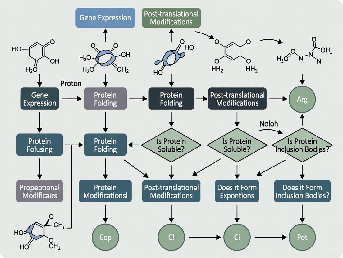
The logical flow of this diagnostic and mitigation process is summarized in the following diagram:
Visualization of Key Concepts
The Protein Secretory Pathway in Filamentous Fungi
The efficient secretion of heterologous proteins in eukaryotic hosts like Aspergillus niger involves a complex, coordinated pathway. Engineering various steps of this pathway is a key strategy for enhancing yield [1] [7].
Metabolic Network Engineering to Reduce Burden
Metabolic burden stems from the reallocation of the host's central metabolic resources. The diagram below illustrates key nodes in the glycolysis and TCA cycle that can be engineered to enhance flux toward heterologous protein production [7].
Troubleshooting Guide: Addressing Protein Toxicity inE. coli
This guide helps diagnose and resolve common issues when producing toxic recombinant proteins in E. coli.
Problem: No Cell Growth or Rapid Culture Collapse After Induction
This indicates severe toxicity where the expressed protein rapidly halts host cell metabolism [11] [12].
| Possible Cause | Diagnostic Experiments | Solution Strategies |
|---|---|---|
| Extreme toxicity of the target protein [12] | Check culture density (OD600) before and after induction. | • Use tightly controlled expression strains (e.g., BL21(DE3)-pLysS) [13] [14].• Switch to a weaker promoter or a promoter induced by a different mechanism (e.g., osmotic shock, temperature shift) [12]. |
| "Leaky" basal expression before induction [13] | Run an uninduced control sample on SDS-PAGE to detect pre-induction protein expression. | • Use strains with plasmid-encoded T7 lysozyme (e.g., pLysS/pLysE), which inhibits T7 RNA polymerase [13] [14].• Add glucose to the growth medium to repress basal expression in T7 systems [12]. |
| Metabolic burden from resource diversion [15] | Monitor growth rate and analyze proteomic changes. | • Optimize induction conditions (cell density, inducer concentration, temperature) [15] [13].• Use richer growth media to provide more resources [15]. |
Problem: Low Yield of Soluble, Active Protein
The protein expresses but is inactive, insoluble, or yields are insufficient [16].
| Possible Cause | Diagnostic Experiments | Solution Strategies |
|---|---|---|
| Aggregation into inclusion bodies [16] | Analyze the soluble and insoluble fractions of cell lysates by SDS-PAGE. | • Reduce induction temperature (e.g., to 25-30°C) [13].• Use fusion tags (e.g., Maltose-Binding Protein, MBP) that enhance solubility [12] [16].• Co-express molecular chaperones to aid folding [17]. |
| Improper protein folding or missing disulfide bonds [17] | Check for activity and use western blot to detect full-length protein. | • Use engineered E. coli strains (e.g., Shuffle T7) with an oxidizing cytoplasm that promotes disulfide bond formation [17].• Target the protein to the periplasm where disulfide bonds form naturally [12]. |
| Host cell toxicity leading to proteolytic degradation or incomplete synthesis [11] | Conduct a time-course experiment to see if the protein degrades over time. | • Use protease-deficient host strains (e.g., BL21).• Shorten the induction time and add protease inhibitors during lysis [16]. |
Frequently Asked Questions (FAQs)
Q1: What are the primary signs that my recombinant protein is toxic to the E. coli host? The main indicators include: severely inhibited cell growth or cell death following induction, a pronounced reduction in final culture density compared to the control, the formation of inclusion bodies for proteins that should be soluble, and the frequent emergence of cells with compensatory mutations that have lost the expression plasmid [11] [15] [12].
Q2: Besides E. coli, what are alternative expression hosts for toxic proteins? No single host is perfect for all toxic proteins, but several alternatives exist:
- Yeast Systems (e.g., Pichia pastoris): Offer the folding machinery of a eukaryote and are generally recognized as safe (GRAS). They can be better suited for producing some eukaryotic toxins [14].
- Baculovirus/Insect Cell Systems: An excellent choice for producing complex eukaryotic proteins that require specific post-translational modifications. They are particularly useful for expressing full-length toxins and immunotoxins [11] [14].
- Mammalian Cell Lines (e.g., CHO, HEK293): Provide the most native environment for expressing human or mammalian toxins, ensuring proper folding, assembly, and activity. However, they are more costly and time-consuming than microbial systems [14].
Q3: How can computational tools help in predicting and mitigating protein toxicity? Advanced computational models like ToxDL 2.0 can predict the potential toxicity of a protein sequence before you even begin lab work. These tools use deep learning to integrate evolutionary, structural, and domain information, helping you identify high-risk motifs in your protein of interest. This allows for the in silico design of deimmunized or less toxic variants by mutating key residues before expression [18].
Q4: My protein is essential but highly toxic. Are there any specialized genetic strategies for its expression? Yes, several strategies are designed specifically for this scenario:
- Use of Weaker Promoters: Avoid strong, constitutive promoters. Use tightly regulated, inducible promoters that allow you to grow the biomass first and induce production later [12].
- Engineering Less Toxic Variants: Identify and mutate the specific amino acid residues responsible for the toxic activity while attempting to retain the protein's structural integrity for study purposes [11].
- Co-expression of Inhibitors: For toxins that inhibit essential host processes (like translation), you can co-express a neutralized version or an inhibitor of the toxin to protect the host until induction [11].
Metabolic Burden and Host Physiology: An Experimental Workflow
Understanding the host's physiological response is key to solving toxicity issues. The following diagram outlines an integrated experimental approach to analyze the impact of recombinant protein production.
Summary of Key Experimental Protocol:
- Culture and Induce: Grow two different E. coli host strains (e.g., M15 and DH5α) in both defined (M9) and complex (LB) media. Induce recombinant protein expression at different growth phases (e.g., early-log phase at OD600 ~0.1 and mid-log phase at OD600 ~0.6) [15].
- Monitor Growth and Expression: Track culture growth (OD600, maximum specific growth rate µmax, dry cell weight) and analyze recombinant protein expression profiles via SDS-PAGE at multiple time points (e.g., mid-log and late-log phase) [15].
- Conduct Proteomic Analysis: Harvest cells from key time points. Perform cell lysis, protein digestion, and Liquid Chromatography-Mass Spectrometry (LC-MS/MS) for Label-Free Quantification (LFQ) proteomics. Compare the proteomes of recombinant cells against control (parental) cells to identify significant changes in protein abundance across different cellular functional categories [15].
- Data Integration: Correlate the proteomic data (changes in transcriptional/translational machinery, stress response proteins) with the observed growth and protein expression parameters. This helps pinpoint the specific metabolic bottlenecks and stress responses caused by the toxic protein [15].
Research Reagent Solutions
The table below lists key reagents and their applications for tackling protein toxicity.
| Research Reagent | Function & Application in Toxicity Mitigation |
|---|---|
| BL21(DE3)-pLysS/E. coli Strain [13] [14] | Host strain; plasmid-encoded T7 lysozyme suppresses basal "leaky" expression of T7 RNA polymerase, essential for toxic gene control. |
| Shuffle T7 E. coli Strain [17] | Engineered host; promotes disulfide bond formation in the cytoplasm, ideal for toxins requiring correct cysteine bridges. |
| Rosetta E. coli Strain | Host strain; supplies tRNAs for rare codons, preventing ribosomal stalling and truncation that can exacerbate toxicity or yield inactive products [12] [13]. |
| pLysS/pLysE Plasmids [13] | Companion plasmids; encode T7 lysozyme for tighter repression in T7 expression systems, can be used in various DE3 strains. |
| Fusion Tags (MBP, GST, SUMO) [12] [16] | Solubility enhancers; fused to the target protein to improve solubility and folding, reducing aggregation and inclusion body formation. |
| Molecular Chaperone Plasmids [17] | Expression vectors; co-express chaperone proteins (e.g., GroEL/GroES, DnaK/DnaJ) to assist in the proper folding of complex or aggregation-prone toxic proteins. |
| ToxDL 2.0 Software [18] | Computational tool; a multimodal deep learning model for predicting protein toxicity from sequence and predicted structure, enabling pre-emptive design. |
Frequently Asked Questions (FAQs)
What are inclusion bodies and why do they form in my protein expression experiments? Inclusion bodies (IBs) are nuclear, cytoplasmic, or periplasmic aggregates of mostly misfolded proteins that lack proper biological function. They form when the rate of recombinant protein expression exceeds the host cell's ability to properly fold the proteins, leading to misfolding where hydrophobic residues normally buried in the native structure become exposed to the aqueous cellular environment. This drives aggregation as these hydrophobic regions interact to shield themselves from water [19]. The aggregation process is primarily driven by these hydrophobic interactions and can be influenced by high expression rates, lack of proper post-translational modification machinery, and specific protein properties [19].
I'm using E. coli as my expression system. Which host strains are recommended to minimize inclusion body formation? The choice of E. coli host strain significantly impacts protein solubility. Strains designed for tight regulation of expression are preferred. For T7-based systems, consider strains that co-express T7 lysozyme (such as lysY or pLysS strains), which inhibits T7 RNA polymerase and reduces basal expression. Additionally, strains lacking proteases (OmpT and Lon) help prevent target protein degradation, and strains carrying the lacIq gene provide enhanced repressor production for tighter control of inducible systems [20]. For proteins requiring disulfide bond formation, specialized strains like SHuffle that allow cytoplasmic disulfide bond formation may be beneficial [20].
What practical steps can I take during my experiment to increase soluble protein yield? Several practical approaches can enhance soluble expression:
- Reduce expression temperature: Inducing protein expression at lower temperatures (15-20°C) slows protein synthesis, allowing more time for proper folding [20].
- Use solubility-enhancing fusion tags: Tags such as Maltose Binding Protein (MBP) can dramatically improve solubility. The pMAL system is one example that facilitates both expression and purification [20].
- Modulate induction conditions: For toxic proteins, use tunable expression systems like the Lemo21(DE3) strain with the rhamnose-inducible PrhaBAD promoter to find the optimal expression level that avoids aggregation [20].
- Consider fusion tag impact: In some cases, commonly used tags like polyhistidine (His-tag) can contribute to insolubility. Testing expression with and without such tags can be beneficial [21].
Are there any sequence-based strategies to prevent aggregation? Yes, optimizing the genetic sequence of your target protein can significantly improve solubility:
- Address mRNA secondary structure: Troublesome secondary structures in the 5' untranslated region, ribosomal binding site, or coding sequence can hinder translation. Altering these sequences, particularly to ensure the ribosomal binding site closely matches the ideal E. coli sequence (AGGAGGT), can help [20].
- Codon optimization: Genes rich in codons corresponding to tRNAs that are low abundance in the host can cause translational stalling. Either co-express rare tRNAs or redesign the gene using host-preferred codons via gene synthesis [20].
- Note of caution: Highly efficient codon optimization can sometimes lead to overly robust expression, creating inclusion bodies. In such cases, pairing optimized sequences with tunable expression systems is recommended [20].
My protein requires disulfide bonds for proper folding. How can I address this in E. coli? Proteins requiring disulfide bonds present a particular challenge in the reducing environment of the E. coli cytoplasm. Strategies include:
- Periplasmic secretion: Using vectors with an N-terminal signal sequence to export the target protein to the oxidative environment of the periplasm, where native Dsb enzymes can catalyze disulfide bond formation [20].
- Engineered cytoplasmic systems: Strains like SHuffle are genetically modified to create a more oxidizing cytoplasm and also express the disulfide bond isomerase DsbC in the cytoplasm, facilitating the correct formation of complex disulfide bonds [20].
- Cell-free systems: Modifying cell-free protein synthesis systems, such as the PURExpress system, by adjusting redox conditions or adding enhancers can also support proper disulfide bond formation [20].
Troubleshooting Guides
Problem: High Basal Expression Leading to Toxicity or Inclusion Bodies
Background: Uninduced expression of the target protein can severely hamper host viability or lead to plasmid loss, often resulting in protein aggregation before controlled induction can even begin [20].
Experimental Protocol:
- Verify the expression system: Ensure your system supplies sufficient LacI repressor protein. Many systems include the
lacIgene on the expression vector. - Switch to a
lacIqhost: Use an expression strain harboring thelacIqgene (e.g., NEB Express Iq). This mutation increases LacI repressor production ten-fold, providing much tighter control [20]. - For T7 systems, employ T7 lysozyme: If using a T7 promoter (e.g., in BL21(DE3)), basal expression from the T7 RNA polymerase is common. Switch to a host that expresses T7 lysozyme (e.g., T7 Express lysY or strains carrying pLysS plasmid), which inhibits T7 RNA polymerase [20].
- Adjust growth medium: For DE3 strains, adding 1% glucose to the medium can decrease basal expression from the lacUV5 promoter by reducing cAMP levels [20].
Problem: Low Solubility of the Target Protein
Background: Some proteins are inherently prone to misfolding and aggregation due to their physicochemical properties, such as large size, multi-domain structure, or stretches of hydrophobic residues [19].
Experimental Protocol:
- First-line approach - lower temperature: Induce expression at a lower temperature (15-20°C). This slows down protein synthesis, giving the cellular folding machinery more time to act [20].
- Use a solubility-enhancing fusion tag: Clone your target gene into a vector that fuses it to a large, highly soluble tag like MBP (Maltose Binding Protein). This can greatly improve the solubility of the fusion partner [20].
- Co-express molecular chaperones: Co-express chaperone systems (e.g., GroEL/GroES or DnaK/DnaJ/GrpE) in the same host. These can assist in the proper folding of the target protein, though note that some target protein may remain complexed with the chaperones and require further separation [20].
- Evaluate fusion tag necessity: Investigate if the purification tag itself is causing insolubility. For example, try expressing the protein without a His-tag, as its removal has been shown to promote the soluble expression of some proteins [21].
Problem: Co-translational Aggregation and Ribosome Stalling
Background: Recent research highlights that aggregation can occur during translation itself ("co-translational"), leading to the sequestration of ribosomal components and mRNAs in amyloid-like inclusion bodies, particularly affecting membrane proteins and those with long-range beta-sheet interactions [22].
Experimental Protocol & Visualization: The diagram below illustrates the mechanism of co-translational aggregation and the ensuing cellular response.
Diagram 1: Mechanism of co-translational aggregation induced by aggregation-prone peptides.
Methodology:
- Detect aggregation: Use super-resolution structured illumination microscopy (SIM) with amyloid-binding dyes like pFTAA (Amytracker) to identify intracellular aggregates with amyloid-like characteristics [22].
- Confirm secondary structure: Employ Atomic Force Microscopy-based Infrared Spectroscopy (AFM-IR) on bacterial sections to analyze the secondary structure of aggregates, specifically looking for a signature peak around 1630 cm⁻¹ indicating high beta-sheet content [22].
- Assess ribosome impact: Monitor activation of the SsrA (tmRNA) ribosome rescue pathway, which is a cellular response to stalled ribosomes, as evidence of co-translational stalling [22].
Table 1: Summary of Key Experimental Findings from Literature
| Experimental Finding | Quantitative/Descriptive Result | Context / System | Source |
|---|---|---|---|
| Non-expression rate in E. coli | Over 20% of >9,000 recombinant proteins failed to express. | Large-scale study (NESG) on diverse proteins in E. coli BL21(DE3) with pET plasmid. | [12] |
| Low expression threshold | Extremely low levels: <0.1 mg per 100 mL of culture medium. | Defined as a critical scenario making subsequent experiments impractical. | [12] |
| His-tag deletion impact | Promoted soluble and highly active expression of uridine phosphorylase and γ-lactamases. | Strategy tested on industrial biocatalysts expressed in E. coli using the pET System. | [21] |
| Antibacterial peptide-induced aggregation | Peptide P33 (from RhtA APR) caused formation of polar inclusion bodies, bactericidal against ESKAPE pathogens. | Induced co-translational aggregation as a broad-spectrum antibacterial mechanism. | [22] |
The Scientist's Toolkit: Key Research Reagent Solutions
Table 2: Essential Materials and Reagents for Addressing Inclusion Body Formation
| Reagent / Tool | Function / Purpose | Example Products / Strains |
|---|---|---|
| Tightly Regulated E. coli Strains | Minimizes basal (uninduced) expression of the toxic target protein, improving cell health and clone stability. | T7 Express lysY, NEB Express Iq, Lemo21(DE3) [20]. |
| Solubility-Enhancing Fusion Tags | Increases the solubility of the fused target protein during expression. | MBP (Maltose Binding Protein) in pMAL system [20]. |
| Chaperone Plasmid Kits | Co-expression of helper proteins (e.g., GroEL, DnaK) that assist in the proper folding of the target protein. | Various commercial chaperone plasmids [20]. |
| Disulfide Bond Engineered Strains | Enables correct formation of disulfide bonds in the E. coli cytoplasm for proteins that require them. | SHuffle strains [20], CyDisCo system [23]. |
| Amyloid-Specific Dyes | Detect and visualize protein aggregates with amyloid-like characteristics in cells. | pFTAA (Amytracker), Thioflavin-T [22]. |
| Tunable Induction Systems | Allows fine control over protein expression levels to find the balance between yield and solubility. | Rhamnose-inducible PrhaBAD promoter in Lemo21(DE3) [20]. |
Heterologous protein production is a cornerstone of modern biotechnology, essential for producing therapeutic enzymes, vaccines, and industrial proteins. However, achieving high yields of functional proteins remains challenging due to molecular bottlenecks that occur at multiple stages: transcription, translation, and post-translational modifications (PTMs). These constraints can drastically reduce protein yield, stability, and biological activity, ultimately impacting research outcomes and commercial viability.
This technical support center provides targeted troubleshooting guides and FAQs to help researchers identify and overcome these critical barriers. The content is framed within the context of systematic approaches for enhancing heterologous protein production, drawing on current advances in genetic engineering, metabolic manipulation, and process optimization.
Troubleshooting Guides & FAQs
Frequently Asked Questions
Q1: I have confirmed that my gene is present in the host, but I detect no protein expression. What are the most common causes?
- A: This common issue can stem from several factors [24] [25]:
- Low Transfection Efficiency: Ensure your transfection protocol is optimized. For stable expression, perform proper selection and use methods that allow examination of individual cells.
- Promoter Strength: The promoter may not be active under your experimental conditions. Consider switching to a stronger or more suitable inducible promoter.
- Detection Method Sensitivity: Your detection protocol (e.g., Western blot) may not be sensitive enough. Optimize your protocol or try a more sensitive method.
- Toxicity of the Protein: Expression of the recombinant protein may be toxic to the host cell, preventing successful colony growth. Using a tightly controlled inducible expression system can help mitigate this.
Q2: My protein is expressed, but it is inactive. What could be wrong?
- A: Expression of a protein does not guarantee functionality [24] [26]:
- Improper Folding: The protein may be misfolded or forming inactive aggregates (inclusion bodies). Consider lowering the induction temperature (e.g., to 30°C) or using a host strain engineered with enhanced chaperone systems.
- Lacking Essential PTMs: Many proteins require specific post-translational modifications (e.g., glycosylation, disulfide bond formation) for activity. Ensure your expression host (e.g., mammalian, insect, or engineered yeast) can perform the necessary modifications that bacterial systems like E. coli typically cannot.
- Truncated Protein: The expressed protein may be truncated due to degradation or premature termination. Check your protein size on an SDS-PAGE gel and verify your plasmid sequence for errors.
Q3: I am working with a fungal expression system like Aspergillus niger, and my protein secretion efficiency is low. What strategies can I use?
- A: Aspergillus niger is a powerful host but faces secretion bottlenecks [7]:
- Secretion Pathway Optimization: Engineer the secretory pathway by overexpressing key components like signal peptides, ER folding chaperones (e.g., BiP), and proteins involved in vesicle trafficking (e.g., SNARE proteins) [7].
- Cell Wall Remodeling: The dense cell wall can trap secreted proteins. Modifying cell wall structure through genetic engineering can facilitate the release of larger proteins [7].
- Signal Peptide Engineering: The native signal peptide of your protein may be inefficient. Screen different natural or engineered signal peptides to find the most efficient one for your protein of interest [7].
Quantitative Data on Common Bottlenecks and Solutions
The table below summarizes key bottlenecks and the efficacy of various strategies based on published research, providing a quick reference for experimental planning.
Table 1: Efficacy of Strategies to Overcome Molecular Bottlenecks
| Bottleneck Category | Specific Challenge | Solution Strategy | Reported Efficacy / Outcome | Key References |
|---|---|---|---|---|
| Transcription | Weak or leaky promoter | Use of strong, inducible promoters (e.g., T7, Tet-On) | Up to 100-fold increase in protein yield | [25] |
| Engineering synthetic promoters | Enables precise spatiotemporal control | [7] | ||
| Translation | Rare codon usage | Host strain engineering (e.g., tRNA supplementation) | Rescues expression of full-length protein | [25] [27] |
| mRNA instability | Codon optimization & 5' GC content adjustment | Improves mRNA half-life & translational efficiency | [25] | |
| Protein Folding & Secretion | Protein misfolding & aggregation | Co-expression of chaperones (e.g., DnaK, BiP); Lowered induction temp | Significant increase in soluble, active protein | [7] [26] [25] |
| Inefficient secretion | Signal peptide engineering; Optimizing ER-Golgi trafficking | Can increase secretion efficiency by over 10-fold | [7] | |
| Disulfide bond formation | Use of SHuffle E. coli or engineered eukaryotic hosts; Optimizing redox | Enabled production of complex antibodies (124 µg/mL IgG) | [28] | |
| Post-Translational Modifications | Lack of glycosylation | Use of eukaryotic hosts (e.g., CHO, P. pastoris) | Essential for therapeutic efficacy & half-life (e.g., EPO) | [26] [27] |
| Methionine oxidation | Media optimization; Use of protective excipients | Preserves anti-elastase activity in α1-antitrypsin | [26] | |
| Deamidation (Asn/Gln) | Control of pH during storage; Formulation optimization | Mitigates loss of bioactivity in IgG1 & Stem Cell Factor | [26] | |
| Host Metabolism | Metabolic burden | Dynamic regulation of central metabolism (e.g., glycolysis, TCA) | Enhanced glycolytic flux & protein yield in A. niger | [7] |
Experimental Protocols for Overcoming Key Bottlenecks
Protocol: Optimizing a CRISPR-Cas System for Multi-Copy Gene Integration inAspergillus niger
Objective: To enhance transcription and gene dosage by integrating multiple copies of a heterologous gene into the genome of A. niger [7].
Materials:
- Aspergillus niger strain susceptible to genetic transformation.
- CRISPR-Cas9 or Cas12 plasmid system optimized for A. niger.
- Donor DNA fragment containing the heterologous gene of interest, flanked by homologous arms targeting a genomic "safe harbor" locus.
- Standard reagents for fungal transformation (e.g., PEG, CaCl₂).
Method:
- Design gRNAs: Design and synthesize guide RNAs (gRNAs) that target specific, non-essential genomic loci suitable for multi-copy integration.
- Prepare Donor DNA: Construct a donor DNA fragment containing your gene of interest, a strong inducible promoter, and a selectable marker.
- Co-transformation: Co-transform the A. niger host strain with the CRISPR-Cas plasmid and the linear donor DNA fragment using protoplast-mediated transformation.
- Selection and Screening: Select transformations on appropriate antibiotic media. Screen resistant colonies via PCR and Southern blotting to confirm multi-copy integration events.
- Expression Validation: Cultivate positive clones and induce expression. Measure transcript levels (qRT-PCR) and protein yield to validate enhanced production.
Protocol: Troubleshooting Protein Solibility and Folding inE. coli
Objective: To recover functional protein from inclusion bodies or prevent their formation [25].
Materials:
- E. coli expression culture (e.g., BL21(DE3) or SHuffle for disulfide bonds).
- IPTG for induction.
- Lysis buffer (e.g., with lysozyme).
- Solubilization buffer (6-8 M Urea or GuHCl).
- Refolding buffer (PBS with reduced denaturant concentration, redox couples like GSH/GSSG).
- SDS-PAGE gel equipment.
Method:
- Test Induction Parameters: Inoculate a small culture and induce at different temperatures (e.g., 18°C, 25°C, 37°C) and IPTG concentrations. Take samples hourly for 4-8 hours.
- Analyze Solubility:
- Pellet 1 mL of induced culture and resuspend in lysis buffer.
- Lyse cells by sonication.
- Centrifuge at high speed to separate soluble (supernatant) and insoluble (pellet) fractions.
- Analyze both fractions by SDS-PAGE.
- If Protein is Insoluble (Inclusion Bodies):
- Solubilize the pellet in 6-8 M Urea.
- Purify the denatured protein under denaturing conditions.
- Refold the protein by slow dialysis or dilution into a refolding buffer.
- If Solubility is Poor, Pre-induction: Use a different host strain (e.g., with chaperone plasmids), change the expression vector, or fuse the protein to a solubility tag (e.g., MBP, GST).
Protocol: Enhancing Thermostability and Pharmacokinetics via PEGylation
Objective: To chemically conjugate polyethylene glycol (PEG) to a therapeutic protein to increase its in vivo half-life, reduce immunogenicity, and improve stability [29].
Materials:
- Purified therapeutic protein.
- Activated PEG derivative (e.g., mPEG-Succinimidyl Carbonate for lysine residues).
- Reaction buffer (e.g., phosphate buffer, pH 8.0-9.0).
- Dialysis membrane or desalting columns.
Method:
- Prepare Protein Solution: Dialyze the purified protein into a suitable reaction buffer (e.g., 50 mM phosphate, 100 mM NaCl, pH 8.5).
- PEGylation Reaction: Add a molar excess of the activated PEG reagent to the protein solution. Gently mix the reaction for several hours at 4°C.
- Quench the Reaction: Stop the reaction by adding a quenching agent like glycine or Tris buffer.
- Purify Conjugate: Separate the PEGylated protein from unreacted PEG and native protein using size-exclusion chromatography or ion-exchange chromatography.
- Characterization: Analyze the conjugate using SDS-PAGE (showing a shift in molecular weight), mass spectrometry, and activity assays to confirm the modification and retained functionality.
Visualization of Bottlenecks and Solutions
The Heterologous Protein Production Cascade
This diagram illustrates the sequential molecular bottlenecks from gene insertion to a functional protein.
Integrated Solutions Workflow
This workflow outlines the multi-strategy approach to overcome the major bottlenecks.
The Scientist's Toolkit: Essential Research Reagents
Table 2: Key Research Reagent Solutions for Heterologous Protein Production
| Reagent / Tool Category | Specific Example | Function & Application | Key References |
|---|---|---|---|
| Advanced Expression Hosts | SHuffle E. coli | Engineered for disulfide bond formation in the cytoplasm; ideal for proteins requiring multiple disulfides. | [28] |
| Pichia pastoris | Eukaryotic host capable of high-density fermentation, secretion, and human-like glycosylation. | [27] | |
| Aspergillus niger | Filamentous fungus; GRAS status; excellent secretor for industrial enzymes and organic acids. | [7] | |
| Genetic Engineering Tools | CRISPR-Cas9/Cas12 Systems | Enables precise gene knock-in, multi-copy integration, and gene repression in fungal and bacterial hosts. | [7] |
| Synthetic Promoters | Engineered for strong, inducible, or tunable control of transcription (e.g., benzoate-activated). | [7] | |
| tRNA Supplementation Plasmids | Provides rare tRNAs to prevent translational stalling and truncation during heterologous expression. | [25] | |
| Folding & Secretion Aids | Chaperone Plasmid Kits | Co-expression plasmids for DnaK/DnaJ/GrpE or GroEL/ES to assist in proper protein folding. | [26] [25] |
| Disulfide Bond Catalysts | Purified DsbC or PDI; added in vitro or co-expressed in vivo to catalyze correct disulfide bond formation. | [28] | |
| PTM Enhancement | Glyco-engineered CHO Cells | Host cells engineered with human glycosyltransferases (e.g., β1,4-GalT, α2,6-SiaT) for human-like glycans. | [26] |
| Cell-Free Protein Synthesis (CFPS) Systems | In vitro system from E. coli, wheat germ, or CHO cells; allows precise control of redox and PTMs. | [28] | |
| Stability & Delivery | PEGylation Reagents | Activated PEG polymers (e.g., mPEG-NHS) for covalent attachment to proteins to enhance half-life. | [29] |
| Formulation Excipients | Sugars, arginine, and other agents used in downstream processing to suppress aggregation and oxidation. | [26] |
The Impact of Gene Source and Sequence Intrinsic Properties
Troubleshooting Guides and FAQs
This technical support center provides targeted guidance for researchers overcoming challenges in heterologous protein production. The following guides address common issues related to gene source and sequence-specific properties that can constrain experimental success and therapeutic development.
Troubleshooting Guide 1: Low Protein Solubility and Yield
Problem: The recombinant protein of interest expresses poorly, forms insoluble inclusion bodies, or yields insufficient quantities for research or development purposes.
Questions & Answers:
Q1: What host-specific factors are critical for improving soluble yield in E. coli? Choosing the correct E. coli host strain is a primary consideration. Strains should be selected to minimize proteolytic degradation and control basal expression. The table below summarizes key host strain features and recommendations [30].
| Host Feature | Function | Recommended Strains/Solutions |
|---|---|---|
| Protease Deficiency | Lacks proteases (e.g., OmpT, Lon) that degrade target proteins. | T7 Express, NEB Express, BL21(DE3) derivatives [30]. |
| Tight Expression Control | Prevents toxic basal expression pre-induction, improving clone stability. | Strains with lacIq gene (increases Lac repressor) or T7 lysozyme (e.g., lysY, pLysS) to inhibit T7 RNA polymerase [30]. |
| Disulfide Bond Formation | Enables correct formation of disulfide bonds in the cytoplasm. | SHuffle strains (oxidizing cytoplasm & disulfide bond isomerase DsbC) [30]. |
| Tunable Expression | Allows fine-tuning of expression level to balance yield and solubility. | Lemo21(DE3) strain using L-rhamnose concentration to modulate expression [30]. |
Q2: Which experimental parameters can be optimized to increase solubility? Several culture and induction conditions can be adjusted to favor proper protein folding [30] [16]:
- Temperature: Inducing protein expression at lower temperatures (15–20°C) often significantly improves yields of properly folded protein.
- Fusion Tags: Fusion partners like Maltose-Binding Protein (MBP) can enhance solubility. Vectors such as the pMAL system are designed for this purpose.
- Chaperone Co-expression: Co-expressing chaperonins like GroEL, DnaK, or ClpB can assist in the proper folding of low-solubility proteins.
Q3: How does the gene source influence the choice of expression system? The intrinsic properties of the protein, dictated by its gene source, determine the required cellular environment for correct folding and function [16].
- Prokaryotic (E. coli) Systems: Ideal for simplicity, speed, and cost-effectiveness. Often unsuitable for complex eukaryotic proteins requiring specific post-translational modifications (e.g., glycosylation).
- Eukaryotic Systems (Yeast, Insect, Mammalian cells): Necessary for proteins requiring disulfide bonds, complex folding, or authentic post-translational modifications. Mammalian cells are the gold standard for producing therapeutic proteins with human-like glycosylation patterns [16].
Troubleshooting Guide 2: Unwanted Cryptic Gene Expression and Toxicity
Problem: The gene of interest is toxic to the host cell, leading to poor host cell growth, genetic instability, plasmid loss, or the expression of unexpected truncated protein products.
Questions & Answers:
Q4: What sequence intrinsic properties can cause toxicity and genetic instability? Unintentional cryptic gene expression is a major cause of toxicity. This occurs when non-native or synthetic DNA sequences introduced into a host are recognized by the host's transcription and translation machinery in unintended ways [31]. This can result in the expression of:
- Truncated peptides or out-of-frame proteins.
- Antisense RNAs that interfere with host genes.
- Burdensome or directly toxic proteins that create strong selection pressure for cells with mutations in your engineered DNA sequence [31].
Q5: What is a "negative design" strategy, and what tools can help? Negative design involves proactively eliminating undesirable sequence features to create more reliable and effective DNA constructs. Instead of just optimizing for high expression, you design to prevent cryptic expression [31].
- Software Tool: CryptKeeper is a pipeline that visualizes predictions of bacterial gene expression signals (promoters, ribosome-binding sites) and estimates the potential translational burden from a DNA sequence.
- Application: It allows researchers to identify and subsequently eliminate unwanted translation initiation sites or promoters before synthesizing a gene, thereby mitigating cloning challenges and experimental failures [31].
Q6: How can codon usage be adapted to manage toxicity? Traditional "codon optimization" that uses only the most frequent codons can lead to excessive expression and toxicity. A more nuanced approach is to design "typical genes" that resemble the codon usage of a specific subset of endogenous host genes (e.g., lowly expressed genes). This strategy can adapt a toxic gene like human α-synuclein for endogenous, low-level expression in yeast, making it possible to work with challenging proteins [32].
Troubleshooting Guide 3: Poor Transfection and Expression in Mammalian Systems
Problem: Low transfection efficiency, high cell toxicity, or undetectable protein expression in mammalian cell cultures.
Questions & Answers:
Q7: What are the common causes of low transfection efficiency and high cell death? The table below outlines frequent causes and their solutions [33] [34].
| Potential Cause | Symptoms | Troubleshooting Solutions |
|---|---|---|
| Poor Cell Health | Low baseline viability, weak adherence. | Use freshly passaged, actively dividing cells. Avoid over-confluent or senescent cultures [34]. |
| Reagent Toxicity | High cell death within 12-24 hours, cell rounding/detachment. | Reduce reagent amount or incubation time. Use low-toxicity, serum-compatible reagents [34]. |
| Incorrect DNA/Reagent Ratio | Low efficiency across all conditions. | Perform a titration experiment to optimize the reagent-to-DNA ratio [34]. |
| Inappropriate Promoter | Low expression in specific cell types. | The CMV promoter can be silenced in some murine cell lines; switch to an alternative promoter like EF-1α [33]. |
Q8: How can I confirm if my protein is being expressed but is simply undetectable?
- Use a Positive Control: Always transfer a control plasmid (e.g., expressing GFP) to verify your transfection protocol is working [33].
- Try a More Sensitive Method: If using Coomassie staining, switch to Western blotting for higher sensitivity. Ensure your primary antibody is specific and validated [33].
- Check Cellular Compartments: For secreted proteins, check both the cellular lysate and the culture medium for the presence of your protein [33].
- Perform a Time-Course: Protein expression over time is protein-dependent. Conduct a pilot time-course assay to find the optimal harvest window [33].
The Scientist's Toolkit: Key Research Reagent Solutions
This table details essential materials and tools used to address the challenges discussed in this guide.
| Item | Function | Key Examples / Notes |
|---|---|---|
| Specialized E. coli Strains | Protein expression with controlled proteolysis, disulfide bond formation, and tight regulation. | SHuffle (disulfide bonds), Lemo21(DE3) (tunable expression), T7 Express lysY (low basal expression) [30]. |
| Cryptic Expression Analysis Tool | Computational prediction of unwanted gene expression signals in DNA constructs. | CryptKeeper software pipeline [31]. |
| "Typical Gene" Design Tool | Designs genes with codon usage resembling a selected subset of host genes (e.g., lowly expressed genes). | Publicly available web-application (e.g., Odysseus) [32]. |
| Solubility Enhancement Tags | Fusion partners that improve solubility and offer a purification handle. | Maltose-Binding Protein (MBP) in the pMAL system [30]. |
| Low-Toxicity Transfection Reagents | Chemical carriers for delivering nucleic acids into sensitive cells, including primary and stem cells. | Lipid-based (e.g., Lipofectamine), Polymer-based (e.g., PEI). Must be selected for cell type and nucleic acid [34]. |
Experimental Workflow and Pathway Diagrams
From Gene Sequence to Functional Protein
This diagram outlines the core experimental workflow for heterologous protein expression and key decision points for troubleshooting.
Mechanisms of Cryptic Gene Expression Toxicity
This diagram illustrates how unintended gene expression arises and impacts the host cell.
Advanced Strategies for Enhanced Protein Expression and Folding
Troubleshooting Guides
Problem 1: Low Heterologous Protein Expression Yield
Potential Causes and Solutions:
Cause A: Suboptimal Codon Usage
- Diagnosis: The codon usage frequency of your gene of interest (GOI) does not match the host's tRNA pool, leading to ribosomal stalling and inefficient translation elongation [35].
- Solution: Implement a context-aware, data-driven codon optimization tool. Avoid solely relying on the Codon Adaptation Index (CAI). Use deep learning frameworks like RiboDecode, which learns from ribosome profiling data to generate sequences that enhance translation, rather than just mimicking host codon bias [36].
- Verification: Compare the predicted translation efficiency (e.g., from RiboDecode's model) of the optimized sequence against the original.
Cause B: Inefficient Translation Initiation
- Diagnosis: A weak or occluded Ribosome Binding Site (RBS) or 5' Untranslated Region (UTR) limits ribosome loading [37].
- Solution: Engineer the 5' UTR. Use pre-validated UTR backbones from highly expressed genes or viruses [38] [39]. For fine-tuning, employ RBS calculators or build 5' UTR libraries to systematically vary translation initiation rates over a 100,000-fold range [37].
- Verification: Measure mRNA levels and protein output. A strong increase in protein with minimal change in mRNA levels confirms improved translation initiation.
Cause C: Poor mRNA Stability
- Diagnosis: The mRNA is rapidly degraded in the host cell, reducing the time available for translation.
- Solution: Incorporate stabilizing elements. Use 3' UTRs from stable mRNAs (e.g., human HBB) or viral RNAs (e.g., MALAT1 ENE) [39]. For the coding sequence, optimize for in-cell stability; highly structured "superfolder" mRNAs can improve both stability and expression [39].
- Verification: Perform an mRNA decay time-course experiment to measure the half-life of your transcript.
Problem 2: Protein Misfolding or Loss of Function
Potential Causes and Solutions:
Cause A: Disruption of Co-Translational Folding
- Diagnosis: Over-optimization for speed, using only the most common codons, can cause ribosomes to move too rapidly and disrupt the precise folding kinetics of the protein [35].
- Solution: Use an optimization tool that preserves functionally important rare codon clusters, especially those known to pause ribosomes at critical folding junctures. Tools like DeepCodon integrate strategies to maintain these conserved rare codons [40].
- Verification: Assess protein activity and solubility. Compare the functionality of the protein expressed from a fully optimized sequence versus one that conserves rare codon clusters.
Cause B: Altered Splicing or Regulatory Motifs
- Diagnosis: Synonymous codon changes can inadvertently create cryptic splice sites, miRNA binding sites, or other regulatory motifs.
- Solution: After in silico optimization, screen the sequence for the accidental creation of these motifs. Use sequence analysis tools to check for off-target regulatory sequences.
- Verification: If possible, check the mRNA product in the host for correct splicing and size.
Problem 3: Inconsistent Performance Across Different Systems
Potential Causes and Solutions:
- Cause: Lack of Cellular Context Consideration
- Diagnosis: An mRNA sequence optimized using a standard, context-free algorithm may not perform well in your specific cell line or tissue type due to differences in tRNA abundance and other cellular machinery [36] [35].
- Solution: Employ context-aware optimization tools. RiboDecode, for instance, can incorporate gene expression profiles from RNA-seq to account for the specific cellular environment, improving performance across different cell lines and for different mRNA formats (unmodified, modified, circular) [36].
- Verification: Validate protein expression in your specific target cell line or tissue, not just in a standard model organism.
Frequently Asked Questions (FAQs)
Q1: What is the most critical factor for maximizing protein expression: codon optimization or UTR engineering? A: While both are crucial, recent high-throughput studies suggest that in-cell mRNA stability is a greater driver of protein output than high ribosome load alone [39]. This means that designing an mRNA with a stable structure (including optimized UTRs and CDS) can be more impactful than only maximizing theoretical translation initiation rates. An integrated approach that optimizes both stability and translation is most effective.
Q2: My codon-optimized gene has a high CAI, but protein expression is still low. Why? A: A high CAI indicates that your sequence uses codons common in highly expressed host genes, but it is a simplistic metric. Low expression can persist due to:
- mRNA Structure: The optimized sequence may have formed stable secondary structures that hide the RBS or start codon [41].
- tRNA Availability: CAI does not fully account for the actual, dynamic availability of tRNAs in your specific host and growth conditions [35].
- Context-Specific Effects: The sequence may be optimal in a general sense but not for your specific cellular context. Shift to tools that use deep learning on empirical data like ribosome profiling (RiboDecode) [36] or that model tRNA competition more accurately.
Q3: How can I design an mRNA sequence that is both highly stable and efficiently translated? A: This was historically challenging due to a perceived trade-off, but it is achievable by:
- Selecting Stabilizing UTRs: Use 5' and 3' UTRs from genes with known high stability (e.g., viral UTRs, human HBB) [39].
- Designing a "Superfolder" CDS: Use platforms like Eterna and models like DegScore to design coding sequences with optimized secondary structures that resist degradation and remain translatable [39].
- Leveraging Nucleoside Modifications: Incorporation of pseudouridine (ψ) can further enhance both the stability and translational capacity of the mRNA [39].
Q4: Can I use codon optimization to control the subcellular localization or timing of protein expression? A: Emerging research suggests yes, through tissue-specific codon optimization. Since tRNA pools can vary between tissues, an mRNA can be optimized to be translated more efficiently in one tissue than another [35]. This is a nascent but promising area for targeted gene therapy.
Experimental Protocols & Data
Protocol 1: Systematic mRNA Optimization using PERSIST-seq
This protocol outlines a high-throughput method for evaluating mRNA designs [39].
- Library Design: Synthesize a DNA library containing your GOI with diverse combinations of 5' UTRs, codon-optimized CDS, and 3' UTRs. Include unique barcodes in the 3' UTR for each variant.
- In Vitro Transcription (IVT): Perform pooled IVT on the library to generate a diverse mRNA pool. Co-transcriptionally add a 5' cap and a 3' poly(A) tail.
- Transfection & Harvest: Transfert the mRNA library into your target cells. Harvest cells at multiple time points.
- Polysome Profiling: Fractionate cell lysates on a sucrose gradient to separate mRNAs based on ribosome load. Sequence the barcodes in each fraction.
- Stability Analysis: Extract total RNA from harvested cells and sequence barcodes to track the abundance of each mRNA variant over time (in-cell stability). Incubate the mRNA pool in a solution mimicking physiological conditions and sequence over time to assess in-solution stability.
- Data Integration: Model protein output based on ribosome load and in-cell stability measurements to identify top-performing constructs.
Protocol 2: Validating RBS/UTR Strength with a Reporter System
- Clone UTR Library: Fuse a library of diverse 5' UTR sequences upstream of a reporter gene (e.g., GFP, luciferase) in your expression vector.
- Transform & Culture: Introduce the plasmid library into your host organism and grow under selective conditions.
- Measure Output: Use flow cytometry (for fluorescent reporters) or enzymatic assays to quantify protein expression for each variant.
- Correlate with Sequence: Sequence the UTR region of clones with high, medium, and low expression to identify optimal sequence features.
Table 1: Performance Comparison of Codon Optimization Tools
| Tool Name | Underlying Approach | Key Feature | Validated Improvement |
|---|---|---|---|
| RiboDecode [36] | Deep Learning (on Ribo-seq data) | Context-aware, generative design | - 10x stronger neutralizing antibodies (in vivo).- Equivalent efficacy at 1/5th mRNA dose (in vivo). |
| DeepCodon [40] | Deep Learning (on natural sequences) | Preserves critical rare codons | Outperformed traditional methods in 9/20 experimental tests. |
| LinearDesign [36] | Linear Programming | Jointly optimizes CAI and MFE | Superior in silico performance over earlier methods. |
Table 2: Key UTR Elements for Expression Optimization
| UTR Element | Type | Function and Application | Key Consideration |
|---|---|---|---|
| AU-rich elements [37] | 5' UTR | Stabilizes mRNA via S1/Hfq proteins, enhancing protein production. | Long AU-rich tracts may increase accessibility to RNases. |
| RG4 Structures [37] | 5' or 3' UTR | Acts as an internal ribosome entry site in 5' UTR; enhances stability in 3' UTR. | Strong structures may potentially inhibit scanning. |
| Synthetic Dual UTRs [37] | 5' & 3' UTR | Concatenated UTRs that enhance both transcription and translation. | Requires screening of large randomized libraries for identification. |
| Viral UTRs (e.g., DENV, TMV) [39] | 5' & 3' UTR | Hijacks host translation machinery for high expression and stability. | May trigger stronger immune responses; requires testing. |
The Scientist's Toolkit: Research Reagent Solutions
Table 3: Essential Reagents and Resources for Cis-Optimization
| Item | Function | Example / Source |
|---|---|---|
| Pre-validated UTR Backbones | Provides a reliable starting point for mRNA design, improving translation efficiency and stability. | Aldevron Blog [38] |
| Ribo-seq Dataset | Provides genome-wide data on ribosome positions, enabling data-driven codon optimization. | Used to train RiboDecode [36] |
| UTR Library Kits | Allows for high-throughput experimental screening of UTR variants to fine-tune expression levels. | Commercially available or custom-built via synthesis [37] |
| In vitro Transcription Kit | For synthesizing mRNA transcripts for validation experiments. | Various commercial suppliers. |
| Pseudouridine (ψ) | A nucleoside modification that decreases immunogenicity and can enhance both stability and translation of mRNA. | Used in PERSIST-seq study [39] |
Workflow and Relationship Visualizations
mRNA Optimization Framework
Key Optimization Factor Relationships
Frequently Asked Questions (FAQs)
1. My protein is toxic to the cells, resulting in no growth after transformation. What can I do? Protein toxicity is a frequent challenge that can inhibit growth or cause cell death [12]. To address this, use expression strains with tighter regulation. For T7-based systems, BL21 (DE3) pLysS or BL21 (DE3) pLysE strains are recommended, as they contain T7 lysozyme inhibitors that suppress basal expression [12] [42]. The BL21-AI strain, which uses arabinose for induction, provides an alternative, tightly-regulated system [42]. Furthermore, you can supplement your growth medium with 0.1-1% glucose to repress basal expression before induction [42].
2. I get good transformation but no protein expression. What are the common causes? This issue can stem from several factors [43]:
- Genetic Sequence: Verify your DNA sequence for frame-shifts or premature stop codons [42].
- Codon Usage: Check for the presence of rare codons that can stall translation. The arginine codons AGG and AGA, for example, are used infrequently in E. coli and can be replaced with more common synonyms [12] [42].
- Plasmid Instability: If using ampicillin resistance, the antibiotic can degrade during culture. Using carbenicillin or a fresh antibiotic dose can help maintain selection pressure [42]. Always use freshly transformed cells for expression experiments.
- Incorrect Assumption: The protein may be expressed but located in the insoluble fraction (inclusion bodies). Always analyze both the soluble and insoluble fractions of the cell lysate [42].
3. My target protein is expressed but entirely in inclusion bodies. How can I improve solubility? Strategies to enhance solubility focus on slowing protein production to allow proper folding [44] [42]:
- Lower Induction Temperature: Reduce the temperature to 30°C, 25°C, or even 18°C after induction. Lower temperatures typically require longer induction times (e.g., overnight at 18°C) [42].
- Reduce Inducer Concentration: Use lower amounts of IPTG (e.g., 0.1 - 1 mM) to moderate expression levels [42].
- Use a Weaker Promoter or Low-Copy Plasmid: This reduces the number of gene copies and the overall expression burden.
- Co-express Chaperones: Use engineered E. coli strains that co-express molecular chaperones like GroEL-GroES or DnaK-DnaJ to assist with protein folding [44] [17].
4. I see multiple protein bands or degradation on my gel. What is happening and how can I prevent it? A single dominant smaller band suggests premature translation termination, often due to codon usage bias, while a ladder of bands typically indicates proteolytic degradation [42]. To prevent degradation:
- Use Protease Inhibitors: Add protease inhibitors like PMSF to your lysis buffer. Note that PMSF is unstable in aqueous solution and should be used fresh [42].
- Perform a Time-Course Experiment: Determine the optimal harvest time by analyzing expression levels at different time points post-induction to avoid prolonged exposure to proteases [42].
- Use Specialized Strains: Consider using protease-deficient host strains (e.g., BL21) to minimize degradation.
Troubleshooting Guides
Guide 1: Overcoming Protein Toxicity and Basal Expression
Problem: The target recombinant protein disrupts the host's normal physiology, leading to inhibited growth or cell death, often due to leaky expression before induction.
Solution Strategy: Implement tighter regulation of expression and consider genetic modifications.
Experimental Protocol:
- Transformation: Clone your gene into a tightly regulated vector (e.g., pET series with T7 promoter, pBAD with arabinose promoter). Transform into a restrictive host like BL21 (DE3) pLysS or BL21-AI [12] [42].
- Plating: Plate transformed cells on LB plates containing the appropriate antibiotic and 0.1-1% glucose. Glucose helps repress basal expression in both T7-lac and pBAD systems [42].
- Culture Growth: Inoculate a primary culture from a fresh colony and grow overnight.
- Expression Induction: Sub-culture the overnight culture into fresh medium. For BL21 (DE3) strains, induce with a low concentration of IPTG (e.g., 0.1 mM) when OD600 reaches ~0.4-0.6. For BL21-AI, induce with L-arabinose (e.g., 0.2%) [42].
- Evaluation: Monitor cell growth post-induction and analyze protein expression via SDS-PAGE.
The following workflow outlines the decision path for addressing toxic protein expression:
Guide 2: Strategies for Enhancing Protein Solubility
Problem: The target protein is expressed but aggregates into insoluble inclusion bodies.
Solution Strategy: Modulate expression conditions and leverage host cell folding machinery to favor correct protein folding.
Experimental Protocol:
- Temperature Screening: Test induction at a range of temperatures (e.g., 37°C, 30°C, 25°C, 18°C). Induce cultures at the target OD600 and continue incubation for 3-4 hours (30°C) or overnight (18°C) [42].
- Inducer Titration: Induce parallel cultures with different IPTG concentrations (e.g., 1.0, 0.5, 0.1 mM) to find the level that minimizes aggregation.
- Chaperone Co-expression: Transform your plasmid into a compatible chaperone plasmid strain (e.g., strains expressing GroEL/ES or Trigger Factor) or include chaperone plasmids in your experiment [44] [17].
- Solubility Analysis: a. Harvest cells by centrifugation. b. Lyse cells using sonication or lysozyme treatment in a suitable buffer. c. Separate the soluble (supernatant) and insoluble (pellet) fractions by centrifugation at >12,000 x g for 10-15 minutes. d. Analyze both fractions by SDS-PAGE to assess solubility.
The table below summarizes key optimization parameters and their effects:
Table 1: Optimization Strategies for Improving Recombinant Protein Solubility
| Parameter | Optimization Strategy | Mechanism of Action | Considerations |
|---|---|---|---|
| Temperature | Lower induction temperature (18-25°C) | Slows translation rate, allowing more time for proper folding | Requires longer induction time (e.g., overnight) [42] |
| Inducer Concentration | Use lower IPTG (0.1 - 0.5 mM) | Reduces transcription/translation burden, minimizing aggregation | May require titration to find optimal level for specific protein [42] |
| Fusion Tags | Use solubility-enhancing tags (e.g., MBP, GST, SUMO) | Acts as a solubility chaperone; can improve folding and yield | May require cleavage and removal for final protein product [44] [12] |
| Chaperone Co-expression | Co-express GroEL/ES, DnaK/DnaJ, etc. | Directly assists in the folding of nascent polypeptides | Requires specialized strains or additional plasmids [44] [17] |
| Media/Cofactors | Use minimal media (e.g., M9); add essential cofactors | Reduces metabolic burden; ensures availability of essential ions/molecules | Can lower overall biomass but increase functional protein yield [42] |
The Scientist's Toolkit: Key Research Reagents
Table 2: Essential Reagents and Strains for Bacterial Trans-Optimization
| Reagent / Material | Function / Purpose | Examples & Notes |
|---|---|---|
| Specialized E. coli Strains | Engineered hosts to address specific challenges like toxicity, disulfide bonds, or difficult codons. | BL21(DE3) pLysS/pLysE: For toxic proteins; suppresses basal expression [12] [42].Origami B: Enhances disulfide bond formation in the cytoplasm [17].Rosetta: Supplies tRNAs for rare codons (AGA, AGG, AUA, CUA, GGA) [12]. |
| Expression Vectors | Plasmids designed for controlled gene expression. | pET series: High-expression, T7 promoter, IPTG-inducible [12].pBAD series: Tightly regulated by arabinose, useful for toxic genes [42]. |
| Fusion Tags | Polypeptide sequences fused to the target protein to aid expression, solubility, or purification. | His-tag: Simplifies purification via immobilized metal affinity chromatography (IMAC).MBP, GST, SUMO: Enhance solubility; can be cleaved off post-purification [44] [12]. |
| Inducers | Chemical molecules that trigger transcription of the target gene. | IPTG: Non-metabolizable inducer for lac/T7-lac promoters [42].L-Arabinose: Inducer for the pBAD promoter system [42]. |
| Protease Inhibitors | Chemicals that inhibit proteolytic enzymes, preventing target protein degradation. | PMSF: Serine protease inhibitor (short half-life in water) [42].Commercial Cocktails: Broad-spectrum inhibitors targeting multiple protease classes. |
Advanced Engineering: Pathway Optimization for Complex Production
Problem: For metabolic engineering beyond single protein production, low yield arises from carbon loss in competing pathways and insufficient supply of key cofactors.
Solution Strategy: Rationally rewire central carbon metabolism using a "host-aware" framework to maximize flux toward the desired product.
Experimental Protocol (Conceptual Workflow for Pathway Engineering):
- Identify Key Nodes: Map the biosynthetic pathway and identify branch points where carbon is diverted (e.g., α-ketoglutarate in the TCA cycle for T-4-HYP production) [45].
- Knock Out Competing Pathways: Use gene knockout techniques (e.g., CRISPR-Cas9, λ-Red recombination) to delete genes encoding enzymes in competing metabolic shunts [45].
- Enhance Precursor Supply: Introduce heterologous pathways to minimize carbon loss. For example, the Non-Oxidative Glycolysis (NOG) pathway can redirect glucose to acetyl-CoA with higher efficiency [45].
- Balance Cofactor Supply: Overexpress enzymes or introduce transhydrogenases to balance cofactors (e.g., increase NADPH supply for reductive biosynthesis) [45].
- Implement Dynamic Control: Engineer genetic circuits that decouple growth from production, allowing cells to first build biomass before switching to a high-production state [46].
- Fed-Batch Fermentation Optimization: In a bioreactor, use continuous feeding of carbon source and key nutrients (e.g., Fe²⁺ for hydroxylases) while controlling dissolved oxygen to support high-density production [45].
The following diagram visualizes this systematic engineering approach:
Core Concepts and Quantitative Foundations
In heterologous protein production, the design of your vector system is a critical determinant of success. Precise control over plasmid copy number (PCN) allows researchers to directly influence gene dosage, thereby optimizing protein expression levels and mitigating host cell metabolic burden [47]. A foundational understanding of these elements is essential for overcoming protein production constraints.
Table 1: Common Origins of Replication and Their Characteristics [48]
| Origin of Replication | Example Vectors | Typical Copy Number (per cell) | Incompatibility Group | Replication Control |
|---|---|---|---|---|
| pUC (pMB1 derivative) | pUC series | 500 - 700 | A | Relaxed |
| pMB1 / ColE1 | pBR322, pET, pGEX | 15 - 20 | A | Relaxed |
| p15A | pACYC | ~10 | B | Relaxed |
| CloDF13 | pCDF | 20 - 40 | D | Relaxed |
| pSC101 | pSC101 | ~5 | C | Stringent |
The following diagram illustrates the fundamental mechanism of copy number control for ColE1-like origins, which form the basis for many common cloning vectors.
Diagram 1: ColE1 replication control mechanism.
Troubleshooting Guides and FAQs
FAQ 1: How do I choose the right plasmid copy number for my experiment?
Selecting the appropriate copy number involves balancing gene dosage with metabolic burden. Key considerations include [47] [48]:
- Protein Properties: For toxic proteins, use low- or medium-copy vectors (e.g., pBR322, pACYC) to prevent host growth inhibition. For high-yield expression of non-toxic proteins, high-copy vectors (e.g., pUC) are preferable.
- Host Strain: The genetic background of your E. coli strain can affect PCN;
endA-strains are recommended for high plasmid yields [48]. - Plasmid Incompatibility: When co-expressing multiple plasmids, ensure they have compatible origins from different incompatibility groups (e.g., Group A pBR322 and Group B pACYC) to maintain stability [48].
FAQ 2: My protein yield is low despite using a high-copy number plasmid. What could be wrong?
This common issue often stems from metabolic burden or protein toxicity. High-copy plasmids can overburden the host, diverting resources away from growth and protein synthesis [47].
Troubleshooting Steps:
- Reduce Metabolic Burden: Switch to a medium- or low-copy number plasmid. The relationship between PCN and growth rate is quantifiable; one study found that each plasmid imposes an additional 0.063% metabolic burden on the host [47].
- Induce at Lower Cell Density: For toxic proteins, induce expression when the culture is in mid-log phase to maximize the number of productive cells before stress impacts yield.
- Use a Tunable System: Employ a plasmid with a tunable copy number (e.g., aTc-inducible priming RNA promoter) [47]. This allows you to start with a low PCN for robust growth, then induce a high PCN for production, optimizing the balance.
FAQ 3: How can I control plasmid copy number dynamically?
Advanced systems now allow for fine-tuned, inducible control of PCN, moving beyond static origins. The table below summarizes key quantitative findings from recent research on tunable systems.
Table 2: Performance of Tunable Plasmid Copy Number Systems [47] [49]
| Control Strategy | Inducer | Plasmid Backbone | Dynamic Range (Copies/Cell) | Key Application/Outcome |
|---|---|---|---|---|
| Inducible priming RNA (RNA-p) promoter | aTc | pUC19 | 1.4 to ~50 | Optimization of violacein pigment production. |
| Inducible inhibitory RNA (RNA-i) | IPTG | pUC19 | ~30 to ~270 | Demonstrated high PCN can correlate with faster growth. |
| Regulation of essential gene (infA) on plasmid | aTc | CloDF13 | 22-fold range | 5.3-fold increase in itaconic acid titer (3 g/L). |
FAQ 4: How can I maintain plasmids without antibiotics, and how does this affect copy number?
Antibiotic-free systems are safer and avoid issues of resistance. One effective method is essential gene complementation, where an essential gene (e.g., infA, encoding translation initiation factor IF-1) is deleted from the host chromosome and placed on the plasmid [49].
Consideration: In these systems, the expression level of the essential gene is inversely correlated with PCN. Lower expression of the essential gene leads to higher copy numbers, and vice versa [49]. This relationship can be leveraged for dynamic control, as shown in the experimental protocol below.
Experimental Protocols
Protocol: Dynamic PCN Control via Essential Gene Complementation
This protocol enables antibiotic-free plasmid maintenance and tunable copy number for metabolic engineering optimization [49].
Workflow Overview:
Diagram 2: Dynamic PCN control workflow.
Detailed Methodology:
Host Strain Engineering:
- Start with E. coli MG1655.
- Use lambda Red recombineering to delete the chromosomal infA gene, replacing it with a selectable marker (e.g., kanamycin resistance).
- The resulting strain is auxotrophic for IF-1 and requires the plasmid for survival.
Plasmid Construction:
- Use a plasmid with a CloDF13 origin.
- Clone the infA gene under the control of the
PphlFpromoter onto this plasmid. - Also clone the metabolic gene of interest (e.g., cad for itaconic acid) onto the same plasmid.
- Include a genetic circuit with
tetRandphlFto regulatePphlF. In this system, adding anhydrotetracycline (aTc) represses infA expression.
Culture and Induction:
- Grow the engineered strain in modified M9 medium with 4 g/L glucose and 7.5 g/L casamino acids at 37°C [49].
- When the culture reaches OD600 ~0.3, add aTc to the final desired concentration (e.g., 0-50 ng/mL). Higher aTc concentrations lower infA expression, thereby increasing PCN.
Analysis:
- PCN Quantification: Use quantitative PCR (qPCR) to measure PCN. Primers are designed for a plasmid-specific sequence and a chromosomal reference gene (e.g., rpoA). PCN is calculated as (plasmid molecules) / (chromosomal molecules) [49].
- Product Titer: Analyze the culture supernatant via HPLC or other relevant methods to measure the yield of the target metabolite (e.g., itaconic acid).
The Scientist's Toolkit
Table 3: Research Reagent Solutions for Vector System Design
| Reagent / Tool | Function / Description | Example Use |
|---|---|---|
| pUC Plasmid Backbone | High-copy vector (~500-700 copies/cell) with a pMB1-derived origin [48]. | General cloning and high-yield protein expression for non-toxic genes. |
| aTc-Inducible Promoter (pTet) | Allows fine-tuning of gene expression with anhydrotetracycline [47]. | Building systems for dynamic PCN control by regulating replication elements. |
| CRISPR/Cas9n System | A nickase variant (Cas9n) enabling efficient and precise genome editing with reduced off-target effects [50]. | Engineering host strains (e.g., deleting chromosomal essential genes like infA). |
| OrthoRep System | A yeast-based (S. cerevisiae) continuous evolution system with tunable plasmid copy number [51]. | Evolving genes encoded on multicopy plasmids; studying evolutionary dynamics. |
| Toxin-Antitoxin System | A mechanism for plasmid maintenance without antibiotics [49]. | Ensuring plasmid stability in large-scale or long-duration fermentations. |
Chaperone Co-expression and Folding Modulators
Troubleshooting Guide: FAQs for Heterologous Protein Production
This guide addresses common challenges researchers face when using chaperone co-expression to improve the functional yield of heterologous proteins in microbial systems like E. coli.
FAQ 1: I co-expressed a chaperone set, but my total recombinant protein yield decreased. Why did this happen?
A decrease in total yield, even with an increase in soluble protein, is a documented side effect of chaperone co-expression. This is frequently due to chaperone-mediated proteolysis rather than a failure of the approach.
- Underlying Cause: Molecular chaperones like DnaK and GroEL have a dual role in the cell. Not only do they assist folding, but they are also integral parts of the protein quality control network and can target unfolded or misfolded polypeptides for degradation by proteases like Lon and ClpP [52]. When chaperones are overproduced, this proteolytic activity can be enhanced, leading to the degradation of your target protein before it can be isolated [52].
- Solutions:
- Modulate Chaperone Levels: High chaperone concentrations can be detrimental. Try using plasmids with tunable promoters (e.g., pBAD, T7 lac) to find the optimal, lower level of chaperone expression that improves solubility without triggering significant degradation [53] [52].
- Use Protease-Deficient Strains: Consider using E. coli host strains deficient in key ATP-dependent proteases like Lon and ClpP. However, note that this can induce other stress responses and may not completely eliminate the issue [52].
- Switch Expression Hosts: As an alternative, insect cell-baculovirus systems have been successfully used to co-express bacterial chaperones (DnaK/DnaJ). In this eukaryotic environment, the folding activity is conserved, but the proteolytic effect is absent because the bacterial-specific proteases are not present, leading to enhanced yield and stability [52].
FAQ 2: How do I select the right chaperone or chaperone combination for my protein of interest?
There is no universal predictor, but selection can be guided by the known functions of different chaperone systems and a strategy of systematic screening.
- Underlying Cause: Different chaperones resolve distinct folding bottlenecks. The major cytoplasmic chaperone systems in E. coli and their primary functions are [53] [54]:
- Trigger Factor (TF): The first chaperone to interact with nascent chains at the ribosome; has peptidyl-prolyl cis-trans isomerase (PPIase) activity.
- DnaK-DnaJ-GrpE (Hsp70 system): Prevents aggregation of newly synthesized polypeptides, facilitates refolding, and can traffic proteins to other systems. It binds short, hydrophobic stretches.
- GroEL-GroES (Hsp60 system): Provides an isolated chamber for single polypeptide chains to fold unimpeded by aggregation, ideal for proteins up to 60 kDa.
- Solutions:
- Match the Chaperone to the Hypothesized Bottleneck: If your protein is proline-rich, systems with PPIase activity (like TF) may be helpful. For proteins prone to aggregation, the Hsp70 system is a good starting point. For complex folding issues, the encapsulated environment of GroEL-GroES may be necessary [53].
- Screen Chaperone "Cocktails": The most common and effective approach is to use available plasmid systems that allow for the co-expression of multiple chaperone sets (e.g., TF, DnaKJE, and GroELS) simultaneously. This non-rational screening leverages the complementary functions of different chaperones and has successfully solved production issues for numerous proteins [53].
- Consider Protein Size: The GroEL cavity has a limited volume and is generally ineffective for folding proteins larger than 60 kDa [52].
FAQ 3: I achieved high solubility with chaperone co-expression, but my protein is inactive. What is wrong?
Increased solubility does not always equate to correct folding and biological activity. Soluble but misfolded or partially folded species, including soluble aggregates, can be present [53] [52].
- Underlying Cause: Chaperones may prevent irreversible aggregation but could stabilize off-pathway folding intermediates that are soluble yet non-native and inactive. The activity of certain chaperones, particularly DnaK, can sometimes lead to the accumulation of these soluble aggregate species with variable specific activity [52].
- Solutions:
- Always Measure Specific Activity: Correlate solubility measurements with a functional assay for your protein.
- Optimize the Chaperone Cocktail: The chaperone set that promotes solubility may not be the one that promotes the final native state. Experiment with different combinations. For instance, some proteins require sequential handling by DnaK and then GroEL for proper folding [53] [54].
- Check for Essential Cofactors: Ensure that any essential metal ions, cofactors, or post-translational modifications required for activity are present or can be simulated in your production host.
Experimental Protocols & Data
Detailed Methodology: Screening Chaperone Plasmids for Cytosolic Protein Production
This protocol outlines the simultaneous co-transformation of a target protein plasmid with various chaperone plasmids to identify the best combination for improving soluble, functional yield [53].
- Preparation of Competent Cells: Use a standard E. coli expression strain (e.g., BL21(DE3)).
- Co-transformation:
- Co-transform your target gene expression plasmid (e.g., pET-based) with a single chaperone plasmid from a compatible set (e.g., the Takara chaperone plasmid set: pGro7 (GroELS), pKJE7 (DnaKJE), pTf16 (Trigger Factor), or pG-Tf2 (GroELS/TF)).
- Include a control co-transformed with an empty vector or a non-chaperone plasmid.
- Plate transformations on LB agar containing the appropriate antibiotics for both plasmids (e.g., Chloramphenicol for Takara chaperone plasmids and Kanamycin for a pET vector).
- Small-scale Expression Test:
- Inoculate 3-5 mL of LB medium (with antibiotics) with a single colony for each chaperone/control condition.
- Grow at a standard temperature (e.g., 37°C) to mid-log phase (OD600 ~0.5-0.6).
- Induce chaperone expression according to the plasmid specifications (e.g., add L-arabinose for pGro7 and pKJE7; add Tetracycline for pTf16).
- After 1 hour, induce target protein expression (e.g., with IPTG for a T7 promoter).
- Incubate for a further 3-5 hours at a temperature optimized for your protein (e.g., 25-30°C to slow folding and reduce aggregation).
- Analysis:
- Harvest cells by centrifugation.
- Lyse cells (e.g., by sonication or lysozyme treatment).
- Separate the soluble (supernatant) and insoluble (pellet) fractions by centrifugation.
- Analyze both fractions by SDS-PAGE to compare total expression and solubility.
- Perform a functional assay on the soluble fraction to confirm the quality of the folded protein.
Quantitative Data on Chaperone Performance
The table below summarizes documented outcomes of chaperone co-expression with various heterologous proteins, illustrating the variable and sometimes conflicting results [53] [52].
Table 1: Documented Effects of Chaperone Co-Expression on Heterologous Proteins
| Chaperone System | Target Protein | Effect on Solubility | Effect on Total Yield | Functional Activity |
|---|---|---|---|---|
| Trigger Factor (TF) | Anti-digoxin Fab antibody fragment | Increased | 4-fold increase in expression | Not specified [53] |
| TF + GroELS | Human lysozyme | Increased | Higher yield | Not specified [53] |
| DnaK-DnaJ-GrpE | Single-chain antibody fragment (scFv) | Increased (reduced aggregation) | Not specified | Presumed functional [53] |
| DnaK-DnaJ-GrpE | Murine endostatin | Increased | Decreased | Not specified [53] |
| DnaK-DnaJ | Human SPARC | Suppressed aggregation | Not specified | Not specified [53] |
| GroELS | Basic fibroblast growth factor | No prevention of IB formation | Complete degradation after IB dissolution | Lost [52] |
Key Signaling Pathways and Workflows
Chaperone-Assisted Protein Folding Pathways in the E. coli Cytoplasm
The following diagram illustrates the collaborative network of major cytoplasmic chaperones in E. coli that can be leveraged for recombinant protein production.
Cytoplasmic Chaperone Network
Experimental Decision Workflow for Chaperone Co-expression
This workflow provides a logical sequence for troubleshooting and optimizing chaperone use in your experiments.
Chaperone Troubleshooting Workflow
The Scientist's Toolkit: Research Reagent Solutions
Table 2: Key Reagents for Chaperone Co-expression Experiments
| Reagent / Tool | Function / Description | Example Use Case |
|---|---|---|
| Chaperone Plasmid Sets | Commercial kits (e.g., from Takara Bio) providing plasmids for TF, DnaKJE, and GroELS under independently inducible promoters. | Enables rapid, systematic, non-rational screening of multiple chaperone combinations with a target protein [53]. |
| Tunable Promoters | Promoters inducible by specific molecules (e.g., pBAD/arabinose, rhamnose) that allow fine control of chaperone expression levels. | Critical for optimizing chaperone levels to avoid toxicity and proteolytic side effects while improving solubility [53] [52]. |
| Protease-Deficient Strains | E. coli host strains with mutations in genes for proteases like Lon and ClpP. | Used to test if chaperone-induced yield loss is due to proteolysis; however, may induce compensatory stress responses [52]. |
| Chemical Chaperones | Small molecules like sorbitol, betaine, and trehalose that stabilize native protein structures and aid refolding in vivo. | Added to culture media to reduce inclusion body formation and osmotically stress cells to induce natural osmolyte production [55]. |
| Alternative Host Systems | Non-bacterial expression systems like the insect cell-baculovirus system. | Allows exploitation of bacterial chaperone folding activity while avoiding bacterial-specific proteolytic machinery [52]. |
Overcoming Disulfide Bond and Membrane Protein Challenges
Troubleshooting Guides
Common Problems and Solutions in Heterologous Protein Production
| Problem Category | Specific Issue | Possible Causes | Recommended Solutions | Key Performance Indicators (When Solution Works) |
|---|---|---|---|---|
| Disulfide Bond Formation | Incorrect pairing of cysteines, leading to misfolding and aggregation in the reducing cytoplasm of E. coli. [56] | Reducing environment of the bacterial cytoplasm; lack of appropriate oxidoreductases. [23] [56] | Use engineered E. coli strains like SHuffle, which provide an oxidizing cytoplasm and express disulfide isomerase (DsbC). [57] [56] Employ the CyDisCo (Cytoplasmic Disulfide bond formation in E. coli) system, which co-expresses sulfhydryl oxidase and a disulfide isomerase. [58] [23] | Yields of soluble, functional nanobodies reaching 100–800 mg/L in shake flasks and >2 g/L in a bioreactor. [58] |
| Low yields of proteins with multiple disulfide bonds. | Overwhelmed native bacterial disulfide formation pathways. | Use of switchable systems that transition the cytoplasm from reducing to oxidizing conditions during the stationary phase. [58] Expression in the bacterial periplasm or using fusion tags like CASPON. [58] | ||
| Membrane Protein Stability & Crystallization | Protein instability and loss of native conformation after isolation from membranes. [59] | Lack of crystal contacts due to small, detergent-covered hydrophilic surfaces; inherent flexibility. [59] | Apply the termini-restraining strategy: fuse a self-assembling soluble coupler protein (e.g., sfGFP) to both the N- and C-termini of the membrane protein. [59] | Increased thermostability and yield; enables crystallization and structure determination of previously intractable proteins (e.g., human CD53, VKOR). [59] |
| Poor functional expression of membrane proteins in heterologous systems (e.g., Bacteriorhodopsin in E. coli). [60] | Issues localized to specific regions of the protein coding sequence that hinder expression. | Use the Complementary Protein Approach (CPA): construct chimeric proteins with parts from the target and a well-expressing homologous protein to identify and rectify problematic regions. [60] | Increased functional expression of Bacteriorhodopsin by two orders of magnitude. [60] | |
| Interplay of PTMs | Improper folding despite the presence of consensus sequences for N-glycosylation and disulfide bonds. [61] [62] [63] | Interdependence between disulfide bond formation and N-glycosylation; lack of one PTM can disrupt the other. [61] [62] | Systematically analyze the relationship between specific glycosylation sites and disulfide bonds. For example, ensure the formation of one disulfide bond may be prerequisite for the proper glycosylation at a specific site. [61] | Correct topological folding of extracellular loops, proper plasma membrane trafficking, and functional expression of ion transport activity. [61] [63] |
Experimental Protocols
Protocol 1: Termini-Restraining for Membrane Protein Stabilization and Crystallization [59]
This protocol stabilizes membrane proteins by tethering their two termini with a self-assembling coupler protein, facilitating biochemical studies and high-resolution structure determination.
- Membrane Protein Engineering (1 week): Clone the gene of interest into an expression vector so that its N- and C-termini are fused to the two parts of a split coupler protein (e.g., superfolder GFP). This requires that the membrane protein has an even number of transmembrane helices, placing both termini on the same side of the membrane.
- Quality Assessment (1-2 weeks): Transfer the constructed plasmid into an appropriate expression host. Analyze small-scale cultures for protein expression and functionality (e.g., fluorescence if using sfGFP) to confirm correct folding and coupling.
- Protein Production (1-4 weeks): Scale up expression. Harvest cells and solubilize the membrane fraction using a suitable detergent. Purify the fused protein using affinity chromatography (e.g., a His-tag on the coupler) followed by size-exclusion chromatography.
- Crystallization (1-2 weeks): Use the purified protein for crystallization trials. The coupler protein provides a large, stable hydrophilic surface to form crystal contacts. Screen crystallization conditions using vapor diffusion or lipidic cubic phase (in meso) methods. Use fluorescence microscopy to easily identify crystal hits if a fluorescent coupler is used.
- Diffraction Improvement (1-3 months): Optimize initial crystal hits by fine-tuning precipitant concentration, pH, and temperature. If diffraction is poor, systematically shorten the linkers between the membrane protein and the coupler to restrict orientations and improve crystal packing.
- Crystallographic Data Analysis (1 week): Collect X-ray diffraction data. Solve the phase problem using molecular replacement with the known structure of the coupler protein. Build and refine the atomic model of the membrane protein.
Protocol 2: Optimizing Cytoplasmic Disulfide Bond Formation Using a Switchable System [58]
This protocol details a method for high-yield production of disulfide-bonded proteins in the E. coli cytoplasm by inducing a switch from reducing to oxidizing conditions.
- Strain Engineering: Use an engineered E. coli strain where genes of the glutaredoxin pathway are deleted and thioredoxin B is fused to a degradation tag. The strain should also harbor plasmids for tunable expression of disulfide bond isomerase (DsbC) and sulfhydryl oxidase (Erv1p).
- Bacterial Culture and Induction:
- Inoculate the culture in a medium containing phosphate. During the exponential growth phase, the cytoplasm remains reducing, supporting normal cell growth.
- As phosphate depletes, it triggers the switch: the degradation of thioredoxin B is induced, and the expression of DsbC and Erv1p is turned on. This converts the cytoplasm to an oxidizing environment.
- Recombinant Protein Expression: Induce the expression of your target protein at the beginning of the stationary phase, coinciding with the oxidizing conditions and the presence of foldases.
- Purification under Oxidizing Conditions: Harvest cells and purify the protein using standard methods (e.g., affinity chromatography). Maintain non-reducing conditions in buffers throughout purification to preserve disulfide bonds.
Workflow Visualization
Decision Workflow for Selecting a Protein Production Strategy
Mechanism of the Termini-Restraining Strategy [59]
FAQs
Q1: What are the most effective strategies for producing a complex human membrane protein with multiple disulfide bonds in E. coli?
A combination of strategies is often required. For the membrane protein aspect, the termini-restraining approach can greatly enhance stability and provide a handle for crystallization [59]. For the disulfide bonds, using an engineered E. coli strain like SHuffle or employing the CyDisCo system is recommended. Recent advances show that switchable systems, which convert the cytoplasm from reducing to oxidizing conditions during fermentation, can yield very high amounts (grams per liter) of functional, multi-disulfide-bonded proteins like nanobodies [58] [23].
Q2: Why does my protein have the correct disulfide bonds according to analysis, but still lacks biological activity?
This can occur if the protein lacks necessary post-translational modifications beyond disulfide bonds, such as N-glycosylation. There is a well-documented but often overlooked interplay between disulfide bonding and N-glycosylation in the endoplasmic reticulum [62]. The formation of a specific disulfide bond can be a prerequisite for the efficient glycosylation of a nearby sequon, and vice-versa [61] [63]. If one modification is missing, the other may not form correctly, leading to a non-native, albeit covalently linked, structure. You should verify the glycosylation status of your protein if it is expected to be glycosylated.
Q3: My target protein is toxic to my E. coli production host. What can I do?
Toxicity often arises from leaky expression of the recombinant protein before induction. The most effective way to suppress this is to use a dual transcriptional-translational control system [23]. This can involve the use of riboswitches, ribozymes, or antisense RNAs that tightly repress both the synthesis of mRNA and its translation until induction. Alternatively, using a fusion tag can sometimes reduce toxicity by sequestering the protein's activity or improving its solubility [23].
Q4: Are there alternatives to traditional E. coli expression for disulfide-rich proteins?
Yes, two main alternatives are:
- Using other prokaryotic strains: Strains like E. coli Rosetta-gami 2, which combine mutations for disulfide bond formation (trxB/gor) with enhanced tRNA availability for mammalian codons, can be effective [57].
- Cell-free protein synthesis: This system separates transcription and translation from cellular metabolism. You have direct control over the redox environment of the reaction mixture, allowing you to create optimal oxidizing conditions for disulfide bond formation without concerns about cell viability or toxicity [23].
The Scientist's Toolkit: Essential Research Reagents
| Reagent / Tool | Function / Application | Key Examples / Strains |
|---|---|---|
| Engineered E. coli Strains | Provide an oxidizing cytoplasm and/or disulfide isomerase activity to promote correct disulfide bond formation. | SHuffle [57] [56], Rosetta-gami 2 [57], Origami [58] [56], FA113 [56] |
| Cytoplasmic Disulfide Systems | Systems co-expressing oxidoreductases to enable disulfide bond formation in the cytoplasm. | CyDisCo system [23], Switchable phosphate-depletion system [58] |
| Fusion Tags & Partners | Enhance solubility, serve as folding nuclei, facilitate detection (e.g., fluorescence), and provide crystal contacts. | superfolder GFP (sfGFP) [59], Maltose-Binding Protein (MBP) [64], CASPON tag [58] |
| Specialized Expression Strains | Address other expression bottlenecks like codon bias or toxicity. | BL21(DE3) (standard workhorse), Rosetta (rare codons), Lemo21(DE3) (toxicity control) [23] |
| Chemical Chaperones & Additives | Added to culture medium to stabilize proteins, reduce aggregation, and promote correct folding. | Betaine, L-arginine, Glycerol, Sorbitol, Ethanol [64] |
Practical Solutions for Low or No Expression Scenarios
In heterologous protein production, the bacteriophage T7 RNA polymerase (T7RNAP) serves as a powerful "resource allocator" for cellular metabolic fluxes [65]. Its exceptional transcriptional rate—approximately five times faster than native E. coli RNA polymerase—and high specificity for T7 promoters make it a cornerstone of recombinant protein expression [65]. However, this very efficiency presents a fundamental constraint: unregulated T7RNAP activity can overwhelm host resources, trigger stress responses, and lead to the accumulation of misfolded proteins or toxic products, ultimately compromising yield and cell viability [65] [66]. Effective regulation of T7RNAP is therefore not merely an optimization step but a prerequisite for overcoming the core bottlenecks in microbial cell factories. This technical support center outlines established and emerging strategies to precisely control T7RNAP activity, providing troubleshooting guides and FAQs to address the key challenges faced by researchers in drug development and industrial biotechnology.
Core Challenges and Regulatory Mechanisms
Fundamental Challenges in T7-Based Expression Systems
The high activity of T7RNAP introduces several critical challenges that can hinder successful heterologous production:
- Toxicity and Metabolic Burden: Rapid, high-level expression of recombinant proteins can suck resources away from cellular growth and essential functions, leading to reduced biomass and poor overall yield [66]. This is particularly problematic for membrane proteins and toxic enzymes [66].
- Leaky Expression: In the widely used BL21(DE3) system, the T7RNAP gene is under the control of the lacUV5 promoter. This promoter exhibits significant leakiness, meaning T7RNAP and the target protein are expressed even in the absence of an inducer [66]. This pre-induction expression can select for plasmid-free or mutant cells that lose the expression plasmid or accumulate mutations in the gene of interest, sabotaging production before it officially begins [66].
- Immunostimulatory Byproducts: In vitro transcription (IVT) using T7RNAP for mRNA vaccine or therapeutic production can generate immunostimulatory double-stranded RNA (dsRNA) byproducts. These impurities require complex and costly downstream purification processes to ensure the safety and efficacy of the final product [65] [67].
Strategies for Regulating T7RNAP In Vivo
Tuning T7RNAP activity in living cells is primarily achieved through genetic engineering of the expression host. The following table summarizes the primary in vivo regulatory strategies.
Table 1: Strategies for Regulating T7RNAP Activity In Vivo
| Strategy | Key Feature | Mechanism | Ideal For |
|---|---|---|---|
| Promoter Engineering [66] | Controls transcription level and leakiness of the T7RNAP gene. | Replacing the native lacUV5 promoter with tighter, inducible promoters (e.g., arabinose -araBAD, rhamnose -rhaBAD, tetracycline -tet promoters). | Producing toxic proteins; improving system stability. |
| T7 Lysozyme Inhibition [66] | Controls T7RNAP activity post-translation. | Co-expressing T7 lysozyme, a natural inhibitor of T7RNAP. The inhibitor's expression can be tightly controlled (e.g., by the rhaBAD promoter) for fine-tuning. | Expressing hard-to-express and membrane proteins. |
| CRISPRi-Based Growth Switches [66] | Decouples cell growth from protein production. | Uses CRISPR interference to downregulate host growth genes, redirecting cellular resources toward T7RNAP and target protein expression after sufficient biomass is achieved. | Maximizing yield for non-toxic, easy-to-express proteins. |
| Chromosomal Integration in Non-Model Hosts [68] [69] | Expands T7 system applicability. | Stably integrating the T7RNAP gene into the chromosome of non-E. coli hosts (e.g., Salmonella enterica, Cupriavidus necator) under a regulated promoter. | Leveraging beneficial traits of alternative hosts (e.g., pathogenicity, lithoautotrophy). |
The logical workflow for selecting and implementing these in vivo strategies, from identifying the problem to validating the solution, is outlined below.
Strategies for Engineering and Controlling T7RNAP In Vitro
For in vitro transcription (IVT) applications, such as mRNA therapeutic production, the focus shifts to engineering the T7RNAP enzyme itself to enhance its properties and reduce impurities.
Table 2: Engineering Strategies for Improved T7RNAP In Vitro Applications
| Strategy | Key Feature | Mechanism | Application/Outcome |
|---|---|---|---|
| Machine Learning (ML)-Guided Engineering [70] | Uses ML models to predict beneficial mutations. | ML algorithms (e.g., MutCompute, Stability Oracle) analyze protein structure and evolutionary data to identify mutations that improve stability, function, or fusion compatibility. | Engineered T7RNAP fused to capping enzymes showed >10-fold improvement in gene expression in yeast [70]. |
| Rational Design to Reduce dsRNA [67] | Targets specific structural domains to minimize byproducts. | Mutations in the C-terminal "foot" (e.g., F884 residue) and N-terminal domain (e.g., G47A) reduce immunostimulatory dsRNA formation by altering polymerase-RNA interactions. | The G47A+F884G double mutant produces mRNA with lower immunostimulatory content, simplifying purification [67]. |
| Target-Dependent RNAP (TdRNAP) [71] | Enables gene expression in response to intracellular molecules. | Splits T7RNAP and fuses fragments to antibody variable domains. Target molecule binding reassembles functional polymerase, activating transcription. | Creates biosensors and smart circuits in human cells that respond to proteins, peptides, RNA, or small molecules [71]. |
Troubleshooting and FAQs
Troubleshooting Guide for Common Experimental Issues
Table 3: Troubleshooting Common T7 System Problems
| Problem | Possible Causes | Solutions & Recommendations |
|---|---|---|
| Low or No Protein Yield (In Vivo) | 1. Host strain leakiness causing pre-growth toxicity [66].2. Protein insolubility (inclusion bodies).3. Codon usage incompatibility in non-model hosts [69]. | 1. Switch to a low-leakage engineered strain (e.g., with rhaBAD or tet promoter) [66].2. Lower induction temperature, use rich medium, co-express chaperones.3. Codon-optimize the gene of interest for the production host [69]. |
| No RNA Transcript (In Vitro) | 1. RNase contamination [72] [73].2. Denatured or inactive T7RNAP [72].3. Poor quality DNA template [73]. | 1. Use RNase inhibitors (e.g., RiboLock RI), work quickly on ice, and use RNase-free techniques [72].2. Aliquot enzyme to minimize freeze-thaw cycles; avoid drastic temperature changes [72].3. Ethanol-precipitate template to remove contaminants like salts [73]. |
| Incorrect RNA Transcript Size (In Vitro) | 1. Incomplete plasmid linearization [73].2. Cryptic termination sites in template [73].3. Template with high GC content causing premature termination [73]. | 1. Run digested template on a gel to confirm complete linearization.2. Subclone template into a different plasmid backbone.3. Lower the IVT reaction temperature (e.g., from 37°C to 28-30°C) [73]. |
Frequently Asked Questions (FAQs)
Q: My target protein is toxic to E. coli. Which T7 host strain should I choose?
- A: For toxic proteins, avoid the standard BL21(DE3) strain due to its leaky lacUV5 promoter. Opt for strains with tighter regulatory control, such as those where T7RNAP is under the control of the rhamnose (rhaBAD) or tetracycline (tet) promoters, which exhibit very low leakage. Alternatively, systems like Lemo21(DE3), where T7 lysozyme expression is tightly controlled, can finely titrate T7RNAP activity [66].
Q: Why is my in vitro transcription reaction producing no RNA, and how can I fix it?
- A: This is a common issue with several potential causes. First, ensure you are working in an RNase-free environment by using RNase inhibitors and decontaminating surfaces. Second, check the viability of your T7RNAP, as it is sensitive to denaturation from repeated freeze-thaw cycles; store it in single-use aliquots. Finally, verify the quality and linearization of your DNA template, as contaminants or an incorrect template can cause complete reaction failure [72] [73].
Q: How can I reduce dsRNA byproducts in mRNA synthesis for therapeutics?
- A: Beyond optimizing IVT conditions and using purification steps, you can now use engineered T7RNAP variants. The double-mutant T7RNAP (G47A + F884G) was rationally designed to produce substantially less immunostimulatory dsRNA during transcription, reducing the burden on downstream purification while maintaining high RNA yield [67].
Q: Can I use the T7 system in bacterial hosts other than E. coli?
- A: Yes. The T7 system has been successfully integrated into the chromosomes of various non-model bacteria to leverage their unique metabolisms. Examples include Salmonella enterica for pathogenicity studies and Cupriavidus necator for its lithoautotrophic growth and high capacity for soluble protein production. Key to success is optimizing codon usage and regulatory elements for the specific host [68] [69].
Essential Experimental Protocols
Protocol: Evaluating Engineered E. coli Strains for Toxic Protein Production
This protocol is adapted from research comparing BL21(DE3) derived strains with different promoters controlling T7RNAP [66].
- Strain Transformation: Transform your plasmid containing the toxic gene of interest (GOI) under a T7 promoter into various BL21(DE3)-derived strains (e.g., with PlacUV5, ParaBAD, PrhaBAD, Ptet).
- Flask Fermentation:
- Inoculate 3 mL of LB liquid medium with a single colony and grow overnight at 37°C.
- Use 300 µL of the overnight culture to inoculate 30 mL of Terrific Broth (TB) medium in a 250 mL shake flask.
- Grow cultures at 37°C with constant shaking (220 rpm) until the OD600 reaches 2–4.
- Induction: Add the appropriate inducer to each strain:
- BL21(DE3) (PlacUV5): 0.3 mM IPTG
- BL21(DE3::ara): 10 mM L-arabinose
- BL21(DE3::rha): 10 mM Rhamnose
- BL21(DE3::tet): 2.4 µM Anhydrotetracycline (aTc)
- Post-Induction: Continue fermentation for an additional 60 hours at 28°C.
- Analysis: Monitor cell density (OD600) and cell survival rate throughout the process. Harvest cells and analyze protein expression yield and solubility via SDS-PAGE and western blotting. Strains with tighter regulation (e.g., PrhaBAD, Ptet) should exhibit higher biomass, higher cell viability, and potentially higher functional protein yield for toxic targets [66].
Protocol: Testing Machine Learning-Guided T7RNAP Mutants
This protocol outlines the process for expressing and testing novel T7RNAP variants, as used in ML-guided engineering studies [70].
- Gene Synthesis and Cloning: Synthesize the gene for the ML-predicted T7RNAP variant (e.g., containing multiple point mutations). Clone it into an appropriate expression vector (e.g., pET series) for production in E. coli.
- Protein Expression and Purification:
- Express the recombinant T7RNAP variant in a suitable E. coli host strain.
- Purify the protein using a standardized protocol, typically involving affinity chromatography (e.g., Ni-NTA for His-tagged proteins), followed by ion-exchange and/or size-exclusion chromatography to achieve high purity.
- In Vitro Transcription (IVT) Assay:
- Set up a standard IVT reaction containing: purified DNA template (linearized plasmid or PCR product with T7 promoter), NTPs (at least 12µM each), reaction buffer, and the purified mutant T7RNAP.
- Incubate the reaction at 37°C for a set time (e.g., 3-6 hours).
- Performance Evaluation:
- RNA Yield: Quantify the total RNA output using a spectrophotometer (e.g., Nanodrop).
- Product Purity: Analyze the RNA product by gel electrophoresis to check for full-length transcripts and the absence of truncated products. Use specialized assays (e.g., RNase T1 digestion, dsRNA ELISA) to quantify immunostimulatory byproducts like dsRNA [67].
- Functional Potency: Transfert the synthesized mRNA into eukaryotic cells (e.g., HEK293) and measure the resulting protein expression level (e.g., via luciferase activity or fluorescence) to assess the functional quality of the mRNA [70].
The Scientist's Toolkit: Key Research Reagents
Table 4: Essential Reagents for T7RNAP-Based Expression and Troubleshooting
| Reagent / Material | Function / Purpose | Examples & Notes |
|---|---|---|
| Engineered E. coli Strains [66] | Provide a chassis with regulated T7RNAP expression. | BL21(DE3::rha): Very low leakiness. BL21(DE3::ara): Good for toxic proteins. Lemo21(DE3): T7 lysozyme-controlled. |
| T7RNAP Mutants [67] | Reduce impurities in IVT or enhance performance in non-standard hosts. | G47A+F884G: Low dsRNA byproduct. ML-engineered variants: For higher yield or specific fusions [70]. |
| RNase Inhibitors [72] [73] | Protect RNA from degradation during IVT and handling. | RiboLock RI: Commonly used. Essential for reliable RNA synthesis. |
| Non-Canonical NTPs [65] | Enable production of modified mRNA therapeutics. | Pseudouridine: Reduces immunogenicity of mRNA vaccines. |
| Inducers for Alternative Promoters [66] | Precisely trigger T7RNAP expression in engineered strains. | Rhamnose: For rhaBAD promoter. Anhydrotetracycline (aTc): For tet promoter. Avoids IPTG toxicity. |
| Codon-Optimized Genes [69] | Maximizes translation efficiency, especially in non-model hosts. | Critical for high-yield production in hosts like Cupriavidus necator. |
| CRISPR/Cas9 System for Strain Engineering [66] | Enables precise chromosomal modifications to create custom T7 hosts. | Used to replace native promoters or integrate T7RNAP into new hosts [66] [68]. |
Precise regulation of T7RNAP activity has evolved from a simple induction concept to a sophisticated toolkit encompassing genetic, enzymatic, and computational strategies. The future of tuning T7 expression systems lies in the integration of machine learning and synthetic biology to create next-generation smart systems [65] [70]. ML models will rapidly predict optimal T7RNAP variants for specific applications, while synthetic biology platforms like the target-dependent TdRNAP will transform the polymerase from a mere expression driver into an intracellular biosensor and logic processor [71]. These advances will profoundly impact heterologous production research, enabling more robust microbial cell factories, simpler and cheaper mRNA therapeutic manufacturing, and novel diagnostic and therapeutic circuits that autonomously respond to disease biomarkers.
Addressing Codon Bias and Rare Codon Clusters
Core Concepts: The "What" and "Why"
What are codon bias and rare codon clusters?
The genetic code is degenerate, meaning most amino acids are encoded by more than one codon (a three-nucleotide sequence); these are called synonymous codons [74]. Codon Usage Bias (CUB) refers to the non-random, preferential use of certain synonymous codons over others in the DNA of an organism [74]. For example, in E. coli, the amino acid alanine can be encoded by four codons (GCT, GCC, GCA, GCG), but they are not used with equal frequency.
A rare codon is a synonymous codon that is used with a low frequency in a particular organism. Contrary to the earlier assumption that these are randomly scattered, research shows they often occur in rare codon clusters—significant groupings within a gene sequence [75].
Why do they matter in heterologous protein production?
When you express a gene in a heterologous host (e.g., a human gene in E. coli), the codon usage of your gene may not match the preferred codon usage of the production host [76]. This mismatch can cause several critical issues:
- Reduced Translation Efficiency: Rare codons can slow down the ribosome as it waits for the less abundant corresponding tRNAs, leading to lower overall protein yield [77] [78].
- Translation Errors: Ribosome stalling at rare codons can increase the chance of misincorporation of amino acids, resulting in an inactive or misfolded protein [78].
- Premature Termination: In severe cases, ribosomal stalling can lead to the incomplete release of the protein chain [75].
- Impaired Protein Folding: Evidence suggests that clusters of rare codons are evolutionarily conserved and may act as "translation pauses" to allow for proper co-translational folding of specific protein domains. Disrupting these clusters, even with synonymous mutations, can lead to a misfolded and non-functional protein [75] [79].
The diagram below illustrates the contrasting outcomes of unoptimized versus optimized gene sequences in a heterologous host.
Troubleshooting Guides & FAQs
Frequently Asked Questions
Q1: My heterologous protein expression yield is very low, but the mRNA level is high. Could codon bias be the issue? A: Yes, this is a classic symptom. High mRNA levels confirm that transcription is not the bottleneck. The problem likely lies in translation, where rare codons in your transcript cause ribosomal stalling and inefficient protein synthesis, leading to low yield [76] [78].
Q2: I expressed a codon-optimized gene and got high protein yields, but the protein is insoluble or inactive. What went wrong? A: This can happen if the optimization algorithm replaced all codons with the most common ones, inadvertently eliminating beneficial rare codon clusters. These clusters can act as natural pauses that allow for proper co-translational folding [75] [79]. Over-optimization can make translation too fast, leading to aggregation and misfolding.
Q3: Are rare codon clusters always detrimental, or do they have a function? A: They are not always "errors" to be fixed. Growing evidence shows they are under evolutionary selection and play functional roles. These roles include:
- Ensuring proper protein folding by pausing translation at critical domain boundaries [75] [79].
- Regulating protein activity or facilitating secretion [75].
- They are found in many highly expressed native genes, including those for ribosomal proteins [75].
Q4: What is the difference between a single rare codon and a rare codon cluster? A: A single rare codon might cause a brief pause with minimal overall impact. A cluster, however, is a concentration of multiple rare codons within a short sequence window. This has a multiplicative effect, causing a significant translational pause that can drastically alter the folding pathway and functionality of the protein [75] [79].
Step-by-Step Troubleshooting Guide
Problem: Low or No Protein Expression
| Step | Action & Description | Key Tools & Reagents |
|---|---|---|
| 1 | Analyze Codon Usage: Calculate the Codon Adaptation Index (CAI) of your gene for the host organism. A CAI < 0.8 suggests suboptimal adaptation [78]. | Bioinformatics tools like Codon Usage (Bioinformatics.org) [80] or the cubar R package [81]. |
| 2 | Identify Rare Codons: Generate a codon frequency table and flag codons with a frequency below 20% in your expression host. | Host-specific Codon Usage Table (from databases like Kazusa or CoCoPUTs) [82]. |
| 3 | Optimize the Sequence: Use a codon optimization tool to replace the identified rare codons with host-preferred synonyms. | IDT Codon Optimization Tool [76], BaseBuddy [82], or DNA Chisel [82]. |
| 4 | Synthesize & Clone: Synthesize the optimized gene and clone it into your expression vector. | Commercial gene synthesis services. |
Problem: Protein is Expressed but Insoluble or Inactive
| Step | Action & Description | Key Tools & Reagents |
|---|---|---|
| 1 | Check for Rare Codon Clusters: Before optimization, analyze the native gene sequence for clusters. Use a sliding-window analysis tool. | %MinMax Algorithm [75] or Sherlocc Program [79]. |
| 2 | Preserve Beneficial Clusters: If a cluster is found in a critical region (e.g., between domains), consider a "harmonization" optimization strategy that matches the host's codon usage frequency without completely eliminating the native sequence's rhythmic pattern [82]. | Codon harmonization tools (e.g., in DNA Chisel or BaseBuddy) [82]. |
| 3 | Validate Experimentally: Express both the fully optimized and the harmonized constructs. Compare protein solubility and activity. | SDS-PAGE, Western Blot, activity assays. |
Optimization Strategies & Experimental Protocols
Comparison of Codon Optimization Strategies
The table below summarizes the main strategies for codon optimization, helping you choose the right approach.
| Strategy | Principle | Pros | Cons | Best For |
|---|---|---|---|---|
| 'One Amino Acid-One Codon' (Use Best Codon) | Replaces all instances of an amino acid with the single most frequent codon in the host [82] [78]. | Maximizes speed; simple to implement. | Can disrupt protein folding; may cause ribosome traffic jams; ignores codon pair bias. | High-throughput screening of many constructs; simple, robust proteins. |
| Match Codon Usage | Redesigns the gene so that its overall codon usage frequency matches the host's genomic average [82]. | Avoids extreme bias; more natural distribution of codons. | May still eliminate functional rare clusters. | General-purpose optimization for soluble, functional expression. |
| Codon Harmonization | Matches the codon usage pattern of the source gene to the frequency of the host, preserving "slow" and "fast" regions [82]. | Preserves co-translational folding signals; maintains function for complex proteins. | More complex design; requires the native source sequence. | Complex proteins (e.g., PKSs, kinases, multi-domain proteins) prone to misfolding [82]. |
Detailed Protocol: Systematic Codon Optimization and Validation
This protocol, adapted from recent studies on Type I Polyketide Synthases (T1PKS), provides a robust framework for optimizing and testing difficult-to-express genes [82].
Objective: To enhance the heterologous expression of a target protein while maintaining its biological activity.
Materials:
- Gene of Interest: Native DNA sequence.
- Software: Codon optimization tool (e.g., BaseBuddy, DNA Chisel).
- Host Organisms: E. coli, C. glutamicum, P. putida, or your chosen host.
- Cloning Reagents: Vectors, primers, DNA assembly mix.
- Analytical Tools: SDS-PAGE, Western Blot, spectrophotometer, activity assay reagents.
Workflow:
In Silico Analysis and Design:
- Obtain the native nucleotide and amino acid sequences.
- Calculate the CAI and GC content for the native sequence in your host.
- Use a tool like Sherlocc or a sliding-window %MinMax analysis to identify conserved rare codon clusters in the native gene [75] [79].
- Design at least three codon variants:
- Variant A (Full Optimization): Using the "Use Best Codon" strategy.
- Variant B (Matched): Using the "Match Codon Usage" strategy.
- Variant C (Harmonized): Using the "Harmonize" strategy to preserve the source organism's codon rhythm [82].
Gene Synthesis and Cloning:
- Synthesize all three codon variants and the wild-type (WT) control gene.
- Clone each gene into an identical expression vector system. Using a Backbone Excision-Dependent Expression (BEDEX) system can facilitate consistent cloning across multiple hosts [82].
Heterologous Expression:
- Transform each plasmid into your expression host(s).
- Induce protein expression under standardized conditions (e.g., same temperature, inducer concentration, and duration).
Phenotypic Characterization:
- Measure Transcript Levels: Use RT-qPCR to ensure mRNA levels are equivalent across all variants. This confirms that differences are translational/post-translational.
- Measure Protein Levels: Analyze cell lysates via SDS-PAGE and Western Blot to quantify total protein yield.
- Assess Protein Solubility & Activity: Perform solubility fractionation and use a specific functional assay to determine if the protein is correctly folded and active.
The following diagram visualizes this multi-variant experimental workflow.
The Scientist's Toolkit
Essential Research Reagents & Computational Tools
| Tool / Reagent Name | Type | Function & Application |
|---|---|---|
| IDT Codon Optimization Tool | Web Tool | User-friendly web interface for optimizing gene sequences for a wide range of host organisms [76]. |
| BaseBuddy | Web Tool | A transparent, highly customizable codon optimization tool with up-to-date codon usage tables (e.g., CoCoPUTs) [82]. |
| DNA Chisel | Python Library | An open-source toolkit for optimizing DNA sequences, offering fine-grained control over strategies like harmonization [82]. |
cubar R Package |
R Package | A versatile package for calculating codon usage indices, sliding-window analyses, and differential usage assessment [81]. |
| Codon Usage (Bioinformatics.org) | Web Tool | A simple online tool to calculate the number and frequency of each codon in a DNA sequence [80]. |
| Sherlocc | Computational Program | Detects statistically relevant, conserved rare codon clusters in protein families, helping identify functional pauses [79]. |
| BEDEX System | Molecular Tool | A Backbone Excision-Dependent Expression system to facilitate consistent cloning and constitutive expression across multiple heterologous hosts [82]. |
| CoCoPUTs Database | Database | An up-to-date and comprehensive database of codon and codon pair usage tables for a wide range of organisms [82]. |
Fusion Tags and Solubility Enhancement Partners
Troubleshooting Guide: Common Issues and Solutions
This guide addresses frequent challenges encountered when using fusion tags to enhance recombinant protein solubility.
Table 1: Troubleshooting Common Fusion Protein Problems
| Problem | Possible Causes | Recommended Solutions |
|---|---|---|
| Low or No Expression | Protein toxicity to host cell; rare codon usage; protein degradation [83]. | Use lower-copy-number plasmids; reduce induction temperature; use protease-deficient host strains (e.g., lon-/ompT-); co-express rare tRNAs [83] [84]. |
| Fusion Protein Insolubility | Misfolding due to rapid synthesis; hydrophobic aggregation-prone regions [64] [83]. | Lower induction temperature (e.g., 15-25°C); extend induction time; fuse to strong solubility-enhancing tags (MBP, NusA); use rich media [83] [84]. |
| Proteolytic Degradation | Exposure to host proteases during lysis or from periplasm [83]. | Use protease-deficient host strains; add protease inhibitor cocktails to lysis buffer; harvest cells promptly after fermentation [83]. |
| Tag Inaccessibility | His-tag buried within protein's 3D structure [85]. | Purify under denaturing conditions (urea/guanidine); introduce a flexible linker (e.g., Gly-Ser); move tag to opposite terminus [85]. |
| Poor Cleavage | Protease site inaccessible due to fusion protein folding [83]. | Add denaturants (e.g., 2M urea); extend linker sequence; add residues to the N-terminus of the target protein [83]. |
| Low Binding to Affinity Resin | For MBP: amylase in media; distorted binding site; detergents [83]. For His-tag: high imidazole in binding buffer; incorrect pH [85]. | For MBP: add glucose to media; try different termini fusion. For His-tag: optimize/remove imidazole from binding buffer; ensure correct pH [83] [85]. |
Frequently Asked Questions (FAQs)
Q1: Which fusion tag is most effective for enhancing solubility? No single tag is universally best, but larger tags like NusA (55 kDa) and Maltose-Binding Protein (MBP, 42 kDa) are often highly effective, with success rates of 60% or higher in high-throughput screens [84]. The solubility enhancement can be protein-dependent, so screening multiple tags (e.g., NusA, MBP, GST, Trx) is recommended for challenging targets [64] [84].
Q2: How can I improve the solubility of a protein that already has a fusion tag? Beyond choosing a potent tag, you can optimize extrinsic factors. Lowering the growth temperature during induction (to 15-25°C) is one of the most effective strategies, as it slows protein synthesis and allows more time for correct folding [83] [84]. You can also modify the culture medium by adding chemical chaperones like arginine, glycerol, or sorbitol, or by co-expressing molecular chaperones like DnaK/DnaJ or GroEL/GroES in the host cell [64].
Q3: Why is my His-tagged protein not binding to the purification resin? The most common reason is a "hidden His-tag," where the tag is buried in the protein's folded structure and is inaccessible [85]. To troubleshoot this:
- Test binding under denaturing conditions (e.g., with 6-8 M urea). If binding occurs, the tag was inaccessible [85].
- Add a flexible linker (e.g., (Gly-Gly-Gly-Gly-Ser)ₙ) between the tag and your protein to provide spatial separation [85].
- Ensure your binding buffer pH and imidazole concentration are appropriate, as low pH or high imidazole can compete for binding [85].
Q4: My fusion protein is expressed but is inactive. What could be wrong? Incorrect folding, even if the protein is soluble, can lead to loss of activity. This can be due to rapid expression at high temperatures (e.g., 37°C) overwhelming the folding machinery [83]. Re-try expression at lower temperatures. Additionally, ensure your protein does not require post-translational modifications (e.g., glycosylation, disulfide bonds) that the prokaryotic host (like E. coli) cannot provide. In such cases, a eukaryotic system (yeast, insect, mammalian cells) may be necessary [86].
Experimental Workflow: A Systematic Approach
The following diagram outlines a logical pathway for troubleshooting and optimizing soluble recombinant protein expression using fusion tags.
Research Reagent Solutions
Table 2: Essential Tools for Fusion Protein Work
| Item | Function | Example/Note |
|---|---|---|
| Solubility-Enhancing Tags | Improve folding and prevent aggregation of the target protein [87]. | NusA, MBP, GST, Thioredoxin (Trx), SUMO [84] [87]. |
| Protease-Deficient E. coli Strains | Minimize proteolytic degradation of the recombinant protein during expression [83]. | Strains lacking Lon and OmpT proteases (e.g., NEB Express, BL21(DE3) gold) [83]. |
| Affinity Resins | Purify the fusion protein based on the tag's properties [87]. | Amylose resin (for MBP), Glutathione resin (for GST), IMAC resin (for His-tag) [87]. |
| Protease Inhibitor Cocktails | Prevent protein degradation during cell lysis and purification [83]. | Added to lysis buffer to inhibit a broad spectrum of proteases [83]. |
| Site-Specific Proteases | Remove the fusion tag from the purified protein to obtain the native sequence [83]. | TEV Protease, Factor Xa, Thrombin (Note: cleavage efficiency can be context-dependent) [83]. |
| Chemical Chaperones | Stabilize proteins in solution and improve folding efficiency [64]. | L-arginine, glycerol, sorbitol, glycine betaine [64]. |
Troubleshooting Guides
Problem 1: Low Yield of Soluble Heterologous Protein
Issue: The target heterologous protein is expressed mostly in an insoluble form (as inclusion bodies) or the overall yield is unacceptably low.
Solutions:
- Adjust the Induction Temperature: Lowering the temperature at the time of induction (e.g., to 18-25°C) can significantly slow down protein synthesis, giving the protein more time to fold correctly and reducing aggregation [88] [89]. A standard protocol involves growing the culture at 37°C until mid-log phase, then cooling the culture to 18°C before adding the inducer for an overnight incubation [88].
- Optimize Inducer Concentration: Using the correct inducer concentration is critical. A Michaelis-Menten estimation for a Lactococcus lactis system showed that the nisin concentration needed for half-maximal protein production was 9.6 ng/mL, with the highest band intensity at 40 ng/mL [90]. Start with a concentration curve to find the optimum for your system.
- Co-express Chaperones or Use Fusion Tags: Co-expression of molecular chaperones, such as cold-adapted GroELS homologs, can assist with proper protein folding, especially at lower temperatures [89]. Alternatively, fuse the target protein to a solubility-enhancing partner like Maltose-Binding Protein (MBP), small ubiquitin-like modifier (SUMO), or thioredoxin (TRX) [89].
Experimental Protocol: Temperature Optimization for Solubility
- Transform your expression vector into an appropriate host strain (e.g., E. coli BL21(DE3)).
- Inoculate a single colony into a starter culture of LB medium with the appropriate antibiotic. Grow overnight at 37°C with shaking.
- Dilute the overnight culture 1:100 into fresh, pre-warmed medium in baffled shaker flasks to increase aeration.
- Grow the culture at 37°C with vigorous shaking (200-250 rpm) until the OD600 reaches approximately 0.6-0.9.
- Split the culture into several smaller flasks.
- Induce protein expression in each flask with IPTG (e.g., 0.1-1 mM), but immediately place the flasks into shakers at different temperatures (e.g., 37°C, 25°C, 18°C).
- Continue incubation with shaking for 4-6 hours (for 37°C) or overnight (for lower temperatures).
- Harvest the cells by centrifugation and analyze the solubility of the expressed protein via SDS-PAGE and western blot of the soluble and insoluble fractions.
Problem 2: Inefficient Induction in Large-Scale or High-Throughput Cultures
Issue: The process of monitoring cell density and adding an inducer like IPTG is cumbersome, costly, and difficult to scale or automate.
Solutions:
- Implement an Auto-Induction System: Use self-inducible systems that do not require manual addition of an inducer. The SILEX system, for example, utilizes the co-expression of human Hsp70, which interacts with bacterial GAPDH to trigger autoinduction of the T7 lacO promoter system without IPTG [91]. This system works in standard media and at various temperatures.
- Use Alternative Inducers: For some systems, lactose can be used as a natural and non-toxic inducer. Alternatively, engineer strains that respond to other sugars like galactose [91].
- Employ a Cold-Shock Inducible System: For proteins that are sensitive to high temperatures, use a system induced by a temperature downshift. The E. coli cold shock protein A (cspA) promoter is induced when the growth temperature is reduced (e.g., from 37°C to 10-25°C), allowing for expression at low temperatures that favor the solubility of psychrophilic or difficult-to-fold proteins [89].
Problem 3: Poor Cell Growth and Low Protein Production Titer
Issue: The culture does not reach a high cell density, thereby limiting the total volumetric yield of the recombinant protein.
Solutions:
- Optimize Media Composition: Standard media like LB may not be optimal. Use statistical design (like Response Surface Methodology) to find the ideal concentrations of carbon sources, nitrogen sources (e.g., yeast extract, tryptone), and salts [92]. For L. lactis, supplementing with 4% (w/v) yeast extract and 6% (w/v) sucrose significantly increased spike protein expression [90].
- Enhance Host Strain Robustness: Engineer host strains to be more resilient to production stresses. A point mutation (N1546K) in the CYR1 gene of Kluyveromyces marxianus, which codes for adenylate cyclase, was shown to simultaneously enhance the yeast's thermotolerance and increase the production of various recombinant proteins by up to 5.5-fold [93]. This mutation reduces cAMP production, leading to a realignment of the cell's energy and stress response systems.
Quantitative Data on Media and Inducer Optimization
Table 1: Optimized Conditions for Spike Protein Expression in Lactococcus lactis [90]
| Parameter | Tested Range | Optimum Value | Effect |
|---|---|---|---|
| Nisin Concentration | 0 - 40 ng/mL | 40 ng/mL | Highest protein band intensity observed. |
| EC50 for Nisin | - | 9.6 ng/mL | Concentration for half-maximal protein production. |
| Incubation Time | 3 - 24 hours | 9 hours | Peak protein expression at this time point. |
| Yeast Extract | Varied | 4% (w/v) | Significantly increased target protein expression. |
| Sucrose | Varied | 6% (w/v) | Significantly increased target protein expression. |
| pH | 4 - 8 | No significant difference | pH variation did not strongly affect expression. |
Table 2: Comparison of Common Protein Expression Systems [88]
| Host System | Average Time of Cell Division | Cost of Expression | Key Advantages | Key Disadvantages |
|---|---|---|---|---|
| E. coli | 30 min | Low | Simple, rapid, robust, high yield, easy labeling. | No complex PTMs, insolubility issues, difficult disulfide bonds. |
| Yeast | 90 min | Low | Simple, low cost, eukaryotic PTMs. | Less PTMs than higher eukaryotes, hyperglycosylation. |
| Insect Cells | 18 hr | High | More complex PTMs. | Slow, expensive, production of membrane proteins is difficult. |
| Mammalian Cells | 24 hr | High | Natural protein configuration, full PTMs. | Very slow, high cost, lower yield. |
Frequently Asked Questions (FAQs)
Q1: What is the single most important factor to adjust first when my protein is insoluble? A1: The induction temperature is often the most impactful first step. A high induction temperature (37°C) can overwhelm the cellular folding machinery. Simply shifting to a lower temperature (e.g., 18-25°C) at the time of induction can dramatically improve solubility by slowing down translation and allowing for proper folding [88].
Q2: Beyond temperature, what other cultivation parameters are critical for maximizing yield? A2: A multi-factorial approach is best. You should simultaneously optimize:
- Inducer concentration: Too little gives low yield; too much can cause toxicity or saturation of synthesis machinery. Determine the EC50 for your system [90].
- Media composition: The carbon and nitrogen sources are crucial. Use statistical design to optimize their concentrations rather than relying on one-factor-at-a-time experiments [90] [92].
- Induction timing: Induce during mid-log phase (OD600 ~0.6-0.8) for healthy, high-yielding cells [88].
Q3: My protein is expressed but is not secreted efficiently. What can I do? A3: Secretion efficiency can be improved by:
- Signal Peptide Engineering: Screen different native or heterologous signal peptides to find the most efficient one for your target protein and host [7].
- Engineer the Secretory Pathway: In eukaryotic hosts like yeast or fungi, overexpress key components of the unfolded protein response (UPR) or engineer chaperones in the endoplasmic reticulum (e.g., BiP) to increase the secretory capacity [7] [94].
- Modify the Host Strain: Use protease-deficient strains to minimize degradation of the secreted protein [88].
Q4: How can I make my protein production process more scalable and cost-effective? A4: To improve scalability:
- Switch to Auto-inducing Systems: Systems like SILEX or those using lactose avoid the need for expensive IPTG and manual monitoring, simplifying large-scale fermentation [91].
- Use Low-Cost Carbon and Nitrogen Sources: In an industrial context, replace defined, expensive components with complex, low-cost nitrogen sources like soybean meal or yeast extract [92] [7].
- Implement High-Density Fermentation: Develop fed-batch processes that allow cell densities to exceed 100 g/L dry cell weight, greatly increasing volumetric yield [95].
The Scientist's Toolkit: Key Research Reagent Solutions
Table 3: Essential Reagents and Their Functions in Heterologous Protein Expression
| Reagent / Tool | Function / Application | Example Hosts |
|---|---|---|
| IPTG (Isopropyl β-D-1-thiogalactopyranoside) | A potent, non-metabolizable inducer for the lac and T7 lac promoter systems. | E. coli [88] |
| Nisin | A food-grade antimicrobial peptide that induces the Nisin-Controlled gene Expression (NICE) system. | Lactococcus lactis [90] |
| CRISPR-Cas Systems | For precise genome editing to create knock-outs, introduce mutations, or insert expression cassettes. | E. coli, Yeast, Aspergillus niger [7] [93] [94] |
| Solubility-Enhancing Fusion Tags (MBP, SUMO, TRX) | Fused to the target protein to improve solubility and correct folding. Can often be cleaved off after purification. | E. coli, Yeast [89] |
| Molecular Chaperones (GroEL/S, DnaK/J) | Co-expressed to assist in the folding of nascent polypeptides, reducing aggregation and inclusion body formation. | E. coli [88] [89] |
| T7 RNA Polymerase / Promoter System | A very strong, tightly regulated system for high-level transcription of the target gene. | E. coli [88] [91] |
Visualization of Key Concepts
Diagram 1: Coupling Thermotolerance and High Protein Yield via CYR1 Mutation
This diagram illustrates the mechanism by which a mutation in the CYR1 gene enhances both heat resistance and recombinant protein production in yeast.
Diagram 2: Multi-Parameter Optimization Workflow for Heterologous Protein Production
This workflow diagram outlines a systematic, experimental approach to optimizing cultivation conditions.
Big Data and Machine Learning in Codon Optimization
Troubleshooting Guides & FAQs
FAQ 1: Why does my codon-optimized gene still fail to express protein inE. coli, and how can machine learning help?
Issue: Despite using traditional codon optimization tools (e.g., those based primarily on Codon Adaptation Index, or CAI), target protein expression remains low or undetectable in E. coli.
Explanation: Traditional optimization often focuses on a single parameter like codon usage bias. However, protein expression is a multi-factorial process. The failure can stem from issues that simple codon matching does not address [12]. Key overlooked factors include:
- Toxicity to the Host: The heterologous protein may interfere with essential host processes, inhibiting growth and expression [12].
- Problematic mRNA Secondary Structure: Overly stable secondary structures around the Ribosomal Binding Site (RBS) or gene start can severely hinder translation initiation [96] [12].
- Codon Pair Bias (CPB): The specific pairing of adjacent codons can affect translational efficiency in ways not captured by individual codon usage [96].
- Depletion of Specific tRNAs: Even with generally "common" codons, a high frequency of a single codon type can exhaust its corresponding tRNA pool [12].
ML-Driven Solution: Next-generation, AI-powered tools overcome these limitations by using deep learning models trained on vast genomic and experimental datasets. They perform multi-parameter optimization, simultaneously balancing codon usage, mRNA structure (minimum free energy - MFE), GC content, and CPB [40] [36]. For example, the DeepCodon framework was trained on 1.5 million natural sequences and fine-tuned on highly expressed genes, allowing it to learn complex, non-linear relationships between sequence features and expression outcomes [40]. Furthermore, models like RiboDecode are trained directly on ribosome profiling (Ribo-seq) data, which provides a genome-wide snapshot of translational activity, enabling the AI to learn the rules of efficient translation directly from biological evidence [36].
Recommendation: If traditional optimization fails, switch to an AI-based platform. When setting up the optimization run, ensure it is configured for your specific host strain (e.g., E. coli BL21(DE3)) and, if possible, enable parameters that control for mRNA secondary structure and CPB.
FAQ 2: How do I validate the performance of an AI-optimized gene sequence before moving to costly synthesis and experimentation?
Issue: AI models can generate novel sequences that are not found in nature. Researchers need cost-effective methods to gain confidence in these designs prior to full-scale gene synthesis and expression trials.
Explanation and Protocol: A tiered validation strategy is recommended, starting with in silico analysis and proceeding to medium-throughput experimental screening.
Step 1: Comprehensive In Silico Analysis Compare the AI-generated sequence against the wild-type and sequences from traditional tools using a standardized set of metrics [96]. The table below outlines key parameters to evaluate.
Table 1: Key Metrics for In Silico Sequence Validation
| Metric | Description | Ideal Range for E. coli | Why It Matters |
|---|---|---|---|
| Codon Adaptation Index (CAI) | Measures the similarity of codon usage to highly expressed host genes [96]. | >0.8 | Higher CAI generally correlates with higher translational efficiency. |
| GC Content | Percentage of Guanine and Cytosine nucleotides in the sequence [96]. | ~50-60% | Extremely high or low GC can affect mRNA stability and transcription. |
| mRNA Stability (ΔG) | Gibbs Free Energy, calculated by tools like RNAfold; indicates stability of secondary structures [96] [36]. | Less stable (higher ΔG) around start codon is often beneficial. | Stable secondary structures can block ribosome binding and scanning. |
| Codon Pair Bias (CPB) | A measure of the preference for specific pairs of adjacent codons in the host genome [96]. | Aligns with host's highly expressed genes. | Non-optimal pairing can cause ribosome stalling and reduced yield. |
Step 2: Medium-Throughput Experimental Screening with a Reporter System To experimentally test multiple sequence variants without full protein purification, use a reporter gene fusion system.
Protocol: Screening AI-Optimized Gene Variants Using a Reporter Assay
- Design & Synthesis: Design several (e.g., 3-5) of the top AI-optimized sequences for your gene of interest, along with a wild-type and a traditionally optimized control. Fuse these sequences to a C-terminal reporter tag, such as Green Fluorescent Protein (GFP) or a luciferase.
- Cloning: Clone these fusion constructs into your standard E. coli expression vector (e.g., a pET series plasmid with a T7 promoter) [88].
- Transformation: Transform the constructs into an appropriate expression host, such as BL21(DE3), which is deficient in lon and ompT proteases to minimize protein degradation [88].
- Cultivation & Expression:
- Inoculate 1-5 mL of culture medium (e.g., LB) in a deep-well plate and grow to mid-log phase (OD600 ~0.6-0.8) [88].
- Induce protein expression with IPTG. Using a lower induction temperature (e.g., 18-25°C) can enhance the solubility of many heterologous proteins [88].
- Continue expression for a set period (e.g., 4-16 hours).
- Measurement & Analysis:
- Measure the fluorescence/luminescence of the reporter directly from cell cultures using a plate reader. This signal serves as a proxy for soluble protein yield.
- Normalize the reporter signal to the cell density (OD600) of each culture.
- The constructs showing the highest normalized reporter activity are the best candidates for follow-up studies with the untagged, full-length protein.
FAQ 3: My protein expresses but is insoluble. Can codon optimization strategies address this?
Issue: The target protein is produced at high levels but forms inactive inclusion bodies.
Explanation: Insolubility is a common challenge, especially for complex eukaryotic proteins expressed in E. coli. The rapid pace of bacterial translation can outpace the folding capacity of the cell, leading to aggregation. While optimization of expression conditions (e.g., lower temperature) is a primary strategy, the genetic sequence itself plays a role [88] [12].
ML and Optimization Solutions: Modern optimization approaches can help mitigate insolubility.
- Preserving Rare Codon Clusters: Some functionally important rare codons can act as "translation pauses," giving a protein domain time to fold correctly before the next domain is synthesized. AI tools like DeepCodon integrate strategies to identify and preserve these evolutionarily conserved rare codon clusters, which are often scrubbed out by traditional optimization methods [40].
- Co-optimization for Folding: Advanced models can be trained not just on expression level data, but also on data correlating sequence features with protein solubility. By tuning parameters that influence translation elongation rates (e.g., codon context and pairing), these tools can generate sequences that promote co-translational folding.
- Using Fusion Tags: As part of your experimental design, consider cloning your AI-optimized gene with an N-terminal solubility tag (e.g., Maltose Binding Protein - MBP, Thioredoxin - Trx). These tags can enhance solubility and serve as a handle for purification [88]. A protease cleavage site (e.g., TEV protease site) can be included for tag removal after purification [88].
Key Experimental Workflows
The following diagram illustrates the core workflow for applying big data and machine learning to codon optimization, from data integration to experimental validation.
The Scientist's Toolkit: Research Reagent Solutions
Table 2: Essential Materials for Codon Optimization and Expression Experiments
| Reagent / Material | Function / Explanation | Example Products / Strains |
|---|---|---|
| AI-Optimization Platform | A software tool that uses deep learning models to design gene sequences for high expression by analyzing multiple complex parameters simultaneously. | DeepCodon [40], RiboDecode [36] |
| Expression Vector | A plasmid DNA designed to carry the gene of interest and enable its controlled expression in the host cell. Contains a strong promoter, selectable marker, and other regulatory elements. | pET series (with T7 promoter) [88] [12] |
| Expression Host Strain | Genetically engineered cells optimized for protein production. Features can include protease deficiencies and plasmids encoding rare tRNAs. | E. coli BL21(DE3), C41(DE3), C43(DE3); BL21(DE3)-RIL (for rare codons) [88] [12] |
| Reporter System | A gene (e.g., for fluorescence or luminescence) fused to the target gene to enable rapid, high-throughput screening of expression levels and solubility without direct protein measurement. | Green Fluorescent Protein (GFP), Luciferase |
| Solubility Enhancement Tags | Proteins fused to the target protein to improve its solubility and stability during expression. Often combined with a protease site for subsequent removal. | Maltose-Binding Protein (MBP), Thioredoxin (Trx), GST, NUS-tag [88] |
| Specialized Growth Media | Formulated media that supports high-density cell growth and induction of protein expression. | LB, TB, Auto-induction Media |
| Analysis Software | Tools for predicting and analyzing mRNA secondary structure and other sequence features as part of the validation process. | RNAfold [96] [36] |
Evaluating Host Systems and Ensuring Product Quality
The successful production of recombinant proteins is a cornerstone of modern biotechnology, with applications ranging from basic research to the industrial manufacturing of therapeutic enzymes and biologics. Selecting the appropriate expression system is a critical first step that dictates the feasibility, cost, and efficiency of this process. Escherichia coli and various yeast species, such as Saccharomyces cerevisiae and Pichia pastoris, are two of the most prevalent microbial hosts for heterologous protein production. This technical support center is designed to guide researchers in selecting between these systems and to provide troubleshooting advice for overcoming common protein production constraints, framed within the broader objective of optimizing recombinant protein yield and functionality. [97] [98] [17]
Comparative Analysis: E. coli vs. Yeast at a Glance
The table below summarizes the key characteristics of E. coli, Pichia pastoris, and Bacillus subtilis to aid in initial system selection. Bacillus subtilis is included as a relevant comparator for its secretion capabilities. [98]
| Aspect | E. coli | Pichia pastoris | Bacillus subtilis |
|---|---|---|---|
| Key Advantages | Rapid growth, easy genetic manipulation, low cost, wide range of molecular tools [97] [98] | High cell density, performs glycosylation, scalable, well-suited for complex proteins [98] | Naturally secretes proteins, GRAS status, suitable for industrial fermentation [98] |
| Key Limitations | Limited PTMs, inclusion body formation for some proteins [97] [98] | Requires precise optimization of growth conditions, higher cost than bacterial systems [98] | Limited PTMs, some proteins require strain-specific optimization [98] |
| Post-Translational Modifications | No (minimal to none) [98] | Yes, performs eukaryotic-like glycosylation [98] | No (minimal to none) [98] |
| Protein Secretion | Limited (usually intracellular) [98] | Moderate, requires specific conditions and signal sequences [98] [99] | High (secretes proteins extracellularly) [98] |
| Growth Rate | Very fast (doubling time ~20 min) [98] | Moderate (doubling time ~2 hours) [98] | Moderate (~30-60 min doubling time) [98] |
| Cost Efficiency | Very Low (most affordable system) [98] | Moderate to High (higher initial investment but scalable for industrial use) [98] | Low to Moderate (competitive for bulk production) [98] |
For a more direct comparison of growth and post-translational modification capabilities between E. coli and yeast, consult the following table. [99]
| Characteristic | E. coli | Yeast | Insect Cells | Mammalian Cells |
|---|---|---|---|---|
| Cell Growth | Rapid (30 min) | Rapid (90 min) | Slow (18–24 hr) | Slow (24 hr) |
| Cost of Growth Medium | Low | Low | High | High |
| Ease of Use | Easy | Easy to medium | Complex | Complex |
| Expression Level | High | Low–high | Low–high | Low–moderate |
| Extracellular Expression | Secretion to periplasm | Secretion to medium | Secretion to medium | Secretion to medium |
| Protein Folding | Refolding usually required | Refolding may be required | Proper folding | Proper folding |
| N-linked Glycosylation | None | High mannose | Simple, no sialic acid | Complex |
| O-linked Glycosylation | No | Yes | Yes | Yes |
Frequently Asked Questions & Troubleshooting Guides
E. coli Expression System
Question: I am getting few or no transformants after my transformation step. What could be the cause? [100] [101]
Answer: This is a common issue with several potential causes related to the competency of your cells, the quality of your DNA, or your technique.
- Check Transformation Efficiency: Always include a positive control (e.g., a known, high-quality plasmid like pUC19) to verify the competence of your cells. Calculate the transformation efficiency to ensure it meets expectations. Store competent cells at –70°C and avoid freeze-thaw cycles. Thaw cells on ice and do not vortex. [100] [101]
- Assess DNA Quality and Quantity: The transforming DNA should be free of contaminants like phenol, ethanol, or detergents. For ligation reactions, avoid using excessive amounts; for chemical transformation, do not use more than 5 µL of ligation mixture for 50 µL of competent cells. Ensure you are using an appropriate amount of DNA (e.g., 1–10 ng for chemically competent cells). [100]
- Review Protocol and Conditions: Ensure you are precisely following the heat-shock or electroporation parameters recommended for your specific competent cells. After transformation, recover cells in a rich medium like SOC for approximately 1 hour to allow for expression of the antibiotic resistance gene before plating. [100] [101]
- Verify Antibiotic Selection: Confirm that the antibiotic in your plates corresponds to the resistance marker on your vector and that the antibiotic is not degraded. Use the correct concentration to prevent lawn growth or satellite colony formation. [100] [101]
Question: My target protein is expressed in E. coli but is mostly found in inclusion bodies. What strategies can I use to obtain soluble, functional protein? [97] [17]
Answer: Inclusion body formation is a frequent challenge when expressing heterologous proteins in E. coli, especially for complex or eukaryotic proteins.
- Lower Growth Temperature: Reduce the incubation temperature after induction (e.g., to 25-30°C or even room temperature). Slower protein production can facilitate proper folding and reduce aggregation. [100]
- Use a Weaker Promoter or Low-Copy Plasmid: Switch to a system with lower expression levels, which can decrease the rate of protein synthesis and allow the folding machinery to keep up. [100]
- Employ Fusion Tags: Utilize tags like thioredoxin (TrxA) or NusA that are known to enhance solubility. These tags can be cleaved off later in purification. [17]
- Co-express Molecular Chaperones: Use engineered E. coli strains that overexpress chaperone proteins (e.g., GroEL/GroES, DnaK/DnaJ) to assist with proper protein folding. [17]
- Optimize Sequence and Codon Usage: Perform multi-parameter codon optimization that goes beyond simple codon adaptation index (CAI). Consider mRNA secondary structure, especially around the Shine-Dalgarno sequence, and harmonize codon usage to introduce strategic pauses for correct folding. [17]
Question: I am not getting any expression of my recombinant protein. What are the potential reasons and solutions? [12]
Answer: A lack of expression can be due to factors at the DNA, RNA, or protein level.
- Protein Toxicity: If the protein is toxic to E. coli cells, it will inhibit growth and prevent expression. Use a tightly regulated inducible promoter (e.g., T7 lac) to minimize basal expression, use a low-copy number plasmid, and grow cells at a lower temperature. For highly toxic proteins, specialized strains (e.g., C41(DE3) or C43(DE3)) can be used. [12] [100]
- Suboptimal Genetic Sequence: The gene sequence itself may be problematic.
- Codon Bias: Replace rare codons in your gene with those more frequently used in E. coli. However, note that recent research shows that simple "codon optimization" is not always the best strategy; sometimes rare codons are needed for proper folding. Use advanced algorithms that consider multiple factors. [12] [17]
- mRNA Secondary Structure: Strong secondary structures in the 5' end of the mRNA (UTR and beginning of the coding sequence) can prevent ribosome binding and scanning. Re-design this region to have minimal structure and a higher adenosine (A) content, which has been correlated with higher expression. [17]
- Use Specialized Strains: For proteins requiring disulfide bonds, use E. coli strains like SHuffle that have an oxidizing cytoplasm to promote correct bond formation. [17]
Yeast Expression System
Question: Should I choose Saccharomyces cerevisiae or Pichia pastoris for my protein expression project? [98] [99]
Answer: The choice depends on the nature of your protein and your project goals.
- Pichia pastoris is often preferred for high-level production of recombinant proteins, especially those requiring eukaryotic post-translational modifications. It can achieve very high cell densities in fermenters, leading to high yields. Its glycosylation patterns, while still high-mannose, are shorter and less antigenic than those from S. cerevisiae, making it more suitable for therapeutic proteins. It also lacks the terminal α1,3 glycan linkages found in S. cerevisiae glycoproteins. [99]
- Saccharomyces cerevisiae is a well-established model organism with extensive genetic tools. It is excellent for functional studies and smaller-scale production. However, it tends to hyperglycosylate proteins, adding long, immunogenic mannose chains, which can be undesirable for therapeutics. [97] [99]
- Decision Workflow: If your priority is high yield of a glycosylated protein for industrial or pre-clinical use, P. pastoris is likely the better choice. If you need a well-characterized, genetically tractable system for functional analysis and yield is less critical, S. cerevisiae is a valid option.
Question: My protein yield in Pichia pastoris is low. How can I optimize expression? [98] [99]
Answer: Low yields in Pichia can be addressed by optimizing the expression construct and culture conditions.
- Secretion Signal: The choice of secretion signal peptide dramatically affects efficiency. The S. cerevisiae α-mating factor (α-MF) prepro-signal is commonly used, but other signals (e.g., P. pastoris native PHO1) may work better for your specific protein. Systems like the PichiaPink offer multiple secretion signals for optimization. [99]
- Gene Copy Number: Integrate multiple copies of your expression cassette into the Pichia genome. Kits like the Multi-Copy Pichia Expression Kit or the PichiaPink system (with high-copy vectors) allow for the selection of strains with multiple gene integrations, which often correlates with higher expression levels. [99]
- Promoter and Induction: For the common AOX1 system (methanol-inducible), ensure precise control over methanol feeding during fermentation. Inadequate or excessive methanol can reduce yields. Alternatively, consider constitutive expression using the GAP promoter to simplify the process and avoid methanol handling. [98]
- Protease Degradation: Use protease-deficient strains (e.g., SMD1163, SMD1168) to minimize proteolytic degradation of your secreted recombinant protein. [99]
Question: The glycosylation pattern on my protein produced in yeast is non-human and affects its function. What can I do? [99]
Answer: This is a known limitation of native yeast glycosylation pathways.
- Use Pichia pastoris over S. cerevisiae: P. pastoris naturally adds shorter, more "human-like" N-linked glycans (8-14 mannose residues) compared to the very long chains (50-150 mannose residues) added by S. cerevisiae. [99]
- Employ Glyco-engineered Strains: Engineered yeast strains (often called "humanized" yeast) are available. These strains have had endogenous glycosylation enzymes knocked out and are engineered to express human glycosylation enzymes, allowing them to produce proteins with complex, terminally sialylated glycans similar to those found in humans. [99]
The Scientist's Toolkit: Essential Research Reagents
The table below lists key reagents and their functions for experiments in heterologous protein expression. [100] [99] [17]
| Reagent / Material | Function / Application |
|---|---|
| Chemically Competent E. coli Cells (e.g., DH5α, BL21(DE3)) | Routine cloning and plasmid propagation (DH5α) or high-level protein expression using T7 RNA polymerase (BL21(DE3)). [100] |
| Electrocompetent E. coli Cells | Higher efficiency transformation, especially for large plasmids or library construction. [100] |
| Specialized E. coli Strains (e.g., SHuffle, Origami) | Engineered for disulfide bond formation in the cytoplasm, improving folding of proteins that require these bonds. [17] |
| Pichia pastoris Strains (e.g., X-33, GS115, KM71H) | Hosts for protein expression with different genotypes (e.g., Mut+ or MutS methanol utilization phenotypes). [99] |
| Protease-deficient Yeast Strains (e.g., SMD1163) | Reduce degradation of secreted recombinant proteins. [99] |
| pET Plasmid Vectors | High-level, inducible expression in E. coli using the T7 promoter/lac operator system. [12] |
| PichiaPink or pPICZ Vectors | Vectors for intracellular or secreted expression in Pichia pastoris, using AOX1 promoter and antibiotic or auxotrophic selection. [99] |
| SOC Medium | Nutrient-rich recovery medium used after bacterial transformation to boost cell viability and outgrowth. [100] [101] |
| Zeocin / Geneticin (G418) | Antibiotics used for selection of transformed Pichia pastoris and S. cerevisiae, respectively. [99] |
Experimental Workflow & Pathway Visualization
E. coli T7 Expression and Optimization Pathway
The following diagram outlines the key steps and decision points in the E. coli T7 expression system, incorporating common challenges and optimization strategies.
Multi-Parameter Codon Optimization Logic
Codon optimization is more complex than simply using the most frequent codons. The following diagram illustrates the key factors to consider for rational sequence design. [17]
Mammalian and Plant-Based Platforms for Complex Proteins
Troubleshooting Guides and FAQs
Mammalian Cell Culture FAQs
Q1: My mammalian cell culture media is changing color rapidly. What could be causing this pH shift?
Rapid pH shifts in cell culture media are commonly caused by incorrect CO₂ levels, contamination, or improper flask venting [102].
- Incorrect CO₂ tension: Ensure the CO₂ percentage in your incubator matches the sodium bicarbonate concentration in your medium. For 2.0-3.7 g/L sodium bicarbonate, use 5-10% CO₂ respectively [102].
- Overly tight caps: Loosen tissue culture flask caps one-quarter turn to allow for proper gas exchange [102].
- Insufficient buffering: Add HEPES buffer to a final concentration of 10-25 mM for additional pH stability [102].
- Bacterial, yeast, or fungal contamination: Discard the culture and medium, then attempt to decontaminate the culture [102].
Q2: My recombinant protein yields from CHO cells are lower than expected. What vector optimization strategies can I use?
Low protein yield is often addressed through systematic vector optimization.
- Codon Optimization: Optimize the codon adaptation index (CAI) to match the host cell's codon usage bias. A higher CAI (closer to 1) significantly enhances translation efficiency and mRNA stability [103] [104].
- Regulatory Elements: Incorporate a Kozak sequence (GCCRCC) upstream of the start codon. Research shows this can increase protein expression by 1.26 to 2.2-fold. Combining Kozak with a Leader sequence can enhance yields even further [105].
- Promoter Selection: Consider using strong, constitutive promoters like hCMV, but be aware they can trigger epigenetic silencing. Alternatively, explore strong endogenous promoters from the host cell to avoid silencing and reduce cellular stress responses [103].
- Fusion Tags: Utilize solubility-enhancing tags like MBP (maltose-binding protein) and affinity tags like His-tags to improve protein folding, solubility, and ease of purification [103].
Q3: How can I prevent cell death and extend production phases in bioreactors?
Inhibiting apoptosis is a key strategy to prolong production and increase volumetric yield.
- Anti-Apoptotic Engineering: Knock out key apoptotic genes such as
Apaf1(apoptotic protease activating factor 1) using CRISPR/Cas9 technology.Apaf1is a central regulator of the mitochondrial apoptosis pathway, and its disruption can significantly reduce programmed cell death, leading to higher recombinant protein production [105]. - Process Parameter Control: Implement a temperature shift from 37°C to 30-35°C around 48 hours post-inoculation. This manipulation of the cell cycle can extend culture longevity and improve production rates in both CHO and HEK293 cells [103].
Plant-Based and Fungal Platform FAQs
Q4: What are the key advantages of using plant-based platforms for therapeutic protein production?
Plant systems offer unique benefits, particularly in safety and cost.
- Safety: Plants cannot harbor human or animal pathogens, a significant advantage over mammalian production systems. This reduces purification costs and minimizes production shutdowns due to contamination [106].
- Cost-Effectiveness: Agricultural production is low-cost, and proteins can be stably stored in plant organs like seeds. For some applications, crude plant materials can be used directly in industrial processes [106].
- Post-Translational Modifications: Plants perform many necessary PTMs. While glycosylation patterns differ from mammals (lacking sialic acid, for example), strategies like RNAi can humanize glycosylation pathways by knocking down plant-specific fucosyl- and xylosyltransferases [106].
Q5: I am using Aspergillus niger for protein expression but getting high background of native proteins and low heterologous yield. What is wrong?
This is a common challenge in fungal systems, but it can be overcome with targeted genetic engineering.
- Reduce Background Secretion: Engineer a chassis strain by deleting multiple copies of highly expressed native genes (e.g., glucoamylase). One study deleted 13 out of 20 copies of the glucoamylase gene, reducing extracellular protein by 61% and creating a low-background host [1].
- Disrupt Proteases: Knock out major extracellular protease genes (e.g.,
PepA) to minimize degradation of your target heterologous protein [1]. - Utilize High-Expression Loci: Integrate your target gene into the genomic loci previously occupied by the deleted, highly expressed native genes to take advantage of strong native regulatory elements [1].
- Enhance Secretion Pathway: Overexpress components of the vesicular trafficking system, such as the COPI component
Cvc2. This has been shown to further increase the yield of a target protein (pectate lyase) by 18% [1].
Quantitative Data for Platform Selection
Table 1: Comparison of Heterologous Protein Production Platforms
| Platform | Typical Yields | Key Strengths | Major Limitations | Best For |
|---|---|---|---|---|
| CHO Cells | Varies; can be high with optimization [105] | Human-like PTMs, industry standard, high productivity [103] | High cost, complex media, slow growth, risk of human pathogens [107] | Complex therapeutic proteins, monoclonal antibodies [108] |
| HEK293 Cells | Varies; can be high with optimization [103] | Human-like PTMs, good for transient expression [103] | High cost, complex media, less scalable than CHO [107] | Research, viral antigens, difficult-to-express proteins [103] |
| Aspergillus niger | 110-400+ mg/L in shake flasks [1] | High secretion capacity, GRAS status, scalable fermentation [1] [7] | High native protein background, proteolysis, complex genetics [1] | Industrial enzymes, bulk protein production [1] [7] |
| Plant-Based Systems | Up to 25% of TSP in leaves; 18% of TSP in seeds [106] | Very low cost, high safety, scalable agriculture [106] | Different glycosylation, public GMO concerns, slower initial strain development [106] | Industrial enzymes, vaccines, biopolymers (e.g., spider silk, collagen) [106] |
Table 2: Impact of Vector Optimization on Protein Expression in CHO Cells [105]
| Optimization Strategy | Target Protein | Fold Increase in Expression (Transient) | Fold Increase in Expression (Stable) |
|---|---|---|---|
| Kozak Sequence | SEAP | 1.37 | 1.49 |
| Kozak + Leader Sequence | SEAP | 1.40 | 1.55 |
| Kozak Sequence | IL-3 | 1.27 | 1.43 |
| Kozak + Leader Sequence | IL-3 | 1.39 | Information Not Provided |
| Kozak Sequence | eGFP | 1.26 (MFI) | Not Measured |
| Kozak + Leader Sequence | eGFP | 2.20 (MFI) | Not Measured |
Detailed Experimental Protocols
This integrated protocol combines vector optimization with CRISPR/Cas9-mediated cell line engineering to significantly enhance recombinant protein production in CHO cells.
1. Vector Optimization with Regulatory Elements
- Backbone Vector: Start with a standard mammalian expression vector (e.g., pCMV-eGFP-F2A-RFP).
- Insert Regulatory Elements: Synthesize and clone the following sequences immediately upstream of your gene's start codon:
- Kozak Sequence: GCCACCATGG
- Combined Kozak + Leader Sequence
- Transfection and Validation:
- Transfect the optimized vectors and control vector into CHO-S cells.
- After 48 hours, analyze expression using fluorescence microscopy (for fluorescent proteins) or flow cytometry to measure Mean Fluorescence Intensity (MFI).
- For secreted proteins (e.g., SEAP, IL-3), collect supernatant and quantify using relevant activity assays or ELISAs.
2. Generation of Apoptosis-Resistant CHO Cell Line via CRISPR/Cas9
- gRNA Design: Design a guide RNA (gRNA) targeting a critical exon of the
Apaf1gene. - CRISPR Transfection: Transfect CHO-S cells with a plasmid expressing Cas9 and the
Apaf1-specific gRNA. - Clonal Selection: Single-cell sort the transfected population and expand clonal lines.
- Validation of Knockout:
- Screen clones by genomic PCR of the
Apaf1locus and sequence the amplified product to confirm indels. - Validate the knockout functionally by challenging clones with an apoptotic stimulus (e.g., staurosporine) and assaying for cell viability compared to wild-type cells.
- Screen clones by genomic PCR of the
3. Protein Production in Engineered System
- Stable Pool Generation: Stably transfect the
Apaf1-KO CHO cell line with your optimized expression vector and select with an appropriate antibiotic (e.g., Blasticidin). - Production and Assay: Expand the stable cell pool in suspension culture and measure the target protein concentration in the conditioned medium. Compare the yield to that from a non-engineered system.
This protocol outlines the creation of a genetically engineered A. niger strain optimized for heterologous protein production by reducing background and enhancing secretion.
1. Generation of a Low-Background Chassis Strain
- Strain Selection: Begin with an industrial A. niger strain with strong native secretion machinery (e.g., strain AnN1).
- CRISPR/Cas9-Mediated Gene Deletion:
- Design gRNAs to target multiple copies of a highly expressed native gene (e.g., 13 out of 20 copies of the glucoamylase
TeGlaAgene). - Design a gRNA to disrupt a major extracellular protease gene (e.g.,
PepA). - Co-transfect the strain with Cas9 and the pool of gRNAs using a marker recycling technique to create the derivative chassis strain (e.g., AnN2).
- Design gRNAs to target multiple copies of a highly expressed native gene (e.g., 13 out of 20 copies of the glucoamylase
- Validation:
- Measure the total extracellular protein of the new chassis strain. A successful engineering step can reduce background protein by over 60% [1].
- Assay for glucoamylase activity to confirm the reduction of the native protein.
2. Targeted Integration and Expression of Heterologous Genes
- Vector Construction: Build a donor DNA plasmid using the native
AAmypromoter andAnGlaAterminator as homologous arms for site-specific integration. - CRISPR-Mediated Integration: Integrate the target heterologous gene into the high-expression loci previously occupied by the deleted glucoamylase genes.
- Small-Scale Production and Validation:
- Cultivate recombinant strains in 50 mL shake flasks for 48-72 hours.
- Analyze the culture supernatant via SDS-PAGE and Western Blot to confirm secretion of the target protein.
- Quantify yield, which can range from 110 to over 400 mg/L for various proteins [1].
3. Enhancement of the Secretory Pathway (Optional)
- To further boost yields, overexpress genes involved in vesicular trafficking (e.g., the COPI component
Cvc2), which can enhance production of specific target proteins by more than 18% [1].
Visualizing Key Workflows and Strategies
Strategic Framework for Enhancing Mammalian Protein Production
A. niger Chassis Strain Construction for High Protein Yield
The Scientist's Toolkit: Essential Research Reagents and Materials
Table 3: Key Reagents for Heterologous Protein Production Research
| Reagent/Material | Function/Application | Example Use Case |
|---|---|---|
| CRISPR/Cas9 System | Precise genomic editing for cell line engineering. | Knocking out the Apaf1 gene in CHO cells to inhibit apoptosis and extend production life [105]. |
| Codon Optimization Tool | In silico optimization of gene sequences for a specific host organism. | Improving the Codon Adaptation Index (CAI) of a human gene for optimal expression in CHO cells [104]. |
| Kozak & Leader Sequences | Regulatory elements that enhance translation initiation and efficiency. | Cloning upstream of the GOI in a mammalian expression vector to boost protein yield by over 2-fold [105]. |
| Chemically Defined Medium (CDM) | Serum-free medium with known composition for consistent cell culture. | Supporting high-density growth of CHO or HEK293 cells in bioreactors while minimizing variability [103]. |
| Signal Peptides | Short peptide sequences that direct proteins for secretion. | Fusing to the N-terminus of a recombinant protein to facilitate its export from A. niger or mammalian cells into the culture medium [1] [7]. |
| Fusion Tags (His-tag, MBP) | Affinity and solubility tags for purification and improved folding. | His-tag for IMAC purification; MBP to enhance solubility of difficult-to-express proteins in mammalian systems [103]. |
| Molecular Chaperones (BiP, PDI) | Proteins that assist in the folding and assembly of other proteins. | Co-expressing in the ER of CHO cells to reduce aggregation and increase titers of complex recombinant proteins [103]. |
| Sodium Bicarbonate Buffer | Essential buffering agent in cell culture media to maintain physiological pH in a CO₂ environment. | Formulating DMEM medium for culturing mammalian cells at 5-10% CO₂ [102] [109]. |
Analytical Methods for Protein Characterization and Validation
Frequently Asked Questions (FAQs)
Fundamental Concepts
Q1: What is the core purpose of protein characterization and validation in heterologous production? Protein characterization and validation are critical for ensuring that a recombinant protein produced in a host organism like E. coli or yeast is correct, pure, functional, and safe. This process confirms the protein's identity, analyzes its physicochemical properties, checks for impurities, and verifies its biological activity. In the context of heterologous production, where the host cell's machinery may not perfectly mimic the native environment, rigorous characterization is essential to overcome constraints related to improper folding, aggregation, or unwanted modifications, thereby ensuring the protein's therapeutic efficacy and stability [110] [111].
Q2: My heterologously expressed protein is insoluble. What are my primary strategies? Insolubility, leading to inclusion body formation, is a common challenge. You can employ several strategies:
- Lower Expression Temperature: Inducing protein expression at a lower temperature (e.g., 15–20°C) can slow down synthesis, allowing the cellular folding machinery to keep up and improve the yield of properly folded, soluble protein [112].
- Use Solubility Fusion Tags: Fusing your target protein to a highly soluble tag, such as Maltose-Binding Protein (MBP) or thioredoxin, can enhance its solubility. Vectors like the pMAL system are designed for this purpose. The tag can often be removed later with a specific protease [112] [4].
- Co-express Chaperones: Overexpressing cellular chaperone proteins like GroEL/S or DnaK/J can assist in the proper folding of the target protein inside the host cell [4].
- Optimize Expression Conditions: Reducing the concentration of the inducer (e.g., IPTG) can slow down protein production, reducing the burden on the folding machinery [112].
Q3: How can I check if my protein is soluble after expression? After cell lysis, centrifuge the sample at high speed. The supernatant contains the soluble fraction. Resuspend the pellet in an equal volume of buffer; this is the insoluble fraction. Analyze both fractions by SDS-PAGE. A band for your target protein in the supernatant indicates soluble expression, while a band in the pellet suggests the protein is in inclusion bodies [4].
Q4: What techniques are used to determine a protein's molecular weight and purity? Several techniques are standard for this:
- SDS-PAGE: Separates proteins by molecular mass, providing an initial estimate of size and purity [110].
- Size Exclusion Chromatography (SEC): Separates proteins in their native state based on their hydrodynamic size, useful for assessing aggregation and purity [110].
- Mass Spectrometry (MS): Techniques like MALDI-TOF MS provide an accurate measurement of the molecular mass, which can confirm the protein's primary structure and identify post-translational modifications [110].
Q5: My protein requires disulfide bonds for activity. Which expression system should I consider? For proteins requiring disulfide bonds, the standard E. coli cytoplasm is reducing and not favorable. Your options are:
- Target to the Periplasm: Use a vector with a signal sequence (e.g., pMAL-p5x) to export the protein to the oxidative periplasm, where disulfide bond formation can occur [112].
- Use Specialized E. coli Strains: Strains like SHuffle are engineered to have an oxidizing cytoplasm and express disulfide bond isomerase (DsbC) in the cytoplasm, promoting correct disulfide bond formation in the cytosolic space [112].
- Eukaryotic Hosts: Consider using yeast systems like Komagataella phaffii, which possess the cellular machinery for complex folding and post-translational modifications [111].
Method Selection
Q6: What is the difference between Edman degradation and Mass Spectrometry for protein identification?
- Edman Degradation: This is a sequential method that cleaves and identifies one amino acid at a time from the N-terminus of a protein or peptide. It is useful for N-terminal sequencing but is less effective for larger proteins or blocked N-termini [110].
- Mass Spectrometry (MS): MS is a high-throughput technique that identifies proteins by measuring the mass-to-charge ratio of ions. It can analyze complex mixtures, identify proteins from peptide fragments (peptide mass fingerprinting), sequence peptides, and characterize post-translational modifications, making it the dominant tool in modern proteomics [110] [113].
Q7: When should I use NMR for protein structure determination? Nuclear Magnetic Resonance (NMR) spectroscopy is ideal for determining the three-dimensional structure and studying the dynamics of proteins in solution. It is best suited for:
- Proteins under 25-30 kDa: Larger proteins cause signal broadening, making analysis difficult without specialized methods like deuteration [114].
- Studying protein dynamics and folding: NMR can probe molecular motions at various timescales [114].
- Mapping binding sites: It is excellent for identifying the site of interaction for small molecules, metals, or other proteins [114]. A prerequisite for protein NMR is labeling the protein with stable isotopes (15N and/or 13C) by growing the expression host on enriched media [114].
Troubleshooting and Validation
Q8: I see no expression of my target protein. What should I do?
- Verify the DNA Construct: Sequence the expression cassette to ensure there are no accidental stop codons or mutations [4].
- Check for Toxicity: If the protein is toxic to the host, it can prevent cell growth and expression. Use tightly regulated expression systems (e.g., strains with T7 lysozyme/pLysS/lacIq for basal repression) or try a cell-free system [112] [12].
- Address Codon Bias: The heterologous gene may contain codons that are rare in your expression host. Use a host strain that supplies additional copies of rare tRNAs (e.g., Rosetta strains) or consider synthesizing the gene with codons optimized for your host [112] [4] [12].
- Try a Different Promoter: Secondary structures in the mRNA can prevent efficient translation. Switching to an alternative promoter can sometimes resolve this [4].
Q9: How do I characterize and control protein aggregation? Protein aggregation is a major concern for stability and efficacy.
- Detection and Analysis: Use Dynamic Light Scattering (DLS) to measure particle size distribution and detect small aggregates. Size Exclusion Chromatography (SEC) coupled with Multi-Angle Light Scattering (MALS) can quantify aggregates and fragments. Advanced imaging techniques like Backgrounded Membrane Imaging (BMI) can count, size, and characterize particles, differentiating protein aggregates from other particles [110].
- Control Strategies: Optimize formulation buffers, pH, and excipients. Use DSC to find the protein's melting temperature (Tm) and assess thermal stability. During expression, lowering the temperature and using chaperones can reduce aggregation [110] [112].
Experimental Protocols
Protocol 1: Assessing Protein Solubility and Purity
Method: SDS-PAGE Analysis of Soluble and Insoluble Fractions This protocol helps determine if your expressed protein is soluble or has formed inclusion bodies.
- Cell Lysis: Resuspend the cell pellet from a small culture (e.g., 1 mL) in lysis buffer. Lyse cells using sonication or lysozyme treatment.
- Separation: Centrifuge the lysate at >12,000 x g for 10-15 minutes at 4°C.
- Fraction Preparation:
- Soluble Fraction (Supernatant): Carefully transfer the supernatant to a new tube.
- Insoluble Fraction (Pellet): Resuspend the pellet in the same volume of lysis buffer as the supernatant.
- Sample Preparation: Mix both fractions with SDS-PAGE loading dye. Heat the samples at 95°C for 5-10 minutes.
- Analysis: Load equal volumes of each fraction on an SDS-PAGE gel. Run the gel and stain with Coomassie Blue or a more sensitive stain. A band for your protein in the soluble fraction indicates success; a band only in the pellet indicates inclusion bodies [4].
Protocol 2: Protein Identification via Peptide Mass Fingerprinting
Method: In-Gel Tryptic Digestion and MALDI-TOF Mass Spectrometry This protocol identifies a protein by matching its peptide masses to a database.
- Gel Electrophoresis: Separate your protein sample using SDS-PAGE. Stain the gel to visualize protein bands.
- Destaining and Digestion:
- Excise the band of interest and destain it.
- Reduce disulfide bonds with dithiothreitol (DTT) and alkylate with iodoacetamide.
- Digest the protein within the gel piece with a protease, typically trypsin, overnight at 37°C.
- Peptide Extraction: Extract the peptides from the gel into an acidic solution.
- Mass Spectrometry:
- Spot the peptide mixture onto a MALDI target plate with a matrix solution.
- Analyze the sample using a MALDI-TOF mass spectrometer to obtain a list of peptide masses (a "mass fingerprint").
- Database Search: Use bioinformatics software to compare the experimental peptide mass list against theoretical digests of proteins in a database. A statistically significant match identifies the protein [110].
Data Presentation
Table 1: Key Protein Characterization Techniques and Applications
| Technique | Primary Principle | Key Applications in Characterization | Sample Requirements & Notes |
|---|---|---|---|
| Mass Spectrometry (MS) [110] | Measures mass-to-charge ratio of ions | Identify protein, sequence peptides, find PTMs, measure molecular weight | Requires pure protein or gel spot; high sensitivity |
| Size Exclusion Chromatography (SEC) [110] | Separates by hydrodynamic size in solution | Assess protein aggregation state, purity, and native oligomeric size | Native conditions; requires soluble protein |
| SDS-PAGE [110] | Separates by molecular weight under denaturing conditions | Check purity, estimate molecular weight, analyze solubility | Denaturing conditions; simple and fast |
| Dynamic Light Scattering (DLS) [110] | Measures fluctuations in scattered light from particles | Determine hydrodynamic radius and size distribution of particles in solution | Rapid analysis of polydispersity and aggregation |
| Circular Dichroism (CD) [110] | Measures differential absorption of left and right-handed circularly polarized light | Determine secondary structure (α-helix, β-sheet), monitor folding/unfolding | Requires transparent solvent; low sample consumption |
| Nuclear Magnetic Resonance (NMR) [114] | Exploits magnetic properties of atomic nuclei in a magnetic field | Determine 3D structure in solution, study dynamics, map interactions | Requires 13C/15N-labeled protein; best for proteins < 25-30 kDa |
| Surface Plasmon Resonance (SPR) [110] | Measures change in refractive index near a sensor surface | Quantify binding kinetics (ka, kd, KD) and affinity for biomolecular interactions | One molecule must be immobilized on a chip |
Table 2: Troubleshooting Guide for Common Heterologous Expression Problems
| Problem | Potential Causes | Recommended Solutions [citations] |
|---|---|---|
| No Expression | Toxic protein, rare codons, mRNA secondary structure, erroneous sequence | 1. Use tighter repression (e.g., pLysS, lacIq strains) [112]. 2. Use codon-optimized gene or tRNA-enhanced strains [112] [12]. 3. Verify DNA construct by sequencing [4]. |
| Protein Insoluble (Inclusion Bodies) | Too-rapid expression, lack of chaperones, hydrophobic protein | 1. Lower induction temperature (15-20°C) [112]. 2. Reduce inducer concentration [4]. 3. Use solubility tags (e.g., MBP) [112] [4]. 4. Co-express chaperones [4]. |
| Low Yield | Protease degradation, poor cell growth, basal expression burden | 1. Use protease-deficient host strains (e.g., ompT-, lon-) [112]. 2. Add protease inhibitors during lysis. 3. Optimize culture medium and aeration. |
| Incorrect Folding / Disulfide Bonds | Reducing cytoplasm, lack of isomerase | 1. Target protein to periplasm with a signal sequence [112]. 2. Use engineered strains (e.g., SHuffle) for cytosolic disulfide bonds [112]. |
| Protein Aggregation | Unstable protein, stress conditions, formulation | 1. Characterize with SEC and DLS [110]. 2. Optimize buffer, pH, and add stabilizers. 3. Use DSC to find optimal storage temperature [110]. |
Visualizations
Protein Characterization Workflow
Troubleshooting Insoluble Protein
The Scientist's Toolkit
Research Reagent Solutions for Protein Characterization
| Tool / Reagent | Function in Characterization & Validation |
|---|---|
| Protease-Deficient E. coli Strains (e.g., BL21 ompT-, lon-) | Host strains that minimize degradation of the target recombinant protein during expression and purification [112]. |
| T7 Express lysY/Iq Competent E. coli | Expression strains designed for tight control of basal expression, crucial for producing toxic proteins that would otherwise inhibit host cell growth [112]. |
| SHuffle T7 E. coli Strain | Specialized strain engineered for cytoplasmic disulfide bond formation, enabling proper folding of proteins that require these bonds for activity [112]. |
| pMAL Protein Fusion System | Vector system for creating fusions with Maltose-Binding Protein (MBP), which enhances the solubility of the target protein and allows purification via amylose resin [112]. |
| Chaperone Plasmid Sets | Kits containing plasmids for co-expressing specific chaperone proteins (e.g., GroEL/S), which can assist in the folding of complex target proteins [4]. |
| Size Exclusion Chromatography (SEC) Columns | Chromatography resins and columns for separating proteins by size, essential for analyzing oligomeric state, removing aggregates, and ensuring purity [110]. |
| Trypsin, MS Grade | High-purity protease used for digesting proteins into peptides for mass spectrometric analysis and protein identification [110] [115]. |
| Stable Isotope-Labeled Media (e.g., 15NH4Cl, 13C-Glucose) | Growth media containing 15N and/or 13C isotopes, required for producing labeled proteins for NMR spectroscopy studies [114]. |
Metabolic Modeling and Constraint-Based Analysis for Strain Validation
Frequently Asked Questions: Troubleshooting Common Issues
Q1: My model predicts growth, but my engineered strain does not grow in vitro. What could be wrong? This common discrepancy can arise from several factors. The model may lack critical genetic or thermodynamic constraints, leading to unrealistic flux predictions. Experimentally, the failure could be due to protein toxicity, where the heterologous protein disrupts the host's physiology [12]. It is also essential to verify that all necessary enzyme cofactors or vitamins are present in your growth medium, as the model might assume their availability.
Q2: How can I improve the expression of a heterologous protein that is toxic to the host? For toxic proteins, consider using specialized E. coli strains. Some strains have a reduced T7 RNA polymerase activity to lessen the metabolic burden and toxicity during overexpression [17]. Alternatively, use strains engineered for disulphide bond formation in the cytoplasm (e.g., SHuffle strains) if toxicity is linked to improper folding [17] [116]. Tightly controlling expression with inducible promoters and optimizing inducer concentration are also critical strategies [12].
Q3: My flux variability analysis (FVA) shows a wide range of possible fluxes. How can I constrain my model further? Wide flux ranges indicate under-constrained models. You can integrate additional biological data to refine the solution space. Consider incorporating transcriptomics or proteomics data using methods like E-flux or iMAT to set context-specific flux bounds [117]. Applying thermodynamic constraints through tools like CycleFreeFlux can eliminate flux cycles that are energetically infeasible [117]. Finally, measure and constrain the model with experimentally determined substrate uptake and secretion rates [118].
Q4: What are the first steps to take when a reconstructed model fails to produce biomass? Begin by checking for gap-filling. Identify and fill metabolic gaps using tools like CarveMe or Model SEED, which can add necessary orphan reactions to connect the network [117] [119]. Ensure that the biomass objective function accurately reflects your specific organism's biomass composition (e.g., nucleotides, amino acids, lipids) [118]. Verify that the medium constraints in your model allow for the uptake of all essential nutrients required for growth [118].
The Scientist's Toolkit: Key Research Reagents & Software
The following table details essential reagents, software, and bacterial strains used in metabolic modeling and strain validation.
| Item Name | Type | Primary Function | Key Features / Applications |
|---|---|---|---|
| COBRApy [117] | Python Package | Core constraint-based modeling | Provides object-oriented framework for FBA, FVA, and gene knockout simulations. |
| CarveMe [117] | Reconstruction Tool | Genome-scale model reconstruction | Uses a top-down, template-based approach for automated model building and gap-filling. |
| cameo [117] | Python Package | Strain design & optimization | Implements methods like OptKnock and OptGene for predicting gene knockouts to overproduce targets. |
| MEMOTE [117] | Testing Tool | Model quality assurance | Assesses and checks the quality and consistency of genome-scale metabolic models. |
| SHuffle E. coli [116] | Bacterial Strain | Difficult protein expression | Engineered for disulfide bond formation in the cytoplasm, ideal for expressing toxic proteins. |
| BL21(DE3) [12] | Bacterial Strain | Standard protein expression | Common host for T7-based recombinant protein expression; multiple derivative strains available. |
| MICOM [120] | Modeling Tool | Microbial community modeling | Models metabolic interactions in multi-species communities, predicting growth and metabolite exchange. |
Essential Quantitative Data for Model Validation
When validating a metabolic model, comparing model predictions against empirical data is crucial. The table below summarizes key quantitative metrics to gather and compare.
| Metric | Experimental Method | Model Prediction | Typical Value Range | Interpretation & Action on Discrepancy |
|---|---|---|---|---|
| Growth Rate | Optical density (OD600) or cell counting | Biomass flux (h⁻¹) | Varies by organism (e.g., 0.1 - 0.8 h⁻¹ for E. coli) | Check biomass reaction and medium constraints. |
| Substrate Uptake Rate | Metabolite analysis (e.g., HPLC) | Exchange flux (mmol/gDW/h) | Glucose: ~10 mmol/gDW/h | Verify transport reaction and ATP maintenance. |
| Product Secretion Rate | Metabolite analysis (e.g., HPLC) | Exchange flux (mmol/gDW/h) | Lactate: 0-15 mmol/gDW/h | Check pathway stoichiometry and redox balance. |
| Gene Essentiality | Gene knockout libraries & growth assays | in silico single-gene deletion | % Essential genes: 5-15% | Curate GPR rules and non-gene-associated reactions. |
| ATP Maintenance (ATPM) | Measurement of energy dissipation during non-growth | Lower-bound flux on ATPM reaction | E. coli: ~3-8 mmol/gDW/h | Adjust the ATPM lower bound to match data [121]. |
Detailed Experimental Protocols
Protocol 1: Genome-Scale Model Reconstruction and Curation This protocol outlines the creation of a species-specific metabolic model from genomic data.
- Draft Reconstruction: Start with an annotated genome. Use an automated reconstruction tool like CarveMe [117] or the Model SEED [119]. Input the genome sequence in FASTA format to generate an initial draft model.
- Manual Curation: Manually review and refine the model. This involves checking Gene-Protein-Reaction (GPR) associations for accuracy and adding missing reactions based on literature and biochemical databases [118].
- Biomass Formulation: Define a detailed biomass objective function. This should include the molar contributions of all major cellular constituents—amino acids, nucleotides, lipids, and cofactors—specific to your organism, often determined experimentally [118].
- Gap-Filling: Identify and resolve dead-ends in the network. Use the gap-filling functions in tools like CarVeMe or the RAVEN Toolbox to add minimal reactions that enable growth on the target medium [117] [119].
Protocol 2: Simulating and Validating Gene Essentiality This protocol describes how to use your model to predict essential genes and validate them experimentally.
- In Silico Prediction: Using COBRApy or a similar tool, perform a single-gene deletion analysis for every gene in the model [117]. Simulate growth on your defined medium. A gene is predicted essential if its knockout leads to zero or severely impaired growth in the model.
- Experimental Validation: Create a corresponding set of single-gene knockout mutants in the lab, for example, using a transposon mutagenesis library.
- Growth Assay: Measure the growth rate of each knockout mutant in the same medium used for the simulation.
- Comparison: Compare the in silico predictions with the experimental growth data. Calculate the accuracy of your model. Discrepancies require re-examination of the model's GPR rules and network connectivity [118].
Protocol 3: Integrating Proteomics Data to Create a Context-Specific Model This protocol constrains a general model using omics data to reflect a specific physiological condition.
- Data Generation: Collect proteomics data (e.g., from mass spectrometry) for your strain under the specific experimental condition of interest.
- Data Mapping: Map the measured protein abundances to the corresponding reactions in the metabolic model using the GPR rules.
- Apply Constraints: Use a context-specific reconstruction algorithm like iMAT or GIMME (available in tools like Troppo [117]) to create a sub-network. These methods force the model to use reactions associated with highly expressed proteins while minimizing fluxes through reactions with low or no expression.
- Simulate & Analyze: Run FBA or FVA on the context-specific model to predict metabolic fluxes. These predictions will be more reflective of the actual condition than the unconstrained model.
Workflow Diagram: From Model to Validated Strain
The diagram below outlines the core iterative workflow for developing and validating a genome-scale metabolic model for strain engineering.
Frequently Asked Questions (FAQs)
What are the most common reasons for low or no expression of recombinant proteins in E. coli? Challenges often include protein toxicity to the host cell, suboptimal mRNA structure or stability, and codon bias where the host's tRNA pools cannot match the sequence of the heterologous gene [12]. For toxic proteins, even small amounts of basal (leakage) expression before induction can inhibit host growth and limit protein yield [122].
Which system should I use for producing proteins requiring multiple disulfide bonds in E. coli? The CyDisCo (cytoplasmic disulfide bond formation in E. coli) system is highly effective. It is based on the co-expression of enzymes that catalyze disulfide bond formation and isomerization and has been successfully used to produce complex proteins with up to 44 disulfide bonds in the otherwise reducing cytoplasm of E. coli BL21(DE3) [122].
How can culture medium composition influence recombinant protein yield and quality? The culture medium is a major cost driver and can account for up to 80% of direct production costs [86]. Components like carbon sources, nitrogen, amino acids, salts, and trace metals directly impact the physicochemical environment (pH, osmolality) and nutrient availability, which in turn affects protein expression, stability, and correct folding [86]. Variability in trace metals due to water sources and raw materials can be a significant source of inconsistency [86].
What strategies can help express proteins that are toxic to the host cell? Several strategies can mitigate toxicity [122] [12]:
- Use tightly controlled inducible systems with dual transcriptional and translational control to completely suppress leakage expression.
- Utilize fusion tags that increase solubility and can reduce toxicity.
- Switch to a cell-free protein synthesis system, which eliminates cellular metabolism and allows full control over the reaction environment.
- Employ specialized bacterial strains engineered for expressing toxic proteins, such as C41(DE3) and C43(DE3).
Troubleshooting Guides
Problem: Low or No Expression of Target Protein
Potential Causes and Recommended Solutions
| Problem Area | Specific Cause | Recommended Solution | Case Study / Example |
|---|---|---|---|
| Protein Toxicity | Basal (leakage) expression inhibits host growth [122]. | Use expression systems with dual transcriptional & translational control (e.g., riboswitches, antisense RNA) [122]. | – |
| Toxic protein disrupts host physiology [12]. | Use strains designed for toxic proteins (C41/C43(DE3)), low-copy plasmids, or cell-free systems [122] [12]. | – | |
| Messenger RNA (mRNA) Issues | Suboptimal mRNA stability or structure [12]. | Optimize the gene sequence to avoid problematic secondary structures near the ribosome binding site [12]. | – |
| Codon Bias | Rare codons in the heterologous gene cause translational stalling [12]. | Perform comprehensive codon optimization, considering factors like tRNA availability and codon context [12]. | – |
| Protein Insolubility | Aggregation into Inclusion Bodies (IBs) [122]. | Co-express molecular chaperones; use fusion tags; refine culture conditions (pH, temperature) [122] [86]. | Mut-F Protein in CHO Cells: Low yield (0.012 mg/L) in flasks. Using a perfusion bioreactor with optimized media for 77 days yielded 220 mg total [123]. |
Problem: Protein Insolubility and Misfolding
Potential Causes and Recommended Solutions
| Problem Area | Specific Cause | Recommended Solution | Case Study / Example |
|---|---|---|---|
| Inclusion Body Formation | Recombinant protein aggregates [122]. | Use fusion tags (e.g., GST, MBP); co-express chaperones; optimize cultivation temperature [122] [12]. | – |
| Low solubility of target protein [122]. | Screen for soluble expression using different tags and strains; use non-denaturing solubilization protocols for IBs [122]. | – | |
| Disulfide Bond Formation | Inability to form correct S-S bonds in E. coli cytoplasm [122]. | Use CyDisCo system or commercial strains (e.g., gor- trxB- mutants) with an oxidizing cytoplasm [122]. | Mammalian ECM Proteins: Successfully produced in E. coli BL21(DE3) using the CyDisCo system despite requiring 8 to 44 disulfide bonds [122]. |
| Culture Conditions | Suboptimal pH, temperature, or feeding strategy [86]. | Implement a Design of Experiments (DoE) approach to optimize conditions for solubility [86]. | HC3 Protein in CHO Cells: Expression suppressed >10 mg/L in flasks. Perfusion bioreactor with custom media for 30 days yielded 4.6 g total protein [123]. |
Experimental Protocols
Protocol 1: Using the CyDisCo System for Disulfide-Rich Proteins inE. coli
Principle: This protocol enables the production of proteins requiring disulfide bonds in the cytoplasm of E. coli by co-expressing a sulfhydryl oxidase and a disulfide isomerase, effectively converting the cytoplasm into an oxidizing environment conducive to proper folding [122].
Methodology:
- Clone the target gene into a standard expression vector for E. coli BL21(DE3). The gene does not require a signal peptide.
- Co-transform the expression plasmid with a second plasmid encoding the CyDisCo system (e.g., genes for a sulfhydryl oxidase and a disulfide isomerase).
- Inoculate and grow the culture in a rich or defined medium. The system is compatible with standard media like LB or TB.
- Induce expression of the CyDisCo enzymes first, or co-induce them simultaneously with the target protein using the appropriate inducers (e.g., IPTG, arabinose).
- Harvest cells and analyze protein expression and activity. The functional, oxidized protein is typically found in the soluble fraction.
This workflow is outlined in the diagram below.
Protocol 2: Cell Culture Media Optimization Using Design of Experiments (DoE)
Principle: This protocol uses a systematic DoE approach to efficiently identify the critical media components and their optimal concentrations for maximizing recombinant protein yield, minimizing experimental time and cost [86].
Methodology:
- Planning: Define the objective (e.g., maximize protein titer) and select the media components (factors) to be tested (e.g., glucose, specific amino acids, trace elements).
- Screening: Perform a screening design (e.g., Plackett-Burman) to identify which components have a statistically significant impact on the response.
- Modeling: Use the screening data to build a predictive model (e.g., Response Surface Methodology) that describes the relationship between component concentrations and protein yield.
- Optimization: Use the model to predict the optimal media composition.
- Validation: Perform a confirmatory experiment using the predicted optimal medium to validate the model's accuracy.
The following diagram illustrates this iterative process.
The Scientist's Toolkit: Key Research Reagent Solutions
Table: Essential Tools for Overcoming Production Challenges
| Reagent / Tool | Function in Production | Example Use Case |
|---|---|---|
| C41(DE3) & C43(DE3) Strains | Specialized E. coli strains for expressing toxic proteins that are difficult to produce in standard BL21(DE3) [12]. | Expression of membrane proteins or other polypeptides that disrupt host cell physiology [12]. |
| CyDisCo System | Plasmid system for producing proteins with disulfide bonds in the E. coli cytoplasm by co-expressing oxidation and isomerization catalysts [122]. | Production of mammalian extracellular matrix proteins or IgG1-based Fc fusion proteins requiring multiple disulfides [122]. |
| Fusion Tags (e.g., GST, MBP, SUMO) | Tags fused to the target protein to improve solubility, enhance expression, and facilitate purification; some can be cleaved off post-purification [12]. | Reducing aggregation and toxicity of difficult-to-express proteins; increasing yields of soluble target [122] [12]. |
| T7 Promoter System | A strong, tightly regulated bacteriophage promoter system widely used in E. coli expression vectors (e.g., pET vectors) [12]. | High-level expression of recombinant proteins in BL21(DE3) and derivative strains; the basis for many optimization studies [12]. |
| Artificial Intelligence/Machine Learning (AI/ML) | Computational models that analyze large datasets to predict optimal DNA sequences, media compositions, and cultivation parameters [86]. | Accelerating the design of high-yield processes by predicting factors like codon usage and media component interactions [86] [12]. |
Conclusion
Overcoming constraints in heterologous protein production requires a multifaceted strategy that integrates foundational understanding with advanced methodological applications. The key takeaways highlight that successful production is not solely about maximizing expression but involves carefully balancing transcription and translation rates, mitigating host burden, and ensuring proper protein folding. Future directions point toward the increased use of synthetic biology for designing tailored expression hosts, the application of AI and machine learning for predictive sequence and strain optimization, and the development of more sophisticated cell-free systems. For biomedical and clinical research, these advances promise to accelerate the production of novel biotherapeutics, including complex proteins previously considered 'undruggable,' ultimately expanding the frontiers of treatable diseases.
References
- 1. Development of an efficient heterologous protein ... [https://pmc.ncbi.nlm.nih.gov/articles/PMC12236015/]
- 2. Metabolic load and heterologous gene expression - PubMed [https://pubmed.ncbi.nlm.nih.gov/14537822/]
- 3. Genome-scale models of metabolism and expression predict the metabolic burden of recombinant protein expression - ScienceDirect [https://www.sciencedirect.com/science/article/pii/S1096717624000788]
- 4. Solved: Heterologous Gene Expression Problems [https://bitesizebio.com/10285/heterologous-gene-expression-problems/]
- 5. Troubleshooting: Protein Expression [https://www.goldbio.com/blogs/articles/troubleshooting-protein-expression?srsltid=AfmBOorhgORnGxqt7Tj5Fg9Zqr2pJvkSNdwVz2pwBncMsQLe6ytep6P_]
- 6. Avoid Common Obstacles in Protein Expression [https://www.neb.com/en/tools-and-resources/feature-articles/bypassing-common-obstacles-in-protein-expression?srsltid=AfmBOoo2FzZpruiA2Te9xfiGXsaD0JU0FZ7TKTHDGyl916o9HyAEvnz_]
- 7. Multidimensional strategies for efficient heterologous ... [https://www.sciencedirect.com/science/article/abs/pii/S0734975025001764]
- 8. Heterologous Metabolic Pathways: Strategies for Optimal Expression in Eukaryotic Hosts - PMC [https://www.ncbi.nlm.nih.gov/pmc/articles/PMC7385092/]
- 9. Challenges and advances in the heterologous expression of ... [https://pmc.ncbi.nlm.nih.gov/articles/PMC4212100/]
- 10. Expression of Difficult Proteins [https://www.neb.com/en-us/applications/protein-expression/expression-of-difficult-proteins?srsltid=AfmBOootZqsgg2yKIKsRg79_9m73ERLyEreLrexYK6WAJBkpP9ysE48q]
- 11. Hosts for Hostile Protein Production: The Challenge of ... [https://pmc.ncbi.nlm.nih.gov/articles/PMC6630761/]
- 12. Strategies to overcome the challenges of low or no ... [https://www.sciencedirect.com/science/article/abs/pii/S0734975024001113]
- 13. Troubleshooting: Protein Expression [https://www.goldbio.com/blogs/articles/troubleshooting-protein-expression?srsltid=AfmBOooaMCccGarRRlKUkfu4kHq4JkAv6tNK63ma3UImrfy4Obaxxszo]
- 14. Hosts and Heterologous Expression Strategies of ... [https://www.mdpi.com/2072-6651/15/12/699]
- 15. Proteomics and metabolic burden analysis to understand ... [https://www.nature.com/articles/s41598-024-63148-y]
- 16. Troubleshooting Guide for Common Recombinant Protein ... [https://synapse.patsnap.com/article/troubleshooting-guide-for-common-recombinant-protein-problems]
- 17. The new strategies to overcome challenges in protein ... [https://pmc.ncbi.nlm.nih.gov/articles/PMC6302713/]
- 18. ToxDL 2.0: Protein toxicity prediction using a pretrained ... [https://pmc.ncbi.nlm.nih.gov/articles/PMC12018212/]
- 19. Challenges Associated With the Formation of Recombinant ... [https://www.frontiersin.org/journals/bioengineering-and-biotechnology/articles/10.3389/fbioe.2021.630551/full]
- 20. Avoid Common Obstacles in Protein Expression [https://www.neb.com/en-us/tools-and-resources/feature-articles/bypassing-common-obstacles-in-protein-expression?srsltid=AfmBOoruvH0YDmohDamNIVLi1YQlrG-mUxgq_NR3Ga_9vi90OarYTMnq]
- 21. A simple and effective strategy for solving the problem of ... [https://pubmed.ncbi.nlm.nih.gov/23250226/]
- 22. Co-translational protein aggregation and ribosome stalling ... [https://www.nature.com/articles/s41467-025-56873-z]
- 23. Heterologous Expression of Difficult to Produce Proteins in ... [https://www.mdpi.com/1422-0067/25/2/822]
- 24. Protein Expression Basics Support—Troubleshooting [https://www.thermofisher.com/us/en/home/technical-resources/technical-reference-library/protein-expression-support-center/expression-basics-support/expression-basics-support-troubleshooting.html]
- 25. Troubleshooting: Protein Expression [https://www.goldbio.com/blogs/articles/troubleshooting-protein-expression?srsltid=AfmBOopwfMK_1ZyuCeFGzaPddFDVpRK72ewVjGD3xZALQB739gjQKuQE]
- 26. Modifications of therapeutic proteins: challenges and ... [https://pmc.ncbi.nlm.nih.gov/articles/PMC2267613/]
- 27. Current state of molecular and metabolic strategies for the ... [https://pmc.ncbi.nlm.nih.gov/articles/PMC10323146/]
- 28. Challenging Post-translational Modifications in the Cell ... [https://www.sciepublish.com/article/pii/56]
- 29. Strategic Approaches for Overcoming Peptide and Protein ... [https://pubmed.ncbi.nlm.nih.gov/41206379/]
- 30. Avoid Common Obstacles in Protein Expression [https://www.neb.com/en/tools-and-resources/feature-articles/bypassing-common-obstacles-in-protein-expression?srsltid=AfmBOoowRuoYSXRlxJwU7UsnSE8I0RrLkXIsJneixzMwxmnSDIJnaAfQ]
- 31. CryptKeeper: a negative design tool for reducing unintentional ... [https://pmc.ncbi.nlm.nih.gov/articles/PMC11398486/]
- 32. Design of typical genes for heterologous gene expression [https://www.nature.com/articles/s41598-022-13089-1]
- 33. Basic Mammalian Expression Support—Troubleshooting [https://www.thermofisher.com/cl/en/home/technical-resources/technical-reference-library/protein-expression-support-center/basic-mammalian-expression-support/basic-mammalian-expression-support-troubleshooting.html]
- 34. Troubleshooting Transfection: Common Problems and Solutions [https://www.yeasenbio.com/blogs/cell/troubleshooting-transfection-common-problems-and-solutions]
- 35. Codon-optimization in gene therapy: promises, prospects ... [https://www.frontiersin.org/journals/bioengineering-and-biotechnology/articles/10.3389/fbioe.2024.1371596/full]
- 36. Deep generative optimization of mRNA codon sequences ... [https://www.nature.com/articles/s41467-025-64894-x]
- 37. Untranslated region engineering strategies for gene ... [https://jmicrobiol.or.kr/journal/view.php?doi=10.71150/jm.2501033]
- 38. Optimizing mRNA Template Design [https://www.aldevron.com/blog/optimizing-mrna-template-design]
- 39. Combinatorial optimization of mRNA structure, stability ... [https://pmc.ncbi.nlm.nih.gov/articles/PMC8940940/]
- 40. DeepCodon: A deep learning codon-optimization model to ... [https://www.sciencedirect.com/science/article/pii/S2693125725000433]
- 41. Generative and predictive neural networks for the design of ... [https://www.nature.com/articles/s41467-025-59389-8]
- 42. Bacteria Expression Support—Troubleshooting [https://www.thermofisher.com/us/en/home/technical-resources/technical-reference-library/protein-expression-support-center/bacterial-expression-support/bacterial-expression-support-troubleshooting.html]
- 43. troubleshooting recombinant protein expression: general [https://pubmed.ncbi.nlm.nih.gov/24674074/]
- 44. Factors involved in heterologous expression of proteins in E ... [https://pmc.ncbi.nlm.nih.gov/articles/PMC10148705/]
- 45. Optimization of trans-4-hydroxyproline synthesis pathway by ... [https://microbialcellfactories.biomedcentral.com/articles/10.1186/s12934-023-02236-6]
- 46. Design principles for engineering bacteria to maximise ... [https://www.nature.com/articles/s41467-024-55347-y]
- 47. A plasmid system with tunable copy number [https://www.nature.com/articles/s41467-022-31422-0]
- 48. Plasmids 101: Origin of Replication [https://blog.addgene.org/plasmid-101-origin-of-replication]
- 49. Dynamic control of the plasmid copy number maintained ... [https://jbioleng.biomedcentral.com/articles/10.1186/s13036-024-00460-1]
- 50. Advancing recombinant protein production by bacteria [https://www.sciencedirect.com/science/article/pii/S2590262825000735]
- 51. The role of plasmid copy number and mutation rate ... - PubMed [https://pubmed.ncbi.nlm.nih.gov/40659873/]
- 52. Side effects of chaperone gene co-expression in recombinant ... [https://microbialcellfactories.biomedcentral.com/articles/10.1186/1475-2859-9-64]
- 53. Use of folding modulators to improve heterologous protein ... [https://pmc.ncbi.nlm.nih.gov/articles/PMC2642769/]
- 54. Catalyzing Protein Folding by Chaperones - PMC [https://pmc.ncbi.nlm.nih.gov/articles/PMC12562196/]
- 55. Stabilization challenges and aggregation in protein-based ... [https://pmc.ncbi.nlm.nih.gov/articles/PMC10711991/]
- 56. Production of disulfide-bonded proteins in Escherichia coli [https://www.sciencedirect.com/science/article/pii/S1046592811003068]
- 57. Disulfide Bond Formation and N-Glycosylation Modulate ... [https://www.nature.com/articles/srep45912]
- 58. Recent advances in recombinant production of soluble ... [https://microbialcellfactories.biomedcentral.com/articles/10.1186/s12934-025-02646-8]
- 59. Stabilization and structure determination of integral membrane ... [https://pmc.ncbi.nlm.nih.gov/articles/PMC11649303/]
- 60. An Approach to Heterologous Expression of Membrane ... [https://journals.plos.org/plosone/article?id=10.1371/journal.pone.0128390]
- 61. Interplay between Disulfide Bonding and N-Glycosylation ... [https://pmc.ncbi.nlm.nih.gov/articles/PMC4342456/]
- 62. Hidden Relationships between N-Glycosylation and ... [https://pmc.ncbi.nlm.nih.gov/articles/PMC8998389/]
- 63. Role of Glycosylation and Disulfide Bond Formation in the ... [https://www.sciencedirect.com/science/article/pii/S0021925818408885]
- 64. Optimizing protein folding in prokaryotes: Strategies to ... [https://www.sciencedirect.com/science/article/abs/pii/S0960852425012337]
- 65. Innovations, Challenges and Future Directions of T7RNA ... [https://pmc.ncbi.nlm.nih.gov/articles/PMC12090346/]
- 66. Regulating the T7 RNA polymerase expression in E. coli BL21 ... [https://microbialcellfactories.biomedcentral.com/articles/10.1186/s12934-021-01680-6]
- 67. An engineered T7 RNA polymerase that produces mRNA ... [https://www.nature.com/articles/s41587-022-01525-6]
- 68. A Chromosomally Integrated T7 RNA Polymerase Enables ... [https://pubmed.ncbi.nlm.nih.gov/41260602/]
- 69. Optimization of a T7 RNA polymerase expression system ... [https://research.rug.nl/en/publications/optimization-of-a-t7-rna-polymerase-expression-system-for-high-yi/]
- 70. Machine learning-guided engineering of T7 RNA ... [https://www.sciencedirect.com/science/article/abs/pii/S1385894725060279]
- 71. Target-dependent RNA polymerase as universal platform ... [https://www.nature.com/articles/s41467-023-42802-5]
- 72. Top Tips for Troubleshooting In Vitro Transcription [https://bitesizebio.com/44813/top-tips-for-troubleshooting-in-vitro-transcription/]
- 73. In Vitro Transcription: Common Causes of Reaction Failure [https://www.promegaconnections.com/in-vitro-transcription-common-causes-of-reaction-failure/]
- 74. Codon usage bias - PMC - PubMed Central - NIH [https://pmc.ncbi.nlm.nih.gov/articles/PMC8613526/]
- 75. Rare Codons Cluster - PMC [https://pmc.ncbi.nlm.nih.gov/articles/PMC2565806/]
- 76. Codon Optimization: Understanding the Basics | IDT [https://www.idtdna.com/page/support-and-education/decoded-plus/codon-optimization-the-basics-explained/]
- 77. the causes and consequences of codon bias [https://www.nature.com/articles/nrg2899]
- 78. Strategies of codon optimization for high-level ... [https://www.sciencedirect.com/science/article/abs/pii/S2452014417300614]
- 79. The Characteristics of Rare Codon Clusters in the Genome ... [https://pmc.ncbi.nlm.nih.gov/articles/PMC4208930/]
- 80. Codon Usage [https://www.bioinformatics.org/sms2/codon_usage.html]
- 81. a versatile package for codon usage bias analysis in R [https://pubmed.ncbi.nlm.nih.gov/41004312/]
- 82. Maximizing Heterologous Expression of Engineered Type I ... [https://pmc.ncbi.nlm.nih.gov/articles/PMC10661030/]
- 83. Troubleshooting Guide for NEBExpress® MBP Fusion and ... [https://www.neb.com/en-us/tools-and-resources/troubleshooting-guides/troubleshooting-guide-for-nebexpress-mbp-fusion-and-purification-system-neb-e8201?srsltid=AfmBOopr25JQC4Dta9V84k-CYHVaVUxaWjv-NS1OSZgCTCDHqOzE4Ved]
- 84. High-throughput screening of soluble recombinant proteins [https://pmc.ncbi.nlm.nih.gov/articles/PMC2373646/]
- 85. When your his-tagged constructs don't bind— ... [https://www.takarabio.com/about/bioview-blog/tips-and-troubleshooting/when-your-his-tagged-constructs-dont-bind?srsltid=AfmBOopGILAOmGj25JARNKCmI4_b4wcRsaDqucDrQ1TscAItY_61EBhL]
- 86. Smart culture medium optimization for recombinant protein ... [https://www.sciencedirect.com/science/article/abs/pii/S0734975025002241]
- 87. Beyond Purification: Evolving Roles of Fusion Tags in ... [https://pmc.ncbi.nlm.nih.gov/articles/PMC12468823/]
- 88. Strategies to Optimize Protein Expression in E. coli - PMC - NIH [https://pmc.ncbi.nlm.nih.gov/articles/PMC7162232/]
- 89. Engineering low-temperature expression systems for ... [https://pmc.ncbi.nlm.nih.gov/articles/PMC4878266/]
- 90. Optimization of growth conditions and the inducer ... [https://pmc.ncbi.nlm.nih.gov/articles/PMC12236166/]
- 91. A self-inducible heterologous protein expression system in ... [https://www.nature.com/articles/srep33037]
- 92. Optimization of medium composition to increase the ... [https://pmc.ncbi.nlm.nih.gov/articles/PMC8055739/]
- 93. Coupling thermotolerance and high production of ... [https://www.nature.com/articles/s42003-024-06341-z]
- 94. Engineering strategies for enhanced heterologous protein ... [https://microbialcellfactories.biomedcentral.com/articles/10.1186/s12934-024-02299-z]
- 95. Strategies for optimizing heterologous protein expression ... [https://www.sciencedirect.com/science/article/abs/pii/S0167779997011554]
- 96. Comparative Analysis of Codon Optimization Tools [https://pmc.ncbi.nlm.nih.gov/articles/PMC12010093/]
- 97. E. coli vs. Yeast: Which Protein Expression System Is Better? [https://synapse.patsnap.com/article/e-coli-vs-yeast-which-protein-expression-system-is-better]
- 98. Compare microbial systems for protein expression [https://cultiply.net/en/comparing-microbial-expression-systems/]
- 99. Yeast Expression Support—Getting Started [https://www.thermofisher.com/us/en/home/technical-resources/technical-reference-library/protein-expression-support-center/yeast-expression-support/yeast-expression-support-getting-started.html]
- 100. Bacterial Transformation Troubleshooting Guide [https://www.thermofisher.com/us/en/home/life-science/cloning/cloning-learning-center/invitrogen-school-of-molecular-biology/molecular-cloning/transformation/bacterial-transformation-troubleshooting-guide.html]
- 101. Troubleshooting: Bacterial Transformation [https://www.goldbio.com/blogs/articles/troubleshooting-bacterial-transformation?srsltid=AfmBOoq0wIJ89NdpHzBzHa6XuuT7XTsSVEXtMxhPqZyOlH5VFuhRnQx-]
- 102. Mammalian Cell Culture Basics Support—Troubleshooting [https://www.thermofisher.com/us/en/home/technical-resources/technical-reference-library/cell-culture-support-center/mammalian-cell-culture-basics-support/mammalian-cell-culture-basics-support-troubleshooting.html]
- 103. Top tips for maximizing protein production in mammalian ... [https://www.eppendorf.com/us-en/lab-academy/life-science/cell-biology/top-tips-for-maximizing-protein-production-in-mammalian-cells/]
- 104. Codon Optimization Tool [https://en.vectorbuilder.com/tool/codon-optimization.html]
- 105. Optimization of a novel expression system for recombinant ... [https://www.nature.com/articles/s41598-024-76995-6]
- 106. Platforms for Plant-Based Protein Production - PMC [https://pmc.ncbi.nlm.nih.gov/articles/PMC7124131/]
- 107. Using cell engineering and omic tools for the improvement of ... [https://pmc.ncbi.nlm.nih.gov/articles/PMC2267617/]
- 108. Protein Expression Technology Market Growth 2025-2035 [https://www.futuremarketinsights.com/reports/protein-expression-technology-market]
- 109. Dmem Cell Culture Medium: The Complete FAQ Guide In ... [https://aipakengineering.com/dmem-cell-culture-medium/]
- 110. Complete Guide to Protein Identification and Characterization [https://www.halolabs.com/blog/protein-identification-and-characterization-guide/]
- 111. Characterizing heterologous protein burden in Komagataella ... [https://pmc.ncbi.nlm.nih.gov/articles/PMC11881926/]
- 112. Avoid Common Obstacles in Protein Expression [https://www.neb.com/en-us/tools-and-resources/feature-articles/bypassing-common-obstacles-in-protein-expression?srsltid=AfmBOooto999_oNcel1ydXP49A341E3Q8kx4DLpz2VQQ9EMmLYTBw4_Y]
- 113. Frequently Asked Questions (FAQs) [https://ms-nmr.alfa-chemistry.com/frequently-asked-questions-faqs.html]
- 114. Frequently Asked Questions | NMR Structural Biology Facility ... [https://health.uconn.edu/structural-biology/frequently-asked-questions/]
- 115. Protein Analysis [https://www.promega.com/protein-analysis/]
- 116. Expression of Difficult Proteins [https://www.neb.com/en-us/applications/protein-expression/expression-of-difficult-proteins?srsltid=AfmBOoq31hNKbgnF6Jg1CrqeT2q-N_YE6lzgHnOfiq-kzVxY2xEEeNzX]
- 117. Open-Source Python Tools and Applications to Cancer - PMC [https://pmc.ncbi.nlm.nih.gov/articles/PMC9303011/]
- 118. Genome‐scale metabolic modeling reveals key features of a ... [https://pmc.ncbi.nlm.nih.gov/articles/PMC8290834/]
- 119. Genome-scale Metabolic Modeling Tools [https://www.secondarymetabolites.org/sysbio/]
- 120. New Metabolic Modeling Tool Allows Microbiome ... [https://isbscience.org/news/health/new-metabolic-modeling-tool-allows-microbiome-researchers-to-map-community-ecology-to-ecosystem-function/]
- 121. Design Principles as a Guide for Constraint Based and ... [https://pmc.ncbi.nlm.nih.gov/articles/PMC4693187/]
- 122. Heterologous Expression of Difficult to Produce Proteins in ... [https://pmc.ncbi.nlm.nih.gov/articles/PMC10815505/]
- 123. How to Optimize Cell Culture Media for High-Yield ... [https://cellculturecompany.com/how-to-optimize-cell-culture-media-for-high-yield-recombinant-protein-production-in-mammalian-cells/]